Embed Size (px)
Citation preview
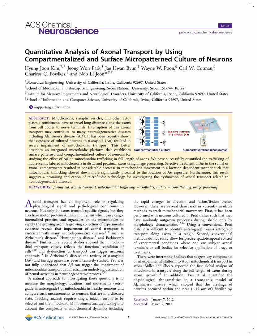
Quantitative Analysis of Axonal Transport by UsingCompartmentalized and Surface Micropatterned Culture of NeuronsHyung Joon Kim,†,⊥ Jeong Won Park,† Jae Hwan Byun,‡ Wayne W. Poon,§ Carl W. Cotman,§
Charless C. Fowlkes,∥ and Noo Li Jeon*,‡,∇
†Biomedical Engineering, University of California, Irvine, California 92697, United States‡School of Mechanical and Aerospace Engineering, Seoul National University, Seoul 151-744, Korea§Institute for Memory Impairments and Neurological Disorders, University of California, Irvine, California 92697, United States∥School of Information and Computer Science, University of California, Irvine, California 92697, United States
*S Supporting Information
ABSTRACT: Mitochondria, synaptic vesicles, and other cyto-plasmic constituents have to travel long distance along the axonsfrom cell bodies to nerve terminals. Interruption of this axonaltransport may contribute to many neurodegenerative diseasesincluding Alzheimer’s disease (AD). It has been recently shownthat exposure of cultured neurons to β-amyloid (Aβ) resulted insevere impairment of mitochondrial transport. This Letterdescribes an integrated microfluidic platform that establishessurface patterned and compartmentalized culture of neurons forstudying the effect of Aβ on mitochondria trafficking in full length of axons. We have successfully quantified the trafficking offluorescently labeled mitochondria in distal and proximal axons using image processing. Selective treatment of Aβ in the somal oraxonal compartments resulted in considerable decrease in mitochondria movement in a location dependent manner such thatmitochondria trafficking slowed down more significantly proximal to the location of Aβ exposure. Furthermore, this resultsuggests a promising application of microfluidic technology for investigating the dysfunction of axonal transport related toneurodegenerative diseases.
KEYWORDS: β-Amyloid, axonal transport, mitochondrial trafficking, microfludics, surface micropatterning, image processing
Axonal transport has an important role in regulatingphysiological signal and pathological conditions in
neurons. Not only do axons transmit specific signals, but theyalso have motor proteins-kinesin and dynein which carry cargo,internalized proteins, and organelles on the microtubules tosupply the growing axon. A growing collection of experimentalevidence reveals that impairment of axonal transport isassociated with many neurodegenerative diseases1−6 such asAlzheimer’s disease,7 Huntington’s disease,8 and Parkinson’sdisease.9 Furthermore, recent studies showed that mitochon-drial transport closely reflects the functional condition ofcells5,10 and dysfunction of transport can trigger neuronalapoptosis.11 In Alzheimer’s disease, the toxicity of β-amyloid(Aβ) and tau aggregates has been intensively studied. Yet, it isnot fully understood that Aβ can trigger the impairment ofmitochondrial transport as a mechanism underlying dysfunctionof neural activities in neurodegenerative process.2,12
A natural approach to investigating these questions is tomeasure the morphology, locations, and movements (retro-grade vs anterograde) of mitochondria in healthy neurons andcompare such measurements to neurons that are in a diseasedstate. Tracking analysis requires single, intact neurons to beselected and the mitochondrial movement analyzed taking intoaccount the complexity of mitochondrial dynamics including
the rapid changes in direction and fusion/fission events.However, there are several drawbacks in currently availablemethods to track mitochondrial movement. First, it has beenperformed with neurons cultured in Petri dishes such that theyhave randomly outgrown processes distinguishable only bymorphologic characteristics.13,14 Using a conventional Petridish, it is difficult to identify anterograde versus retrogradetransport along axons in a tangle. Second, conventionalmethods do not easily allow for precise spatiotemporal controlof experimental conditions where one can subject axonalterminals or cell bodies for selective application of drugs ortreatments.There were interesting findings that suggest key components
of an experimental platform to study mitochondrial transport inaxons. Miller and Sheetz reported the first global analysis ofmitochondrial transport along the full length of axons duringaxonal growth.15 In addition, Tsai et al. quantified thephysiological abnormalities in a transgenic model ofAlzheimer’s disease, which showed that the breakage ofneurites occurred within and near (∼15 μm of) fibrillar Aβ
Received: January 7, 2012Accepted: March 9, 2012
Letter
pubs.acs.org/acschemicalneuroscience
© XXXX American Chemical Society A dx.doi.org/10.1021/cn3000026 | ACS Chem. Neurosci. XXXX, XXX, XXX−XXX

deposits.16 Previously, we reported that the microfluidic-basedneuron culture platform could generate a fluidically isolatedmicroenvironment between the soma and axonal compart-ment.17,18 By utilizing this microfluidic-based neuron cultureplatform, we have demonstrated neuron-to-cell spread of alpha-herpes virus,19 the involvement of local protein synthesis inaxonal growth,20 and the identification of axonal mRNA incortical mammalian axon.21 Recent studies have showed the useof a microfluidic device to quantify neurite outgrowth towardsurface gradient22 and to study axonal transport of NGF,23
BDNF,24 and tau proteins.25 Especially in these studies foraxonal transport, they solely used a microfluidic neuron cultureplatform such that they may bring similar difficulties inmitochondrial trafficking for regions that are not sitting in astraight microgroove region as in a random culture with aconventional Petri dish. In order to address drawbacks inpreviously developed methods,23−25 we employed two method-ologies: (1) integrated microfluidic device with surfacemicropatterning as a physical/biochemical cue for acquiringcontrolled axonal outgrowth and fluidic isolation and (2)automated tracking of mitochondrial movement by imageprocessing. Through making these innovations, we developedan integrated microfluidic platform and automated image-processing for better understanding of the role of Aβ and
correlation between mitochondrial transport in axons and manyneurodegenerative diseases. This platform allowed us toquantify the mitochondrial trafficking under localized exposureof Aβ at specific regions of straightly outgrowing axons.
■ RESULTS AND DISCUSSIONTo apply drugs at various distances away from the somas, wesecured an optimized design of a multicompartment neuronculture chamber on the poly-L-lysine (PLL) strips surfacepattern based on previous studies.26,27 Figure 1 shows aschematic view of the experimental setup including controlledaxonal outgrowth and fluidic isolation property. Because of thedifference (30-fold) in height between the multicompartmentalchannels (100 μm high) and the microgrooves (3 μm high),high fluidic resistance was achieved and one could apply drugsonly at a local region of neurons, for example, proximal, distalaxon, or growth cones. Also, cell bodies were plated on the PLLstrip pattern and axonal outgrowth was guided along thissurface pattern. We showed the fluidic isolation by adding 10kDa of FITC-dextran at the right chamber and imaged thefluorescence signal at the opposite chamber. As shown inFigure 1C, there was no fluorescence signal at the left chamberfrom FITC-dextran, but at the right chamber through middlechambers even after 24 h. We finally confirmed the fluidic
Figure 1. Schematic diagram of experimental setup. (A) Procedure for micropatterning of PLL strip on a substrate and its integration with acompartmentalized microfluidic neuron culture device. A PDMS stamp with embossed line pattern (30 × 50 μm) was placed on a PLL precoatedglass coverslip. Then reactive oxygen plasma selectively removed PLL in regions that were not covered by PDMS. The substrate patterned with thePLL strip was aligned and bonded to PDMS neuron culture device. (B) Micrograph showing primary neurons cultured in the integrated microfluidicmulticompartment chamber with PLL strip. Surface patterned PLL was used to control growth of axons. (C) Fluidic isolation was demonstrated byadding 10 kDa of FITC-dextran to the axonal chamber. No fluorescence was observed in the somal chamber for 24 h. (D) Neurons transfected withGFP on the left side of the device (green), and neurons transfected on the right side of the device with RFP (red). Compartmentalization and fluidicisolation allow for the transfection of two different proteins simultaneously.
ACS Chemical Neuroscience Letter
dx.doi.org/10.1021/cn3000026 | ACS Chem. Neurosci. XXXX, XXX, XXX−XXXB
isolation in a microfluidic neuron culture platform bytransfecting different fluorescence proteins on each compart-ment (Figure 1D). These results clearly show that we can applydrugs selectively at specific regions of cultured neurons and alsomaintain fluidic isolation for further quantitative assay. Inaddition, this experimental platform is suitable for studyingcomplex and dynamic behavior of mitochondria such asanterograde versus retrograde trafficking in axons due to theconfined axonal outgrowth onto the micropatterned PLL strips.Overall, we can investigate the effect of selectively applied drugson the mitochondrial movement along multiple locations ofaxons.We established an automated image-processing algorithm to
quantify mitochondrial movement in axons. A mito-GFPmammalian expression vector was transfected into rat corticalneurons at DIV7. The mito-GFP localized mitochondria due toa cytochrome c mitochondrial localization sequence linked tothe GFP. So, we could investigate the movement ofmitochondrial localized GFP with time-lapse imaging. Figure2A shows the automatic extraction of axon and a displacement-
time plot showing fluorescence along the extracted axoncenterline as function of time. The displacement and time ofKymograph represented the length of an axon (400 μm) andduration of time-lapse images (5 min) on the measurement,respectively. In comparison with this Kymograph, there was nosignificant difference in one produced by the use of painstakinghand-labeling of axons in each video frame. Also, the automatedapproach has generated equivalent visualizations in ∼10 s, andoperating autonomously on time-lapse video data. After theidentification of the axon, tracking can be formulated as a one-
dimensional problem of specifying mitochondrial locationsalong the axon at each time point. As shown in Figure 2B, wehave decomposed the space−time data into two components,static mitochondria and dynamic mitochondria. We observed17 nonmoving mitochondria among total 45 mitochondria inthis Kymograph. In order to decompose static and dynamicentities, we extract out traces of mitochondria that haveminimum displacement, 10 μm. Such decomposition will beuseful in initializing tracks and counting the total number ofmitochondria present.Individual mitochondria exhibit a variety of dynamic
behaviors as shown in Supporting Information Movie 1. Forexample, some mitochondria are largely static while othersmove at high speed. Mitochondria may abruptly stop andhesitate before moving on, or reverse direction entirely. It alsoappeared that around 40% of the neurons exhibit mitochondriathat are nearly stationary, yet those neurons were still vital.Mitochondria also showed interesting interactions includingapparent fusion and fission. It has been suggested thatmitochondrial fission may facilitate cellular apoptosis, whereasmitochondrial fusion may protect the neuron from cellulardysfunction.28 An appropriate algorithm for analyzing such datamust take into account this range of behaviors as well asexperimental artifacts, and it will automatically extract the axonlocation and extent based on fluorescence observed across allimages in the time-lapse movie.We analyzed the trafficking of mitochondria in multiple
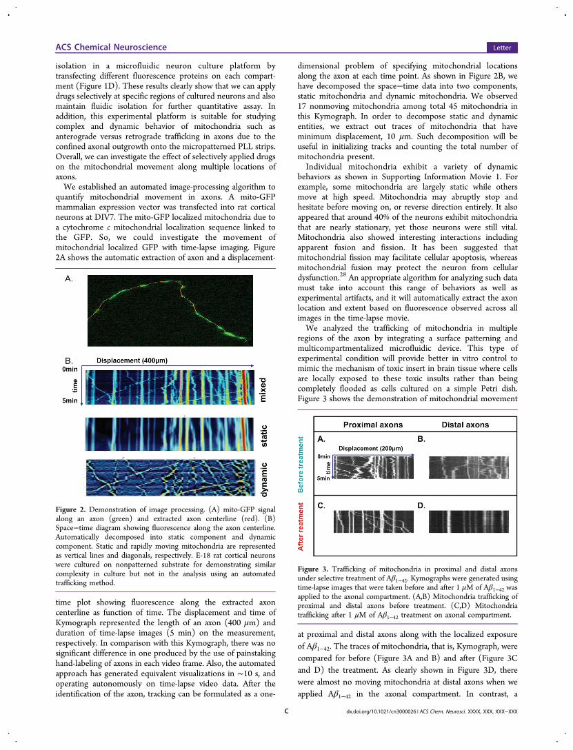
regions of the axon by integrating a surface patterning andmulticompartmentalized microfluidic device. This type ofexperimental condition will provide better in vitro control tomimic the mechanism of toxic insert in brain tissue where cellsare locally exposed to these toxic insults rather than beingcompletely flooded as cells cultured on a simple Petri dish.Figure 3 shows the demonstration of mitochondrial movement
at proximal and distal axons along with the localized exposureof Aβ1−42. The traces of mitochondria, that is, Kymograph, werecompared for before (Figure 3A and B) and after (Figure 3Cand D) the treatment. As clearly shown in Figure 3D, therewere almost no moving mitochondria at distal axons when weapplied Aβ1−42 in the axonal compartment. In contrast, a
Figure 2. Demonstration of image processing. (A) mito-GFP signalalong an axon (green) and extracted axon centerline (red). (B)Space−time diagram showing fluorescence along the axon centerline.Automatically decomposed into static component and dynamiccomponent. Static and rapidly moving mitochondria are representedas vertical lines and diagonals, respectively. E-18 rat cortical neuronswere cultured on nonpatterned substrate for demonstrating similarcomplexity in culture but not in the analysis using an automatedtrafficking method.
Figure 3. Trafficking of mitochondria in proximal and distal axonsunder selective treatment of Aβ1−42. Kymographs were generated usingtime-lapse images that were taken before and after 1 μM of Aβ1−42 wasapplied to the axonal compartment. (A,B) Mitochondria trafficking ofproximal and distal axons before treatment. (C,D) Mitochondriatrafficking after 1 μM of Aβ1−42 treatment on axonal compartment.
ACS Chemical Neuroscience Letter
dx.doi.org/10.1021/cn3000026 | ACS Chem. Neurosci. XXXX, XXX, XXX−XXXC
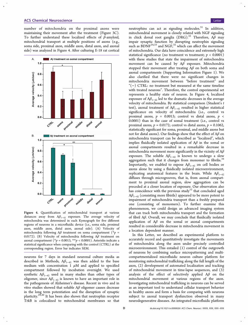
number of mitochondria on the proximal axons weremaintaining their movement after the treatment (Figure 3C).To further understand these localized effects of β-amyloid,mitochondrial transport at multiple positions of axons (e.g.,soma side, proximal axon, middle axon, distal axon, and axonalside) was analyzed in Figure 4. After culturing E-18 rat cortical
neurons for 7 days in standard neuronal culture media asdescribed in Methods, Aβ1−42 was then added to the basemedium with concentration 1 μM and applied to specifiedcompartment followed by incubation overnight. We usedsynthetic Aβ1−42 used in many studies than other types ofoligomer, since Aβ1−42 is known to have an important role inthe pathogenesis of Alzheimer’s disease. Recent in vivo and invitro studies showed that soluble Aβ oligomer causes decreasein the long term potentiation and the disruption of synapticplasticity.29,30 It has been also shown that neutrophin receptorTrkB is colocalized to mitochondrial membranes so that
neutrophins can act as signaling molecules.31 In addition,mitochondrial movement is closely related with NGF signalingin chick dorsal root ganglia (DRG).32 Therefore, Aβ mayimpair synaptic function by disrupting neutrophin signalingsuch as BDNF24,33 and NGF,32 which can affect the movementof mitochondria. Our data have coincidence and extremely highstatistical significance (no treatment vs treatment; p < 0.0001)with these studies that state the impairment of mitochondriamovement can be caused by Aβ exposure. Mitochondriastopped their movement after treating Aβ on both soma andaxonal compartments (Supporting Information Figure 1). Wealso clarified that there were no significant changes inmitochondria movement between “before treatment” and“(−) CTRL: no treatment but measured at the same timelinewith treated neurons”. Therefore, the control experimental setrepresents a healthy state of neuron. In Figure 4, localizedexposure of Aβ1−42 led to the dramatic decreases in the averagevelocity of mitochondria. By statistical comparison (Student’s ttest), axonal treatment of Aβ1−42 resulted in higher statisticalsignificance on velocity of mitochondria (i.e., control vsproximal axons, p = 0.0013; control vs distal axons, p <0.0001) than in the case of somal treatment (i.e., control vsproximal axons, p = 0.0172; control vs distal axons, p = 0.1062,statistically significant for soma, proximal, and middle axons butnot for distal axons). Our findings show that the effect of Aβ onmitochondria transport can be described as “localized”, whichimplies fluidically isolated application of Aβ in the somal oraxonal compartments resulted in a remarkable decrease inmitochondria movement more significantly in the vicinity of Aβexposure. The soluble Aβ1−42 is known to undergo a slowaggregation such that it changes from monomer to fibrilis.34
Importantly, we enabled to expose Aβ1−42 on cell bodies oraxons alone by using a fluidically isolated microenvironment,replicating anatomical features in the brain. While Aβ1−42diffuses through microgrooves, that is, from axonal compart-ment to proximal axonal region, slow aggregation can bepreceded at a closer location of exposure. Our observation alsohas coincidence with the previous study12 that concluded agedAβ1−42 (consisting more fibirils) appeared to be more potent toimpairment of mitochondria transport than a freshly preparedone (consisting of monomers). To further examine thisphenomenon, we could design an advanced imaging systemthat can track both mitochondria transport and the formationof fibril Aβ. Overall, we may conclude that fluidically isolatedapplication of Aβ on the somal or axonal compartmentsresulted in considerable decrease in mitochondria movement ina location dependent manner.In this Letter, we described an experimental platform to
accurately record and quantitatively investigate the movementsof mitochondria along the axon under precisely controlledmicroenvironment. This entailed (1) control of the outgrowthof neurons by combining surface micropatterning and multi-compartmentalized microfluidic neuron culture platform formonitoring mitochondrial trafficking along the full length of theaxon, (2) development of automated localization and trackingof mitochondrial movement in time-lapse sequences, and (3)analysis of the effect of selectively applied Aβ on themitochondrial movement at various regions of the axon.Investigating mitochondrial trafficking in neurons can be servedas an important tool to understand cellular transport behaviorin healthy axons and form a basis for comparing with neuronssubject to axonal transport dysfunction observed in manyneurodegenerative diseases. An integrated microfluidic platform
Figure 4. Quantification of mitochondrial transport at variousdistances away from Aβ1−42 exposure. The average velocity ofmitochondria was determined in each Kymograph for five differentregions of neurons in a microfluidic device (i.e., soma side, proximalaxon, middle axon, distal axon, axonal side). (A) Velocity ofmitochondria following Aβ treatment on soma compartment (*p =0.0172). (B) Velocity of mitochondria following Aβ treatment onaxonal compartment (*p = 0.0013, **p < 0.0001). Asterisks indicate astatistical significance when comparing with the control (CTRL) at thecorresponding region. Error bar indicates SEM.
ACS Chemical Neuroscience Letter
dx.doi.org/10.1021/cn3000026 | ACS Chem. Neurosci. XXXX, XXX, XXX−XXXD
in this study not only is critical to form a basic understanding ofaxonal transport but also paves the way to large scaleexperiments such as screening large drug libraries wheremanual analysis of subtle effects is entirely infeasible.
■ METHODSPreparation of Microfluidic Culture Devices. The PDMS
(Sylgard 184, Dow Corning, MI) chambers were made using softlithography and replica molding as described previously.17,18 Briefly,photolithography was used to make two layers of negative photoresiston a silicon wafer, resulting in a master with positive relief patterns ofcell culture compartments (1.5 mm wide, 7 mm long, 100 μm high)and microgrooves (7.5 μm wide, 3 μm high). A PDMS−prepolymermixture was poured over the positive relief master to obtain a negativereplica-molded piece. After curing, the PDMS was peeled away fromthe master. The reservoirs were punched with a sharpened needle andthen sterilized by autoclaving.Preparation of PLL Patterned Glass Coverslip and Its
Integration with Microfluidic Devices. A detailed schematicprocedure is shown in Figure 1A. The glass coverslip was coatedwith 0.5 mg/mL poly-L-lysine (PLL, MW 70 000−150 000, Sigma, St.Louis, MO) for overnight. They were washed twice with sterilizedwater for 30 min to remove remaining poly-L-lysine. A PDMS stamphaving the desired surface embossed pattern was fabricated by thesame process as PDMS culture devices and placed on the PLLprecoated glass coverslip. It was exposed to reactive oxygen plasma (30W, 200−600 mTorr, Harrick Scientific, Pleasantville, NY) for 10 s tocreate a PLL stripe micropattern on the substrate. The sterilizedPDMS devices were aligned and assembled with the PLL stripemicropattern on the glass coverslips immediately after oxygen plasmatreatment to form an irreversible seal.18,27,35
Cortical Neurons Preparation. In accordance with AAALACguidelines, rodents were housed in the vivarium of the GillespieNeuroscience Research Laboratories at the University of California,Irvine. We prepared cortical dissociated neurons from embryonic rat(E18) as described previously. Briefly, cortexes of E18 rat embryoswere dissected and resuspended in a trypsin solution (0.125% trypsinin CMF-HBSS containing 0.5 mM EDTA) for 7 min at 37 °C or 25min at ambient temperature. Trypsinization was stopped withDulbecco’s modified Eagle’s medium (DMEM) containing 10% fetalbovine serum (FBS), the tissue was centrifuged at 1000 rpm for 1 min,and the resulting cell pellet was resuspended in 2 mL of culturemedium (neurobasal medium, Gibco 21103, containing B27 supple-ment, Gibco 17504, GlutaMAX, Gibco 35050, and penicillin-streptomycin, Gibco 15070). Cells were plated at densities of 3−5 ×10⁶ cells/mL in microfluidic devices.Plasmids and Proteins. Vectors were purchased, subcloned in
house, or received as kind gifts from other researchers. The pEGFP-N1 vector was purchased from Clontech (Mountain View, CA). TheroGFP-mito that is a modified version of pEYFP-mito (Clontech,Mountain View, CA) was kindly provided by Dr. Lin from the HowardHughes Medical Institute (San Diego, CA). The RFP gene containedin the pDsRed1 vector (Clontech, Mountain View, CA) was subclonedinto a mammalian expression vector pcDNA3.1+ (Invitrogen,Carlsbad, CA).Fluorescent Labeling of Mitochondria. Neurons were trans-
fected after 7 days of culture. For one device, 0.8 μg of the appropriatevector was mixed with 50 μL of OptiMEM (Invitrogen, Carlsbad, CA)and incubated at room temperature (RT) for 5 min. In a separate tube,50 μL of OptMEM was mixed with 2 μL of lipofectamine 2000 (L2K)(Invitrogen, Carlsbad, CA) and incubated at RT for 5 min. After the 5min incubation of each individual tube, the DNA/OptiMEM andL2K/OptiMEM were mixed and incubated for an additional 20 min atRT. The mixture of DNA/L2K/OptiMEM was added to the soma sideof the device. After adding the transfection mixture, the neurons wereincubated at 37 °C, 5% CO2 for 3 h. After that, the tranfection mixturewas removed and fresh media was added twice for washing. And thenthe neurons were incubated for at least 48 h before imaging.
Preparation of Aβ Oligomer. Lyophilized Aβ oligomer as a HFIPfilm (Chemicon, Temecula, CA) was stored at −80 °C until used. Aβwas dissolved in neat, sterile dimethyl sulfoxide (DMSO; 5 mM) anddiluted in phosphate buffered saline (PBS), pH 7.4 to 100 mM andaged overnight (4 °C), centrifuged (14 000g, 10 min, 4 °C), and thesupernatants transferred to fresh Eppendorf tubes and stored at 4 °Cuntil use.
Quantifying Mitochondrial Movement Using Image Pro-cessing. Fluorescently labeled mitochondria were imaged 48 h aftertransfection. A Nikon TE 200 Eclipse (Nikon, Melville, NY)microscope equipped with a CoolSnap CCD camera (Photometrics,Tucson, AZ) was used to take time-lapse images. Images were taken atthe same axon/location before and after Aβ treatment in every 3 sduring 5 min. In order to track the movement of mitochondria byhand, we used the z-projection function and multiple Kymographplug-in of ImageJ (National Institutes of Health, Bethesda, MD). Forautomatic trafficking of mitochondria, a MATLAB algorithm wascoded to extract axon segment centerlines from the time-lapse movie.Then, it performed initial one-dimensional tracking and generatedKymographs. A MATLAB algorithm was validated by comparing theresult using ImageJ and automatically generated data. Kymographswere generated for each axon, and the average velocity of mitochondriawas evaluated from each track. Quantification was performed for atleast three axons per movie, and each axon (in field of view) has about10−15 mitochondria such that the average velocity was determined for30−45 mitochondria, and a statistical comparison of control (beforetreatment, i.e., same set of neurons in healthy state) versus Aβ treatedneurons.
■ ASSOCIATED CONTENT*S Supporting InformationFigure 1: Quantitative measurement of mitochondria transporton the control experiment. Asterisks indicate a statisticalsignificance when comparing “before treatment” and thenegative control with positive control (both side treatment).Error bar indicates SEM. Movie 1: Live cell imaging showsfluorescence labeled mitochondria in the microgrooves ofmicrofluidics device. Mitochondria trafficking on three axons inthis field of view could be evaluated. This material is availablefree of charge via the Internet at http://pubs.acs.org.
■ AUTHOR INFORMATIONCorresponding Author*Telephone 1-82-2-880-7111. Fax: 1-82-2-880-7119. E-mail:[email protected] Addresses⊥Laboratory of Genetics, The Salk Institute for BiologicalStudies, 10010 North Torrey Pines Road, La Jolla, California92037, United States.∇WCU Multiscale Mechanical Design, School of Mechanicaland Aerospace Engineering, Seoul National University, Seoul151-744, Korea.Author ContributionsH.J.K. contributed to experimental design, performed research,analyzed data, and contributed to writing of the manuscript.J.W.P., J.H.B., and W.W.P. contributed to experimental designand manuscript preparation. C.C.F. contributed to data analysisusing MATLAB. C.W.C. and N.L.J. provided research over-sight, technical direction, and manuscript preparation.FundingThis work was supported by Roman Reed Spinal Cord InjuryResearch Fund of California, NIH/NIA AG00538, theGraduate Studies Abroad Fellowship (KRF-2005-215-D00030), WCU (World Class University) program throughthe Korea Research Foundation funded by the Ministry of
ACS Chemical Neuroscience Letter
dx.doi.org/10.1021/cn3000026 | ACS Chem. Neurosci. XXXX, XXX, XXX−XXXE

Education, Science and Technology (R31-2008-000-10083-0),and Biomembrane Plasticity Research Center (2011-0000841)through the National Research Foundation (NRF) funded bythe Ministry of Education, Science and Technology (MEST).
NotesThe authors declare no competing financial interest.
■ REFERENCES(1) Chevalier-Larsen, E., and Holzbaur, E. L. (2006) Axonal transportand neurodegenerative disease. Biochim. Biophys. Acta 1762, 1094−1108.(2) Morfini, G. A., Burns, M., Binder, L. I., Kanaan, N. M., LaPointe,N., Bosco, D. A., Brown, R. H. Jr., Brown, H., Tiwari, A., Hayward, L.,Edgar, J., Nave, K. A., Garberrn, J., Atagi, Y., Song, Y., Pigino, G., andBrady, S. T. (2009) Axonal transport defects in neurodegenerativediseases. J. Neurosci. 29, 12776−12786.(3) Chang, D. T., Honick, A. S., and Reynolds, I. J. (2006)Mitochondrial trafficking to synapses in cultured primary corticalneurons. J. Neurosci. 26, 7035−7045.(4) Ebneth, A., Godemann, R., Stamer, K., Illenberger, S., Trinczek,B., and Mandelkow, E. (1998) Overexpression of tau protein inhibitskinesin-dependent trafficking of vesicles, mitochondria, and endoplas-mic reticulum: implications for Alzheimer’s disease. J. Cell Biol. 143,777−794.(5) Malaiyandi, L. M., Honick, A. S., Rintoul, G. L., Wang, Q. J., andReynolds, I. J. (2005) Zn2+ inhibits mitochondrial movement inneurons by phosphatidylinositol 3-kinase activation. J. Neurosci. 25,9507−9514.(6) Szeto, H. H. (2006) Mitochondria-targeted peptide antioxidants:novel neuroprotective agents. AAPS J. 8, E521−531.(7) Lustbader, J. W., Cirilli, M., Lin, C., Xu, H. W., Takuma, K.,Wang, N., Caspersen, C., Chen, X., Pollak, S., Chaney, M., Trinchese,F., Liu, S., Gunn-Moore, F., Lue, L. F., Walker, D. G., Kuppusamy, P.,Zewier, Z. L., Arancio, O., Stern, D., Yan, S. S., and Wu, H. (2004)ABAD directly links Aβ to mitochondrial toxicity in Alzheimer’sdisease. Science 304, 448−452.(8) Bae, B. I., Xu, H., Igarashi, S., Fujimuro, M., Agrawal, N., Taya, Y.,Hayward, S. D., Moran, T. H., Montell, C., Ross, C. A., Snyder, S. H.,and Sawa, A. (2005) p53 mediates cellular dysfunction and behavioralabnormalities in Huntington’s disease. Neuron 47, 29−41.(9) Valente, E. M., Abou-Sleiman, P. M., Caputo, V., Muqit, M. M.,Harvey, K., Gispert, S., Ali, Z., Del Turco, D., Bentivoglio, A. R., Healy,D. G., Albanese, A., Nussbaum, R., Gonzalez-Maldonado, R., Deller,T., Salvi, S., Cortelli, P., Gilks, W. P., Latchman, D. S., Harvey, R. J.,Dallapiccola, B., Auburger, G., and Wood, N. W. (2004) Hereditaryearly-onset Parkinson’s disease caused by mutations in PINK1. Science304, 1158−1160.(10) Hollenbeck, P. J., and Saxton, W. M. (2005) The axonaltransport of mitochondria. J. Cell Sci. 118, 5411−5419.(11) Wallace, D. C. (2001) Mitochondrial defects in neuro-degenerative disease. Ment. Retard. Dev. Disability Res. Rev. 7, 158−166.(12) Rui, Y., Tiwari, P., Xie, Z., and Zheng, J. Q. (2006) Acuteimpairment of mitochondrial trafficking by beta-amyloid peptides inhippocampal neurons. J. Neurosci. 26, 10480−10487.(13) Hollenbeck, P. J. (1996) The pattern and mechanism ofmitochondrial transport in axons. Front. Biosci. 1, d91−102.(14) Ligon, L. A., and Steward, O. (2000) Role of microtubules andactin filaments in the movement of mitochondria in the axons anddendrites of cultured hippocampal neurons. J. Comp. Neurol. 427,351−361.(15) Miller, K. E., and Sheetz, M. P. (2006) Direct evidence forcoherent low velocity axonal transport of mitochondria. J. Cell Biol.173, 373−381.(16) Tsai, J., Grutzendler, J., Duff, K., and Gan, W. B. (2004) Fibrillaramyloid deposition leads to local synaptic abnormalities and breakageof neuronal branches. Nat. Neurosci. 7, 1181−1183.(17) Taylor, A. M., Blurton-Jones, M., Rhee, S. W., Cribbs, D. H.,Cotman, C. W., and Jeon, N. L. (2005) A microfluidic culture platform
for CNS axonal injury, regeneration and transport. Nat. Methods 2,599−605.(18) Park, J. W., Vahidi, B., Taylor, A. M., Rhee, S. W., and Jeon, N.L. (2006) Microfluidic culture platform for neuroscience research. Nat.Protoc. 1, 2128−2136.(19) Liu, W. W., Goodhouse, J., Jeon, N. L., and Enquist, L. W.(2008) A microfluidic chamber for analysis of neuron-to-cell spreadand axonal transport of an alpha-herpesvirus. PLoS ONE 3, e2382.(20) Hengst, U., Deglincerti, A., Kim, H. J., Jeon, N. L., and Jaffrey, S.R. (2009) Axonal elongation triggered by stimulus-induced localtranslation of a polarity complex protein. Nat. Cell Biol. 11, 1024−1030.(21) Taylor, A. M., Berchtold, N. C., Perreau, V. M., Tu, C. H., Jeon,N. L., and Cotman, C. W. (2009) Axonal mRNA in uninjured andregenerating cortical mammalian axons. J. Neurosci. 29, 4697−4707.(22) Millet, L. J., Stewart, M. E., Nuzzo, R. G., and Gillette, M. U.(2010) Guiding neuron development with planar surface gradients ofsubstrate cues deposited using microfluidic devices. Lab Chip 10,1525−1535.(23) Zhang, K., Osakada, Y., Vrljic, M., Chen, L., Mudrakola, H. V.,and Cui, B. (2010) Single-molecule imaging of NGF axonal transportin microfluidic devices. Lab Chip 10, 2566−2573.(24) Poon, W. W., Blurton-Jones, M., Tu, C. H., Feinberg, L. M.,Chabrier, M. A., Harris, J. W., Jeon, N. L., and Cotman, C. W. (2011)beta-Amyloid impairs axonal BDNF retrograde trafficking. Neurobiol.Aging 32, 821−833.(25) Stoothoff, W., Jones, P. B., Spires-Jones, T. L., Joyner, D.,Chhabra, E., Bercury, K., Fan, Z., Xie, H., Bacskai, B., Edd, J., Irimia,D., and Hyman, B. T. (2009) Differential effect of three-repeat andfour-repeat tau on mitochondrial axonal transport. J. Neurochem. 111,417−427.(26) Rhee, S. W., Taylor, A. M., Cribbs, D. H., Cotman, C. W., andJeon, N. L. (2007) External force-assisted cell positioning insidemicrofluidic devices. Biomed. Microdevices 9, 15−23.(27) Rhee, S. W., Taylor, A. M., Tu, C. H., Cribbs, D. H., Cotman, C.W., and Jeon, N. L. (2005) Patterned cell culture inside microfluidicdevices. Lab Chip 5, 102−107.(28) Chen, H., and Chan, D. C. (2005) Emerging functions ofmammalian mitochondrial fusion and fission. Hum. Mol. Genet. 14,R283−289.(29) Billings, L. M., Oddo, S., Green, K. N., McGaugh, J. L., andLaFerla, F. M. (2005) Intraneuronal Abeta causes the onset of earlyAlzheimer’s disease-related cognitive deficits in transgenic mice.Neuron 45, 675−688.(30) Cleary, J. P., Walsh, D. M., Hofmeister, J. J., Shankar, G. M.,Kuskowski, M. A., Selkoe, D. J., and Ashe, K. H. (2005) Naturaloligomers of the amyloid-beta protein specifically disrupt cognitivefunction. Nat. Neurosci 8, 79−84.(31) Wiedemann, F. R., Siemen, D., Mawrin, C., Horn, T. F., andDietzmann, K. (2006) The neurotrophin receptor TrkB is colocalizedto mitochondrial membranes. Int. J. Biochem. Cell Biol. 38, 610−620.(32) Chada, S. R., and Hollenbeck, P. J. (2003) Mitochondrialmovement and positioning in axons: the role of growth factorsignaling. J. Exp. Biol. 206, 1985−1992.(33) Tong, L., Balazs, R., Thornton, P. L., and Cotman, C. W. (2004)Beta-amyloid peptide at sublethal concentrations downregulates brain-derived neurotrophic factor functions in cultured cortical neurons. J.Neurosci. 24, 6799−6809.(34) Parbhu, A., Lin, H., Thimm, J., and Lal, R. (2002) Imaging real-time aggregation of amyloid beta protein (1−42) by atomic forcemicroscopy. Peptides 23, 1265−1270.(35) Duffy, D. C., McDonald, J. C., Schueller, O. J. A., andWhitesides, G. M. (1998) Rapid prototyping of microfluidic systems inpoly(dimethylsiloxane). Anal. Chem. 70, 4974−4984.
ACS Chemical Neuroscience Letter
dx.doi.org/10.1021/cn3000026 | ACS Chem. Neurosci. XXXX, XXX, XXX−XXXF