Embed Size (px)
Citation preview
Quantitative Imaging of Neuroreceptors in the Living Human Brain
Henry N. Wagner , Jr
Pos i t ron emission t o m o g r a p h y (PET) makes it possi- ble for the f i rs t t ime to examine in l iving humans the chemistry of the brain, which relates the structures of the brain to the functions of the mind. PET scans make it possible to assess the state of neurotrans- mi t ter receptors , such as the dopamine, sero ton in , muscar in ic chol inergic, opiate, and benzodiazapine receptors , in different regions in normal persons and pat ien ts w i th neuropsychiatr ic diseases. One of the most striking findings to date is t ha t dopamine recep to rs fal l significantly be tween the ages of 19 and 73 years, to a greater degree in men than in
women. The effects of neurotropic drugs, such as haloperidol and methadone, can be assessed in an individual pat ient , providing for the f irst t ime an objective indicator of the specific desired effects of such drugs in the t rea tment of nervous or mental disease. Studies of pat ients w i th diseases such as Parkinson's disease, Huntington's disease, Alzheim- er 's disease, bipolar disease, and schizophrenia are in progress. A t present , the pat ients are being exam- ined as part of research protocols, but it is likely that clinical t r ia ls wi l l not be far off. �9 1986 by Grune & Strat ton, Inc.
E ACH N E U R O N of the brain receives input from thousands or even tens of thousands of
synaptic contacts with other neurons converging on it. Through its own axon, and the thousands of divergent axonal branches, each neuron sends output to thousands of other neurons. One of the most important findings of neurobiology is that information transfer across a synapsis is by the process of chemical neurotransmission. The arri- val of electrical impulses at the end of an axonal branch causes the release of chemical neuro- transmitters, such as dopamine, serotonin, or acetylcholine. Chemical receptors on the synap- tic membrane of the downstream neurons bind these, or the more than 30 to 50 other neuro- transmitters, to identify the information con- tained in the neurotransmitters. The combina- tion of transmitters with receptors affects the subsequent depolarization process in the postsy- naptic neurons. Certain receptors, such as the acetylcholine receptor, are believed to exist in two states: "activable" or "nonactivable" allo- steric forms that together provide an effective regulator of the process of synaptic transmission. The synapse often contains an enzyme, such as acetylcholinesterase that destroys the neuro- transmitter and terminates its action. Blocking the enzyme causes an increase of the neurotrans- mitter in the cleft and prolongs the response.
The involvement of chemical transmitters and receptors in the transfer of information from one neuron to another has important practical conse- quences in medicine, not the least of which is making possible the use of drugs to stimulate or block the neurotransmission process by stimulat-
ing or blocking the neuroreceptors. 1 Many of the most widely used drugs in medical practice today act in this way. For example, the drug cimetidine blocks histamine receptors; propranolol blocks beta adrenergic receptors; and haloperidot blocks dopamine receptors. The widely used drug Val- ium (Hoffmann-LaRoche, Nutley, N J) stimu- lates benzodiazepine receptors that have an inhibitory effect on neurotransmission.
Much of what is now known about the chemis- try of neurotransmission is based on the use of radioactive tracers. 2 Although chemical trans- mission of information within the nervous system had been recognized since early in the 20th century, the development of tritiated norepi- nephrine made possible the detailed study of the intermediary metabolism of this neurotransmit- ter. 3 One of the early discoveries was that after a neurotransmitter such as norepinephrine is released into the synapse and brings about propa- gation of presynaptic impulses, the action of the neurotransmitter is terminated by its reuptake into the axons of the presynaptic neuron. Knowl-
From the Divisions of Nuclear Medicine and Radiation Health Sciences, The Johns Hopkins Medical Institutions, Baltimore.
Supported by US Public Health Service Grants No. NS 15080 and CA 32845.
Address reprint requests to Henry N. Wagner, Jr, MD, Divisions of Nuclear Medicine and Radiation Health Sciences, The Johns Hopkins Medical Institutions, 615 N Wolfe St, Room 2001, Baltimore, MD 21205-2179.
�9 1986 by Grune & Stratton, Inc. 0001-2998/86/1601-0003505.00/0
Seminars in Nuclear Medicine, Vol XVI, No 1 (January), 1986: pp 51-62 51
52
edge of such mechanisms is of practical impor- tance, because some drugs can act by blocking the reuptake process, thus prolonging the action of the neurotransmitter. The use of imipramine to increase catecholamine concentrations at the synapse in patients with depression is an exam- ple.
Reserpine was the first of a long series of drugs that have revolutionized the care of patients with psychiatric disorders. Discovered as a potent hypotensive agent, reserpine was observed to have a tranquilizing effect without associated sedation to the degree associated with the use of barbiturates. Monoamine oxidase inhibitors were also found to have an antidepressant effect and chlorpromazine was found to be an effective antipsychotic. Pharmacologic research that soon followed resulted in the development of tricyclic antidepressants, lithium for the treatment of bipolar depression, and chlordiazepoxide for the treatment of anxiety. Such drugs replaced seda- tives and hypnotics and made possible the release of many patients from mental hospitals. The discovery that dopamine concentrations were low in the caudate nucleus and putamen of patients with Parkinson's disease brought about the first rational neurotherapy: the use of L-dopa to increase the concentration of dopamine in the caudate nucleus and putamen and relieve many of the symptoms of Parkinson's disease. Other neurotropic drugs included methyldopa for the treatment of hypertension, propranolol for the treatment of arrhythmias, and a host of other psychopharmaceutical agents.
In addition to the practical uses of these newly developed drugs, studies of their mechanism of action resulted in many advances in neurobiolo- gy. For example, it was found that reserpine profoundly depleted the brain of all of its bio- genic amines, ie, dopamine, serotonin, and nor- epinephrine. Studies of neurotransmitter recep- tor binding in the mammalian brain derived from initial investigations of the opiate receptor. 4 The general approach is to measure the binding of an appropriate drug with high affinity for the recep- tor and labeled with a high specific activity. The most important technical hurdle to overcome in demonstrating specific receptor binding is to minimize the amount of nonspecific binding with other membrane components, since nonspecific sites generally exceed by a large margin the
HENRY N. WAGNER
number of true receptors. Using low concentra- tions of relatively selective drugs and by washing rapidly but thoroughly to remove nonspecific binding while preserving specific binding to the receptors, it was possible to demonstrate binding of radioactive opiates to pharmacologically rele- vant opiate receptors. Subsequently, numerous other neurotransmitter receptors in the brain could be demonstrated, including receptor sites for serotonin, norepinephrine, glycine, gamma- aminobutyric acid (GABA), dopamine, and vari- ous peptides, such as angiotensin, neurotensin, and thyrotropin-releasing hormone.
One of the most striking findings subsequent to the discovery of neuroleptic drug receptors was that one could predict clinical potency of neuroleptic drugs of diverse chemical structures from in vitro effects of the drugs in binding to neurotransmitter receptors. 5 The close correla- tion between the clinical effects and the affinity of the diverse drugs for dopamine receptors was of great significance for pharmaceutical research because it greatly extended the study of the effects of the drugs on animal behavior. The relative binding properties of the neuroleptic drugs to the receptors could be correlated with pharmacologic actions in animals. For example, apomorphine induces a stereotyped sniffing, lick- ing, and gnawing behavior in rodents, which derives from stimulation of the postsynaptic dop- amine receptors in the corpus striatum. The ability of neuroleptic drugs to block apomor- phine-induced stereotyped behavior predicts the clinical potencies of drugs and has been used by the pharmaceutical industry as a screening test.
Progress in nuclear medicine has now made possible another important breakthrough in pharmacology, the direct measurement in living human beings of the specific effects of neuro- tropic drugs on receptors.
POSITRON EMISSION TOMOGRAPHY
The ability to study the process of chemical neurotransmission within the brain of living human beings by positron emission tomography (PET) makes it possible to relate chemical pro- cesses involving neurotransmitters to anatomic structures within the human brain as well as to mental and behavioral functions. 6 The recent development of suitable ligands labeled with single photon emitting tracers, such as iodine

IMAGING OF NEURORECEPTORS IN HUMAN BRAIN 53
123, makes it likely that such studies will soon be available using single photon emission computed tomography (SPECT) as well. PET, in the study of neuroreceptors, involves drugs labeled with carbon 11 or fluorine 18, tracers that require a cyclotron within or very near a hospital for their production. In a typical study, the cyclotron- produced radioactive atom is incorporated into a drug, such as the neuroleptic N-methyl spipe- rone, The intravenously (IV) injected drug is bound to neuroreceptors, such as dopamine, sero- tonin, opiate, benzodiazepine, or muscarinic cholinergic receptors. A radioactive atom, such as ~C, which has a half-life of 20 minutes, emits positrons in the process of radioactive decay. The positron travels for 1 or 2 mm within the brain before combining with an electron to yield two gamma rays that are emitted in opposite direc- tions through the skull; the spatial resolution of PET imaging today is between 5 and 8 ram. The ultimate resolution is limited by the range of positrons in tissue before combining with an electron, ie, between 1 and 2 mm.
For a radioactive drug to be identified as binding specifically to a neuroreceptor, certain criteria must be fulfilled: (1) the binding must be saturable at low concentrations of drug because of the small number of receptor sites; (2) the binding must be blocked by high concentrations of the drug; and (3) the distribution within the brain should correspond to known sites of the receptors. For example, in the human brain, most dopamine (D0 receptors are found in the cau- date nucleus and putamen with fewer D 2 sites in other regions] Serotonin ($2) receptors, on the other hand, are found widely distributed within the cerebral cortex] The autoradiographic stud- ies of brain slices of animals and of human autopsy paved the way for the extension of these approaches to living humans by means of PET. Kuhar et al were able to carry out the autoradio- graphic studies 8 on the basis of the development of tritiated spiperone by Laduron and Leysen. 9 The autoradiographic studies of Kuhar et al stimulated the attempt to extend these studies to the living human brain.
D 2 DOPAMINE RECEPTORS
~lC-N-methyl spiperone is an analogue of spi- perone, a butyrophenone neuroleptic drug with an extremely high affinity for dopamine recep-
tors. The synthesis of the radiolabeled analogue 3-N-methyl spiperone was based on the rapid introduction of 11C into the spiperonc molecule by methylation of the amide nitrogen with 1~C methyl iodide] ~ The ~C methyl iodide was pre- pared from HC dioxide.
Chemical purification of HC-N-methyl spipe- rone ([UC]NMSP) was simplified by synthesiz- ing a byproduct of this compound, heptyl deriva- tized spiperone, that was more lipophilic and eluted with a longer retention volume than the desired product, which is [I~C]NMSP. The frac- tion of the eluant containing the labeled 3- N-methyl spiperone was collected and evapo- rated to dryness, dissolved in a mixture of sterile saline/bicarbonate, and assayed for sterility, apyrogenicity, and radiochemical purity. ~~
Due to the short half-life of 1~C, and the need to know the mass of byproducts and starting materials, it was necessary to develop a fast, reliable method for determining the specific activity of the final product and the presence of any contaminants. We used analytic high perfor- mance liquid chromatography (HPLC) and an automated integrating recorder because of its speed and reproducibilityJ ~ With this system we determined the specific activity of the (3-N [~ ~C]- methyl)spiperone in the final injectable solution within four minutes. The average specific activ- ity of the first 72 consecutive preparations that we have studied was 270 mCi/umole at end of synthesis (EOS) (1,080 mCi/~zmole at end of bombardment). Since then, even higher specific activities have been obtained.
VALIDATION OF [11C]NMSP AS A D 2 LIGAND
An essential first step is the selection of a radioactive ligand with good in vitro binding properties; that is, it has a high affinity for the receptors when studied in vitro. Studies in rodent brains indicated that tritiated spiperone and ben- peridol had high affinities, while drugs such as chlorpromazine and haloperidol did not. In vivo studies in rodents next indicated that the binding of [I~C]NMSP was greatest in the striatum, which is rich in dopamine receptors, and least in the cerebellum in which there are few. 7 Thus, the radioactivity in the cerebellum could be taken to represent nonspecific binding. In the first six minutes after injection of the tracer [~C]NMSP, the concentrations in the striatum were equal to
54 HENRY N. WAGNER
those in the cerebellum, but the activity in the striatum increased for the next two hours while that in the cerebellum fell, indicating more pre- ferential binding to the dopamine receptors as time went by. Injection of the rodents with the antipsychotic drug (+)-butaclamol reduced the striatal level to that in the cerebellum. The inactive ( - ) form of butaclamol did not have this effect. The distribution of the [~C]NMSP within the brain corresponded exactly to the sites where dopamine receptors are present in high quantities. Thus, all criteria of receptor binding had been satisfied in the case of spiperone and its analogue, [~C]N MSP, including saturability of high doses of the drug. m
NORMAL PERSONS
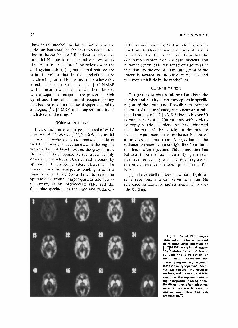
Figure 1 is a series of images obtained after IV injection of 20 mCi of [11C]NMSP. The initial images, immediately after injection, indicate that the tracer has accumulated in the regions with the highest blood flow, ie, the gray matter. Because of its lipophilicity, the tracer readily crosses the blood-brain barrier and is bound by specific and nonspecific sites. Thereafter the tracer leaves the nonspecific binding sites at a rapid rate as blood levels fall, the serotonin specific sites (frontal temporoparietal and occipi- tal cortex) at an intermediate rate, and the dopamine-specific sites (caudate and putamen)
at the slowest rate (Fig 2). The rate of dissocia- tion from the D2 dopamine receptor binding sites is so slow that the tracer activity within the dopamine-receptor rich caudate nucleus and putamen continues to rise for several hours after injection. By the end of 90 minutes, most of the tracer is located in the caudate nucleus and putamen with little in the cerebellum.
QUANTIFICATION
Our goal is to obtain information about the number and affinity of neuroreceptors in specific regions of the brain, and if possible, to estimate the rates of release of endogenous neurotransmit- ters. In studies of [ I IC]NMSP kinetics in over 50 normal persons and 200 patients with various neuropsychiatric disorders, we have observed that the ratio of the activity in the caudate nucleus or putamen to that in the cerebellum, as a function of time after IV injection of the radioactive tracer, was a straight line for at least two hours after injection. This observation has led to a simple method for quantifying the rela- tive receptor density within various regions of interest. In essence, the assumptions are as fol- lows:
(1) The cerebellum does not contain D2 dopa- mine receptors, and can serve as a suitable reference standard for metabolites and nonspe- cific binding.
Fig 1. Serial PET images obtained at the times indicated in minutes after injection of [11C]NMSP. In the initial images the distribution of the tracer ref lects the distribution of blood f low. Therea f te r the tracer progressively accumu- lates in the D z dopamine recep- tor-rich regions, the caudate nucleus, and putamen, and falls rapidly in the regions contain- ing nonspecific binding sites. By 60 minutes after injection, most of the tracer is bound to and putamen, (Reprinted with permission, zs)

IMAGING OF NEURORECEPTORS IN HUMAN BRAIN
A c t i v i t y I
- 0 .
: = Caudo te and Putamen
o---o O c c i p i t a l V i sua l C o r t e x
x--x Fronta l Lobe Cor tex C e r e b e l l a r Cortex
A---J C e r e b e l l o r White M a t t e r
0 12 36 6 0 84 108
T i m e ( m l n u t e s )
Fig 2. T ime course of the tracer [11C]NMSP radioactiv- ity in t he s t ruc tu res ind icated at t he var ious t imes af ter IV injection. (Reprinted w i t h permiss ion. 2s)
(2) The rate of dissociation of the NMSP tracer from the receptors in the caudate nucleus and putamen is negligible (otherwise the cau- date/cerebellar curve would bend downward rather than remain linear with time). This assumption has been supported by measurement of caudate activity for periods as long as four hours after injection of ]SF NMSP.
(3) Metabolites of NMSP behave with a con- stant relationship in both the cerebellum and caudate nucleus; that is, with respect to the fraction crossing the blood-brain barrier and their subsequent kinetics.
A complete mathematical model that makes possible calculation of absolute receptor density and affinity has been developed, but the simplest method for assessing receptor availability in a region such as the caudate nucleus or putamen is as follows: Serial PET scans are obtained contin- uously for two hours after injection. Binding to t h e D 2 dopamine receptor is estimated by the ratio of radioactivity in the caudate to that in the
5 5
cerebellum (Ac,/Acb , expressed as mean counts per minute per computer picture element) and by the ratio of radioactivity in the putamen to that in the cerebellum, expressed as a function of time after injection of the tracer. Binding to the $2 serotonin receptor is estimated by the ratio of radioactivity in the frontal cortex to that in the cerebellum (Afr/Acb) as a function of time.
In 50 normal subjects and over 200 patients with neuropsychiatric disorders studied to date, the Aca/Acb ratio has invariably increased linearly with time after injection. The slope of this line reflects the rate of [11C]NMSP binding to the D 2 receptor from the exchangeable tissue pool (including nonspecific binding) (Fig 3).
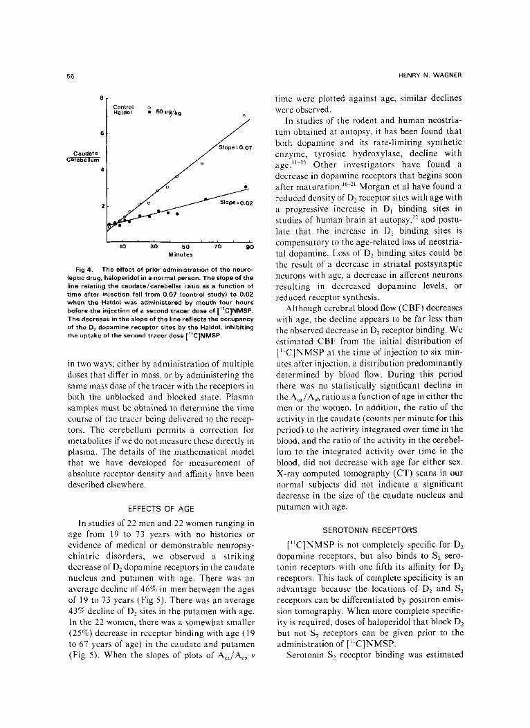
The slope of the line relating the caudate/ cerebellar ratio to time can be used directly as an index of relative receptor density if we assume that the affinity of the ligand for the receptor is unaffected by the condition under study. Thus, we have observed that receptor blocking doses of neuroleptic drugs, such as haloperidol (Fig 4), result in a decrease in the slope of the straight line, whereas increased receptor density is reflected in an increased slope. Whereas the slope of the Acd/Aob V time line can be taken to represent relative receptor availability, absolute receptor density and affinity can be determined
6
O
i i i i i i 20 40 60 80 100 120
Time (minutes)
Fig 3. The caudate /cerebel lar ra t io at the t imes indi- cated after IV injection of [11C]NMSP in a no rma l person. The s t ra igh t l ine relationship af ter the first ten minu tes was observed in over 50 normal persons and 200 pat ien ts w i t h var ious neuropsych ia t r i c d isorders. The slope of t he l ine is a reflection of the relative number of unoccupied binding si tes in the D 2 dopamine receptor-r ich caudate nucleus. (Repr in ted w i t h permiss ion. TM)
56 HENRY N. WAGNER
6
Caudate Cerebellum
4
Control Haldol ~ 50 u g/kg e
i i i i i lJO 3~0 5~0 70 90
Minutes
Fig 4. The effect of prior administrat ion of the neuro- leptic drug, haloperidol in a normal person. The slope of the line relating the cauda te /ce rebe l l a r ratio as a function of t ime af ter injection fell f rom 0 .07 (control study) to 0.02 w h e n the Haldol was administered by mouth four hours before the injection of a second t racer dose of [ l lC]NMSP. The decrease in the slope of the line ref lects the occupancy of the D 2 dopamine receptor sites by the Haldol, inhibiting the uptake of the second t racer dose [11C]NMSP.
in two ways; either by administration of multiple doses that differ in mass, or by administering the same mass dose of the tracer with the receptors in both the unblocked and blocked state. Plasma samples must be obtained to determine the time course of the tracer being delivered to the recep- tors. The cerebellum permits a correction for metabolites if we do not measure these directly in plasma. The details of the mathematical model that we have developed for measurement of absolute receptor density and affinity have been described elsewhere.
EFFECTS OF AGE
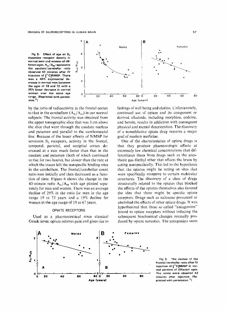
In studies of 22 men and 22 women ranging in age from 19 to 73 years with no histories or evidence of medical or demonstrable neuropsy- chiatric disorders, we observed a striking decrease of O 2 dopamine receptors in the caudate nucleus and putamen with age. There was an average decline of 46% in men between the ages of 19 to 73 years (Fig 5). There was an average 43% decline of D2 sites in the putamen with age. In the 22 women, there was a somewhat smaller (25%) decrease in receptor binding with age (19 to 67 years of age) in the caudate and putamen (Fig 5). When the slopes of plots of Aca/Acb v
time were plotted against age, similar declines were observed.
In studies of the rodent and human neostria- turn obtained at autopsy, it has been found that both dopamine and its rate-limiting synthetic enzyme, tyrosine hydroxylase, decline with age. ~M5 Other investigators have found a decrease in dopamine receptors that begins soon after maturation. ~6-2~ Morgan et al have found a reduced density of O 2 receptor sites with age with a progressive increase in D~ binding sites in studies of human brain at autopsy, 22 and postu- late that the increase in Dt binding sites is compensatory to the age-related loss of neostria- tal dopamine. Loss of D2 binding sites could be the result of a decrease in striatal postsynaptic neurons with age, a decrease in afferent neurons resulting in decreased dopamine levels, or reduced receptor synthesis.
Although cerebral blood flow (CBF) decreases with age, the decline appears to be far less than the observed decrease in D2 receptor binding. We estimated CBF from the initial distribution of [~C]NMSP at the time of injection to six min- utes after injection, a distribution predominantly determined by blood flow. During this period there was no statistically significant decline in the Aca/Acb ratio as a function of age in either the men or the women. In addition, the ratio of the activity in the caudate (counts per minute for this period) to the activity integrated over time in the blood, and the ratio of the activity in the cerebel- lum to the integrated activity over time in the blood, did not decrease with age for either sex. X-ray computed tomography (CT) scans in our normal subjects did not indicate a significant decrease in the size of the caudate nucleus and putamen with age.
SEROTONIN RECEPTORS
[~IC]NMSP is not completely specific for D 2 dopamine receptors, but also binds to $2 sero- tonin receptors with one fifth its affinity for D 2 receptors. This lack of complete specificity is an advantage because the locations of D2 and $2 receptors can be differentiated by positron emis- sion tomography. When more complete specific- ity is required, doses of haloperidol that block D2 but not $2 receptors can be given prior to the administration of [I~C]NMSP.
Serotonin $2 receptor binding was estimated
IMAGING OF NEURORECEPTORS IN HUMAN BRAIN 57
Fig 5. Effect of age on D 2 4 dopamine receptor density in normal men and women of dif- ferent ages. A~/As=, represents < the caudate/cerebel lar rat io "~ 3 observed 43 minutes after IV "~ injection of [I~C]NMSP. There was a 46% exponential de-
2 crease in normal men between the ages of 19 and 72 wi th a 25% linear decrease in normal women over the same age 0 range. (Reprinted w i th permis- sion. TM )
i
0
Males
�9 Q e - , /
i L i
20 4'0 6'0 8+0 0
Age (years)
F e m a l e s
20 40 60 80
by the ratio of radioactivity in the frontal cortex to that in the cerebellum (Afr/Acb) in our normal subjects. The frontal activity was obtained from the upper tomographic slice that was 3 cm above the slice that went through the caudate nucleus and putamen and parallel to the: canthomeatal line. Because of the lesser affinity of NMSP for serotonin $2 receptors, activity in the frontal, temporal, parietal, and occipital cortex de- creased at a rate much faster than that in the caudate and putamen (both of which continued to rise for two hours), but slower than the rate at which the tracer left the nonspecific binding sites in the cerebellum. The frontal/cerebellar count ratio rose initially and then decreased as a func- tion of time. Figure 6 shows the change in the 43-minute ratio Afr/Acb with age plotted sepa- rately for men and women. There was an average decline of 29% in the ratio for men in the age range 19 to 73 years and a 19% decline for women in the age range of 19 to 67 years.
OPIATE RECEPTORS
Used as a pharmaceutical since classical Greek times, opium relieves pain and gives rise to
feelings of well being and elation. Unfortunately, continued use of opium and its component or derived alkaloids, including morphine, codeine, and heroin, results in addiction with consequent physical and mental deterioration. The discovery of a nonaddictive opiate drug remains a major goal of modern medicine.
One of the characteristics of opiate drugs is that they produce pharmacologic effects at extremely low chemical concentrations that dif- ferentiates them from drugs such as the anes- thetic gas diethyl ether that affects the brain by acting nonspecifically. This led to the hypothesis that the opiates might be acting on sites that were specifically receptive to certain molecular structures. The discovery of a class of drugs structurally related to the opiates that blocked the effects of the opiates themselves also favored the idea that there might be specific opiate receptors. Drugs such as naloxone prevented or abolished the effects of other opiate drugs. It was hypothesized that these so called "antagonists" bound to opiate receptors without inducing the subsequent biochemical changes normally pro- duced by opiate narcotics. The antagonists seem
J~
<
A 2
1
M a l e s
A r l i i i |
20 4 0 O0
B �9 F e m a l e s
.-.
1
B I i L l
80 0 20 Age ( y e e r e )
I I i i
40 60
Fig 6. The decline of the f rontal /cerebe| |ar ratio after IV injection of [11C]NMSP in nor- mal persons of different ages. The ratios were obtained 43 minutes after injection. (Re- printed w i th permission. TM)
58 HENRY N. WAGNER
to prevent the access of the morphine-like drugs to the opiate receptor.
For these reasons many scientists devoted many years to the search for the opiate receptor. The approach was to measure the binding of radiolabeled drugs, but the problem was that most chemicals that are lipophilic such as opiate drugs bind nonspecifically to many kinds of molecules on the surface of nerve membranes. The first successful demonstration of the exis- tence of opiate receptors was accomplished by Candace Pert and Solomon Snyder at Johns Hopkins in 1973. They used high-specific activ- ity opiates and perfected procedures for washing away the nonspecific binding rapidly and thor- oughly while preserving specific binding to the receptors. Independently, Lars Terenius at the University of Uppsala (Sweden) and Eric Simon at New York University (New York) obtained similar results in the same year. In other experi- ments Kuhar, Pert, and Snyder examined where in the brain opiate receptors were located. They dissected many regions of the brain and mea- sured the binding of radioactivity labeled opiate drugs in each region. For example, the medial portion of the thalamus contained high concen- trations of opiate receptors whereas the lateral thalamus had few. It was hypothesized that the abundance of opiate receptors in the medial thalamus accounts, at least in part, for the ability of opiates to relieve pain. Using autoradiogra- phy, Michael Kuhar extended his studies to microscopic levels of the brain. For example, he found dense bands of opiate receptors in the spinal cord, providing strong evidence that opiates act on the spinal cord as well as the brain. The amygdala had virtually the highest concen- trations of opiate receptors in the brain, a site that may be involved in the euphoric effect of these drugs.
PET
One year after the successful imaging of D 2
dopamine receptors in the brain of living human beings, PET was used to obtain quantitative imaging of opiate receptors in the human brain.
Imaging of opiate receptors was accomplished by developing 4-carbo-methoxy-fentanyl, called carfentanil, with a potency over 7,000-fold greater than that of morphine. 23'24 The average specific activity was 1,145 mCi/#mol at the end
of synthesis, which corresponds to an average specific activity at the end of bombardment of 3,270 mCi/umol.
Carfentanil binds chiefly to mu type opiate receptors that are involved in pain perception (we give evidence below). The mu subtype specificity of carfentanil was ascertained by measuring its effect on receptor binding of [3H]-dihydromor- phine, a highly u-selective ligand, as well as other ligands that have relatively high specificity for and K receptors. The calculated affinity con- s tants of carfentanil for u, 6, and K opiate recep- tors at 37 ~ are 0.051 nmol, 4.7 nmol, and 13 nmol. In baboon studies, highest concentrations of radioactivity were found in the thalamus and caudate nucleus, with intermediate concentra- tions in the frontal cerebral cortex and a low concentration in the cerebellum. Pretreatment with naloxone resulted in a markedly reduced accumulation of the tracer in the thalamus, caudate nucleus, cerebral cortex, and amygdala but not in the cerebellum.
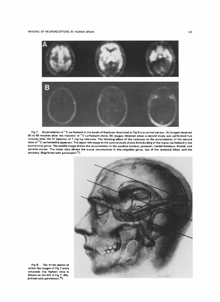
Images obtained in a normal human being 30 to 60 minutes after injection of ~C carfentanil are shown in Fig 7. The levels at which these images were obtained is shown in Fig 8. The top slice is on the left in Fig 7; the bottom slice on the far right. Radioactivity was highly concentrated in the medial thalamus with substantially lesser levels in the lateral thalamus. The caudate nucleus and putamen displayed a similarly high level of radioactivity. The frontal and temporal parietal cerebral cortex had somewhat less radioactivity but substantially more than the occipital cortex. A high level of tracer accumula- tion was also observed in the pituitary region and temporal lobes of the cerebral cortex, while the cerebellum had much lower levels of radioactivi- ty. The postcentral gyrus accumulated low quan- tities of the tracer. Following the blocking dose of naloxone, the initial images obtained immedi- ately after L~C carfentanil injection revealed a distribution of tracer activity that corresponded to the known distribution of regional blood flow, but subsequent images indicated that the uptake by opiate receptors was essentially eliminated by the prior administration of naloxone. The decrease in ~C-carfentanil accumulation in the caudate nucleus and thalamus as a function of the administered dose of naloxone is illustrated in Fig 9.
IMAGING OF NEURORECEPTORS IN HUMAN BRAIN 59
Fig 7. Accumulat ion of 11C carfentanil at the levels of the brain i l lustrated in Fig 8 in a normal person. (A) Images obtained 30 to 60 minutes after the injection of 11C carfantanil atone; |B) images obtained when a second study was performed five minutes after the IV injection of 1 mg /kg naloxone. The blocking effect of the naloxone on the accumulation of the second dose of ~C carfentanil is apparent. The upper left image in the control study shows l i t t le binding of the tracer carfentanil in the postcentral gyrus. The middle image shows the accumulation in the caudate nucleus, putamen, medial thalamus, frontal, and parietal cortex. The lower slice shows the tracer accumulated in the cingulate gyrus, t ips of the temporal lobes, and the pituitary. (Reprinted w i th permission. 24)
Fig 8. The three planes at which the images of Fig 7 were obtained; the highest slice is shown on the left in Fig 7. (Re- pr inted w i th permission, ze)

60 HENRY N, WAGNER
6 -
.E 5 - E O 1o 6 ~. 4
g
3 Z
i I
::::::~:2~ ~te~':: Tha lamus ~ : . . , .
i ! | |
1.0 0.1 0.0 i 0.0 01
N a l o x o n e ( m g / k g )
Fig 9. On the vertical axis is the rat io of the ~1C carfentanil activity in the caudate or thalamus w i t h o u t / w i th the prior administration of naloxone. At the higher doses of naloxone the accumulation of the ~1C carfentanil was reduced to less than 20% of the unblocked levels. (Reprinted wi th permission. =s)
APPLICATIONS
An immediate application of measurements of receptor binding of drugs such as carfentanil or methyl spiperone that bind to opiate and D2 dopamine receptors, respectively, is to monitor the state of the receptors in the course of treat- ment of patients with neurotropic drugs. It is now possible to directly measure the specific effects on the receptors of drugs designed to block them. One example is the monitoring of the degree of occupancy of opiate receptors by administration of methadone. The dose that is used to treat patients for drug addiction varies from 10 to 80 mg/d. We hypothesize that the degree of recep- tor blockade may vary much less and provide a useful means of monitoring the effect of the methadone. In an analogous way, it may be useful to monitor the blocking effect of neurolep- tic drugs o n D 2 dopamine receptors in patients with schizophrenia.
To facilitate such measurements, we have developed a simplified dual detector system that, in some ways, is analogous to the measurement of the thyroidal accumulation of radioiodine to
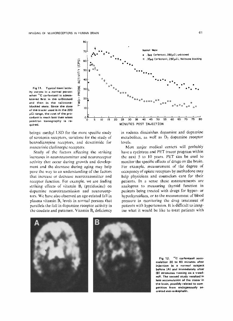
detect increased or decreased thyroidal function. Such a device does not replace PET, but permits the extension of the use of positron tracers in a way that is much less complex and expensive. A schematic representation of the dual detector device is shown in Fig 10, with a typical result shown in Fig 11. The vertical axis indicates the activity recorded by the dual detectors at the times indicated after the 1V injection of 1~C carfentanil, the upper curve in the unblocked state and the lower curve after the prior adminis- tration of naloxone.
CURRENT RESEARCH
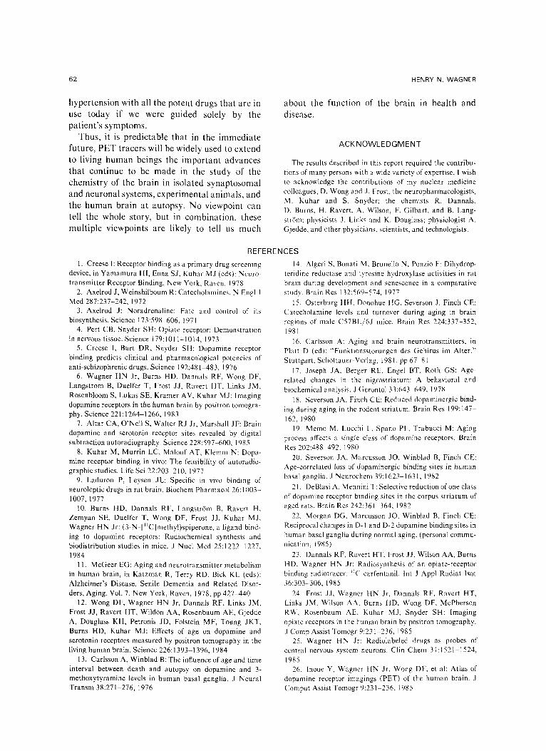
In addition to measuring the density and affin- it 3 of receptors within the living human brain, and assessing the effects of drugs such as opiates and neuroleptics, our goal is to attempt to assess the rates of release of endogenous neurotransmit- *ers. An example of such a preliminary study is assessment of the effect of strenuous exercise on a normal person. Fig 12A shows the accumula- tion of JIC carfentanil when the normal subject was injected at rest. Fig 12B shows the same subject injected with the tracer dose of 11C carfentanil immediately after exercise. The results are consistent with, but do not prove, the hypothesis that the exercise was associated with the release of endogenous enkephalin that com- peted for the carfentanil for accumulation by the **-type opiate receptors. Further studies of these types are in progress, and must include measure- ment of other effects of exercise on the brain and other metabolic sites.
Three new labeled ligands have been devel- oped for assessing other receptors in human
P h - - ~ ~ ~Cyli~dricol Bore For No I End ~ Defector Insertion View [ ] I 1
Lucite "j ~ Lucite
CoNes
Potient Stretcher
Top View
ttPb Cothmotors Positioned On Stretcher
Fig 10. A schemat ic diagram of a t w o detector positron system that is used to monitor the t ime course of a PET t racer w i th in the brain. The device costs about 1 /100 that of a PET scanner and is used when a high degree of spatial resolut ion in the measurement is not required.
IMAGING OF NEURORECEPTORS IN HUMAN BRAIN 61
Fig 11. Typical t ime/activ- ity curves in a normal person when 11C carfentanil is admin- istered f i r s t in the unblocked and then in the na loxone- blocked state. Since the dose of the tracer used is in the 200 p.Ci range, the cost of the pro- cedure is much less than when positron tomograpby is re- quired.
80
70 �9 �9
~ 6o 0
•
~ 50 ~ ~ ~ o
~ 4 0 (.9
W ~ 5O
~ ao
p--
I0
i
O0 5
~ �9 o o
o @ � 9
�9 O~o
~ 0
0
0 o
0 o
0 0 0 0
I I 1
10 15 20
Normol MGLe
�9 .Sp.g Corfentonil, 280~Ci, unblocked
o .55p.9 Corfentanil, 280p.Ci, Noloxone blocking
000 OOQQ
OOODO 000
~00 0000 O0
000o o
0 Q �9
�9 000 �9 O
O O 000 ~ o 0
ODO O0 0 o
beings: methyl LSD for the more specific study of serotonin receptors, suriclone for the study of benzodiazepine receptors, and dexetimide for muscarinic cholinergic receptors.
Study of the factors affecting the striking increases in neurotransmitter and neuroreceptor activity that occur during growth and develop- ment and the decrease during aging may help pave the way to an understanding of the factors that increase or decrease neurotransmitter and receptor function. For example, we are finding striking effects of vitamin B6 (pyridoxine) on dopamine neurotransmission and neurorecep- tots. We have also observed an age-related fall in plasma vitamin B 6 levels in normal persons that parallels the fall in dopamine receptor activity in the caudate and putamen. Vitamin B 6 deficiency
. . . . ' 5 5' 6'0 6' ' ' J 25 :50 35 40 0 5 5 70 75 80
M I N U T E S POST I N J E C T I O N
in rodents diminishes dopamine and dopamine metabolites, as well as D 2 dopamine receptor levels.
Most major medical centers will probably have a cyclotron and PET tracer program within the next 5 to 10 years. PET can be used to monitor the specific effects of drugs on the brain. For example, measurement of the degree of occupancy of opiate receptors by methadone may help physicians and counselors care for their patients. In a sense these measurements are analogous to measuring thyroid function in patients being treated with drugs for hyper- or hypothyroidism, or to the measurement of blood pressure in monitoring the drug treatment of patients with hypertension. It is difficult to imag- ine what it would be like to treat patients with
Fig 12. 11C carfentanil accu- mulation 30 to 60 minutes after injection in a normal subject before (A) and immediately after (B) strenuous running on a tread- mill. The second study resulted in less accumulation of the tracer in the brain, possibly related to com- petition from endogenously se- creted met-enkaphalin.
62 HENRY N. WAGNER
hypertension with all the potent drugs that are in use today if we were guided solely by the patient's symptoms.
Thus, it is predictable that in the immediate future, PET tracers will be widely used to extend to living human beings the important advances that continue to be made in the study of the chemistry of the brain in isolated synaptosomal and neuronal systems, experimental animals, and the human brain at autopsy. No viewpoint can tell the whole story, but in combination, these multiple viewpoints are likely to tell us much
about the function of the brain in health and disease.
ACKNOWLEDGMENT
The results described in this report required the contribu- tions of many persons with a wide variety of expertise. 1 wish to acknowledge the contributions of my nuclear medicine colleagues. D. Wong and J. Frost, the neuropharmacologists, M. Kuhar and S. Snyder; the chemists R. Dannals, D. Burns, H. Ravert, A. Wilson, F. Gilbart, and B. Lang- str~Sm: physicists J. Links and K. Douglass; physiologist A. Gjedde, and other physicians, scientists, and technologists.
REFERENCES
1. Creese I: Receptor binding as a primary drug screening device, in Yamamura HI, Enna S J, Kuhar MJ (eds): Neuro- transmitter Receptor Binding. New York, Raven, 1978
2. Axelrod J, Weinshilboum R: Catecholamines. N Engl J Med 287:237-242, 1972
3. Axelrod J: Noradrenaline: Fate and control of its biosynthesis. Science 173:598-606, 1971
4. Pert CB, Snyder SH: Opiate receptor: Demonstration in nervous tissue. Science 179:1011-1014, 1973
5. Creese I, Butt DR, Snyder SH: Dopamine receptor binding predicts clinical and pharmacological potencies of anti-schizophrenic drugs. Science 192:481-483, 1976
6. Wagner HN Jr, Burns HD, Dannals RF, Wong DF, Langstrom B, Duelfer T, Frost J J, Ravert HT, kinks JM, Rosenbloom S, Lukas SE, Kramer AV, Kuhar M J: Imaging dopamine receptors in the human brain by positron tomogra- phy. Science 221:1264-1266, 1983
7. Altar CA, O'Neil S, Walter RJ Jr, Marshall JF: Brain dopamine and serotonin receptor sites revealed by digital subtraction autoradiography. Science 228:597-600, 1985
8. Kuhar M, Murrin LC, Malouf AT, Klemm N: Dopa- mine receptor binding in vivo: The feasibility of autoradio- graphic studies. Life Sci 22:203-210, 1977
9. Laduron P, Leysen Jk: Specific in vivo binding of neuroleptic drugs in rat brain. Biochem Pharmacol 26:1003- 1007, 1977
10. Burns HD, Dannals RF, Langstr6m B, Ravert H, Zemyan SE, Duelfer T, Wong DF, Frost J J, Kuhar M J, Wagner HN Jr: (3-N-[llC]methyl)spiperone, a ligand bind- ing to dopamine receptors: Radiochemical synthesis and biodistribution studies in mice. J Nucl Med 25:1222 1227, 1984
11. McGeer EG: Aging and neurotransmitter metabolism in human brain, in Katzman R, Terry RD, Bick KL (eds): Alzheimer's Disease, Senile Dementia and Related Disor- ders, Aging, Vol. 7. New York, Raven, 1978, pp 427-440
12. Wong DF, Wagner HN Jr, Dannals RF, kinks JM, Frost J J, Ravert HT, Wildon AA, Rosenbaum AE, Gjedde A, Douglass KH, Petronis JD, Folstein MF, Toung JKT, Burns HD, Kuhar M J: Effects of age on dopamine and serotonin receptors measured by positron tomography in the living human brain. Science 226:1393 1396, 1984
13. Carlsson A, Winblad B: The influence of age and time interval between death and autopsy on dopamine and 3- methoxytyramine levels in human basal ganglia. J Neural Transm 38:271-276, 1976
14, Algeri S, Bonati M, Brunello N, Ponzio F: Dihydrop- teridine reductase and tyrosine hydroxylase activities in rat brain during development and senescence in a comparative study. Brain Res 132:569-574, 1977
15, Osterburg HH, Donohue HG, Severson J, Finch CE: Catecholamine levels and turnover during aging in brain regions of male C57BL/6J mice. Brain Res 224:337-352, 1981
16. Carlsson A: Aging and brain neurotransmitters, in Platt D (ed): "Funktionsstorungen des Gehirus im Alter." Stuttgart, Schottauer-Verlag, 1981, pp 67 81
17. Joseph JA. Berger RE, Engel BT, Roth GS: Age- related changes in the nigrostriatum: A behavioral and biochemical analysis. J Gerontol 33:643 649, 1978
18. Severson JA, Finch CE: Reduced dopaminergic bind- ing during aging in the rodent striatum. Brain Res 199:147- 162, 1980
19. Memo M, Lucchi L, Spano PF, Trabucci M: Aging process affects a single class of dopamine receptors. Brain Res 202:488 492, 1980
20. Severson JA, Marcusson JO, Winblad B, Finch CE: Age-correlated loss of dopaminergic binding sites in human basal ganglia. J Neurochem 39:1623-1631, 1982
21. DeBlasi A, Mennini T: Selective reduction of one class of dopamine receptor binding sites in the corpus striatum of aged rats. Brain Res 242:361 364, 1982
22. Morgan DG, Marcusson JO, Winblad B, Finch CE: Reciprocal changes in D-1 and D-2 dopamine binding sites in human basal ganglia during normal aging. (personal commu- nication, 1985)
23. Dannals RF, Ravert HT. Frost J J, Wilson AA, Burns HD. Wagner HN Jr: Radiosynthesis of an opiate-receptor binding radiotracer: HC-carfentanil. lnt J Appl Radiat Isot 36:303-306, 1985
24. Frost J J, Wagner HN Jr, Dannals RF, Ravert HT, Links JM, Wilson AA, Burns HD, Wong DF, McPherson RW, Rosenbaum AE, Kuhar M J, Snyder SH: Imaging opiate receptors in the human brain by positron tomography. J Comp Assist Tomogr 9:231 236, 1985
25. Wagner HN Jr: Radiolabeled drugs as probes of central nervous system neurons. Clin Chem 31:1521 1524, 1985
26. lnoue Y, Wagner HN Jr. Wong DF, et al: Atlas of dopamine receptor imagings (PET) of the human brain. J ComputAssistTomogr9:231 236,1985