Embed Size (px)
Citation preview
RESEARCH ARTICLE Open Access
Quantitative radiologic criteria for the diagnosisof lumbar spinal stenosis: a systematic literaturereviewJohann Steurer1*, Simon Roner1, Ralph Gnannt2, Juerg Hodler2 and forOn behalf of the LumbSten Research Collaboration, Zurich, Switzerland
Abstract
Background: Beside symptoms and clinical signs radiological findings are crucial in the diagnosis of lumbar spinalstenosis (LSS). We investigate which quantitative radiological signs are described in the literature and whichradilogical criteria are used to establish inclusion criteria in clincical studies evaluating different treatments inpatients with lumbar spinal stenosis.
Methods: A literature search was performed in Medline, Embase and the Cochrane library to identify papersreporting on radiological criteria to describe LSS and systematic reviews investigating the effects of differenttreatment modalities.
Results: 25 studies reporting on radiological signs of LSS and four systematic reviews related to the evaluation ofdifferent treatments were found. Ten different parameters were identified to quantify lumbar spinal stenosis. Most oftenreported measures for central stenosis were antero-posterior diameter (< 10 mm) and cross-sectional area (< 70 mm2)of spinal canal. For lateral stenosis height and depth of the lateral recess, and for foraminal stenosis the foraminaldiameter were typically used. Only four of 63 primary studies included in the systematic reviews reported onquantitative measures for defining inclusion criteria of patients in prognostic studies.
Conclusions: There is a need for consensus on well-defined, unambiguous radiological criteria to define lumbarspinal stenosis in order to improve diagnostic accuracy and to formulate reliable inclusion criteria for clinical studies.
BackgroundSpinal lumbar stenosis is the most frequent indication forspine surgery in patients older than 65 years of age [1]. Inclinical medicine lumbar spinal stenosis is defined as “but-tock or lower extremity pain, which may occur with orwithout low back pain, associated with diminished spaceavailable for the neural and vascular elements in the lum-bar spine”[2]. This definition includes two aspects: mor-phological abnormalities and clinical manifestations,neurogenic claudication, caused by the somatic anomaly.From a radiological perspective, emphasizing the
underlying structural anomaly, stenosis of the spinalcanal with or without clinical manifestations is a more
appropriate definition. The condition underlying the clin-ical manifestations, as the term implies, is a stenosis ofthe spinal canal and it is well known that not all patientswith a narrowing of the spinal canal, verified by an ima-ging procedure, suffer from neurogenic claudication[3-6].Proper research in patients with a particular illness
requires a precise definition of the illness at issue - pre-ferentially the underlying somatic anomaly - in order toformulate sensible and reliable inclusion criteria [7]. In arecent review Genevay [8] reported that researchersused quite different combinations of symptoms, clinicalsigns and radiological criteria to set up inclusion criteriafor trials in patients with lumbar spinal stenosis. Impre-cise definitions limit the interpretability and clinicalrelevance of trial results. In the case of lumbar spinalstenosis radiologic criteria are as relevant as clinical
* Correspondence: [email protected] Centre for patient oriented research and knowledge transfer,University Zurich, Raemistrasse 100, CH 8091 Zurich, SwitzerlandFull list of author information is available at the end of the article
Steurer et al. BMC Musculoskeletal Disorders 2011, 12:175http://www.biomedcentral.com/1471-2474/12/175
© 2011 Steurer et al; licensee BioMed Central Ltd. This is an Open Access article distributed under the terms of the Creative CommonsAttribution License (http://creativecommons.org/licenses/by/2.0), which permits unrestricted use, distribution, and reproduction inany medium, provided the original work is properly cited.

signs to characterize these patients. The North Ameri-can Spine Society states in their guideline that imagingis the key noninvasive test for lumbar spinal stenosis,but they provide no radiological criteria for stenosis inthese guidelines [2].In the present study radiological criteria were col-
lected that were published in the literature to describeand quantify lumbar spinal stenosis and we investigatedwhich radiological signs have been used to establishinclusion criteria for prognostic studies in patients withthis disorder.
MethodsLiterature searchA three-step literature search was performed to identifypotentially fitting studies. The search was performed byan experienced librarian (MG) with special training andskills in literature search. In a first step she searched inMedline and Embase (from 1974 to July 2010) for stu-dies potentially reporting on radiological criteria fordescribing lumbar stenosis. Search was performedincluding the following MESH terms: “spinal stenosis”,“lumbar vertebrae”, “Magnetic resonance imaging”,“Tomography X-Ray Computed” and “sensitivity andspecificity”. The search was restricted to English andGerman language. The search strategy for Medline isshown in additional file 1.In a second phase the reference lists of papers identi-
fied in the first search was scanned for further publica-tions about radiological signs of lumbar stenosis.In a third phase systematic reviews were searched in
Medline (from 1974 to July 2010) and the CochraneLibrary related to prognostic issues in patients with lum-bar spinal stenosis. Search was performed including thefollowing terms: “spinal stenosis”, “lumbar vertebrae”,“treatment”, and “systematic review”. Original studiesincluded in the systematic reviews were ordered in paperform. The rationale for evaluating original studies is theexpectation that eligibility and inclusion criteria aredescribed precisely, including precise radiological criteria,in studies fulfilling the quality criteria to be included in asystematic review.
Eligibility criteriaOnly studies reporting on preoperative imaging, usingmagnetic resonance imaging (MRI), computer tomogra-phy (CT), conventional myelography, or computertomography-myelography (CT-myelography) wereincluded if they described at least one radiological signof spinal stenosis in quantitative terms. Conventionalradiography is not an imaging modality of first choice inthe diagnosis of lumbar spinal stenosis and studies onthis method were not included in the review.
Data extractionData extraction involved reviewing the method section toidentify radiological criteria by two reviewers indepen-dently (SR and JS). Bibliographic information, detailsabout the radiological method (MRI, CT, myelography,CT myelography) site of measurement (distances, areas,angles) and cut-off values for defining stenosis wereextracted in a purpose defined form. We extracted datadescribing stenosis quantitatively.
Statistical methodsData are presented in a descriptive way. Additional sta-tistical analysis was not performed.
ResultsThe search in Medline and Embase, described as step one,yielded, after excluding duplicates, 170 publications. Afterreading title and abstract 103 papers were excluded. Forthe remaining 67 publications full text versions wereordered. After reading the publications 7 papers fulfilledthe inclusion criteria, and 60 papers were excludedbecause they did not report on any quantitative radiologi-cal sign of lumbar spinal stenosis. Based on the referencelists of the included papers 18 further papers were identi-fied that were eligible for inclusion in the structured litera-ture review (in total 25). Flowchart is shown in Figure 1.In Medline and the Cochrane Library four systematic
reviews including 63 original studies were identified[8-11]. Only 4 of the 63 original articles included patientswith lumbar spinal stenosis and reported on quantitativeradiological details in defining eligibility criteria. Threeprimary studies were included in more than one systema-tic review [12-14].In the included studies, ten various parameters were
applied with regard to lumbar stenosis. These measuresmight be distinguished into descriptors for central, lat-eral and foraminal stenosis, respectively.
Descriptors for central stenosisFor all methods, myelography, CT myelography, CT orMRI, different measurements are reported: Transverse(Figure 2) and antero-posterior (Figure 3) diameter ofthe osseous spinal canal, ligamentous interfacet distance(Figure 4) (distance between the inner surface of flavalligaments on a line connecting the joint space of thefacet joints at the level of the intervertebral disc), andcross sectional area of the spinal canal (Figure 5) [15].Distances or areas were reported either in absolutenumbers or in relative changes compared to specifiedreference measurements. Values for the antero-posteriordiameter of the osseous spinal canal were reported andstenosis is defined by some authors by a distance of lessthan 10 mm, by others below 7 mm. In the majority
Steurer et al. BMC Musculoskeletal Disorders 2011, 12:175http://www.biomedcentral.com/1471-2474/12/175
Page 2 of 9
most papers reporting a cross sectional area of the duralsac of less than 100 mm2 indicated central stenosis.Detailed quantitative information about descriptors ofcentral spinal stenosis is given in table 1.
Descriptors for lateral stenosisHeight and depth of the lateral recess, and lateralrecess angle are criteria to describe lateral stenosis.The depth of the lateral recess is measured betweenthe superior articular facet and the top part of the
Search results (n= 170)134 Medline36 Embase
Excluded based on title and abstract: 103
Papers retrieved for more detailed evaluation (n= 67)
Excluded (n= 60)No quantitative information about radiologicalcriteria of lumbar spinal stenosis (n= 60)
Included in the review (n = 25)From Medline and Embase (n = 7)From reference lists of all 67 papers retrievedfor more detailed evaluation (n = 18)
Figure 1 Flowchart of search results in Medline, Embase andbibliographies.
Figure 2 Transaxial computed tomography image of thelumbar spine at the level of L4. The white arrow indicates thetransverse diameter of the osseous spinal canal.
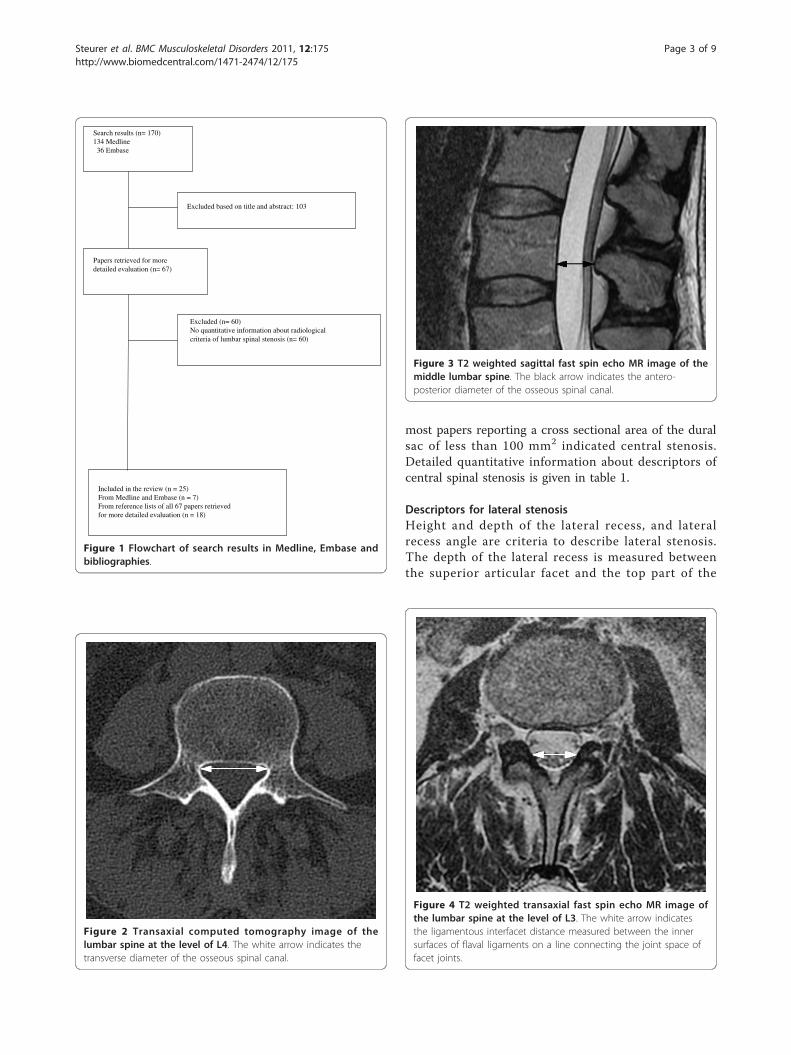
Figure 3 T2 weighted sagittal fast spin echo MR image of themiddle lumbar spine. The black arrow indicates the antero-posterior diameter of the osseous spinal canal.
Figure 4 T2 weighted transaxial fast spin echo MR image ofthe lumbar spine at the level of L3. The white arrow indicatesthe ligamentous interfacet distance measured between the innersurfaces of flaval ligaments on a line connecting the joint space offacet joints.
Steurer et al. BMC Musculoskeletal Disorders 2011, 12:175http://www.biomedcentral.com/1471-2474/12/175
Page 3 of 9

pedicle (Figure 6). Recess height is specified as distancebetween the most anterior point of the superior articu-lar facet and the posterior border of the vertebral bodyand the lateral recess angle as the angle between thelines parallel to the floor and the roof of the lateralrecess (Figure 7) [16]. A lateral recess height ≤ 2 mmand/or lateral recess depth ≤ 3 mm or a lateral recessangle < 30° has been described as diagnostic for lateralrecess stenosis. Detailed information is given in table 2.
Descriptors for foraminal stenosisThe only quantitative criterion was the diameter of theforamen. A diameter of 2 to 3 mm is considered to indi-cate stenosis [17].
Descriptors applied in primary prognostic studies,including surgery and drug treatmentOnly in four of 20 primary studies [14,18-20] detailedradiological information for spinal stenosis was pro-vided. Applied quantitative criteria for inclusion ofpatients in clinical studies were: diameter of spinal canal[14,19-21] and cross-sectional area of the dural tube[18,21]. Criteria for description applied in studies
Figure 5 T2 weighted transaxial fast spin echo MR image ofthe lumbar spine at the level of L1. Cross sectional area of thespinal canal is indicated by the white hatched area.
Table 1 Sites of measurement, measurement points and radiologic definitions for central lumbar spinal stenosis
Imagingmethod
Author Site ofmeasurement
Level, where measured (measurement points) Definition of stenosis (cut-offvalues)
MRI
Antero-posteriordiameter of spinalcanal
Fukusaki[22]
Not reported < 15 mm
Koc [23] Not reported < 12 mm
Mid-sagittaldiameter of thecalsac
Herzog [15] Midbody of each vertebra Compression of thecal sac area in % ofnormal mid-sagittal diameter:Grade 1: anterior < 15%posterior < 10%Grade 2: anterior 15 - 30%posterior 10 - 20%Grade 3: anterior > 30%posterior > 20%
Cross-sectionalarea of dural tubeor sac
Hamanishi[24]
Intervertebral levels: L2/3, L3/4, L4/5 < 100 mm2, at more than two of threeintervertebral levels
Mariconda[25]
Not reported < 130 mm2
Laurencin[26]
Motion segment: Intervertebral disc level coincident withflexible joint;Stable segment:Level coincident with the mid-pedicle unaffected bystenosis
Stenosis ratio:Cross-sectional area of dural sac ofmotion segment divided by stablesegment cross-sectional dural sac area:Level: L3-L4 < 0.66L4-L5 < 0.62L5-S1 < 0.73
Steurer et al. BMC Musculoskeletal Disorders 2011, 12:175http://www.biomedcentral.com/1471-2474/12/175
Page 4 of 9

Table 1 Sites of measurement, measurement points and radiologic definitions for central lumbar spinal stenosis(Continued)
Ligamentousinterfacet distance
Herzog [15] Distance between the innner surface of flaval ligaments ona line connecting the joint space of facet joints at thelevel of the intervertebral disc
< 10 mm (L2 - L3)< 10 mm (L3 - L4)< 12 mm (L4 - L5)< 13 mm (L5 - S1)
Transversediameter of spinalcanal
Koc [23] Not reported < 15 mm
Ullrich [27] 4 zones of measurement:upper, middle, lower zone of vertebral body and diskspace
< 16 mm
CT
Antero -posteriordiameter of spinalcanal
Bolender[28]
5 mm intervals from L2 to L5 < 13 mm
Haig [4] Not reported ≤ 11.95 mm
Lee [29] Not reported < 15 mm (suggesting narrowing)< 10 mm (usually diagnostic)
Ullrich [27] Four zones of measurement:Upper, middle, lower zone of vertebral body and diskspace
< 11.5 mm
Verbiest [30] Not reported < 12 mm (relative)< 10 mm (absolute)
Antero-posteriordiameter of duralsac
Kalichman[3]
Midvertebral body level 10 - 12 mm (relative)< 10 mm (absolute)
Herzog [15] Midbody of each vertebra Compression of thecal sac area in % ofnormal mid-saggital diameter:Grade 1: anterior < 15%posterior < 10%Grade 2: anterior 15 - 30%posterior 10 - 20%Grade 3: anterior > 30%posterior > 20%
Jönsson [31] Disc level ≤ 10 mm
Cross-sectionalarea of dural sac
Bolender[28]
5 mm intervals from L2 to L5 100 - 130 mm2 (early stenosis)< 100 mm2 (present stenosis)
Laurencin[26]
Motion segment: Intervertebral disc level coincident withflexible jointstable segment:Level coincident with the mid-pedicle unaffected bystenosis
Stenosis ratio:Area of motion segment divided bystable segment areaLevel: L3-L4 < 0.66L4-L5 < 0.62L5-S1 < 0.73
Schönström[32]
On each CT scan slice < 100 mm2
Schönström[33]
Not reported 75 - 100 mm2 (moderate)< 75 mm2 (severe)
Ullrich [27] 4 zones of measurement:Upper, middle, lower zone of vertebral body and diskspace
< 145 mm2
Steurer et al. BMC Musculoskeletal Disorders 2011, 12:175http://www.biomedcentral.com/1471-2474/12/175
Page 5 of 9

Table 1 Sites of measurement, measurement points and radiologic definitions for central lumbar spinal stenosis(Continued)
Ligamentousinterfacet distance
Herzog [15] Intervertebral disc level < 10 mm (L2-L3)< 10 mm (L3-L4)< 12 mm (L4-L5)< 13 mm (L5-S1)
Wilmink [34] Pedicular, infrapedicular and/or disc level < 11 mm (L4-L5)
Myelography
conventional Antero-posteriordiameter ofcontrast column
Airaksinen[35]
Narrowest point > 12 mm10 - 12 mm< 10 mmSubtotal blockTotal block
Bolender[28]
Intervertebral level < 13 mm
Herno [36] Not reported < 12 mm
Jönsson [31] Disc level ≤ 10 mm
Sortland [37] Disc level < 10.5 mm (lower limit)< 5.5 - 7 mm (considerable)
Verbiest [38] Superior and inferior boarders of the laminae 10 - 12 mm (relative)< 10 mm (absolute)
Myelo-CT Mariconda[25]
Not reported < 130 mm2
Figure 6 T2 weighted transaxial fast spin echo MR image ofthe lumbar spine at the level of L5. The depth of the lateralrecess is measured between the superior articular facet and the toppart of the pedicle marked with the black arrow.
Figure 7 Transaxial computed tomography image of the lumbarspine at the level of L3. Left side: The lateral recess angle is definedas the angle between the lines parallel to the floor and the roof of thelateral recess. Right side: The height of the lateral recess is defined asthe shortest distance from the most anterior point of the superiorarticular process to the posterior border of the vertebral body.
Steurer et al. BMC Musculoskeletal Disorders 2011, 12:175http://www.biomedcentral.com/1471-2474/12/175
Page 6 of 9

investigating patients with spinal stenosis are given inadditional file 2.
DiscussionThe result of this literature review documents a remark-able list of various quantitative radiologic criteria appliedto describe lumbar spinal stenosis. Measurement ofantero-posterior diameter and the cross sectional area ofspinal canal with varying cut-off levels are the most oftenapplied criteria for central stenosis; height and length ofthe recess for lateral stenosis and foraminal diameter forforaminal stenosis. Only in a minority of primary prognos-tic studies, included in systematic reviews evaluating dif-ferent treatment modalities, distinct and reliable criteriawere used to set up eligibility criteria for patients includedin these studies.To our knowledge no structured and systematic review
collecting radiological criteria applied for defining lumbarspinal stenosis has been published to date. A structuredreview focusing on clinical eligibility criteria was recentlypublished showing a high degree of variability in inclusioncriteria between studies [8]. The finding of our study,focusing on radiological criteria, strengthens the conclu-sion from Genevay [8] that there is a need for a consensuson criteria to define and classify lumbar spinal stenosis.A vague definition of an illness and imprecise criteria to
either rule-in or rule-out an illness, as a consequence of
that, poses a major problem on performing research inpatients with such a disorder. It limits the accuratedescription of patients enrolled in a study and thereforeconfines the interpretation and applicability of theobtained results in medical practice. The primary goal ofclinical studies is to gain results needed to inform physi-cians about the intended and adverse effects of differenttreatment modalities. Our findings indicate shortcomingsin defining and classifying lumbar spinal stenosis resultingin imprecise and variable definitions of inclusion criteriain studies evaluating the natural course of the illness orthe effect of different treatment modalities. This lack ofdistinct criteria impairs the interpretation of the studyresults and the comparability of the findings between dif-ferent studies.Two questions are of particular importance concerning
diagnosis in patients with clinically suspected lumbarspinal stenosis: First, can a lumbar spinal stenosis be veri-fied by radiological measures? Second, if stenosis is verifiedby an imaging procedure, are the symptoms and clinicalsigns caused by the identified somatic anomaly? There is aneed to formulate a code of practice, based on sharedexpert’s belief, to set measurement points in the lumbarspine to describe and quantify structural anomalies and ina further step to establish norm- and cut-off values. Theaim of further studies might be the development of validmethods to assess the relationship between structural
Table 2 Sites of measurement, measurement points and radiologic definitions for lateral lumbar spinal stenosis
Imagingmethod
Author Site ofmeasurement
Level, where measured Definition ofstenosis (cut-offvalues)
CT Lateral recessheight
Ciric[39]
Not reported 5 mm (normal)≤ 3 mm (highlyindicative)≤ 2 mm (diagnostic)
Strojnik[16]
between the most medial point of the superior articular facet and the posterior pointof the vertebral body
≤ 3.6 mm
Depth oflateral recess
Dincer[40]
Between superior articular facet and the top part of the pedicle. > 5 mm (normal)4 - 5 mm (Group 3)3 - 4 mm (Group 2)2 - 3 mm (Group 1)
Mikhael[41]
Between the posterior surface of the vertebral body and the anteromedial portion ofthe superior articular facet at the level of the superior border of the correspondinglevel
> 5 mm (normal)3 - 5 mm(suggestive)≤ 3 mm (definitive)
Lateral recessangle
Strojnik[16]
Between the bottom and the roof of the triangular space (= lateral recess) < 30°
Steurer et al. BMC Musculoskeletal Disorders 2011, 12:175http://www.biomedcentral.com/1471-2474/12/175
Page 7 of 9

anomalies, symptoms and clinical signs. A difficulty toassess the association of clinical manifestations and anato-mical anomalies is the fact that there is no simple refer-ence test for lumbar spinal stenosis. The most obviousreference standard, at least in patients undergoing surgery,could be improvement after surgery. However, surgery atthe lumbar spine might have an inadvertent beneficialeffect on other mechanical pain generators. Innovativemethods have to be developed to overcome theseproblems.Although a thorough search in Medline, Embase and
the Cochrane Library was performed papers reportingon descriptors of spinal stenosis different from thoseincluded in the list we present may have been missed.However, in the primary studies, included in the sys-tematic reviews no additional descriptors not alreadyfound in the first search were identified. This indicatesthat relevant radiologic descriptors of lumbar spinal ste-nosis have most probably not been missed. A shortcom-ing of this review is the sole focus on quantitativeparameters. Beside them qualitative parameters, e.g.,disc protrusion or hypertrophic facet joint degeneration,are used to describe abnormalities in the lumbar spine.Lumbar spinal stenosis is a common disorder and the
most frequent indication for lumbar spine surgery in theelderly. Due to the demographic changes the number ofpatients with this disorder will increase. There is a need fora consensus among experts on well defined, unambiguousradiological and clinical criteria to define lumbar spinal ste-nosis. The criteria, reported in this paper, can be used as asource for the development of radiological criteria.
ConclusionsThere is a need for consensus on well-defined, unambig-uous radiological criteria to define and characterize lum-bar spinal stenosis in order to improve diagnosticaccuracy and to formulate reliable inclusion criteria forclinical studies.
Additional material
Additional file 1: Search strategy in Medline. Search terms,combination of the terms and number of identified publications arereported.
Additional file 2: Radiologic descriptors applied in the originalstudies. Radiologic descriptors form the the 20 primary studiescomparing various treatment modalities in patients with lumbar spinalstenosis included in four systematic reviews.
Acknowledgements and FundingThe authors thank Dr. sc. nat. ETH Martina Gosteli for performing theliterature search and PD Dr. Gustav Andreisek for helpful comments inpreparing the manuscript. There was no funding for the preparation of thismanuscript.
Author details1Horten Centre for patient oriented research and knowledge transfer,University Zurich, Raemistrasse 100, CH 8091 Zurich, Switzerland. 2Diagnosticand Interventional Radiology, University Hospital Zurich, Raemistrasse 100,CH 8091 Zurich, Switzerland.
Authors’ contributionsJS has been involved in the conception, acquisition and interpretation ofdata and was involved in writing the manuscript, SR has been in invovledacquisition and interpretation of data and was involved in writing themanuscript, RG made substantial contributions in the interpretation of dataand was involved in writing the manuscript, and JH has been involved inthe conception, acquisition and interpretation of data and was involved inwriting the manuscript. All authors have given final approval of this versionof the manuscript.
Competing interestsThe authors declare that they have no competing interests.
Received: 7 March 2011 Accepted: 28 July 2011 Published: 28 July 2011
References1. Deyo RA: Treatment of lumbar spinal stenosis: a balancing act. Spine J
2010, 10(7):625-627.2. North American Spine Society: Evidence Based Clinical Guidelines for
Multidisciplinary Spine Care: Diagnosis and Treatment of DegenerativeLumbar Spinal Stenosis. In. Burr Ridge, IL.: North American Spine Society;2007.
3. Kalichman L, Cole R, Kim D, Li L, Suri P, Guermazi A, Hunter D: Spinalstenosis prevalence and association with symptoms: the FraminghamStudy. Spine J 2009, 9(7):545-550.
4. Haig AJ, Geisser ME, Tong HC, Yamakawa KS, Quint DJ, Hoff JT, Chiodo A,Miner JA, Phalke VV: Electromyographic and magnetic resonance imagingto predict lumbar stenosis, low-back pain, and no back symptoms.J Bone Joint Surg Am 2007, 89(2):358-366.
5. Boden S, Davis D, Dina T, Patronas N, Wiesel S: Abnormal magnetic-resonance scans of the lumbar spine in asymptomatic subjects. Aprospective investigation. J Bone Joint Surg Am 1990, 72(3):403-408.
6. Wiesel S, Tsourmas N, Feffer H, Citrin C, Patronas N: A study of computer-assisted tomography. I. The incidence of positive CAT scans in anasymptomatic group of patients. Spine (Phila Pa 1976) 1984, 9(6):549-551.
7. Miettinen OS, Flegel KM: Elementary concepts of medicine: III. Illness:somatic anomaly with. J Eval Clin Pract 2003, 9(3):315-317.
8. Genevay S, Atlas SJ, Katz JN: Variation in eligibility criteria from studies ofradiculopathy due to a herniated disc and of neurogenic claudicationdue to lumbar spinal stenosis: a structured literature review. Spine (PhilaPa 1976) 2010, 35(7):803-811.
9. Aalto T, Malmivaara A, Kovacs F, Herno A, Alen M, Salmi L, Kröger H,Andrade J, Jiménez R, Tapaninaho A, Turunen V, Savolainen S, Airaksinen O:Preoperative predictors for postoperative clinical outcome in lumbarspinal stenosis: systematic review. Spine (Phila Pa 1976) 2006, 31(18):E648-663.
10. Gibson JN, Waddell G: Surgery for degenerative lumbar spondylosis:updated Cochrane Review. Spine (Phila Pa 1976) 2005, 30(20):2312-2320.
11. Coronado-Zarco R, Cruz-Medina E, Arellano-Hernandez A, Chavez-Arias D,Leon-Hernandez SR: Effectiveness of calcitonin in intermittentclaudication treatment of patients with lumbar spinal stenosis: asystematic review. Spine (Phila Pa 1976) 2009, 34:(22):E818-822.
12. Amundsen T, Weber H, Nordal H, Magnaes B, Abdelnoor M, Lilleâs F:Lumbar spinal stenosis: conservative or surgical management?: Aprospective 10-year study. Spine (Phila Pa 1976) 2000, 25(11):1424-1435.
13. Herkowitz H, Kurz L: Degenerative lumbar spondylolisthesis with spinalstenosis. A prospective study comparing decompression withdecompression and intertransverse process arthrodesis. J Bone Joint SurgAm 1991, 73(6):802-808.
14. Tafazal S, Ng L, Sell P: Randomised placebo-controlled trial on theeffectiveness of nasal salmon calcitonin in the treatment of lumbarspinal stenosis. Eur Spine J 2007, 16(2):207-212.
15. Herzog RJ, Kaiser JA, Saal JA, Saal JS: The importance of posterior epiduralfat pad in lumbar central canal stenosis. Spine (Phila Pa 1976) 1991, 16(6Suppl):S227-233.
Steurer et al. BMC Musculoskeletal Disorders 2011, 12:175http://www.biomedcentral.com/1471-2474/12/175
Page 8 of 9

16. Strojnik T: Measurement of the lateral recess angle as a possiblealternative for evaluation of the lateral recess stenosis on a CT scan.Wien Klin Wochenschr 2001, 113(Suppl 3):53-58.
17. Beers GJ, Carter AP, Leiter BE, Tilak SP, Shah RR: Interobserverdiscrepancies in distance measurements from lumbar spine CT scans.AJR Am J Roentgenol 1985, 144(2):395-398.
18. Yukawa Y, Lenke L, Tenhula J, Bridwell K, Riew K, Blanke K: Acomprehensive study of patients with surgically treated lumbar spinalstenosis with neurogenic claudication. J Bone Joint Surg Am 2002, 84-A(11):1954-1959.
19. Eskola A, Pohjolainen T, Alaranta H, Soini J, Tallroth K, Slätis P: Calcitonintreatment in lumbar spinal stenosis: a randomized, placebo-controlled,double-blind, cross-over study with one-year follow-up. Calcif Tissue Int1992, 50(5):400-403.
20. Grob D, Humke T, Dvorak J: Degenerative lumbar spinal stenosis.Decompression with and without arthrodesis. J Bone Joint Surg Am 1995,77(7):1036-1041.
21. Malmivaara A, Slätis P, Heliövaara M, Sainio P, Kinnunen H, Kankare J, Dalin-Hirvonen N, Seitsalo S, Herno A, Kortekangas P, Niinimäki T, Rönty H,Tallroth K, Turunen V, Knekt P, Härkänen T, Hurri H, Finnish Lumbar SpinalResearch Group: Surgical or nonoperative treatment for lumbar spinalstenosis? A randomized controlled trial. Spine (Phila Pa 1976) 2007,32(1):1-8.
22. Fukusaki M, Kobayashi I, Hara T, Sumikawa K: Symptoms of spinal stenosisdo not improve after epidural steroid injection. Clin J Pain 1998,14(2):148-151.
23. Koc Z, Ozcakir S, Sivrioglu K, Gurbet A, Kucukoglu S: Effectiveness ofphysical therapy and epidural steroid injections in lumbar spinalstenosis. Spine (Phila Pa 1976) 2009, 34(10):985-989.
24. Hamanishi C, Matukura N, Fujita M, Tomihara M, Tanaka S: Cross-sectionalarea of the stenotic lumbar dural tube measured from the transverseviews of magnetic resonance imaging. J Spinal Disord 1994, 7(5):388-393.
25. Mariconda M, Fava R, Gatto A, Longo C, Milano C: Unilateral laminectomyfor bilateral decompression of lumbar spinal stenosis: a prospectivecomparative study with conservatively treated patients. J Spinal DisordTech 2002, 15(1):39-46.
26. Laurencin C, Lipson S, Senatus P, Botchwey E, Jones T, Koris M, Hunter J:The stenosis ratio: a new tool for the diagnosis of degenerative spinalstenosis. Int J Surg Investig 1999, 1(2):127-131.
27. Ullrich C, Binet E, Sanecki M, Kieffer S: Quantitative assessment of thelumbar spinal canal by computed tomography. Radiology 1980,134(1):137-143.
28. Bolender N, Schönström N, Spengler D: Role of computed tomographyand myelography in the diagnosis of central spinal stenosis. J Bone JointSurg Am 1985, 67(2):240-246.
29. Lee B, Kazam E, Newman A: Computed tomography of the spine andspinal cord. Radiology 1978, 128(1):95-102.
30. Verbiest H: The significance and principles of computerized axialtomography in idiopathic developmental stenosis of the bony lumbarvertebral canal. Spine (Phila Pa 1976) 1979, 4(4):369-378.
31. Jönsson B, Annertz M, Sjöberg C, Strömqvist B: A prospective andconsecutive study of surgically treated lumbar spinal stenosis. Part I:Clinical features related to radiographic findings. Spine (Phila Pa 1976)1997, 22(24):2932-2937.
32. Schonstrom N, Bolender N, Spengler D: The pathomorphology of spinalstenosis as seen on CT scans of the lumbar spine. Spine (Phila Pa 1976)1985, 10(9):806-811.
33. Schonstrom N, Willen J: Imaging lumbar spinal stenosis. Radiol Clin NorthAm 2001, 39(1):31-53.
34. Wilmink J, Korte J, Penning L: Dimensions of the spinal canal inindividuals symptomatic and non-symptomatic for sciatica: a CT study.Neuroradiology 1988, 30(6):547-550.
35. Airaksinen O, Herno A, Turunen V, Saari T, Suomlainen O: Surgical outcomeof 438 patients treated surgically for lumbar spinal stenosis. Spine (PhilaPa 1976) 1997, 22(19):2278-2282.
36. Herno A, Airaksinen O, Saari T: Computed tomography after laminectomyfor lumbar spinal stenosis. Patients’ pain patterns, walking capacity, andsubjective disability had no correlation with computed tomographyfindings. Spine (Phila Pa 1976) 1994, 19(17):1975-1978.
37. Sortland O, Magnaes B, Hauge T: Functional myelography withmetrizamide in the diagnosis of lumbar spinal stenosis. Acta Radiol Suppl1977, 355:42-54.
38. Verbiest H: Chapter 16. Neurogenic intermittent claudication in caseswith absolute and relative stenosis of the lumbar vertebral canal (ASLCand RSLC), in cases with narrow lumbar intervertebral foramina, and incases with both entities. Clin Neurosurg 1973, 20:204-214.
39. Ciric I, Mikhael MA, Tarkington JA, Vick NA: The lateral recess syndrome. Avariant of spinal stenosis. J Neurosurg 1980, 53(4):433-443.
40. Dincer F, Erzen C, Basgöze O, Özker R, Celiker R: Lateral recess syndromeand computed tomography. Turkish Neurosurgery 1991, 2:30-35.
41. Mikhael M, Ciric I, Tarkington J, Vick N: Neuroradiological evaluation oflateral recess syndrome. Radiology 1981, 140(1):97-107.
Pre-publication historyThe pre-publication history for this paper can be accessed here:http://www.biomedcentral.com/1471-2474/12/175/prepub
doi:10.1186/1471-2474-12-175Cite this article as: Steurer et al.: Quantitative radiologic criteria for thediagnosis of lumbar spinal stenosis: a systematic literature review. BMCMusculoskeletal Disorders 2011 12:175.
Submit your next manuscript to BioMed Centraland take full advantage of:
• Convenient online submission
• Thorough peer review
• No space constraints or color figure charges
• Immediate publication on acceptance
• Inclusion in PubMed, CAS, Scopus and Google Scholar
• Research which is freely available for redistribution
Submit your manuscript at www.biomedcentral.com/submit
Steurer et al. BMC Musculoskeletal Disorders 2011, 12:175http://www.biomedcentral.com/1471-2474/12/175
Page 9 of 9