Embed Size (px)
Citation preview
Quantitative Textures of Calcitic Prismatic Layers in Trichites and
Bivalve Molluscs D. Chateigner, M. Morales, A. Jouanneaux Lab. de Physique de l’Etat Condensé, Le Mans France F. Denis, M. Laulier Lab. de Biologie et Génétique Evolutive, Le Mans France L. Harper Dept. of Earth Sciences, Cambridge U.K. [email protected]
Context Fragments of the large bivalve Trichites are relatively abundant
in shallow marine sediments from the Middle to Upper Jurassic of Europe, Asia and Africa, however entire individuals are rare and the palaeobiology of the genus is poorly understood because of this. The specimens which are found show that the shell was thick, with some fragments measuring up to 3 cm in thickness and are composed of a coarse simple prismatic calcite. It is also clear from particularly well preserved specimens that there was an inner aragonite shell layer, at least in dorsal regions, which was probably made up of nacre and which has typically been lost by diagenetic dissolution. Loss of these aragonite shell layers has made interpretation of the musculature and the hinge structures difficult. Since these are characters which are important in establishing the phylogenetic relationships of bivalves, the taxonomic position of Trichites has remained problematic. [email protected]
Traditionally, Trichites is placed within the Pinnoidea, a pteriomorph group which normally have regular ham-shaped shells and live partially embedded in soft sediments, anchored by byssal threads. Pinnoids do have flexible shells comprised of thin layers of calcite prisms, lined internally by nacre. Trichites appears to adopt a rather different life habit, lying on one side on top of the sediment and is frequently highly distorted and display torsion, therefore implying that if it is indeed a pinnoid it is a rather aberrant form.
Further examination of the crystallographic textures of Trichites and comparison of those from Recent pinnoids and also other pteriomorphians may provide a better means of determining their relationships.
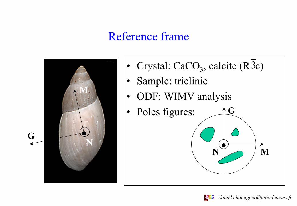
• Crystal: CaCO3, calcite (R c) • Sample: triclinic • ODF: WIMV analysis • Poles figures:
Reference frame
. N
M
G . N M
G
3
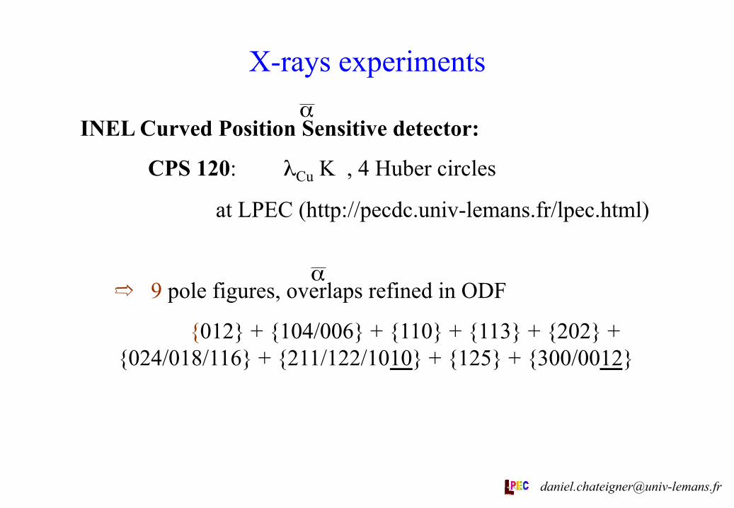
X-rays experiments
INEL Curved Position Sensitive detector:
CPS 120: λCu K , 4 Huber circles
at LPEC (http://pecdc.univ-lemans.fr/lpec.html)
➱ 9 pole figures, overlaps refined in ODF
{012} + {104/006} + {110} + {113} + {202} + {024/018/116} + {211/122/1010} + {125} + {300/0012}
α
α
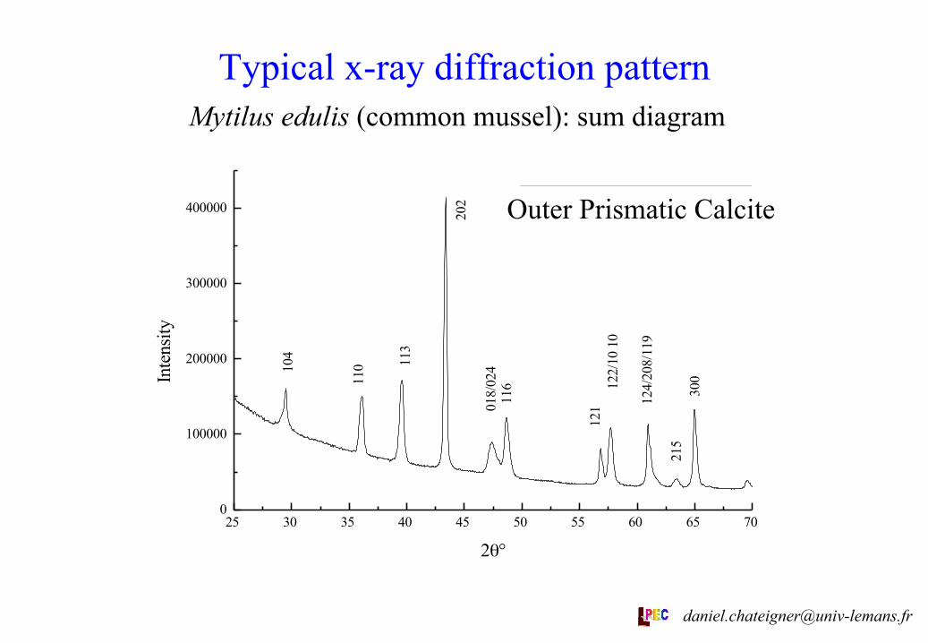
Typical x-ray diffraction pattern Mytilus edulis (common mussel): sum diagram
25 30 35 40 45 50 55 60 65 700
100000
200000
300000
400000
300
215
124/
208/
119
122/
10 1
012
1
116
018/
024
202
113
11010
4
outer foliated calcite
Inte
nsity
2θ°
Outer Prismatic Calcite
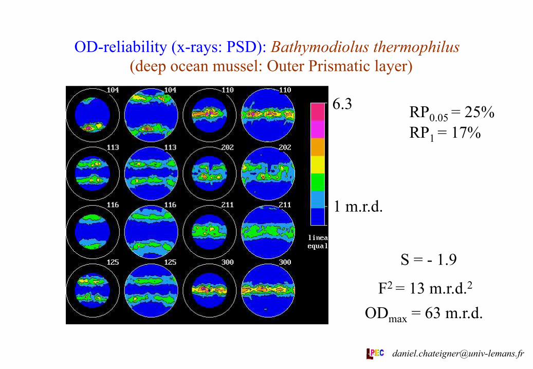
OD-reliability (x-rays: PSD): Bathymodiolus thermophilus (deep ocean mussel: Outer Prismatic layer)
1 m.r.d.
6.3 RP0.05 = 25% RP1 = 17%
S = - 1.9
F2 = 13 m.r.d.2 ODmax = 63 m.r.d.
Lin. scale
Eq. area proj.
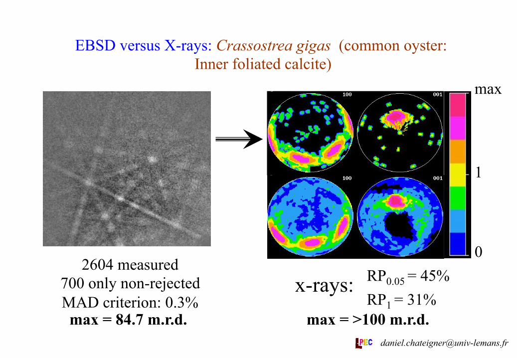
EBSD versus X-rays: Crassostrea gigas (common oyster: Inner foliated calcite)
2604 measured 700 only non-rejected MAD criterion: 0.3%
RP0.05 = 45% RP1 = 31%
x-rays:
max
1
0
max = 84.7 m.r.d. max = >100 m.r.d. [email protected]
Microstructure versus texture
In Bathymodiolus thermophilus, the prismatic calcite layer exhibits a microstructure (as seen with SEM) which does not show a simple relationship between individual crystals and their crystal lattice (revealed by texture experiments): Texture analysis shows c-axes parallel to G, which is not the long axis of the calcite prisms in the SEM image.
There is no apparent relationship between the nacre (aragonite) crystals and the calcite prisms.
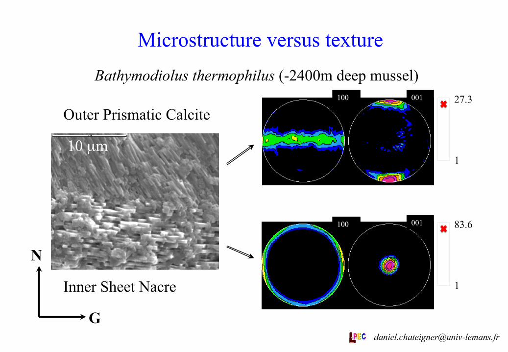
Microstructure versus texture Bathymodiolus thermophilus (-2400m deep mussel)
10 µm
83.6
1
27.3
1
N
G
100 001
100 001
Outer Prismatic Calcite
Inner Sheet Nacre
The x-ray texture analyses show relatively good reliabilities in their ODFs (see figures and RP factors). All the textures show high strengths, as in aragonite layers [1], with ODF minima of 0 for all examined layers. Comparison between EBSD and x-ray measurements on Crassostrea gigas show that EBSD « forgets » a lot of crystallites, and consequently introduces errors. The inner foliated calcite examined has a much higher texture strength than all the measured prismatic layers. Pinna nobilis and Pteria penguin prismatic calcite layers exhibit close textures, with c-axes oriented parallel to N and a-axes at random around c, only different by their dispersions (differences in texture strengths).
Texture Analysis results
Trichites shows inclined c-axes, though only 15° of deviation from N, and a much higher texture strength as P. nobilis and P. penguin. Both examined mussels, Mytilus edulis and Bathymodiolus thermophilus, show very inclined c-axes, with a single-crystal-like distribution of the a-axes. This is also the texture characteristic of Amussium parpiraceum. These three species have texture strengths lower than P. nobilis, P. penguin, Trichites and foliated calcite, and each of them a distinct textural signal, considering their c- and a-axis distributions. Also, the crystal prisms as seen using SEM look elongated along an axis which does not correspond to any of the a, b or c axes of calcite, as prove by the texture analyses. This is an example of the non-trivial relationship existing between SEM and texture investigations, already demonstrated for aragonite [2,3], and proving the non-redundancy of the two approaches that may be used in phylogenetic discussions. [email protected]
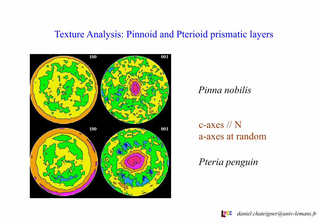
Texture Analysis: Pinnoid and Pterioid prismatic layers
Pinna nobilis
Pteria penguin
c-axes // N a-axes at random
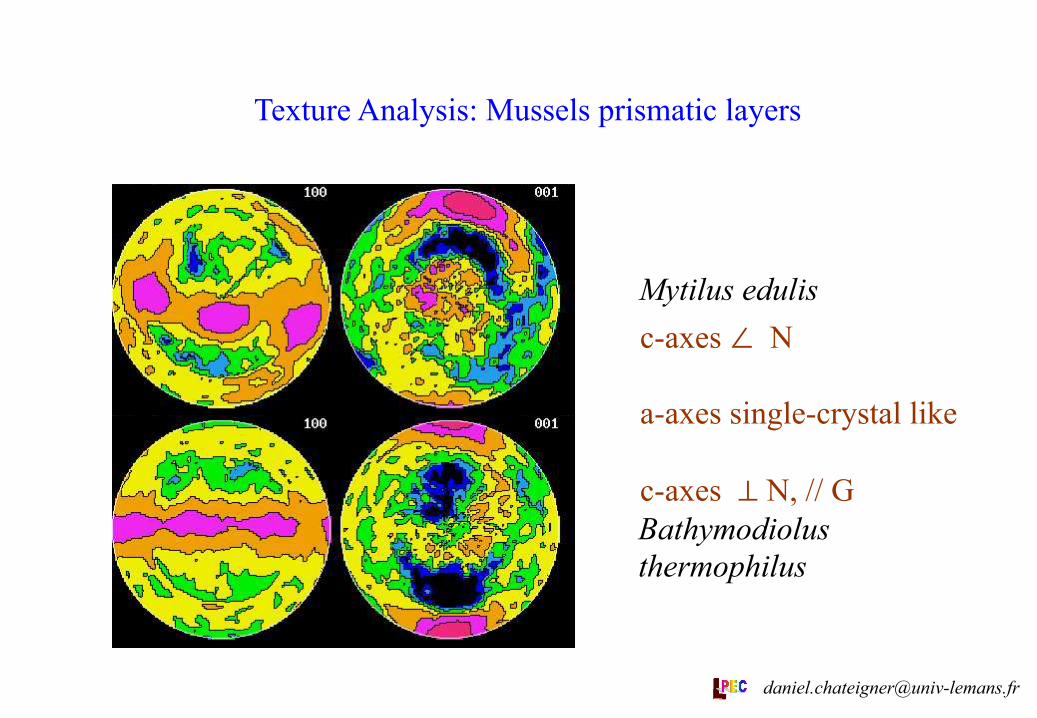
Texture Analysis: Mussels prismatic layers
Mytilus edulis
Bathymodiolus thermophilus
c-axes ∠ N a-axes single-crystal like c-axes ⊥ N, // G
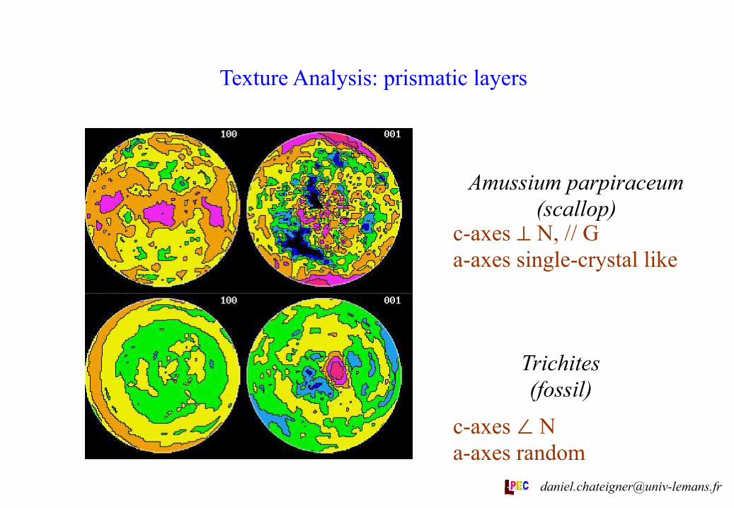
Texture Analysis: prismatic layers
c-axes ⊥ N, // G a-axes single-crystal like
c-axes ∠ N a-axes random
Amussium parpiraceum (scallop)
Trichites (fossil)
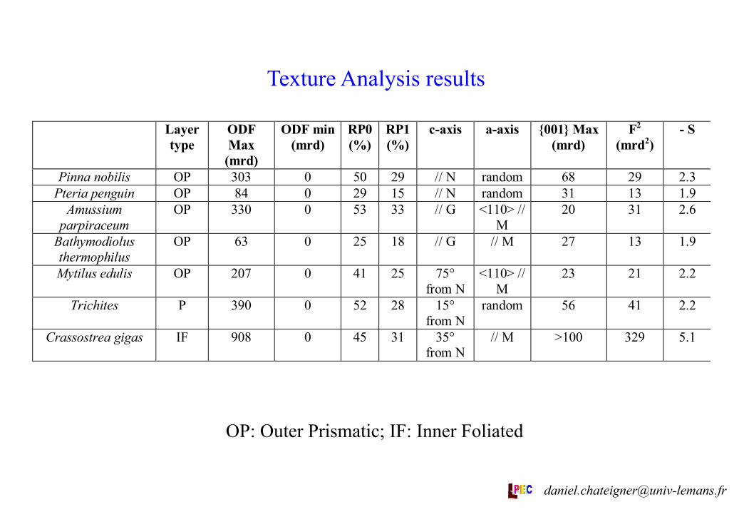
Texture Analysis results
OP: Outer Prismatic; IF: Inner Foliated
Layertype
ODFMax
(mrd)
ODF min(mrd)
RP0(%)
RP1(%)
c-axis a-axis {001} Max(mrd)
F2
(mrd2)- S
Pinna nobilis OP 303 0 50 29 // N random 68 29 2.3Pteria penguin OP 84 0 29 15 // N random 31 13 1.9
Amussiumparpiraceum
OP 330 0 53 33 // G <110> //M
20 31 2.6
Bathymodiolusthermophilus
OP 63 0 25 18 // G // M 27 13 1.9
Mytilus edulis OP 207 0 41 25 75°from N
<110> //M
23 21 2.2
Trichites P 390 0 52 28 15°from N
random 56 41 2.2
Crassostrea gigas IF 908 0 45 31 35°from N
// M >100 329 5.1
Discussion There is good evidence to suggest that the earliest bivalves were
all entirely aragonitic and of rather simple construction from two microstructural types. However, later bivalves evolved a further 5 microstructural types (2 of them calcitic) and utilised these in a variety of different arrangements. The result is that the bivalve microstructure and mineralogy is extremely diverse across the class and is, therefore, thought to be a valuable character for phylogenetic analysis. Two major types of calcite microstructure are identified: prisms, which usually occur on the outside of the valve, and foliae which tends to occupy the bulk of a shell (e.g. oysters: C. gigas). Of these calcite prisms are perhaps the most interesting because there is good evidence that calcitic prisms have evolved at least four times independently within the Bivalvia, in the mussels, pterioids, chamids and the extinct rudists.
The data presented here is from the calcitic prismatic layer of bivalves from members of the sub-class Pteriomorphia. This broad group encompasses two of the calcite secreting clades, the mussels and the pterioids. It is therefore, of great value to compare the form of the calcite crystals within members of these taxa. B. themophilus and M. edulis are both mussels, the former being an important component of deep sea vent faunas. Although both have calcitic outer shell layers it is clear from texture analysis (see Table) that the two are separate innovations of the calcitic shell layer, with different alignment schemes of their a-axes.
The pterioids are represented by P. nobilis, P. penguin., Trichites sp., and A. papiraceum. Although these ostensibly all belong to the same clade of calcite secreters it is important to establish the degree of similarity or difference between the various taxa.
In particular, recent molecular schemes have placed the pteriids (i.e. P. penguin) as a sister-group to the pinnoids (Pinna and possibly Trichites). All three species exhibit similar textures, but Trichites, if we except its slightly inclined c-axis distribution, shows an ODF maximum closer to the pinnoids than to the pteriids.
A. parpiraceum belongs to the superfamily Pectinoidea which, according to some phylogenetic schemes at least, is further removed from the pteriids and pinnoids, than are the mussels. If this is so, then calcite secretion in the pterioid group may be polyphyletic and thus we might expect the Amussium to display different textures to both the Pinna + Pteria and the mussels. This is actually what we have measured as textures in Amussium, which presents a clearly not random alignment of its a-axes (i.e. is different of Trichites, Ptrids and Pinnoids), a c-axis distribution in the plane of the shell (not like M. edulis) and <110> directions aligned with M (where B. thermophilus has its a-axes). [email protected]
Conclusion
• The calcite layers of the taxa analysed here show different texture patterns that allow us to put some light on their place in the phylogeny. • The SEM-microstructure and texture characterisations provide non-redundant information. • The six analysed taxa show six different texture patterns of prismatic calcite layers, which are also distinguishable from the one of the foliated calcite. • These texture patterns present common characteristics, as in the case of aragonitic layers of molluscs [1,2,3]. • The foliated calcite layer appears to be much strongly textured than all the prismatics. • No apparent crystalline epitaxial-like relationship is detected between the aragonitic inner and the calcitic outer layers of B. thermophilus.
References [1]: Texture analysis of a gastropod shell, Cypraea testudinaria: D. Chateigner, C. Hedegaard & H.-R. Wenk, "Textures of Materials” Ed. Z. Liang, L. Zuo & Y. Chu, Int. Academic Publishers, vol. 2, pp 1221-1226, (1996) [2]: Quantitative characterisation of mollusc shell textures: D. Chateigner, C. Hedegaard, H.-R. Wenk, "Textures of Materials" (Ed. J.A. Szpunar), NRC Research Press, vol. 2, pp 1495-1500 (1999) [3]: Mollusc shell microstructures and crystallographic textures: D. Chateigner, C. Hedegaard, H.-R. Wenk, accepted J. Structural Geology 2000.
Acknowledgements Authors thanks H.-R. Wenk (Berkeley, USA) for giving us access to the
EBSD experimental facility at Dept of Geology qnd Geophysics, University of California at Berkeley.