Embed Size (px)
Citation preview
Quantum Strong Coupling with Protein Vibrational ModesRobrecht M. A. Vergauwe,† Jino George,† Thibault Chervy,† James A. Hutchison,† Atef Shalabney,‡
Vladimir Y. Torbeev,*,† and Thomas W. Ebbesen*,†
†University of Strasbourg, CNRS, ISIS, 8 allee Gaspard Monge, 67000 Strasbourg, France‡Department of Physics and Optical Engineering, Ort Braude College, Karmiel 21982, Israel
*S Supporting Information
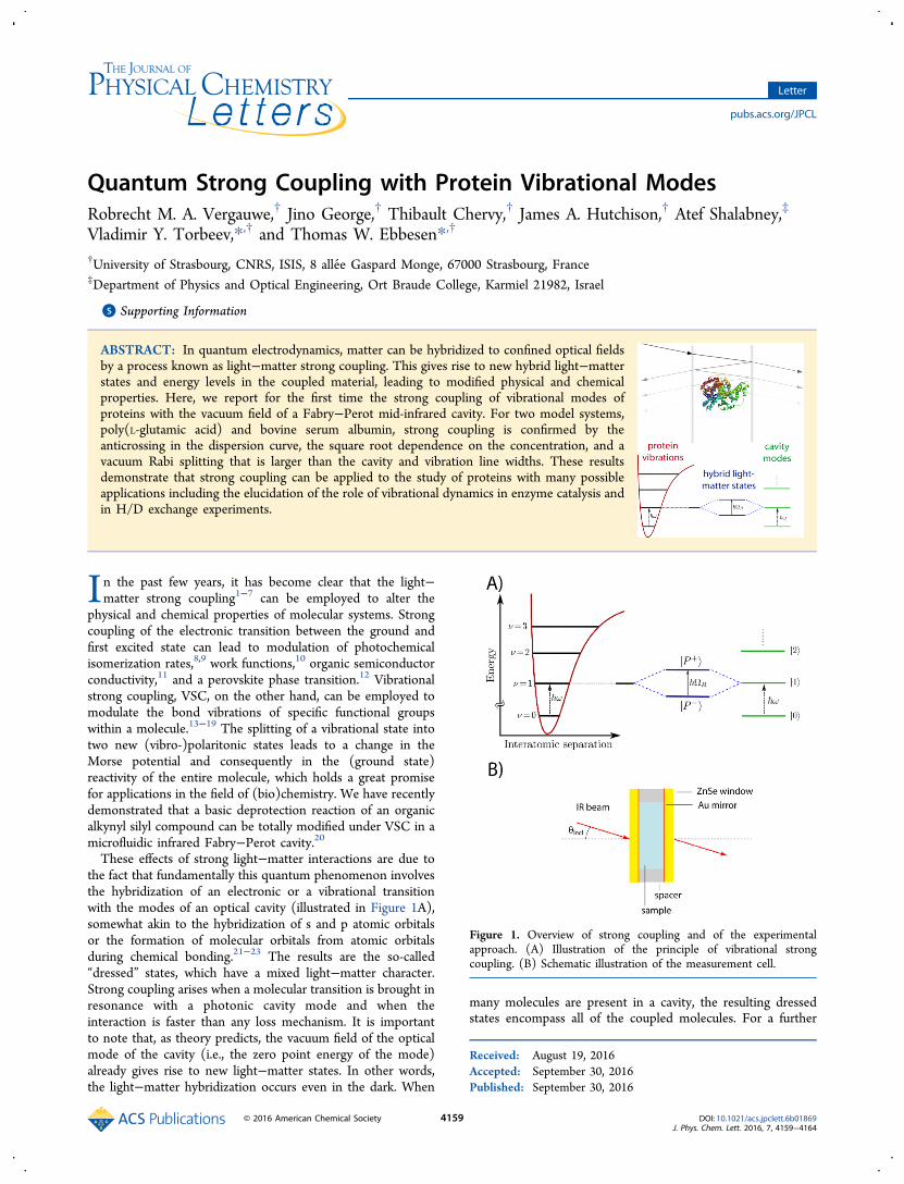
ABSTRACT: In quantum electrodynamics, matter can be hybridized to confined optical fieldsby a process known as light−matter strong coupling. This gives rise to new hybrid light−matterstates and energy levels in the coupled material, leading to modified physical and chemicalproperties. Here, we report for the first time the strong coupling of vibrational modes ofproteins with the vacuum field of a Fabry−Perot mid-infrared cavity. For two model systems,poly(L-glutamic acid) and bovine serum albumin, strong coupling is confirmed by theanticrossing in the dispersion curve, the square root dependence on the concentration, and avacuum Rabi splitting that is larger than the cavity and vibration line widths. These resultsdemonstrate that strong coupling can be applied to the study of proteins with many possibleapplications including the elucidation of the role of vibrational dynamics in enzyme catalysis andin H/D exchange experiments.
In the past few years, it has become clear that the light−matter strong coupling1−7 can be employed to alter the
physical and chemical properties of molecular systems. Strongcoupling of the electronic transition between the ground andfirst excited state can lead to modulation of photochemicalisomerization rates,8,9 work functions,10 organic semiconductorconductivity,11 and a perovskite phase transition.12 Vibrationalstrong coupling, VSC, on the other hand, can be employed tomodulate the bond vibrations of specific functional groupswithin a molecule.13−19 The splitting of a vibrational state intotwo new (vibro-)polaritonic states leads to a change in theMorse potential and consequently in the (ground state)reactivity of the entire molecule, which holds a great promisefor applications in the field of (bio)chemistry. We have recentlydemonstrated that a basic deprotection reaction of an organicalkynyl silyl compound can be totally modified under VSC in amicrofluidic infrared Fabry−Perot cavity.20These effects of strong light−matter interactions are due to
the fact that fundamentally this quantum phenomenon involvesthe hybridization of an electronic or a vibrational transitionwith the modes of an optical cavity (illustrated in Figure 1A),somewhat akin to the hybridization of s and p atomic orbitalsor the formation of molecular orbitals from atomic orbitalsduring chemical bonding.21−23 The results are the so-called“dressed” states, which have a mixed light−matter character.Strong coupling arises when a molecular transition is brought inresonance with a photonic cavity mode and when theinteraction is faster than any loss mechanism. It is importantto note that, as theory predicts, the vacuum field of the opticalmode of the cavity (i.e., the zero point energy of the mode)already gives rise to new light−matter states. In other words,the light−matter hybridization occurs even in the dark. When
many molecules are present in a cavity, the resulting dressedstates encompass all of the coupled molecules. For a further
Received: August 19, 2016Accepted: September 30, 2016Published: September 30, 2016
Figure 1. Overview of strong coupling and of the experimentalapproach. (A) Illustration of the principle of vibrational strongcoupling. (B) Schematic illustration of the measurement cell.
Letter
pubs.acs.org/JPCL
© 2016 American Chemical Society 4159 DOI: 10.1021/acs.jpclett.6b01869J. Phys. Chem. Lett. 2016, 7, 4159−4164
discussion of the physics of quantum strong light−matterinteraction, the reader is referred to one of the reviews22,23 or toa recent perspective oriented toward molecular and materialscience.21
Although electronic strong coupling of biomolecules hasbeen demonstrated,24,25 VSC has not yet been achieved to thebest of our knowledge.13,14,20,21 The main challenge inperforming VSC of proteins is the 2 orders of magnitudesmaller extinction coefficient of vibrational transitions com-pared to those of electronic transitions, which necessitates theuse of very high sample concentrations. To date, the VSC ofpure molecular liquids, polymer films, and organic substances
dissolved in solutions has been reported.13−17,19,26 Workingwith proteins at very high concentrations can lead to structuraland functional alterations and even detrimental effects such asaggregation.27−30 Here, we demonstrate the vibrational strongcoupling of poly(L-glutamic acid) (PLGA) and of bovine serumalbumin (BSA) under conditions where the native fold of thelatter is largely preserved. The PLGA peptide homopolymerand serum protein are attractive model systems because theyare both well characterized, highly water-soluble, and exhibitone or more intense infrared absorption bands (PLGA, amide Iand γ-COO− asymmetric stretching; BSA, amide I).27,31,32
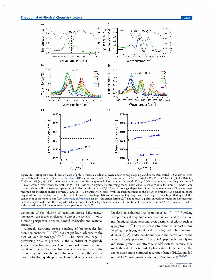
Figure 2. FTIR spectra and dispersion data of poly(L-glutamic acid) in a cavity under strong coupling conditions. Deuterated PLGA was insertedinto a Fabry−Perot cavity (illustrated in Figure 1B) and measured with FTIR spectrometer. (A−C) Data for PLGA at 5% (w/v). (D−F) Data forPLGA at 15% (w/v). (A,D) IR transmission spectrum for cavity tuned close to either the amide I′ or γ-COO− asymmetric stretching vibration ofPLGA. Green curves: resonance with the γ-COO− side-chain asymmetric stretching mode. Blues curve: resonance with the amide I′ mode. Graycurves: reference IR transmission spectrum of PLGA outside a cavity. (B,E) Data of the angle-dependent dispersion measurements. IR spectra wererecorded for incidence angles between 0° and 18°. (C,F) Dispersion curves with the peak positions of the polariton branches as a function of themagnitude of the in-plane wave vector (k∥). To avoid misinterpretations, strong coupling dispersion data is preferentially plotted against thiscomponent of the wave vector (see Supporting Information for the conversion formula).22 The measured polariton peak positions are denoted withdark blue open circles and the coupled oscillator model fit with a light blue solid line. The location of the amide I′ and γ-COO− modes are markedwith dashed lines. All measurements were performed in D2O.
The Journal of Physical Chemistry Letters Letter
DOI: 10.1021/acs.jpclett.6b01869J. Phys. Chem. Lett. 2016, 7, 4159−4164
4160
Deuterium-exchanged PLGA dissolved in D2O under theconditions used here (c = 2.5−15% (w/v), measured pD =7.6−7.8) displays an intense amide I′ and γ-COO− asymmetricstretching bands at 1647 and 1565 cm−1, respectively (Figure2A and D gray curve). To achieve vibrational strong coupling,Fabry−Perot cavities were fabricated consisting of two parallelZnSe windows coated with a reflective Au thin film andseparated by a spacer (schematically shown in Figure 1B). Thetransmission spectrum of such an empty cavity exhibits a typicalprogression of transmission peaks representing the opticalmodes generated by the cavity (see Supporting Information).After introducing a 5% (w/v) PLGA solution and tuning thecavity resonance close to the PLGA γ-COO− mode, the spectralsignature of strong light−matter coupling is observed. Thetransmission spectrum of the sample (shown in Figure 2A,green curve) displays two peaks symmetrically spaced aroundthe peak position of the PLGA γ-COO− mode. These twopeaks are the spectral signature of the two new polaritonicstates (conventionally denoted |P+⟩ and |P−⟩). However, initself this observation does not constitute sufficient proof ofstrong coupling because a similar splitting can be generatedsimply by an absorber overlapping with a transmission mode.For a further confirmation of the presence of strong light−
matter coupling, the angle-dependent dispersion of the sampleneeds to be examined.13,14,21,22 Polaritonic states inherit thedispersive properties of the empty cavity with a characteristicanticrossing at the intersection with the absorbing transitionresulting in the two polaritonic branches.21−23 To observe thisbehavior, the IR spectrum of the sample is measured at differentincidence angles of the probe beam in the FTIR. The spectrafor PLGA recorded between 0° to 18° are displayed in Figure2B. The dimensions of the sample holder prohibit investigationof higher angles. As the angle of incidence is varied, the lowerbranch shifts to higher wavenumbers up to a limiting value andloses intensity. At the same time, the upper branch gains inintensity and disperses to higher wavenumbers.13,14 When thelocations of the peak positions are plotted as a function of themagnitude of the in-plane wave vector (k∥) of the incominglight (Figure 2C), an anticrossing is indeed observed. Fittingthis data with the coupled oscillator model21−23 allows theextraction of the vacuum Rabi splitting, which is the energydifference between the two new vibro-polariton states at exactresonance. The vacuum Rabi splitting amounts here to 55.4cm−1, which corresponds to about 3.5% of the transition energyof the PLGA γ-COO− mode. Finally, the strong couplingregime requires that the line width of the bare moleculartransition and the cavity mode are both less than the vacuumRabi splitting.21−23 This is indeed the case here since they areestimated to be 45.2 and 48.9 cm−1, respectively. Therefore, thestrong coupling conditions are all met for this system.When PLGA at a concentration of 10 or 15% (w/v) is
inserted in a cavity near resonance with either the amide I′ or γ-COO− asymmetric stretching mode of poly(L-glutamic acid),three new bands appear instead of just the two branchesexpected for the coupling of a single molecular vibration withone cavity mode (see Figure 2D for the data for 15% w/v; seeFigure S2D for the data for 10% w/v). Angle-resolvedmeasurements reveal the dispersive behavior of the threebranches as shown in Figures 2E−F and S2E−F. The dispersivebehavior of the different curves is explained by thesimultaneous, independent coupling of both vibrationalmodes to the cavity vacuum field. This is a common effectwhen two transitions are close in energy and couple to the same
optical mode.14,17,33−35 Vacuum Rabi splittings associated withboth vibrations are consequently the minimal energy differ-ences between the respective branches.21−23 Because these Rabisplittings are high compared to the energy difference betweenthe two (bare) transitions, simultaneous coupling occurs evenwhen, for instance, the cavity is blue-detuned with respect tothe higher-lying amide I′ transition. Comparison of the Rabisplittings with the line widths of the coupled cavity mode andmolecular vibrations again confirms that the strong couplingregime is reached. The values are summarized in Table 1. It
should be noted that the reasons that no secondary splitting isobserved at 5% (w/v) PGLA (Figure 2A and B) is due to thefact the coupling was tuned to the γ-COO− and that the Rabisplitting is so small that the higher vibro-polariton branch doesnot reach the amide 1′ band within the angular range of ourexperiment. This exemplifies the sensitivity of the observationof strong coupling to cavity detuning.As the concentration is lowered to 2.5%, a weak branch
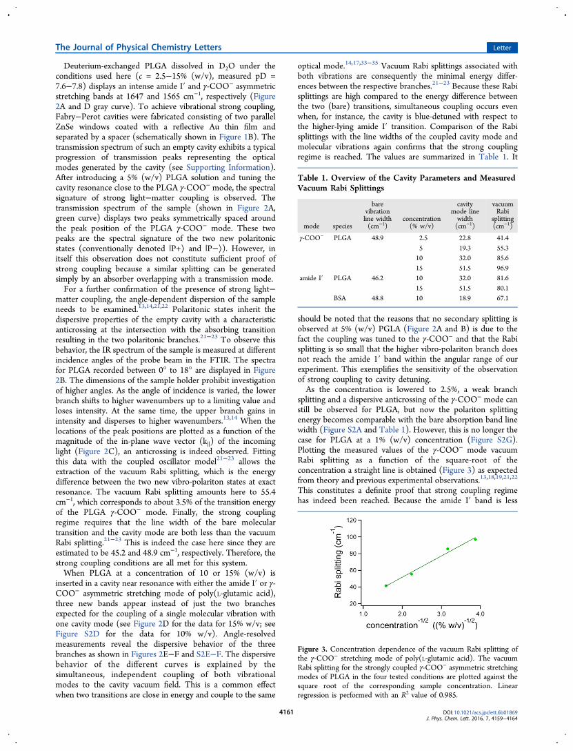
splitting and a dispersive anticrossing of the γ-COO− mode canstill be observed for PLGA, but now the polariton splittingenergy becomes comparable with the bare absorption band linewidth (Figure S2A and Table 1). However, this is no longer thecase for PLGA at a 1% (w/v) concentration (Figure S2G).Plotting the measured values of the γ-COO− mode vacuumRabi splitting as a function of the square-root of theconcentration a straight line is obtained (Figure 3) as expectedfrom theory and previous experimental observations.13,18,19,21,22
This constitutes a definite proof that strong coupling regimehas indeed been reached. Because the amide I′ band is less
Table 1. Overview of the Cavity Parameters and MeasuredVacuum Rabi Splittings
mode species
barevibrationline width(cm−1)
concentration(% w/v)
cavitymode linewidth(cm−1)
vacuumRabi
splitting(cm−1)
γ-COO− PLGA 48.9 2.5 22.8 41.45 19.3 55.310 32.0 85.615 51.5 96.9
amide I′ PLGA 46.2 10 32.0 81.615 51.5 80.1
BSA 48.8 10 18.9 67.1
Figure 3. Concentration dependence of the vacuum Rabi splitting ofthe γ-COO− stretching mode of poly(L-glutamic acid). The vacuumRabi splitting for the strongly coupled γ-COO− asymmetric stretchingmodes of PLGA in the four tested conditions are plotted against thesquare root of the corresponding sample concentration. Linearregression is performed with an R2 value of 0.985.
The Journal of Physical Chemistry Letters Letter
DOI: 10.1021/acs.jpclett.6b01869J. Phys. Chem. Lett. 2016, 7, 4159−4164
4161
intense than the γ-COO− stretching band, the threshold for thestrong coupling regime will be higher for the former than forthe latter.Although poly(L-glutamic acid) is an adequate polypeptide
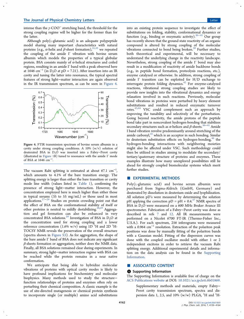
model sharing many important characteristics with naturalproteins (e.g., α-helix and β-sheet formation),31,32 we repeatedthe coupling of the amide I′ vibration with bovine serumalbumin which models the properties of a typical globularprotein. BSA consists mainly of α-helical structures and coiledregions, resulting in an amide I′ band with a peak observed hereat 1646 cm−1 (in D2O at pD = 7.21). After insertion into an IRcavity and tuning the latter into resonance, the typical spectralfeatures of strong light−matter interaction are again observedin the IR transmission spectrum, as can be seen in Figure 4.
The vacuum Rabi splitting is estimated at about 67.1 cm−1,which amounts to 4.1% of the bare transition energy. Thesplitting energy is larger than either the bare transition or cavitymode line width (values listed in Table 1), confirming thepresence of strong light−matter interaction. However, theconcentration employed here is much higher than either thosein typical serums (35 to 55 mg/mL) or those used in mostapplications.27−29 Studies on protein crowding point out thatthe effect of BSA on the conformational stability of itself orother proteins is neutral to mildly destabilizing.28,29 Aggrega-tion and gel formation can also be enhanced in veryconcentrated BSA solutions.29 Investigation of BSA in D2O atthe concentration used for strong coupling and a lowerreference concentration (1.6% w/v) using 1D 1H and 2D 1H-TOCSY NMR reveals the preservation of the overall structure(spectra shown in Figure S3). As for aggregation, the shape ofthe bare amide I′ band of BSA does not indicate any significantβ-sheets formation or aggregation, neither does the NMR data.Finally, all BSA solutions remained clear during experiments. Insummary, strong light−matter interaction regime with BSA canbe reached while the protein remains in a near nativeconformation.We anticipate that being able to hybridize molecular
vibrations of proteins with optical cavity modes is likely tohave profound implications for biochemistry and molecularbiophysics. Many methods used to study the structure−function relationships of proteins and enzymes often rely onperturbing their chemical composition. A classic example is theuse of site-directed mutagenesis or chemical protein synthesisto incorporate single (or multiple) amino acid substitutions
into an existing protein sequence to investigate the effect ofsubstitutions on folding, stability, conformational dynamics orfunction (e.g., binding or enzymatic activity).36−41 Our grouphas recently shown that the ground state reactivity of an organiccompound is altered by strong coupling of the molecularvibrations connected to bond being broken.20 Further studies,both theoretical and experimental, will be necessary tounderstand the underlying change in the reactivity landscape.Nevertheless, strong coupling of the amide I′ bond may alsoresult in a modification of reactivity of amide backbone bonds(e.g., in peptide bond formation, proteolysis reactions, etc.),enzyme catalyzed or otherwise. In addition, strong coupling ofamide I′ transition can be exploited for H/D exchange tointerrogate protein folding dynamics.42 For enzyme-catalyzedreactions, vibrational strong coupling studies are likely toprovide new insights into the vibrational dynamics and energyrelaxation involved in such reactions. Previously, molecularbond vibrations in proteins were perturbed by heavy elementsubstitutions and resulted in reduced enzymatic turnoverrates.43,44 VSC could complement such an approach byimproving the tunability and selectively of the perturbation.Going beyond reactivity, the amide protons of the peptidebond take part in noncovalent hydrogen-bonding that stabilizessecondary structures such as α-helices and β-sheets.36−41 AmideI band vibration revolve predominantly around stretching of theamide carbonyl,45 which is an acceptor in such bonding. Similarto deuterium substitution effects on hydrogen-bonding,46 thehydrogen-bonding interactions with neighboring moietiesmight also be affected under VSC. Such methodology couldthen be utilized in studies aiming to modulate the secondary/tertiary/quaternary structure of proteins and enzymes. Theseexamples illustrate how many unexplored possibilities still lieahead for strongly coupled biomolecular systems which meritfurther studies.
■ EXPERIMENTAL METHODSPoly(L-glutamic acid) and bovine serum albumin werepurchased from Sigma-Aldrich (GmbH, Germany) anddeuterated by dissolution in deuterium oxide and lyophilization.All solution pD’s were measured by determining the solutionpH applying the correction pD = pH + 0.4.47 NMR spectra ofBSA in D2O were measured on a 600 MHz Bruker Avance IIIspectrometer. Fabrication of a Fabry−Perot cavity was done asdescribed in refs 7 and 12. All IR measurements wereperformed on a Nicolet 6700 FT-IR (Thermo-Fisher Inc.,U.S.A.). For each spectrum, 256 inferograms were measuredwith a 0.964 cm−1 resolution. Extraction of the polariton peakpositions was done by manually fitting of the polariton bandswith a Gaussian model. Fitting of the dispersion curves wasdone with the coupled oscillator model with either 1 or 2independent excitons in order to retrieve the vacuum Rabisplitting energy. Additional experimental details and informa-tion on the data analysis can be found in the SupportingInformation.
■ ASSOCIATED CONTENT*S Supporting InformationThe Supporting Information is available free of charge on theACS Publications website at DOI: 10.1021/acs.jpclett.6b01869.
Supplementary methods and materials, empty Fabry−Perot cavity transmission spectrum, spectra and dis-persion data 1, 2.5, and 10% (w/w) PLGA, 1H and 1H-
Figure 4. FTIR transmission spectrum of bovine serum albumin in acavity under strong coupling conditions. A 10% (w/v) solution ofdeuterated BSA in D2O was inserted into a Fabry−Perot cavity(illustrated in Figure 1B) tuned to resonance with the amide I′ modeof BSA at 1646 cm−1.
The Journal of Physical Chemistry Letters Letter
DOI: 10.1021/acs.jpclett.6b01869J. Phys. Chem. Lett. 2016, 7, 4159−4164
4162
TOCSY NMR spectra BSA and examples of curve fittingresults for polariton peak extraction. (PDF)
■ AUTHOR INFORMATIONCorresponding Authors*E-mail: [email protected].*E-mail: [email protected] authors declare no competing financial interest.
■ ACKNOWLEDGMENTSJeremy Ruiz is gratefully acknowledged for help with preparingthe deuterated samples and Anoop Thomas for helpfuldiscussions. We acknowledge support from the InternationalCenter for Frontier Research in Chemistry (icFRC, Stras-bourg), the ANR Equipex Union (ANR-10-EQPX-52-01), theLabex NIE projects (ANR-11-LABX-0058 NIE) and CSC(ANR-10-LABX-0026 CSC) within the Investissement d’Ave-nir program ANR-10-IDEX-0002-02.
■ REFERENCES(1) Aberra Guebrou, S.; Symonds, C.; Homeyer, E.; Plenet, J. C.;Gartstein, Y. N.; Agranovich, V. M.; Bellessa, J. Coherent Emissionfrom a Disordered Organic Semiconductor Induced by StrongCoupling with Surface Plasmons. Phys. Rev. Lett. 2012, 108, 066401.(2) Shi, L.; Hakala, T. K.; Rekola, H. T.; Martikainen, J. P.; Moerland,R. J.; Torma, P. Spatial Coherence Properties of Organic MoleculesCoupled to Plasmonic Surface Lattice Resonances in the Weak andStrong Coupling Regimes. Phys. Rev. Lett. 2014, 112, 153002.(3) Berrier, A.; Cools, R.; Arnold, C.; Offermans, P.; Crego-Calama,M.; Brongersma, S. H.; Gomez-Rivas, J. Active Control of the StrongCoupling Regime between Porphyrin Excitons and Surface PlasmonPolaritons. ACS Nano 2011, 5, 6226−6232.(4) Zengin, G.; Johansson, G.; Johansson, P.; Antosiewicz, T. J.; Kall,M.; Shegai, T. Approaching the Strong Coupling Limit in SinglePlasmonic Nanorods Interacting with J-Aggregates. Sci. Rep. 2013, 3,3074.(5) Nagasawa, F.; Takase, M.; Murakoshi, K. Raman Enhancementvia Polariton States Produced by Strong Coupling between a LocalizedSurface Plasmon and Dye Excitons at Metal Nanogaps. J. Phys. Chem.Lett. 2014, 5, 14−19.(6) Hao, Y. W.; Wang, H. Y.; Jiang, Y.; Chen, Q. D.; Ueno, K.; Wang,W. Q.; Misawa, H.; Sun, H. B. Hybrid-State Dynamics of GoldNanorods/dye J-Aggregates under Strong Coupling. Angew. Chem., Int.Ed. 2011, 50, 7824−7828.(7) Vasa, P.; Wang, W.; Pomraenke, R.; Maiuri, M.; Manzoni, C.;Cerullo, G.; Lienau, C. Optical Stark Effects in J-Aggregate-MetalHybrid Nanostructures Exhibiting a Strong Exciton-Surface-Plasmon-Polariton Interaction. Phys. Rev. Lett. 2015, 114, 036802.(8) Schwartz, T.; Hutchison, J. A.; Genet, C.; Ebbesen, T. W.Reversible Switching of Ultrastrong Light-Molecule Coupling. Phys.Rev. Lett. 2011, 106, 196405.(9) Hutchison, J. A.; Schwartz, T.; Genet, C.; Devaux, E.; Ebbesen, T.W. Modifying Chemical Landscapes by Coupling to Vacuum Fields.Angew. Chem., Int. Ed. 2012, 51, 1592−1596.(10) Hutchison, J. A.; Liscio, A.; Schwartz, T.; Canaguier-Durand, A.;Genet, C.; Palermo, V.; Samorì, P.; Ebbesen, T. W. Tuning the Work-Function via Strong Coupling. Adv. Mater. 2013, 25, 2481−2485.(11) Orgiu, E.; George, J.; Hutchison, J. A.; Devaux, E.; Dayen, J. F.;Doudin, B.; Stellacci, F.; Genet, C.; Schachenmayer, J.; Genes, C.; et al.Conductivity in Organic Semiconductors Hybridized with the VacuumField. Nat. Mater. 2015, 14, 1123−1129.(12) Wang, S.; Mika, A.; Hutchison, J. A.; Genet, C.; Jouaiti, A.;Hosseini, M. W.; Ebbesen, T. W. Phase Transition of a PerovskiteStrongly Coupled to the Vacuum Field. Nanoscale 2014, 6, 7243−7248.
(13) Shalabney, A.; George, J.; Hutchison, J.; Pupillo, G.; Genet, C.;Ebbesen, T. W. Coherent Coupling of Molecular Resonators with aMicrocavity Mode. Nat. Commun. 2015, 6, 5981.(14) George, J.; Shalabney, A.; Hutchison, J. A.; Genet, C.; Ebbesen,T. W. Liquid-Phase Vibrational Strong Coupling. J. Phys. Chem. Lett.2015, 6, 1027−1031.(15) Shalabney, A.; George, J.; Hiura, H.; Hutchison, J. A.; Genet, C.;Hellwig, P.; Ebbesen, T. W. Enhanced Raman Scattering from Vibro-Polariton Hybrid States. Angew. Chem., Int. Ed. 2015, 54, 7971−7975.(16) Long, J. P.; Simpkins, B. S. Coherent Coupling between aMolecular Vibration and Fabry−Perot Optical Cavity to GiveHybridized States in the Strong Coupling Limit. ACS Photonics2015, 2, 130−136.(17) Muallem, M.; Palatnik, A.; Nessim, G. D.; Tischler, Y. R. StrongLight-Matter Coupling and Hybridization of Molecular Vibrations in aLow-Loss Infrared Microcavity. J. Phys. Chem. Lett. 2016, 7, 2002−2008.(18) Pino, J. D.; Feist, J.; Garcia-Vidal, F. J. Quantum Theory ofCollective Strong Coupling of Molecular Vibrations with a MicrocavityMode. New J. Phys. 2015, 17, 053040.(19) Simpkins, B. S.; Fears, K. P.; Dressick, W. J.; Spann, B. T.;Dunkelberger, A. D.; Owrutsky, J. C. Spanning Strong to WeakNormal Mode Coupling between Vibrational and Fabry-Perot CavityModes through Tuning of Vibrational Absorption Strength. ACSPhotonics 2015, 2, 1460−1467.(20) Thomas, A.; George, J.; Shalabney, A.; Dryzhakov, M.; Varma, S.J.; Moran, J.; Chervy, T.; Zhong, X.; Devaux, E.; Genet, C.; et al.Ground-State Chemical Reactivity under Vibrational Coupling to theVacuum Electromagnetic Field. Angew. Chem., Int. Ed. 2016, 55,11462−11466.(21) Ebbesen, T. W. Hybrid Light−Matter States in a Molecular andMaterial Science Perspective. Acc. Chem. Res. Submitted forpublication.(22) Torma, P.; Barnes, W. L. Strong Coupling between SurfacePlasmon Polaritons and Emitters: A Review. Rep. Prog. Phys. 2015, 78,013901.(23) Agranovich, V. M.; Gartstein, Y. N.; Litinskaya, M. HybridResonant Organic−Inorganic Nanostructures for OptoelectronicApplications. Chem. Rev. 2011, 111, 5179−5214.(24) Coles, D. M.; Yang, Y.; Wang, Y.; Grant, R. T.; Taylor, R. A.;Saikin, S. K.; Aspuru-Guzik, A.; Lidzey, D. G.; Tang, J. K.-H.; Smith, J.M. Strong Coupling between Chlorosomes of Photosynthetic Bacteriaand a Confined Optical Cavity Mode. Nat. Commun. 2014, 5, 5561.(25) Dietrich, C. P.; Steude, A.; Tropf, L.; Schubert, M.; Kronenberg,N. M.; Ostermann, K.; Hofling, S.; Gather, M. C. An Exciton-PolaritonLaser Based on Biologically Produced Fluorescent Protein. Sci. Adv.2016, 2, e1600666.(26) Galego, J.; Garcia-Vidal, F. J.; Feist, J. Cavity-InducedModifications of Molecular Structure in the Strong-Coupling Regime.Phys. Rev. X 2015, 5, 41022.(27) Barbosa, L. R. S.; Ortore, M. G.; Spinozzi, F.; Mariani, P.;Bernstorff, S.; Itri, R. The Importance of Protein-Protein Interactionson the pH-Induced Conformational Changes of Bovine SerumAlbumin: A Small-Angle X-Ray Scattering Study. Biophys. J. 2010,98, 147−157.(28) Miklos, A. C.; Sarkar, M.; Wang, Y.; Pielak, G. J. ProteinCrowding Tunes Protein Stability. J. Am. Chem. Soc. 2011, 133, 7116−7120.(29) Guo, J.; Harn, N.; Robbins, A.; Dougherty, R.; Middaugh, C. R.Stability of Helix-Rich Proteins at High Concentrations. Biochemistry2006, 45, 8686−8696.(30) Ellis, R. J. Macromolecular Crowding: Obvious but Under-appreciated. Trends Biochem. Sci. 2001, 26, 597−604.(31) Itoh, K.; Foxman, B. M.; Fasman, G. D. The Two Beta Forms ofPoly(L-Glutamic Acid). Biopolymers 1976, 15, 419−455.(32) Hernik, A.; Puławski, W.; Fedorczyk, B.; Tymecka, D.; Misicka,A.; Filipek, S.; Dzwolak, W. Amyloidogenic Properties of Short α-L-Glutamic Acid Oligomers. Langmuir 2015, 31, 10500−10507.
The Journal of Physical Chemistry Letters Letter
DOI: 10.1021/acs.jpclett.6b01869J. Phys. Chem. Lett. 2016, 7, 4159−4164
4163

(33) Lidzey, D. G.; Bradley, D. D. C.; Armitage, A.; Walker, S.;Skolnick, M. S. Photon-Mediated Hybridization of Frenkel Excitons inOrganic Semiconductor Microcavities. Science 2000, 288, 1620−1623.(34) Wenus, J.; Parashkov, R.; Ceccarelli, S.; Brehier, A.; Lauret, J. S.;Skolnick, M. S.; Deleporte, E.; Lidzey, D. G. Hybrid Organic-InorganicExciton-Polaritons in a Strongly Coupled Microcavity. Phys. Rev. B:Condens. Matter Mater. Phys. 2006, 74, 235212.(35) Hakala, T. K.; Toppari, J. J.; Kuzyk, A.; Pettersson, M.;Tikkanen, H.; Kunttu, H.; Torma, P. Vacuum Rabi Splitting andStrong-Coupling Dynamics for Surface-Plasmon Polaritons andRhodamine 6G Molecules. Phys. Rev. Lett. 2009, 103, 053602.(36) Matouschek, A.; Kellis, J. T.; Serrano, L.; Bycroft, M.; Fersht, A.R. Transient Folding Intermediates Characterized by ProteinEngineering. Nature 1990, 346, 440−445.(37) O’Neil, K. T.; DeGrado, W. F. How Calmodulin Binds ItsTargets: Sequence Independent Recognition of Amphiphilic Alpha-Helices. Trends Biochem. Sci. 1990, 15, 59−64.(38) Clackson, T.; Wells, J. A. A Hot Spot of Binding Energy in aHormone-Receptor Interface. Science 1995, 267, 383−386.(39) Andrews, F. H.; McLeish, M. J. Using Site-SaturationMutagenesis to Explore Mechanism and Substrate Specificity inThiamin Diphosphate-Dependent Enzymes. FEBS J. 2013, 280, 6395−6411.(40) Torbeev, V. Y.; Raghuraman, H.; Hamelberg, D.; Tonelli, M.;Westler, W. M.; Perozo, E.; Kent, S. B. H. Protein ConformationalDynamics in the Mechanism of HIV-1 Protease Catalysis. Proc. Natl.Acad. Sci. U. S. A. 2011, 108, 20982−20987.(41) Torbeev, V. Y.; Hilvert, D. Both the Cis-Trans Equilibrium andIsomerization Dynamics of a Single Proline Amide Modulate β2-Microglobulin Amyloid Assembly. Proc. Natl. Acad. Sci. U. S. A. 2013,110, 20051−20056.(42) Englander, S. W.; Mayne, L.; Kan, Z.-Y.; Hu, W. ProteinFoldingHow and Why: By Hydrogen Exchange, FragmentSeparation, and Mass Spectrometry. Annu. Rev. Biophys. 2016, 45,135−152.(43) Suarez, J.; Schramm, V. L. Isotope-Specific and Amino Acid-Specific Heavy Atom Substitutions Alter Barrier Crossing in HumanPurine Nucleoside Phosphorylase. Proc. Natl. Acad. Sci. U. S. A. 2015,112, 11247−11251.(44) Silva, R. G.; Murkin, A. S.; Schramm, V. L. FemtosecondDynamics Coupled to Chemical Barrier Crossing in a Born-Oppenheimer Enzyme. Proc. Natl. Acad. Sci. U. S. A. 2011, 108,18661−18665.(45) Barth, A.; Zscherp, C. What Vibrations Tell about Proteins. Q.Rev. Biophys. 2002, 35, 369−430.(46) Singh, S.; Rao, C. N. R. Deuterium Isotope Effects on HydrogenBonding. Can. J. Chem. 1966, 44, 2611−2615.(47) Glasoe, P. K.; Long, F. A. Use of Glass Electrodes To MeasureAcidities in Deuterium Oxide. J. Phys. Chem. 1960, 64, 188−190.
The Journal of Physical Chemistry Letters Letter
DOI: 10.1021/acs.jpclett.6b01869J. Phys. Chem. Lett. 2016, 7, 4159−4164
4164