Embed Size (px)
Citation preview

Electronic Supporting Information
Antibacterial activity evaluation and mode of action study of novel thiazole-
quinolinium derivatives
Ying Lia,1, Ning Suna,b,c,1,*, Hooi-Leng Sera,d,1, Wei Longa, Yanan Lie, Cuicui Chena,
Boxin Zhenga, Xuanhe Huanga, Zhihua Liub,*, Yu-Jing Lua,*
a School of Biomedical and Pharmaceutical Sciences, Guangdong University of
Technology, Guangzhou 510006, PR China;
b The Fifth Affiliated Hospital of Guangzhou Medical University, Guangzhou 510700,
PR China;
c The State Key Laboratory of Chemical Biology and Drug Discovery, Department of
Applied Biology and Chemical Technology, The Hong Kong Polytechnic University,
Hung Hom, Kowloon, Hong Kong, China;
d Novel Bacteria and Drug Discovery (NBDD) Research Group, Microbiome and
Bioresource Research Strength, Jeffrey Cheah School of Medicine and Health
Sciences, Monash University Malaysia, 47500 Bandar Sunway, Selangor Darul Ehsan,
Malaysia;
e Department of Pharmacy, The Fifth Affiliated Hospital of Sun Yat-sen University,
Zhuhai, 519000, P. R. China.
1These authors contributed equally to this work.
*Corresponding author:
Dr. Ning. SUN, Email: [email protected]
Dr. Zhihua LIU, Email: [email protected]
Dr. Yu-Jin LU, Email: [email protected]
Electronic Supplementary Material (ESI) for RSC Advances.This journal is © The Royal Society of Chemistry 2020

List of contents:
1. Visualization of bacterial morphology
2. Visualization of bacterial cell membrane
3. Light-scattering assay of 4b4, 4e1 and 4e3
4. GTPase activity assay of 4a4 and 4b4
5. Visualization of Z-ring in bacterial cells
6. Hemolytic activity of 4a4 and 4b4
7. Drug resistance study of 4a4 and 4b4
8. Molecular modeling studies of 4a4 and 4e1 with FtsZ protein
9. 1H NMR (DMSO-d6), 13C NMR (DMSO-d6) and HRMS spectra of compounds
4a1, 4a3-4a4, 4b3-4b4 and 4c1-4c2, 4d1-4d3, 4e1-4e4
1. Visualization of bacterial morphology
Fig. S1. Morphology analysis of B. subtilis 168. Cells were grown in the presence of
4a1-4a3 and 4b1-4b4. Scale bar=20 μm.
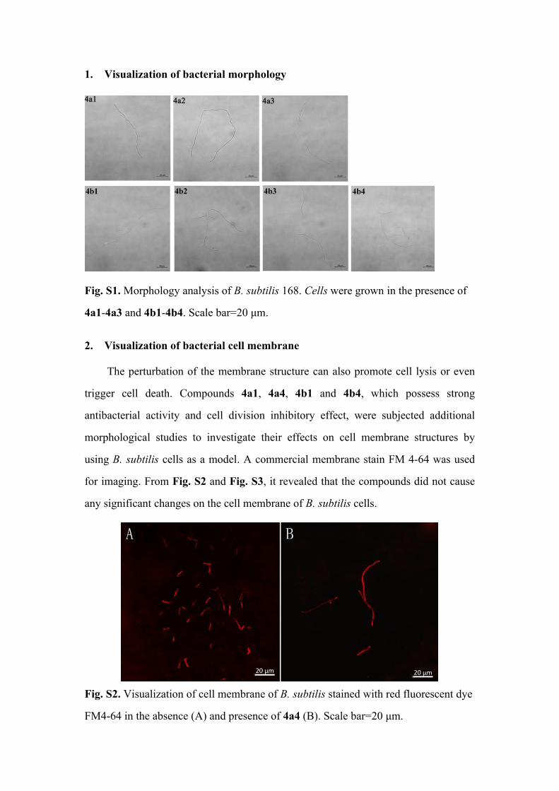
2. Visualization of bacterial cell membrane
The perturbation of the membrane structure can also promote cell lysis or even
trigger cell death. Compounds 4a1, 4a4, 4b1 and 4b4, which possess strong
antibacterial activity and cell division inhibitory effect, were subjected additional
morphological studies to investigate their effects on cell membrane structures by
using B. subtilis cells as a model. A commercial membrane stain FM 4-64 was used
for imaging. From Fig. S2 and Fig. S3, it revealed that the compounds did not cause
any significant changes on the cell membrane of B. subtilis cells.
Fig. S2. Visualization of cell membrane of B. subtilis stained with red fluorescent dye
FM4-64 in the absence (A) and presence of 4a4 (B). Scale bar=20 μm.

Fig. S3. Visualization of cell membrane of B. subtilis stained with red fluorescent dye
FM4-64 in the presence of 4a1, 4b1, 4b4. Scale bar=20 μm.
3. Light-scattering assay of 4b4, 4e1 and 4e3
Fig. S4. Effect of 4e1, 4e3 and 4b4 on the polymerization of FtsZ at a concentration
of 0.125-0.5 µg/mL.

4. GTPase activity assay of 4a4 and 4b4
The dynamic assembly of FtsZ is strictly regulated by its GTPase activity [1, 2].
Two compounds, 4a4 and 4b4 were selected to explore their potential in disrupting
GTPase activity of FtsZ. The results showed that these compounds did not have any
significant effect on the GTPase activity of SaFtsZ (Fig. S5). As a matter of fact,
same phenomenon also occurs in the reported FtsZ-inhibitor 2,6-difluoro-3-
aminobenzamide derivative [3] and a conversion product of PC190723 [4]; these
compounds were reported to bind to the interdomain cleft of FtsZ without interfering
GTPase activity.
Fig. S5. Inhibition of GTPase activity of FtsZ by compounds 4a4 and 4b4.
5. Visualization of Z-ring in bacterial cells
Fig. S6. The perturbation of the cytokinetic Z-ring in B. subtilis. Cells of B. subtilis
were grown in the presence of 4a1, 4b1, 4b4. Scale bar=10 μm.

6. Hemolytic activity of 4a4 and 4b4
Hemolytic activity of compounds 4a4 and 4b4 was conducted using human
erythrocytes. While hemolysis rate of more than 5% indicates break down of
erythrocytes [5], results showed that these compounds did not reflect significant
hemolysis effect. The hemolysis rates of 4a4 and 4b4 at 32×MIC (MICs for S.
aureus ATCC 29213 were 1 μg/mL and 2 μg/mL, respectively) were lower than
5%, and in the previous reports [6], the cells treated by Triton X-100 (0.002 to 1%)
were completely hemolyzed under the same conditions, suggesting that
compounds 4a4 and 4b4 did not display cytotoxicity against human erythrocytes.
Fig. S7. Hemolytic activity of compound 4a4 and 4b4. Human erythrocytes were
treated with compounds 4a4 and 4b4 (0.125~64 µg/mL).
7. Drug resistance study of 4a4 and 4b4
Fig. S8. Bacterial resistance study of compound 4a4 and 4b4 against B. subtilis 168.

Fig. S9. Bacterial resistance study of compound 4a4 and 4b4 against E. coli ATCC
25922.
8. Molecular modeling studies of 4a4 and 4e1 with FtsZ protein

Fig. S10. (A) Molecular modeling studies of 4a4 (green), 4b4 (yellow) and 4e1
(purple) with FtsZ protein; (B) Predicted interactions between 4b4 and the amino
acids of FtsZ; (C) Predicted interactions between 4e1 and the amino acids of FtsZ.

9. 1H NMR, 13C NMR and HRMS spectra of compounds 4a1, 4a3-4a4, 4b3-4b4 and 4c1-4e4
Fig.S11. 1H NMR (DMSO-d6), 13C NMR (DMSO-d6) and HRMS spectra of
compound 4a1.
N
S
N
I
N
N
S
N
I
N

Fig.S12. 1H NMR (DMSO-d6), 13C NMR (DMSO-d6) and HRMS spectra of compound 4a3.
N
S
N
I
N
N
S
N
I
N

Fig.S13. 1H NMR (DMSO-d6), 13C NMR (DMSO-d6) and HRMS spectra of
compound 4a4.
N
S
N
I
N
N
S
N
I
N

Fig.S14. 1H NMR (DMSO-d6), 13C NMR (DMSO-d6) and HRMS spectra of
N
S
N
I
N
N
S
N
I
N

compound 4b3.
Fig.S15. 1H NMR (DMSO-d6), 13C NMR (DMSO-d6) and HRMS spectra of
N
S
N
I
N
N
S
N
I
N

compound 4b4.
N
SN
N
INH
O
N
SN
N
INH
O

Fig.S16. 1H NMR (DMSO-d6), 13C NMR (DMSO-d6) and HRMS spectra of
compound 4c1.

N
SN
N
OHI
O
N
SN
N
OHI
O

Fig.S17. 1H NMR (DMSO-d6), 13C NMR (DMSO-d6) and ESI-MS spectra of
compound 4c2.
N
SN
N
INH
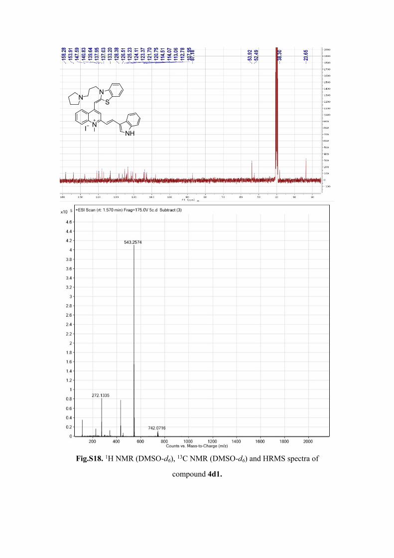
Fig.S18. 1H NMR (DMSO-d6), 13C NMR (DMSO-d6) and HRMS spectra of
compound 4d1.
N
SN
N
INH

N
SN
N
OHI
N
SN
N
OHI

Fig.S19. 1H NMR (DMSO-d6), 13C NMR (DMSO-d6) and HRMS spectra of
compound 4d2.
N
SN
N
SI

Fig.S20. 1H NMR (DMSO-d6), 13C NMR (DMSO-d6) and ESI-MS spectra of
compound 4d3.
N
SN
N
SI

N
SN
N
NHI
N
SN
N
NHI

Fig.S21. 1H NMR (DMSO-d6), 13C NMR (DMSO-d6) and HRMS spectra of
compound 4e1.
N
SN
N
OHI

Fig.S22. 1H NMR (DMSO-d6), 13C NMR (DMSO-d6) and HRMS spectra of
compound 4e2.
N
SN
N
OHI

N
SN
N
SI
N
SN
N
SI

Fig.S23. 1H NMR (DMSO-d6), 13C NMR (DMSO-d6) and HRMS spectra of
compound 4e3.
N
SN
N
NI

Fig.S24. 1H NMR (DMSO-d6), 13C NMR (DMSO-d6) and HRMS spectra of
compound 4e4.
N
SN
N
NI

References
[1] Y. Li, J. Hsin, L. Zhao, Y. Cheng, W. Shang, K.C. Huang, H.-W. Wang, S. Ye, FtsZ Protofilaments Use a Hinge-Opening Mechanism for Constrictive Force Generation, Science., 341 (2013) 392-394.[2] D.J. Haydon, N.R. Stokes, R. Ure, G. Galbraith, J.M. Bennett, D.R. Brown, P.J. Baker, V.V. Barynin, D.W. Rice, S.E. Sedelnikova, J.R. Heal, J.M. Sheridan, S.T. Aiwale, P.K. Chauhan, A. Srivastava, A. Taneja, I. Collins, J. Errington, L.G. Czaplewski, An inhibitor of FtsZ with potent and selective anti-staphylococcal activity, Science., 321 (2008) 1673-1675.[3] H.K. Lui, W. Gao, K.C. Cheung, W.B. Jin, N. Sun, J.W.Y. Kan, I.L.K. Wong, J. Chiou, D. Lin, E.W.C. Chan, Y.-C. Leung, T.H. Chan, S. Chen, K.-F. Chan, K.-Y. Wong, Boosting the efficacy of anti-MRSA β-lactam antibiotics via an easily accessible, non-cytotoxic and orally bioavailable FtsZ inhibitor, Eur. J. Med. Chem., 163 (2019) 95-115.[4] M. Kaul, Y.Z. Zhang, A.K. Parhi, E.J. LaVoie, D.S. Pilch, Inhibition of RND-type efflux pumps confers the FtsZ-directed prodrug TXY436 with activity against Gram-negative bacteria, Biochem. Pharmacol., 89 (2014) 321-328.[5] S. Lim, S. Lee, S.H. Yi, Y.S. Son, S.M. Choi, Y.K. Kim, The Biological Safety of Stainless Steel Needles Used in Warm-needling, Evid-Based. Compl. Alt., 7 (2010) 259-264.[6] Z.J. Zheng, N. Tharmalingam, Q.Z. Liu, E. Jayamani, W. Kim, B.B. Fuchs, R.J. Zhang, A. Vilcinskas, E. Mylonakis, Synergistic Efficacy of Aedes aegypti Antimicrobial Peptide Cecropin A2 and Tetracycline against Pseudomonas aeruginosa, Antimicrob. Agents. Ch., 61 (2017).