Embed Size (px)
Citation preview
Bouali et al., J Spine 2014, 3:6DOI: 10.4172/2165-7939.1000193
Case Report Open Access
Volume 3 • Issue 6 • 1000193J Spine, an open access journalISSN: 2165-7939
Primary Spinal Epidural Rhabdomyosarcoma of the Upper Thoracic SpineSofiene BOUALI, Asma BOUHOULA, Adenane BOUBAKER, Nidhal MAATAR, Imed Ben Said, Jalel KALLEL, Lassaad AOUIJ and Hafedh JEMELDepartment of neurosurgery, National Institute of Neurology”Mongi Ben Hmida”, TunisFaculty of Medicine, University of Tunis el MANAR
AbstractIntroduction: Rhabdomyosarcoma is a highly aggressive and rapidly growing sarcoma with skeletal origin that
occasionally appears in the spinal epidural space.
Method: We report a 20-year-old girl who presented with back pain, progressive paraparesis, and urinary retention.She had muscular weakness in her lower extremities and absent deep tendon reflex.An epidural dumbbell-shape mass at T3-T4 level was observed on MRI.The patient underwent T3-T5 hemilaminectomy.Result: Histopathological examination Immunohistochemical staining confirmed the diagnosis of alveolar
Rhabdomyosarcoma. She received radiotherapy and chemotherapy. The patient died 3 months after.
Conclusion: Primary spinal epidural RMS is an extremely rare and very aggressive tumor. The treatment should focus on extensive resection with intensive combination of radiotherapy and chemotherapy.
*Corresponding author: Bouali Sofiene, Department of Neurosurgery, NationalInstitute of Neurology Tunis, Tunisia Faculty of Medicine, University of Tunis elMANAR, Tunisia, Tel: 21671872253; E-mail: [email protected]
Received September 15, 2014; Accepted November 17, 2014; Published November 19, 2014
Citation: Sofiene B, Asma B, Adenane B, Nidhal M, Jalel BSK,et al. (2014) Primary Spinal Epidural Rhabdomyosarcoma of the Upper Thoracic Spine. J Spine 3: 193. doi:10.4172/2165-7939.1000193
Copyright: © 2014 Sofiene B, et al. This is an open-access article distributed under the terms of the Creative Commons Attribution License, which permits unrestricted use, distribution, and reproduction in any medium, provided the original author and source are credited.
Keywords: Rhabdomyosarcoma; Thoracic spine; Epidural tumor
IntroductionRhabdomyosarcoma (RMS) is a highly aggressive and rapidly
growing neoplasm of skeletal muscle origin that occasionally appears in the vertebral column and spinal epidural space [1]. This report deals with a case of rhabdomyosarcoma in the upper thoracic spine with a particular interest, not only for the rarity of type but also the location of this tumour.
Case ReportWe present a rare case of Rhabdomyosarcoma in a 20 years young
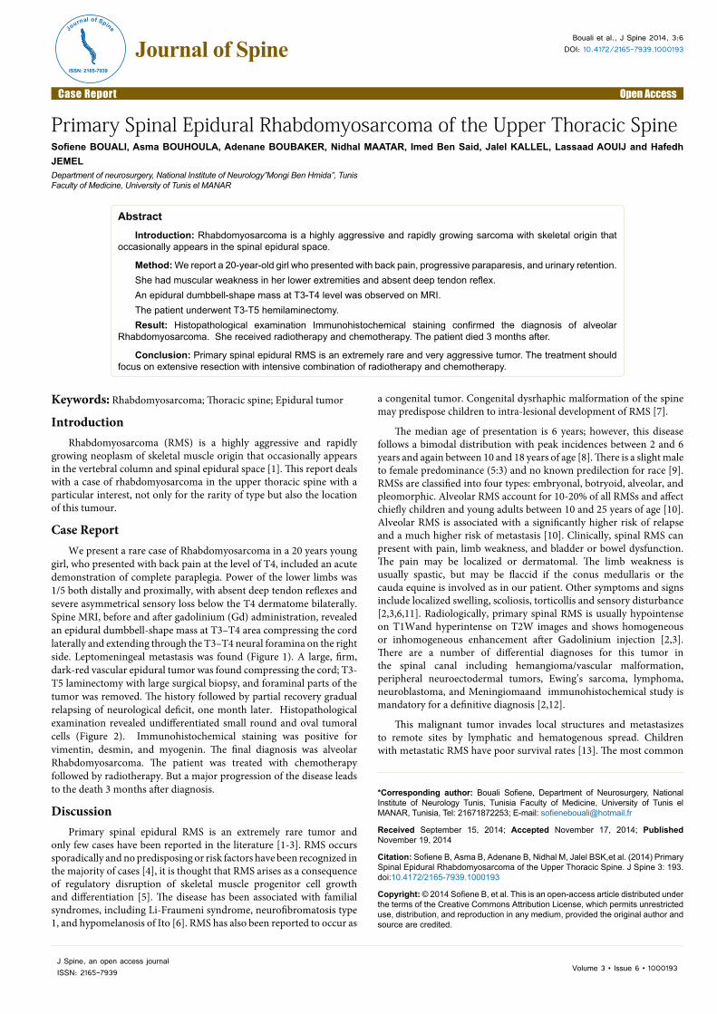
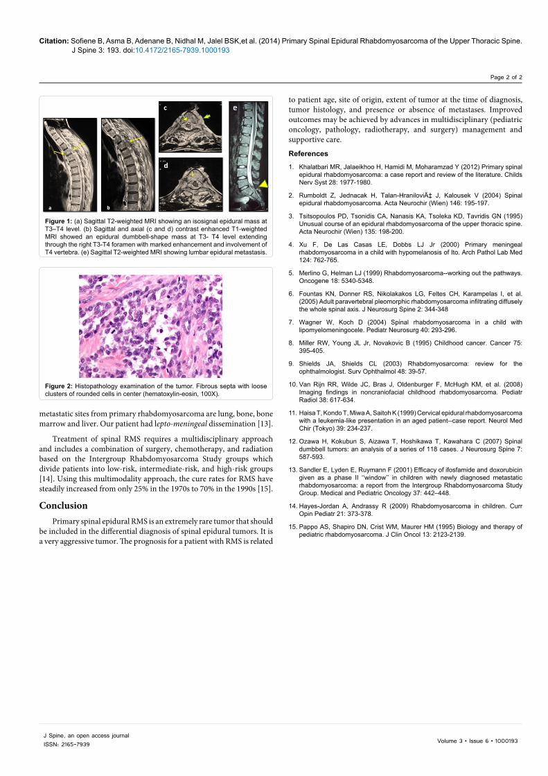
girl, who presented with back pain at the level of T4, included an acute demonstration of complete paraplegia. Power of the lower limbs was 1/5 both distally and proximally, with absent deep tendon reflexes and severe asymmetrical sensory loss below the T4 dermatome bilaterally. Spine MRI, before and after gadolinium (Gd) administration, revealed an epidural dumbbell-shape mass at T3–T4 area compressing the cord laterally and extending through the T3–T4 neural foramina on the right side. Leptomeningeal metastasis was found (Figure 1). A large, firm, dark-red vascular epidural tumor was found compressing the cord; T3-T5 laminectomy with large surgical biopsy, and foraminal parts of the tumor was removed. The history followed by partial recovery gradual relapsing of neurological deficit, one month later. Histopathological examination revealed undifferentiated small round and oval tumoral cells (Figure 2). Immunohistochemical staining was positive for vimentin, desmin, and myogenin. The final diagnosis was alveolar Rhabdomyosarcoma. The patient was treated with chemotherapy followed by radiotherapy. But a major progression of the disease leads to the death 3 months after diagnosis.
DiscussionPrimary spinal epidural RMS is an extremely rare tumor and
only few cases have been reported in the literature [1-3]. RMS occurs sporadically and no predisposing or risk factors have been recognized in the majority of cases [4], it is thought that RMS arises as a consequence of regulatory disruption of skeletal muscle progenitor cell growth and differentiation [5]. The disease has been associated with familial syndromes, including Li-Fraumeni syndrome, neurofibromatosis type 1, and hypomelanosis of Ito [6]. RMS has also been reported to occur as
a congenital tumor. Congenital dysrhaphic malformation of the spine may predispose children to intra-lesional development of RMS [7].
The median age of presentation is 6 years; however, this disease follows a bimodal distribution with peak incidences between 2 and 6 years and again between 10 and 18 years of age [8]. There is a slight male to female predominance (5:3) and no known predilection for race [9]. RMSs are classified into four types: embryonal, botryoid, alveolar, and pleomorphic. Alveolar RMS account for 10-20% of all RMSs and affect chiefly children and young adults between 10 and 25 years of age [10]. Alveolar RMS is associated with a significantly higher risk of relapse and a much higher risk of metastasis [10]. Clinically, spinal RMS can present with pain, limb weakness, and bladder or bowel dysfunction. The pain may be localized or dermatomal. The limb weakness is usually spastic, but may be flaccid if the conus medullaris or the cauda equine is involved as in our patient. Other symptoms and signs include localized swelling, scoliosis, torticollis and sensory disturbance [2,3,6,11]. Radiologically, primary spinal RMS is usually hypointense on T1Wand hyperintense on T2W images and shows homogeneous or inhomogeneous enhancement after Gadolinium injection [2,3]. There are a number of differential diagnoses for this tumor in the spinal canal including hemangioma/vascular malformation, peripheral neuroectodermal tumors, Ewing’s sarcoma, lymphoma, neuroblastoma, and Meningiomaand immunohistochemical study is mandatory for a definitive diagnosis [2,12].
This malignant tumor invades local structures and metastasizes to remote sites by lymphatic and hematogenous spread. Children with metastatic RMS have poor survival rates [13]. The most common
Journal of Spine
ISSN: 2165-7939
Journal of Spine
Citation: Sofiene B, Asma B, Adenane B, Nidhal M, Jalel BSK,et al. (2014) Primary Spinal Epidural Rhabdomyosarcoma of the Upper Thoracic Spine. J Spine 3: 193. doi:10.4172/2165-7939.1000193
Page 2 of 2
Volume 3 • Issue 6 • 1000193J Spine, an open access journalISSN: 2165-7939
to patient age, site of origin, extent of tumor at the time of diagnosis, tumor histology, and presence or absence of metastases. Improved outcomes may be achieved by advances in multidisciplinary (pediatric oncology, pathology, radiotherapy, and surgery) management and supportive care.
References
1. Khalatbari MR, Jalaeikhoo H, Hamidi M, Moharamzad Y (2012) Primary spinal epidural rhabdomyosarcoma: a case report and review of the literature. Childs Nerv Syst 28: 1977-1980.
2. Rumboldt Z, Jednacak H, Talan-Hranilović J, Kalousek V (2004) Spinal epidural rhabdomyosarcoma. Acta Neurochir (Wien) 146: 195-197.
3. Tsitsopoulos PD, Tsonidis CA, Nanasis KA, Tsoleka KD, Tavridis GN (1995) Unusual course of an epidural rhabdomyosarcoma of the upper thoracic spine. Acta Neurochir (Wien) 135: 198-200.
4. Xu F, De Las Casas LE, Dobbs LJ Jr (2000) Primary meningeal rhabdomyosarcoma in a child with hypomelanosis of Ito. Arch Pathol Lab Med 124: 762-765.
5. Merlino G, Helman LJ (1999) Rhabdomyosarcoma--working out the pathways. Oncogene 18: 5340-5348.
6. Fountas KN, Donner RS, Nikolakakos LG, Feltes CH, Karampelas I, et al. (2005) Adult paravertebral pleomorphic rhabdomyosarcoma infiltrating diffusely the whole spinal axis. J Neurosurg Spine 2: 344-348
7. Wagner W, Koch D (2004) Spinal rhabdomyosarcoma in a child with lipomyelomeningocele. Pediatr Neurosurg 40: 293-296.
8. Miller RW, Young JL Jr, Novakovic B (1995) Childhood cancer. Cancer 75: 395-405.
9. Shields JA, Shields CL (2003) Rhabdomyosarcoma: review for the ophthalmologist. Surv Ophthalmol 48: 39-57.
10. Van Rijn RR, Wilde JC, Bras J, Oldenburger F, McHugh KM, et al. (2008) Imaging findings in noncraniofacial childhood rhabdomyosarcoma. Pediatr Radiol 38: 617-634.
11. Haisa T, Kondo T, Miwa A, Saitoh K (1999) Cervical epidural rhabdomyosarcoma with a leukemia-like presentation in an aged patient--case report. Neurol Med Chir (Tokyo) 39: 234-237.
12. Ozawa H, Kokubun S, Aizawa T, Hoshikawa T, Kawahara C (2007) Spinal dumbbell tumors: an analysis of a series of 118 cases. J Neurosurg Spine 7: 587-593.
13. Sandler E, Lyden E, Ruymann F (2001) Efficacy of ifosfamide and doxorubicin given as a phase II ‘‘window’’ in children with newly diagnosed metastaticrhabdomyosarcoma: a report from the Intergroup Rhabdomyosarcoma Study Group. Medical and Pediatric Oncology 37: 442–448.
14. Hayes-Jordan A, Andrassy R (2009) Rhabdomyosarcoma in children. Curr Opin Pediatr 21: 373-378.
15. Pappo AS, Shapiro DN, Crist WM, Maurer HM (1995) Biology and therapy of pediatric rhabdomyosarcoma. J Clin Oncol 13: 2123-2139.
Figure 1: (a) Sagittal T2-weighted MRI showing an isosignal epidural mass at T3–T4 level. (b) Sagittal and axial (c and d) contrast enhanced T1-weighted MRI showed an epidural dumbbell-shape mass at T3- T4 level extending through the right T3-T4 foramen with marked enhancement and involvement of T4 vertebra. (e) Sagittal T2-weighted MRI showing lumbar epidural metastasis.
Figure 2: Histopathology examination of the tumor. Fibrous septa with loose clusters of rounded cells in center (hematoxylin-eosin, 100X).
metastatic sites from primary rhabdomyosarcoma are lung, bone, bone marrow and liver. Our patient had lepto-meningeal dissemination [13].
Treatment of spinal RMS requires a multidisciplinary approach and includes a combination of surgery, chemotherapy, and radiation based on the Intergroup Rhabdomyosarcoma Study groups which divide patients into low-risk, intermediate-risk, and high-risk groups [14]. Using this multimodality approach, the cure rates for RMS have steadily increased from only 25% in the 1970s to 70% in the 1990s [15].
ConclusionPrimary spinal epidural RMS is an extremely rare tumor that should
be included in the differential diagnosis of spinal epidural tumors. It is a very aggressive tumor. The prognosis for a patient with RMS is related