Embed Size (px)
Citation preview

*For correspondence:
Competing interests: The
authors declare that no
competing interests exist.
Funding: See page 22
Received: 06 February 2020
Accepted: 13 July 2020
Published: 14 July 2020
Reviewing editor: Mone Zaidi,
Icahn School of Medicine at
Mount Sinai, United States
Copyright Hasan et al. This
article is distributed under the
terms of the Creative Commons
Attribution License, which
permits unrestricted use and
redistribution provided that the
original author and source are
credited.
RAB23 coordinates early osteogenesis byrepressing FGF10-pERK1/2 and GLI1Md Rakibul Hasan1, Maarit Takatalo1, Hongqiang Ma1, Ritva Rice1,Tuija Mustonen1, David PC Rice1,2*
1Craniofacial Development and Malformations research group, Orthodontics, Oraland Maxillofacial Diseases, University of Helsinki, Helsinki, Finland; 2Oral andMaxillofacial Diseases, Helsinki University Hospital, Helsinki, Finland
Abstract Mutations in the gene encoding Ras-associated binding protein 23 (RAB23) cause
Carpenter Syndrome, which is characterized by multiple developmental abnormalities including
polysyndactyly and defects in skull morphogenesis. To understand how RAB23 regulates skull
development, we generated Rab23-deficient mice that survive to an age where skeletal
development can be studied. Along with polysyndactyly, these mice exhibit premature fusion of
multiple sutures resultant from aberrant osteoprogenitor proliferation and elevated osteogenesis in
the suture. FGF10-driven FGFR1 signaling is elevated in Rab23-/-sutures with a consequent
imbalance in MAPK, Hedgehog signaling and RUNX2 expression. Inhibition of elevated pERK1/2
signaling results in the normalization of osteoprogenitor proliferation with a concomitant reduction
of osteogenic gene expression, and prevention of craniosynostosis. Our results suggest a novel
role for RAB23 as an upstream negative regulator of both FGFR and canonical Hh-GLI1 signaling,
and additionally in the non-canonical regulation of GLI1 through pERK1/2.
IntroductionRas-associated binding protein 23 (RAB23) belongs to the RAB family of small GTPases, which func-
tion during several steps in cell vesicle trafficking (Evans et al., 2003). RAB23 has 35–40% sequence
homology with other RABs, it localizes to the plasma membrane and is proposed to regulate cargo
internalization and relocation in the cell (Evans et al., 2003; Leaf and Von Zastrow, 2015; Lim and
Tang, 2015; Olkkonen et al., 1994). RAB23 is unusual amongst RABs as it is involved in embryogen-
esis, for instance in heart and limb patterning, neural tube closure and skeletal development
(Eggenschwiler and Anderson, 2000; Eggenschwiler et al., 2001; Fuller et al., 2014;
Gunther et al., 1994; Hor and Goh, 2018). RAB23 negatively regulates Hh signaling during mouse
neural tube development (Eggenschwiler and Anderson, 2000), and it controls nodal signaling
independently of Hh signaling during vertebrate left-right patterning (Fuller et al., 2014). RAB23 is
involved in trafficking D1-type dopaminergic receptors (Leaf and Von Zastrow, 2015) and kinesin-2
motor protein Kif-17 (Lim and Tang, 2015) to the primary cilium. However, direct ciliary function of
RAB23 is unclear as cilium length and function are reported normal in Rab23-/- mice (Evans et al.,
2003; Fuller et al., 2014).
Multiple mutations in RAB23 have been reported in patients with the autosomal recessive Car-
penter syndrome; the majority of mutations result in nonsense-mediated decay while others include
missense and in-frame deletions (Jenkins et al., 2011). Carpenter syndrome (CS, MIM #201000) or
Acrocephalopolysyndactyly type II is a multi-organ developmental disorder with polysyndactyly, con-
genital heart defects, mental retardation, obesity and craniosynostosis as central features (Carpen-
ter, 1909; Jenkins et al., 2007).
Craniosynostosis is the premature fusion of one or more craniofacial sutures that results in major
disruption of face and skull growth. Mesenchymal cells in the center of the suture must be kept in an
Hasan et al. eLife 2020;9:e55829. DOI: https://doi.org/10.7554/eLife.55829 1 of 26
RESEARCH ARTICLE

undifferentiated state to maintain suture patency, while progenitor cells at the osteogenic fronts
proliferate and differentiate to facilitate bone growth. Suture biogenesis is dependent on the correct
patterning of the skeletal elements, as well as the regulation of the mesenchymal stem cell niche,
osteogenic condensation formation, osteoprogenitor proliferation and differentiation (Rice and
Rice, 2008; Twigg and Wilkie, 2015). These developmental processes are regulated to permit coor-
dinated craniofacial growth, without the fusion of the neighboring bones and consequent cessation
of growth.
Mutations in FGFRs are a major cause of syndromic craniosynostosis, with many of the mutations
conferring a gain of function (Hajihosseini, 2008; Wilkie et al., 2017). FGFRs isoforms show a dis-
tinct FGF ligand binding affinity and biological functions (Ornitz and Itoh, 2015). However, studies
have shown that in an Apert syndrome (FGFR2 mutation) mouse model isoform FGFR2c loses ligand
specificity, and is able to bind with cognate FGF10 (Ibrahimi et al., 2001; Ibrahimi et al., 2004;
Johnson and Wilkie, 2011; Yu et al., 2000). Interestingly, genetic knockdown of Fgf10 in this
mouse model could rescue the premature fusion (Hajihosseini et al., 2009). FGF signaling pathway
members have not been linked to RAB23-mediated trafficking. However, study suggests that RAB23
resides in the plasma membrane and proposed to be involved in endocytosis (Evans et al., 2003). In
this context, RAB23 might have a direct role in growth factor receptor recycling and turnover, and
therefore regulate the availability of the FGF receptors at the cell surface (Langemeyer et al., 2018;
Zerial and McBride, 2001).
Similar to Carpenter syndrome, several craniosynostosis syndromes caused by mutations in FGFRs
are characterized by patients exhibiting syndactyly and occasionally polysyndactyly (Goos and
Mathijssen, 2019; Mantilla-Capacho et al., 2005). Also, mutations in several hedgehog (Hh) path-
way members cause polydactyly (Malik, 2014; Ullah et al., 2019). Notably, Greig cephalopolysyn-
dactyly syndrome (MIM # 175700) is caused by haploinsufficiency of the Hh signaling negative
regulator, GLI3 (Vortkamp et al., 1991). In addition to the polysyndactyly, some patients with Greig
cephalopolysyndactyly syndrome exhibit craniosynostosis, and the mouse model for Greig cephalo-
polysyndactyly syndrome (Gli3Xt-J/Xt-J) shows complete phenotypic penetrance for both craniosynos-
tosis and polydactyly (Hurst et al., 2011; Rice et al., 2010). Also, a recurrent mosaic mutation in the
hedgehog receptor Smoothened (SMO) causes craniosynostosis and polysyndactyly (Twigg et al.,
2016). These overlapping skeletal phenotypes caused by mutations in FGFRs, Hh regulators and
RAB23 are suggestive of common etiological mechanisms. FGF and Hh signaling have well-defined
roles during intramembranous osteogenesis. FGF signaling regulates many stages including
eLife digest In many animals, the skull is made of several separate bones that are loosely joined
during childhood and only fuse into one piece when the animal stops growing. A genetic disease
called Carpenter syndrome causes the bones of the skull to fuse early in life, stopping it from
growing correctly. Carpenter syndrome is often caused by changes to the gene responsible for
making a protein called RAB23.
RAB23 helps move other molecules and cell components between different parts of the cell, and
is therefore involved in a number of cellular processes. Previous studies suggest that RAB23 has a
role in many parts of the body during development. Yet, it is unclear which cells in the skull depend
on RAB23 activity and how this protein is controlled.
To answer this question, Hasan et al. grew pieces of developing skull bones that had been taken
from mice lacking the RAB23 protein in the laboratory. Examining these samples revealed that
RAB23 is active in cells called osteoblasts that add new bone to the edge of each piece of the skull
as it grows. Hasan et al. also found that RAB23 regulates two cellular signaling pathways – called the
hedgehog pathway and the fibroblast growth factor pathway – that interact with one another and
co-ordinate skull development.
These findings show how RAB23 controls the growth and fusion of skull bones in developing
animals. This could improve our understanding of the role RAB23 plays in other processes during
development. It also sheds light on the mechanisms of Carpenter syndrome which may inform new
approaches for treating patients.
Hasan et al. eLife 2020;9:e55829. DOI: https://doi.org/10.7554/eLife.55829 2 of 26
Research article Developmental Biology Medicine

mesenchymal condensation formation, osteoprogenitor proliferation and differentiation and activa-
tion of the osteogenic transcription factor RUNX2 (Debiais et al., 1998; Kim et al., 1998;
Ornitz and Itoh, 2015; Yoon et al., 2014). IHH positively regulates osteoprogenitor recruitment to
the osteogenic front and GLI transcription factors regulate stem cell maintenance and osteoprogeni-
tor proliferation (Lenton et al., 2011; Rice et al., 2010; Veistinen et al., 2012; Zhao et al., 2015).
Interestingly, RAB23 regulates GLI1 in a Su(Fu)-dependent manner (Chi et al., 2012) and GLI1-posi-
tive cells have been identified in the suture as the main source of mesenchymal stem cells that plays
crucial role in suture patency (Zhao et al., 2015).
The aim of this study was to determine the role of RAB23 during intramembranous bone develop-
ment. Previously, it has not been possible to study skeletal development in RAB23 deficient mice
due to their early lethality. Here, we generated RAB23 deficient (Rab23-/-) mice that survived until
embryonic day (E) 18.5, and this allowed us to investigate how RAB23 regulates bone formation.
Similar to Carpenter syndrome patients, RAB23 deficient mice exhibited polysyndactyly and multiple
craniosynostoses. We show that disruption of Rab23 leads to an upregulation of Pitx2, Fgf10 and
Fgfr1b expression, decreased p38 and enhanced pERK1/2-RUNX2 signaling along with elevated
osteoprogenitor proliferation. In addition, Hh signaling was amplified with increased expression of
GLI1. During in vitro culture, inhibition of elevated pERK1/2 normalized osteoprogenitor prolifera-
tion, corrected the aberrant GLI1 and RUNX2 expressions, and rescued the lambdoid suture fusion.
Our results suggest a novel role for RAB23 as an upstream regulator of both FGF10-pERK1/2 and
Hh-GLI1, and the additional regulation of GLI1 by pERK1/2, to coordinate the initiation of
osteogenesis.
Results
Rab23-/- mice exhibit craniosynostosis in multiple suturesRab23 open brain 2 (opb2) homozygous mutant mice did not produce RAB23 protein in primary
cells, analyzed by western blot (Figure 1—figure supplement 1) and will therefore be referred to as
Rab23-/- mice. Rab23-/- C3 Heb/FeJ mice, generated through an ENU screen, survive until E12.5 ren-
dering full analysis of organogenesis impossible (Eggenschwiler et al., 2001; Kasarskis et al.,
1998). To obviate the gestation lethality before skeletal development, we backcrossed Rab23-/- C3
Heb/FeJ mice onto the C57Bl/6 strain. After six generations, the survival of the Rab23-/- homozygote
embryos were prolonged to E18.5, and this allowed us to study skeletal development of these mice.
Rab23-/- mice at embryonic day E18.5 exhibited craniosynostosis in the coronal, parietal-tempo-
ral, fronto-nasal and in the lambdoid sutures when compared to their wild type (Wt) littermates
(Figure 1A–E). The prevalence of premature suture fusion in Rab23-/- mice represented as percent-
age and only parieto-temporal sutures showed bi-lateral suture fusion (100%) in all the samples
(Figure 1F). Along with craniosynostosis, Rab23-/- mice showed skeletal patterning defects in the
forelimbs and hindlimbs. Rab23-/- mice showed pre-axial polydactyly (seven digits) of the forelimb
and preaxial polysyndactyly (seven digits) of the hindlimbs (Figure 1G). Polydactyly and craniosynos-
tosis were observed (100%) in all Rab23-/- samples examined.
At E18.5, 5% of Rab23-/- lambdoid sutures exhibited unilateral suture fusion (Figure 1F). Com-
pared to Wt samples (Figure 1I), unfused Rab23-/- lambdoid sutures showed abnormal bony protru-
sions from parietal bones toward interparietal bone (Figure 1J) or ectopic bones in the lambdoid
suture (Figure 1K). At this stage, Rab23-/- lambdoid sutures were narrower than Wt lambdoid
sutures (Figure 1L). As 95% of Rab23-/- lambdoid sutures were consistently patent at E18.5 they
were chosen as a model to study the role of RAB23 in osteogenesis. Rab23-/- mice die neonatally,
therefore, analysis of Rab23-/- lambdoid suture beyond the E18.5 stage was assessed by in vitro cal-
varial explant culture (Rice et al., 2003b; Figure 1M). Wt and patent Rab23-/- lambdoid sutures har-
vested for culture at E18.5, Rab23-/- lambdoid sutures fused predictably after 3 days of culture
(Figure 1N,O).
Rab23 expression in calvaria and suturesRab23 mRNA expression was analyzed in whole calvaria and tissue sections by in situ hybridization
and protein expression by immunohistochemical staining and western blotting. Rab23 was
expressed in suture mesenchyme with the strongest signal in interfrontal, sagittal and coronal
Hasan et al. eLife 2020;9:e55829. DOI: https://doi.org/10.7554/eLife.55829 3 of 26
Research article Developmental Biology Medicine

Figure 1. Deficiency of RAB23 causes premature fusion of multiple sutures and polysyndactyly. (A–E) Analysis of
Wt and Rab23-/- skulls by m-CT (A–C) and alizarin red, alcian blue staining (D–E) at E18.5. Rab23-/- skulls show
fusion in the parieto-temporal suture (B-a, black arrow), coronal suture (B-a, green arrow and B-b, m-CT slice,
green arrow), frontonasal suture (C-a, arrow) and lambdoid suture (E-a, arrow). Wt sutures were open at this
embryonic stage (A–a, A–b) (n = 10 for each age and genotype). fb: frontal bone, pb: parietal bone. Scale bar: 1
mm. (F) The prevalence of suture fusion in Rab23-/- mouse at E18.5 shown in percentage. Only parieto-temporal
suture showed bi-lateral suture fusion in all the samples. (n = 10 for sagittal and interfrontal suture, n = 40 for
lambdoid suture and n = 20 for other sutures). (G) Skeletal Analysis of the limbs in Rab23-/- mouse show pre-axial
Figure 1 continued on next page
Hasan et al. eLife 2020;9:e55829. DOI: https://doi.org/10.7554/eLife.55829 4 of 26
Research article Developmental Biology Medicine

sutures and parietal bone side of the lambdoid suture (Figure 1P–a). Analysis of tissue sections
revealed Rab23 expression in the osteogenic fronts of the calvarial sutures and throughout the
sutural mesenchyme (Figure 1P–b). RAB23 was expressed in the lambdoid sutural osteogenic fronts
and in the underlying cartilage. Co-expression with RUNX2 confirmed the cells in the osteogenic
front to be osteoprogenitor or osteoblast (Figure 1P–c). The Rab23 expression pattern was consis-
tent with the phenotype of synostosed calvarial sutures observed in the Rab23-/- mice (Figure 1B,C
and E). Western blotting of proteins extracted from E15.5 Wt calvarial frontal, sagittal and lambdoid
sutures along with their osteogenic fronts confirmed the presence of RAB23 protein in the suture
(Figure 1P–d).
Rab23-/- mice exhibit elevated levels of Fgf10 and Pitx2 in the calvariaand sutureTo investigate differentially expressed genes between Wt and Rab23-/- calvaria and to find candidate
genes responsible for the craniosynostosis in the RAB23-deficient mice, we performed a microarray-
based gene expression analysis of whole Wt and Rab23-/- calvaria samples excluding the skin and
underlying brain at E15.5 (Figure 2A). Our analysis revealed 223 significantly differentially expressed
genes between the Wt and Rab23-/- calvaria (t-test, p<0.05) (Figure 2B, Supplementary file 1,
MIAME-compliant data has been deposited in GEO database as source data, GEO accession
GSE140884). Among these genes, 115 genes were upregulated (Figure 2B, red) and 108 genes
were downregulated (Figure 2B, green). Fgf10 was found to be overexpressed in the microarray in
Rab23-/- calvaria (Figure 2C). Fgf10 was selected as a candidate for further analysis as it has previ-
ously been shown to be expressed early in calvarial development and has been implicated in cranio-
synostosis pathogenesis (Hajihosseini et al., 2009; Veistinen et al., 2009). Fgf10 expression was
elevated in whole calvarial mesenchyme, lambdoid sutural mesenchymal tissue and in calvaria
derived (CD) mesenchymal cells at E15.5 as assessed by RT-qPCR (Figure 2D). Our microarray analy-
sis also showed that the transcription factor Pitx2 was elevated in Rab23-/- calvaria (Figure 2C). Pitx2
is a well-documented upstream regulator of Fgf10 and may act in a reciprocal regulatory loop with
FGF10 during early organogenesis (Al Alam et al., 2012). We found that Pitx2 was overexpressed
both in the Rab23-/- calvaria and in Rab23-/- lambdoid sutures (Figure 2E). Pitx2 overexpression was
Figure 1 continued
polydactyly of the fore limb (FL, 1a, 2a) and pre axial polysyndactyly of the hind limb (HL, 1a, 2a) at E18.5 (n = 10
for each age and genotype. Scale bar: 1.5 mm. (H–K) E18.5 mouse calvaria indicating fb: frontal bone, pb: parietal
bone, ipb: interparietal bone and ls: lambdoid suture (H). Analysis of Wt and Rab23-/- calvaria by m-CT at E18.5
shows Rab23-/- lambdoid sutures form bony protrusions from parietal bones project towards the interparietal
bones (J, arrow), or ectopic bony islands in the mid-sutural mesenchyme (K, arrow) (n = 6 for each age and
genotype). Scale bar: 500 mm. (L) Measurements of the lambdoid suture shows Rab23-/- lambdoid sutures are
narrower as compare to the Wt samples at E18.5 (n = 6 for each age and genotype). Data represented as
mean ± SD, paired Student’s t-test was used. Statistical significance was defined as a p-value < 0.05 (*). (M) In vitro
calvaria culture system. This system was used to culture Wt and Rab23-/- calvaria containing patent lambdoid
sutures at E17.5 and E18.5. (N, O) Represents E18.5 Wt and patent lambdoid suture containing Rab23-/- calvaria
culture in vitro for 3 days in presence of alizarin red. Rab23-/- lambdoid suture shows fusion at day 3 (N, alizarin red
bone staining, O, m-CT images), whereas Wt controls remain open (n = 10 for each genotype). Scale bar: 500 mm.
(P) Rab23 expression in the whole calvarial tissue at E16.5 is shown by digoxigenin labeled whole mount in situ
hybridization (P–a) and shown at E15.5 sutural tissue sections by RNAscope (P–b). Co-expression of RAB23 (green)
and osteoblast marker RUNX2 (red) in the calvarial sutural section at E17.5 is shown by immunohistochemical
staining (P–c), nuclear staining (blue). RAB23 protein expression in the sutures at E15.5 is shown by western
blotting (P–d). fb: frontal bone, pb: parietal bone, ipb: interparietal bone, fs: frontal suture, ss: sagittal suture, ls:
lambdoid suture. Scale bar: 500 mm (a), 100 mm (b, c).
The online version of this article includes the following figure supplement(s) for figure 1:
Figure supplement 1. RAB23 protein expression in WT and Rab23-/- mouse calvaria derived primary cells.
Hasan et al. eLife 2020;9:e55829. DOI: https://doi.org/10.7554/eLife.55829 5 of 26
Research article Developmental Biology Medicine
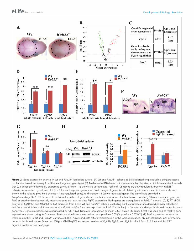
Figure 2. Gene expression analysis in Wt and Rab23-/- lambdoid suture. (A) Wt and Rab23-/- calvaria at E15.5 (dotted ring, excluding skin) processed
for Illumina based microarray (n = 3 for each age and genotype). (B) Analysis of mRNA based microarray data by Chipster, a bioinformatics tool, reveals
that 223 genes are differentially expressed (t-test, p<0.05, 115 genes are upregulated, red and 108 genes are downregulated, green) in Rab23-/-
calvaria, represented by volcano plot (n = 3 for each age and genotype). Fold change of genes is calculated by arithmetic mean in linear scale and
shown in the volcano plot. Fold change >1 (up-regulated gene), fold change < 1 (down-regulated gene). The gene list is provided in
Supplementary file 1. (C) Represents individual searches of genes based on their contribution of suture fusion reveals Fgf10 as a candidate gene and
Pitx2 as another developmentally important gene that can regulate Fgf10 expression. Both genes are upregulated in Rab23-/- calvaria. (D, E) RT-qPCR
analysis of Fgf10 (D) and Pitx2 (E) mRNA extracted from E15.5 Wt and Rab23-/- calvaria (excluding skin), cultured calvaria derived primary cells (CDC)
and from lambdoid sutural tissue reveals that Fgf10 and Pitx2 are overexpressed in Rab23-/- samples (n = 3 calvaria and eight lambdoid sutures for each
genotype). Gene expressions were normalized by 18S rRNA. Data are represented as mean ± SD, paired Student’s t-test was used and as relative gene
expression is shown using DDCт values. Statistical significance was defined as a p-value <0.05 (*), p-value <0.005 (**). (F) Pitx2 expression analysis by
whole-mount ISH in Wt and Rab23-/- calvaria at E15.5. Arrows indicate Pitx2 overexpression in the lambdoid suture. pb: parietal bone, ipb: interparietal
bone, ls: lambdoid suture. Scale bar: 500 mm. (G) RT-qPCR expression analysis of Fgfr1b, Fgfr2b and Fgfr2c mRNA from E15.5 Wt and Rab23-/-
Figure 2 continued on next page
Hasan et al. eLife 2020;9:e55829. DOI: https://doi.org/10.7554/eLife.55829 6 of 26
Research article Developmental Biology Medicine

further observed in Rab23-/- calvaria by whole-mount in situ hybridization in and surrounding the
interparietal bone (Figure 2F).
FGF10-induced upregulation of Fgfr1b expression in Rab23-/- lambdoidsutureWe assessed the expression of FGF10-specific receptors Fgfr1b, Fgfr2b, also Fgfr2c in Wt and
Rab23-/- lambdoid sutural mesenchymal tissue. Our analysis showed elevated Fgfr1b expression and
a non-significant upregulation of Fgfr2b in Rab23-/- samples (Figure 2G). FGFR1 protein levels were
found to be elevated in Rab23-/- lambdoid sutures compared to Wt samples when analyzed by west-
ern blotting (Figure 2H,I) and by immunohistochemical staining (Figure 2—figure supplement 1).
The expression of Fgfr2c was unchanged (Figure 2G). To test whether the upregulation of Fgfr1b
and Fgfr2b expression was due to the high levels of FGF10 ligand present in the Rab23-/- lambdoid
suture, we isolated E15.5 wild-type calvaria derived (CD) mesenchymal cells and treated them with
exogenous FGF10 (250 ng/ml) for 3 hr. Fgfr1b and Fgfr2b expression levels were then assessed by
RT-qPCR. We found a significant (p=0.05) upregulation of Fgfr1b expression and a slight upregula-
tion of Fgfr2b expression in the cultured CD mesenchymal cells after exogenous FGF10 stimulation,
indicating FGFR1b as a target of FGF10 in the calvarial mesenchyme (Figure 2J).
Opposing MAPK-RUNX2 and Hh-GLI1 signaling in Rab23-/- lambdoidsutureFGF signaling has been shown to activate MAPK signaling pathway subtypes pERK and p38 in suture
morphogenesis and osteoblast differentiation (Kyono et al., 2012; Pfaff et al., 2016; Figure 3A). In
Rab23-/- lambdoid suture, we found decreased p38 and elevated pERK1/2 and RUNX2 expressions
compare to Wt samples (Figure 3B–G). To test whether the upregulation of RUNX2 expression was
due to the high levels of FGF10 present in the Rab23-/- lambdoid suture, we isolated E15.5 Rab23-/-
calvaria derived (CD) mesenchymal cells and treated them with exogenous FGF10 (500 ng/ml) for 2
and 4 hr. Runx2 expression levels were then assessed by RT-qPCR. We found a significant (p<0.05)
upregulation of Runx2 expression at both time points compare to untreated samples, indicating that
RUNX2 as a target of FGF10 in the calvarial mesenchyme (Figure 3H). Since ERK1/2 signaling regu-
lates Hh and GLI expression, and RAB23 is a known negative regulator of Hh signaling
(Eggenschwiler et al., 2001), we further assayed Hh, Gli1, Gli2, and Gli3 transcripts extracted from
Wt and Rab23-/- lambdoid sutural mesenchyme at E15.5. We detected a dramatic increase in the
expression of Hh (Figure 3I) and Gli1 (Figure 3J), a sutural stem cell marker (Zhao et al., 2015).
Although Gli3 expression is not changed, a functional change in the ratio of GLI3FL to GLI3R may
still possible (Veistinen et al., 2017). As TGFb-superfamily members have also been shown to regu-
late suture patency (Kim et al., 1998; Komatsu et al., 2013; Pan et al., 2017), we assayed both
pSMAD1/5/8 and pSMAD2/3 expressions but found no difference between Wt and mutant samples
(Figure 3—figure supplement 1).
Figure 2 continued
lambdoid suture reveals Fgfr1b overexpression in Rab23-/- sample (n = 8 for each genotype). Gene expressions were normalized by 18S rRNA. Data are
represented as mean ± SD, paired Student’s t-test was used and relative gene expression is shown using DDCт values. Statistical significance was
defined as a P-value <0.05 (*). (H, I) Western blotting of proteins extracted from Wt and Rab23-/- lambdoid suture at E15.5 (H) and relative intensity
measurement (I) reveals over expression of FGFR1 in the Rab23-/- lambdoid suture (n = 6 for each genotype). Data represented as mean ± SD, paired
Student’s t-test was used. Statistical significance was defined as a p-value <0.05 (*). (J) Exogenous FGF10 treatment for 3 hr on Wt calvaria derived (CD)
cells and subsequent Fgfr1b and Fgfr2b mRNA analysis by q-PCR shows induction of Fgfr1b expression in FGF10 treated cells compare to untreated
Wt CD cells (n = 3 for each genotype). Gene expressions were normalized by 18S rRNA. Data are represented as mean ± SD, paired Student’s t-test
was used and relative gene expression is shown using DDCт values. Statistical significance was defined as a P-value.
The online version of this article includes the following figure supplement(s) for figure 2:
Figure supplement 1. FGFR1 expression in Wt and Rab23-/- lambdoid suture.
Hasan et al. eLife 2020;9:e55829. DOI: https://doi.org/10.7554/eLife.55829 7 of 26
Research article Developmental Biology Medicine

Figure 3. Analysis of MAPK signaling in Wt and Rab23-/- lambdoid suture. (A) Represents two downstream pathway subtypes p38 and ERK of MAPK
signaling involve in RUNX2 activation and suture fusion. (B, C) Western blotting analysis of pERK44/42 and a-ERK44/42 protein levels extracted from Wt
and Rab23-/- lambdoid suture at E15.5 (B) and relative intensity measurement (C) shows higher pERK44 and pERK42 levels in Rab23-/- lambdoid suture
(n = 6 for each genotype). Data represented as mean ± SD, paired Student’s t-test was used. Statistical significance was defined as a P-value <0.05 (*).
(D, E) Western blotting analysis of phospho-p38 and p38 protein levels extracted from Wt and Rab23-/- lambdoid suture at E15.5 (D) and relative
intensity measurement (E) shows lower phospho-p38 and p38 levels in Rab23-/- lambdoid suture (n = 6 for each genotype). Data represented as
Figure 3 continued on next page
Hasan et al. eLife 2020;9:e55829. DOI: https://doi.org/10.7554/eLife.55829 8 of 26
Research article Developmental Biology Medicine

Rab23-/- lambdoid suture and MEF cells show increasedosteoprogenitor cell proliferationAs FGF10-pERK1/2 signaling and RUNX2 are known to regulate suture osteoprogenitor proliferation
(Kim et al., 1998; Qin et al., 2019), we assessed co-expression of EdU and RUNX2 in the WT and
Rab23-/- lambdoid suture. EdU and RUNX2 co-localized in the osteogenic fronts indicating that the
proliferating cells are osteoprogenitors (Figure 4A,B). We analyzed the proliferation in Wt and
Rab23-/- lambdoid sutures (Figure 4C–H). EdU pulsed Rab23-/- samples showed more proliferating
cells in the osteogenic fronts compared to Wt (Figure 4C–H). In addition, increased level of cell pro-
liferation was observed in Rab23-/- derived mouse embryonic fibroblast (MEF) cells (Figure 4I).
Rab23-/- calvaria derived cells also had an elevated number of total cells at different passages (P1 to
P3) when compared to Wt calvaria derived cells (Figure 4J). Increased proliferation in the sutures
can result in a larger pool of osteoprogenitor cells and enhanced osteogenesis (Lana-Elola et al.,
2007; Rice et al., 2003b). Taken together, this data shows that RAB23 is needed to repress prolifer-
ation in undifferentiated mesenchymal cells.
RAB23 regulates suture patency through a MEK-driven mechanismOur data suggest that Rab23-/- lambdoid suture undergoes fusion through aberrant FGF signaling,
specifically by activating pERK1/2 signaling, RUNX2 and enhanced cell proliferation of osteoprogeni-
tor cells. To test this, we aimed to normalize the Rab23-/- lambdoid suture by inhibiting the activa-
tion of MEK1/2, a downstream component of the MAPK-ERK1/2 signaling pathway by using MEK1/2
inhibitor U0126. We cultured E18.5 Wt and Rab23-/- whole calvaria in osteogenic growth medium up
to 72 hr. To confirm that lambdoid sutures were patent at time point zero, the vital bone dye calcein
green was used to detect the bone edges. Calcein green was administered to pregnant females 24
hr before samples were taken. Half of the Rab23-/- calvaria were treated with U0126 and half of the
Rab23-/- calvaria samples were untreated (Figure 5A,B). None of the untreated Rab23-/- (n = 0/12)
and 84.34% of U0126 treated Rab23-/- (n = 11/12) lambdoid sutures were rescued (Figure 5A,B). To
confirm whether bones were fused or not, Wt, U0126 treated and untreated Rab23-/- samples were
further analyzed in tissue sections and by m-CT (Figure 5A–C, Figure 5—figure supplement 1 and
Figure 5—Videos 1–3). None of the Wt lambdoid sutures were fused (n = 0/12). The same experi-
ment (n = 4 in each genotype) was carried out with E17.5 samples and cultured for 4 days. Untreated
Rab23-/- lambdoid sutures underwent suture fusion by day 4, while U0126-treated Rab23-/- lambdoid
sutures remained patent during the culture period (Figure 5—figure supplement 2). The effect of
U0126 on sutural cells was also analyzed by cell death assay and no effect was observed in U0126-
treated lambdoid sutural cells (Figure 5—figure supplement 3). U0126 treatment not only pre-
vented suture fusion but it also normalized the level of osteoprogenitor cell proliferation
(Figure 5D–G). EdU incorporation revealed that U0126 treated Rab23-/- lambdoid sutures (n = 8)
Figure 3 continued
mean ± SD, paired Student’s t-test was used. Statistical significance was defined as a p-value <0.05 (*), p-value <0.005 (**). (F, G) Western blotting
analysis of RUNX2 protein level extracted from Wt and Rab23-/- lambdoid suture at E15.5 (F) and relative intensity measurement (G) shows higher
RUNX2 level in Rab23-/- lambdoid suture (n = 6 for each genotype). Data represented as mean ± SD, paired Student’s t-test was used. Statistical
significance was defined as a p-value <0.05 (*). (H) Runx2 expression analysis by RT-qPCR in exogenous FGF10 treated (500 ng/ml) and untreated
Rab23-/- calvaria derived cells at 2 and 4 hr. (n = 3 for each genotype). Gene expressions were normalized by 18S rRNA. Data are represented as
mean ± SD, paired Student’s t-test was used and relative gene expression is shown using DDCт values. Statistical significance was defined as a P-value
<0.05 (*), P-value <0.005 (**). (I, J) RT-qPCR expression analysis of hedgehog signaling components Hh (H), Gli1, Gli2 and Gli3 (I) in the Wt and Rab23-/-
lambdoid suture reveals overexpression of Hh and Gli1 in Rab23-/- lambdoid suture (n = 8 for each genotype). Gene expressions were normalized by
18S rRNA. Data are represented as mean ± SD, paired Student’s t-test was used and relative gene expression is shown using DDCт values. Statistical
significance was defined as a P-value <0.05 (*), P-value <0.005 (**). n.s: non-significant.
The online version of this article includes the following figure supplement(s) for figure 3:
Figure supplement 1. pSMAD1/5/8 and pSMAD2/3 expression in Wt and Rab23-/- lambdoid suture.
Hasan et al. eLife 2020;9:e55829. DOI: https://doi.org/10.7554/eLife.55829 9 of 26
Research article Developmental Biology Medicine

Figure 4. Cell proliferation analysis in Wt and Rab23-/- lambdoid sutural cells and MEF cells. (A–H) EdU pulsed assay in the Wt and Rab23-/- lambdoid
sutures at E17.5 shows proliferating cells in red color (A–F). Proliferating cells show co-localization with osteoprogenitor and osteoblast marker RUNX2
(green) in the osteogenic front (inset, white arrow) in both Wt and Rab23-/- lambdoid suture (A, B). Analysis of EdU pulsed cells (C–F) together with
nuclear staining (E, F) in the Wt and Rab23-/- lambdoid suture and subsequent quantification revealed that Rab23-/- sutures show higher cell
Figure 4 continued on next page
Hasan et al. eLife 2020;9:e55829. DOI: https://doi.org/10.7554/eLife.55829 10 of 26
Research article Developmental Biology Medicine
exhibited reduced proliferation when compared to Rab23-/- controls (n = 8) (Figure 5D–G) and nor-
malized it to Wt levels (n = 8) after 24 hr of culture (Figure 5D,E). The proliferation in all genotypes
decreased over time (Figure 5D–G). This indicates that RAB23 regulates suture patency through a
MAPK-MEK-ERK-driven mechanism. pERK1/2 regulates RUNX2 and GLI1 expression in calvaria
derived mesenchymal cells.
We analyzed the effect of the MEK1/2 inhibitor U0126, cyclopamine and the combined action of
cyclopamine and U0126 on pERK1/2, RUNX2 and GLI1 expression levels in Rab23-/- CD cells at 6, 24
and 48 hr of culture (Figure 6). After 6 hr of culture, the relative pERK1/2 levels were significantly
reduced in U0126-treated Rab23-/- cells compare to untreated Rab23-/- cells. The downregulation of
pERK1/2 continued in U0126-treated Rab23-/- cells at 24 and 48 hr of culture compare to the
untreated Rab23-/- cells (Figure 6A,B). Whereas, RUNX2 expression significantly reduced only after
48 hr of U0126-treated Rab23-/- cells (Figure 6A,C). U0126-treated Rab23-/- cells showed gradual
reduction of GLI1 expressions at 6 and 24 hr; however, it showed drastic reduction at 48 hr com-
pared to untreated Rab23-/- cells (Figure 6A,D). Further immunohistochemical expression of GLI1
was also found reduced in the U0126 treated Rab23-/- lambdoid sutures at 24 and 48 hr compared
to untreated Rab23-/- lambdoid sutures (Figure 6—figure supplement 1). The effect of cyclopamine
and combined action of cyclopamine and U0126 on these cells showed that cyclopamine has no
effect on pERK1/2 expression and can reduce RUNX2 expression only after prolonged exposure (48
hr) (Figure 6E-H). However, the combined action of cyclopamine and U0126 caused a rapid downre-
gulation of RUNX2 (24 hr) (Figure 6E-H). These findings collectively indicate that pERK1/2 regulates
RUNX2 and GLI1 expressions. Whereas, RUNX2 is a predominant downstream target of pERK1/2
and a weak target of GLI1.
DiscussionThe calvarial suture allows us to study many developmental processes that regulate osteogenesis
and skull morphogenesis. Normal development is ensured by the correct temporal and spatial
coordination of skeletal patterning, mesenchymal stem cell niche regulation, condensation forma-
tion, osteoprogenitor expansion, and osteoblast differentiation and function. In this study, we
generated Rab23-deficient mice that survive into late gestation and they exhibit multiple suture
craniosynostosis. We show that RAB23 regulates the sutural stem cell niche marker GLI1,
represses Hh, and inhibits osteoprogenitor expansion. RAB23 also represses FGF10-driven
pERK1/2 signaling which has the multiple effect of directly regulating osteoprogenitor develop-
ment, RUNX2-mediated differentiation and of repressing GLI1. Therefore, RAB23 coordinates
suture biogenesis by regulating FGF-ERK1/2-RUNX2 activity, as well as the activation of GLI1. In
an attempt to study complex developmental events, we have utilized both primary cells and
organ culture approaches. However, one can also take a more cell biological approach using
immortalized cells remote from the external factors and by using this standardized approach it
may be easier to understand the effects of a specific intervention. This will be help deciphering
the role of RAB23, specifically during endocytosis.
Figure 4 continued
proliferation as percentage of EdU-positive cells compare to total cells (G) and percentage of EdU-positive cells only (H) in those sutures are higher
compare to Wt samples (n = 4 for each genotype). Data represented as mean ± SD, paired Student’s t-test was used. Statistical significance was
defined as a P-value <0.005 (**). pb: parietal bone, ipb: interparietal bone, ls: lambdoid suture, of: osteogenic front. Scale bar: 100 mm. (I) EdU
incorporation in the cultured Wt and Rab23-/- MEF cells isolated from E13.5 embryos show 8–15% more cell proliferation (DNA duplication) in Rab23-/-
samples compare to corresponding Wt samples (n = 3 for each genotype). Data represented as mean ± SD, paired Student’s t-test was used. Statistical
significance was defined as a p-value <0.05 (*). (J) Represents passaging (P1 to P3 ) of Wt and Rab23-/- calvaria derived primary cells in the culture show
the total number of Rab23-/- cells (ml�1) in each passage increases more rapidly than Wt cells, while the cell viability (determined by trypan blue) and
cell size were similar in all cell lines. (n = 3 for each genotype). Data represented as mean ± SD, paired Student’s t-test was used. Statistical significance
was defined as a p-value <0.005 (**), p-value <0.001 (***).
Hasan et al. eLife 2020;9:e55829. DOI: https://doi.org/10.7554/eLife.55829 11 of 26
Research article Developmental Biology Medicine

Figure 5. Rescuing Rab23-/- lambdoid suture fusion with pERK1/2 inhibitor. (A–C) In vivo calcein green incorporation followed by in vitro calvaria
culture in osteogenic medium and adding alizarin red in medium allowed to follow the bone growth in the sutural site. Layout of lambdoid suture
rescue study (A). E18.5 Wt and a double number of Rab23-/- calvaria were cultured in vitro for 3 day. Half of the Rab23-/- calvaria were taken for control
(DMSO) treatment and the other half were treated with ERK1/2 inhibitor U0126. 100% of Rab23-/- control sutures are fused by day 3 of culture (A, B,
Figure 5 continued on next page
Hasan et al. eLife 2020;9:e55829. DOI: https://doi.org/10.7554/eLife.55829 12 of 26
Research article Developmental Biology Medicine

FGF10 signaling regulates early osteogenesis: it is expressed in the osteogenic condensation,
and is implicated in the pathogenesis of several craniosynostotic models (Twigg and Wilkie, 2015;
Veistinen et al., 2009). Here, we show that Fgf10, its receptor FGFR1b and the FGF activator Pitx2,
are all upregulated in Rab23-deficient calvaria. It is surprising that the FGFR1b splice form, which is
normally expressed in epithelia, is upregulated in the mesenchymal suture. However, this is not with-
out precedent. While FGFRc isoforms are predominantly expresses in the mesenchyme and b iso-
forms in the epithelium, their expression is not mutually exclusive and FGFR1b is known to be
expressed by late embryonic murine lung mesenchymal cells and also in the zebrafish in the sutural
mesenchyme (Al Alam et al., 2015; Rice et al., 2003b; Rice et al., 2004; Topczewska et al., 2016).
Under pathological/force conditions, mis-expression of splice forms can occur. Occasionally, Apert
craniosynostosis syndrome is caused by heterozygous Alu-insertions or large deletions in the FGFR2c
domain. These rare mutations induce abnormal expression of the FGFR2b splice form alongside
FGFR2c splice form in mesenchymal cells. This permits cells to respond to both b- and c-activating
FGF ligands including FGF10 (Bochukova et al., 2009; Oldridge et al., 1999). Analyzing the MAPK
pathway in Rab23-/- lambdoid sutures we show that p38 signaling is decreased and pERK1/2
increased. FGFR activation of pERK1/2 is important for several stages of osteoblastogenesis, includ-
ing osteoprogenitor proliferation, and in the induction and stabilization of RUNX2 (Choi et al.,
2008; Yoon et al., 2014). Augmentation of pERK1/2 signaling either through overactivation of
FGFRs, downregulation of its inhibitors, or downregulation of ERF that regulates the export of pERK
from the nucleus, all result in craniosynostosis (Lee et al., 2018a; Lee et al., 2018b;
Timberlake et al., 2017; Twigg et al., 2013).
Although RUNX2 is a substrate for both p38 and pERK1/2, pERK1/2 can activate RUNX2 on aver-
age six times more efficiently than p38 (Ge et al., 2012). In addition, ERK1/2 binding to RUNX2
through MAPK D site has greater affinity than p38 binding and more sensitive to osteoblast differen-
tiation during calvaria explant or primary osteoblast culture (Franceschi and Ge, 2017; Ge et al.,
2012). The importance of pERK1/2 regulation of RUNX2 during osteogenesis is shown in this study
by the reversal of the craniosynostotic phenotype in Rab23-/- mice. p38 has been implicated in the
skull phenotypes of craniosynostotic mice where it may regulate the development and growth of car-
tilage elements (Wang et al., 2010; Wang et al., 2012). Opposing roles of MAPK pathway subtypes
p38 and pERK1/2 in cartilage development are well documented (Ma et al., 2019; Oh et al., 2000;
Stanton et al., 2003).
Figure 5 continued
alizarin red bone staining and C, m-CT images) while 83.34% of Rab23-/- suture with U0126 treated shows no fusion (A, B). # indicates dropout of two
samples from each group due to technical hindrance. Dotted lines indicate bone edge. pb: parietal bone, ipb: interparietal bone. Scale bar: 500 mm (B,
C). (D–G) Represents EdU incorporation in the E18.5 calvaria after 24 hr and 48 hr of culture. EdU was added in the culture medium for additional 2 hr
and cell proliferation analyzed in the sectioned Wt, control Rab23-/- and U0126 treated Rab23-/- lambdoid sutural samples (D, F). U0126 reduced the cell
proliferation in Rab23-/- samples compare to untreated Rab23-/- samples at 24 hr and 48 hr. U0126 treated Rab23-/- suture shows dramatic reduction of
cell proliferation compare to untreated Rab23-/- suture (D, E). The reduction of cell proliferation has shown consistent at 48 hr (F, G) (n = 8 for each
genotype). Data represented as mean ± SD, paired Student’s t-test was used. Statistical significance was defined as a p-value <0.05 (*), p-value <0.005
(**), p-value <0.001 (***). Scale bar: 200 mm (D, F).
The online version of this article includes the following video and figure supplement(s) for figure 5:
Figure supplement 1. Rescuing suture fusion in Rab23-/- calvaria.
Figure supplement 2. Rescuing suture fusion in E17.5 Rab23-/- mice.
Figure supplement 3. Effect of U0126 on Wt and Rab23-/- lambdoid suture.
Figure 5—video 1. Wt lambdoid suture after 72 hr in vitro culture.
https://elifesciences.org/articles/55829#fig5video1
Figure 5—video 2. Rab23-/- lambdoid suture fused after 72 hr in vitro culture.
https://elifesciences.org/articles/55829#fig5video2
Figure 5—video 3. U0126 treated Rab23-/- lambdoid suture remained open after 72 hr in vitro culture.
https://elifesciences.org/articles/55829#fig5video3
Hasan et al. eLife 2020;9:e55829. DOI: https://doi.org/10.7554/eLife.55829 13 of 26
Research article Developmental Biology Medicine

Figure 6. RUNX2 and GLI1 are downstream target of pERK1/2. (A–D) Time-dependent effect of U0126 in
regulating pERK1/2, RUNX2 and GLI1 expressions has been analyzed by western blotting in the proteins extracted
from E15.5 Rab23-/- CD cells. These cells were treated with or without U0126. Relative pERK1/2 levels in Rab23-/-
CD cells are drastically reduces upon U0126 exposure at 6 hr and also shows downregulation of pERK1/2 at 24
Figure 6 continued on next page
Hasan et al. eLife 2020;9:e55829. DOI: https://doi.org/10.7554/eLife.55829 14 of 26
Research article Developmental Biology Medicine

Commonality of etiological mechanisms is suggested by the overlapping skeletal phenotypes
exhibited in Carpenter syndrome and Grieg cephalopolysyndactyly syndrome patients as well as
their mouse disease models, Gli3Xt�J/Xt�J (Rice et al., 2010) and Rab23-/- (this study). Specifically,
postaxial polydactyly in the forelimb, preaxial polydactyly in the hindlimb and lambdoid and inter-
frontal suture craniosynostosis (Eggenschwiler et al., 2001; Jenkins et al., 2007; Kalff-Suske et al.,
1999; Vortkamp et al., 1991). Collectively suggest that craniosynostosis and polysyndactyly are
overlapping phenotypes of aberrant FGF and Hh signaling. Aberrant osteogenesis and suture oblit-
eration in the Gli3Xt�J/Xt�J mouse is caused by concomitant derepression of GLI3 and augmentation
of IHH which activates the osteogenic master regulatory transcription factor RUNX2
(Veistinen et al., 2017). Analysis of early Rab23-/- embryos suggests that RAB23 also has a role in
promoting the production of GLI3 repressor (Eggenschwiler et al., 2006). In both Gli3Xt�J/Xt�J and
Rab23-/- calvaria elevated Hh signaling results in an upregulation of the transcription factor Gli1, and
presumably the mesenchymal stem niche which it labels.
Previously, we have shown that FGF10 upregulates Hh and its receptor Patch1 during facial devel-
opment (Rice et al., 2004). Here, we show FGF10 upregulates Runx2. Our study revealed that inhibi-
tion of pERK1/2 prevented Rab23-/- lambdoid suture fusion by decreasing osteoprogenitor
proliferation and GLI1 expression (Figures 5 and 6). We further showed that combined and individ-
ual inhibition of GLI1 and pERK1/2 in Rab23-/- calvarial cells downregulated RUNX2 and GLI1 (Fig-
ure 6) and that this effect was exaggerated in Rab23-/- cells. Phenotypic analyses of compound
Rab23;Smoothened early embryonic mice shows that RAB23 can regulate GLI independently of
Smoothened (Eggenschwiler et al., 2006), and the regulation of GLI1 by pERK1/2 suggests a mech-
anism how this might occur (Figure 7). Non-canonical regulation GLI1 through tyrosine kinase activ-
ity has been documented during carcinogenesis, with activation of GLI1 promoting stemness
through an increase in transcriptional activity, nuclear localization or protein stability
(Pietrobono et al., 2019). However, the non-canonical repression of GLI1 has not been well
described in a developmental context.
We show that RAB23 represses both FGF and Hh/GLI signaling (Figure 7). RAB23 coordi-
nates suture morphogenesis and controls suture patency through three mechanisms: FGF10-
pERK1/2 signaling, the repression of Hh targets through a canonical pathway, and through the
Figure 6 continued
and 48 hr upon U0126 exposure compare to corresponding untreated Rab23-/- CD cells (A, B). RUNX2 expression
significantly reduces in Rab23-/- CD cells only after 48 hr of U0126 exposures (A, C). GLI1 expression in Rab23-/- CD
cells sequentially downregulated upon U0126 exposures at 6, 24 and 48 hr (A, D) (n = 3 blots for each time points).
(E–H) Time-dependent effect of cyclopamine (10 mM) and combined effect of cyclopamine (10 mM) and U0126 (5
mM) in regulating GLI1, pERK1/2, and RUNX2 expressions has been analyzed by western blotting in the proteins
extracted from E15.5 Rab23-/- CD cells. These cells were treated with or without cyclopamine and combined
cyclopamine and U0126 for 6, 24 and 48 hr. Relative GLI1 levels are significantly reduces at every time points upon
cyclopamine treatment and show further gradual reduction of GLI1 upon combined treatment of cyclopamine with
U0126 (E–H). Relative pERK1/2 levels in Rab23-/- CD cells are drastically reduces upon combined cyclopamine and
U0126 exposure at 6 hr (E, F) and also shows downregulation of pERK1/2 at 24 and 48 hr upon combined
cyclopamine and U0126 exposure compare to corresponding untreated Rab23-/- CD cells (E, G, H). However,
pERK1/2 level remain unchanged at every time points upon cyclopamine treatment alone. RUNX2 expression
significantly reduces in Rab23-/- CD cells after 24 hr by combined treatment of cyclopamine and U0126 (E, G, H),
and only significantly reduces after 48 hr upon cyclopmine treatment alone. (n = 3 blots for each time points). Data
represented as mean ± SD, paired Student’s t-test was used. Statistical significance was defined as a p-value <0.05
(*), p-value <0.005 (**), p-value <0.001 (***).
The online version of this article includes the following figure supplement(s) for figure 6:
Figure supplement 1. GLI1 is a downstream target of pERK1/2 Immunohistochemical staining shows GLI1
expression in Rab23-/- and U0126-treated Rab23-/- lambdoid sutural sections at 24 and 48 hr of in vitro culture of
E18.5 samples.
Hasan et al. eLife 2020;9:e55829. DOI: https://doi.org/10.7554/eLife.55829 15 of 26
Research article Developmental Biology Medicine

repression of GLI1 by pERK1/2. GLI1 contributes in the regulation of the mesenchymal stem
cell niche. FGF10-pERK1/2 and also Hh-GLI1 signaling regulate osteoprogenitor cell
proliferation.
Figure 7. Model: regulation of suture patency by RAB23. In the developing calvaria RAB23 regulates FGFR
signaling by repressing FGF10 expression. This regulates the delicate balance of osteoprogenitor proliferation and
differentiation. RAB23 also known as a negative regulator of Hh signaling. The regulation of GLI1 through ERK1/2
and Hh in the mesenchymal stem cell niche is maintained along with RUNX2 during osteoprogenitor proliferation
and differentiation. Thus, through a combination of FGF and Hh signaling, RAB23 tightly synchronizes suture
morphogenesis and patency.
Hasan et al. eLife 2020;9:e55829. DOI: https://doi.org/10.7554/eLife.55829 16 of 26
Research article Developmental Biology Medicine

Materials and methods
Key resources table
Reagent type(species) orresource Designation
Source orreference Identifiers
Additionalinformation
Strain, strainbackground(M. musculus,C3Heb/FeJ)
Rab23opb2mice C3Heb/FeJ
Eggenschwileret al., 2001; Kasarskiset al., 1998
Strain, strainbackground(M. musculus,C57/Bl6)
Rab23opb2mice C57Bl/6
This paper
Cell lines (Mus musculus) This paper
Antibody RabbitpolyclonalRAB23
Proteintech Cat#11101–1-AP WB: 1:1000,IHC: 1:250
Antibody RabbitmonoclonalFGFR1
Cell signalingtechnology
Cat#9740 WB: 1:1000,IHC: 1:250
Antibody Rabbitmonoclonalp44/42
Cell signalingtechnology
Cat#4965S WB: 1:1000
Antibody Rabbitmonoclonalphospho-p44/42
Cell signalingtechnology
Cat#9101 WB: 1:1000
Antibody Rabbitmonoclonalphospho-p38
Cell signalingtechnology
Cat#4631S WB: 1:1000
Antibody Rabbitpolyclonal p38
Cell signalingtechnology
Cat#9212 WB: 1:1000
Antibody RabbitmonoclonalRUNX2
Cell signalingtechnology
Cat#8486 WB: 1:1000,IHC: 1:250
Antibody MousemonoclonalGLI1
Cell signalingtechnology
Cat#2643 WB: 1:1000,IHC: 1:250
Antibody RabbitmonoclonalpSMAD2/3
Cell signalingtechnology
Cat#8828 WB: 1:1000
Antibody RabbitpolyclonalpSMAD1/5/8
Millipore Cat#AB3848 WB: 1:1000
Antibody MousemonoclonalRUNX2
Santa CruzBiotechnology
Cat# sc-390351 WB: 1:1000,IHC: 1:250
Antibody MouseaTubulin
Sigma-Aldrich Cat#T6199 1:2000
Antibody Goat anti-rabbitIgG (H+L),Alexa 488
ThermoFisher Scientific
Cat#A-11008 1:500
Antibody Goat anti-mouseIgG (H+L),Alexa 546
ThermoFisher Scientific
Cat#A-110003 1:500
Antibody Goat anti-mouseIgG (H+L),Alexa 488
ThermoFisher Scientific
Cat#A-11001 1:500
Antibody Goat anti-rabbit 680LT
LI-COR Cat#925–68021 1:5000
Continued on next page
Hasan et al. eLife 2020;9:e55829. DOI: https://doi.org/10.7554/eLife.55829 17 of 26
Research article Developmental Biology Medicine

Continued
Reagent type(species) orresource Designation
Source orreference Identifiers
Additionalinformation
Antibody Goat anti-rabbit 800CW
LI-COR Cat#925–32211 1:5000
Antibody Goat anti-mouse IRDye800CW
LI-COR Cat#925–32210 1:5000
Commercial assay or kit Enzmet HRPdetection kit
Nanoprobes Cat#6001
Commercial assay or kit Hoechst 33342 ThermoFisher Scientific
Cat# H3570 1:2000
Commercial assay or kit Pierce IPlysis buffer
ThermoFisher Scientific
Cat#87787
Commercial assay or kit RevertAidreversetranscriptase
ThermoFisher Scientific
Cat#EP0441
Commercial assay or kit Randomhexamer primer
ThermoFisher Scientific
Cat#SO142
Commercial assay or kit RibolockRNase inhibitor
ThermoFisherScientific
Cat#EO0381
Commercial assay or kit Odysseyblocking buffer
LI-COR 927–40100
Commercial assay or kit FGF10 R and D Cat#6224-FG-025
Commercial assay or kit Complete proteaseinhibitor cocktail
Roche Cat#04693116001
Commercial assay or kit Completephosphataseinhibitor cocktail
Roche Cat#04906845001
Commercial assay or kit Nucleospin RNA Biotop Cat#740955.250
Commercial assay or kit Click-iT EdUAlexa Fluor 594
Invitrogen Cat#C10339
Commercial assay or kit EdU (5-ethynyl-2´-deoxyuridine)
Sigma-Aldrich Cat#A10044
Commercial assay or kit U0126 Sigma-Aldrich Cat#662005
Commercial assay or kit Alizarin-3-methyliminodiaceticacid
Sigma-Aldrich Cat#A3882
Commercial assay or kit Calcein green Sigma-Aldrich Cat#C0875
Commercial assay or kit Apoptosisdetection kit
Abcam Cat#ab206386
Commercial assay or kit Pierce BCAprotein assay kit
ThermoFisher Scientific
Cat#23225
Commercial assay or kit KAPA SYBR FASTqPCR mastermix (2X)
KAPA Biosystems Cat#KK4601
Commercial assay or kit 4–20% Mini-PROTEAN TGX Gels
BIORAD Cat#456–1094
Sequence-based reagent Pitx2 This study NM_001286942.1 F: CCGCCTGGCAGTCACCR: CTCCATTCCCGGTTATCGGC
Sequence-based reagent Fgf10 This study NM_008002.4 F: AGGCTGTTCTCCTTCACCAAGR:ATGTTATCTCCAGGACACTGTACG
Sequence-based reagent Gli1 This study NM_010296.2 F: CAGCATGGGAACAGAAGGACTR: CTCTGGCTGCTCCATAACCC
Continued on next page
Hasan et al. eLife 2020;9:e55829. DOI: https://doi.org/10.7554/eLife.55829 18 of 26
Research article Developmental Biology Medicine

Continued
Reagent type(species) orresource Designation
Source orreference Identifiers
Additionalinformation
Sequence-based reagent Gli2 This study NM_001081125.1 F: AACTTTTGTCTCCTCGGGTCCR: CTGCTGTCCTCCAAGAGACC
Sequence-based reagent Gli3 This study NM_008130.3 F: AAGCCCATGACATCTCAGCCR: CTCGAGCCCACTGTTGGAAT
Sequence-based reagent Hh This study XM_006535649.4 F: AAGCAGGTTTCGACTGGGTCR: CCACGGAGTTCTCTGCTTTCA
Sequence-based reagent Fgfr1b Lee et al., 2008 F: GGGAATTAATAGCTCGGATGAR: ACGCAGACTGGTTAGCTTCA
Sequence-based reagent Fgfr2b This study NM_201601.2 F: TCAAGGTCCTGAAGCACTCGR: CAGCATCCATCTCCGTCACA
Sequence-based reagent Fgfr2c This study NM_010207.2 F: AACCAGAAGAGCCACCAACCR: TAGTCCAACTGATCACGGCG
Sequence-based reagent Runx2 This study NM_001271631.1 F: CAGTCCCAACTTCCTGTGCTR: CCCATCTGGTACCTCTCCGA
Sequence-based reagent 18S rRNA Nishioka et al., 2010 F: AAACGGCTACCACATCCAAGR: CAATTACAGGGCCTCGAAAG
Software Fiji (Image J) NationalInstitute ofHealth
https://fiji.sc/
Software Desktopmicro-CTsystem
Bruker SkyScan1272
Software Nrecon,Desktopmicro-CTsystem
Bruker SkyScan1272
Software Odyssey infraredimaging system
LI-CORBiosciences
Model 9120
Study approvalAnimal experiments were approved by the University of Helsinki, Helsinki University Hospital and the
Southern Finland Council Animal Welfare and Ethics committees. Permit ESAVI/11956/04.10.07/
2017 (external) and KEK17-008 (internal).
MiceRab23opb2/opb2 (Rab23-/-) mice were originally in C3Heb/Fej background were crossed into C57Bl/6J
mouse strain (Charles River) for six or more generations with speed congenic method. The animals
for breeding were selectively based on highest amount of C57 SNP markers (Illumina mouse chip
array) to speed up strain change. After four generations more than 89% of SNPs were on C57. The
genotype of all mouse and embryos were verified with PCR-based genotyping as previously
described (Eggenschwiler and Anderson, 2000; Kasarskis et al., 1998).
Microarray and RT-qPCRAnalysis of the RNA expression was performed using Illuminan Mouse WG-6v2 microchips. RNA was
isolated from E15 calvaria from Rab23-/- embryos and Wt littermates using Machrey-Nagel Nucleo-
spin RNA II isolation kit. Labeling and chip run were performed by the Technology Centre, Institute
for Molecular Medicine Finland (FIMM), University of Helsinki. The data analysis and normalization
was performed using Chipster 3.16.0. Fold change of genes is calculated by arithmetic mean in lin-
ear scale and shown in the volcano plot. Fold change >1 (up-regulated gene), fold change < 1
(down-regulated gene). The microarray dataset has been deposited in MIAME-compliant GEO pub-
lic database as source data. Accession number: GEO accession GSE140884. For quantitative PCR,
RNA was isolated individually from whole calvaria and from lambdoid suture of Rab23-/- and Wt
embryos at E15.5. 1 mg of RNA from calvarial (n = 3+3) and 500 ng of RNA from lambdoid suture
Hasan et al. eLife 2020;9:e55829. DOI: https://doi.org/10.7554/eLife.55829 19 of 26
Research article Developmental Biology Medicine
(n = 8+8) were reverse-transcribed into cDNA using random hexamer primer and RevertAid reverse
transcriptase (Thermo scientific). 2 ml of diluted cDNA and Brilliant III ultra-fast SYBR-green qPCR
master mix with QuantiTect Primers (Qiagen).
RNAscope in situ hybridizationWe performed the experiments as previously described (Sanz-Navarro et al., 2019). In single-gene
in situ RNAscope transcripts appear in red. A customized 20ZZ probe named Mm-Rab23 targeting
45–1330 of NM_008999.4 was purchased from Advanced Cell Diagnostics to perform Rab23 expres-
sion analysis on mouse calvarial suture. Images were obtained using a bright field microscope (BX61,
Olympus).
In situ hybridization on whole mountsIn situ hybridizations on whole embryonic samples was performed using digoxigenin-UTP labeled
Rab23 probe and Pitx2 probe according to the protocol and modification described (Kim et al.,
1998).
ImmunohistochemistryImmunohistochemistry on lambdoid sutural samples was performed using primary anti-RAB23, GLI1,
RUNX2 and FGFR1 antibody. After incubation with HRP conjugated or alexa fluor conjugated sec-
ondary antibody Enzmet detection kit was used for GLI1 and FGFR1 detection and alexa fluor conju-
gated 488, 546 secondary antibody was used to detect RAB23 and RUNX2 signals. The samples
were permeabilized by triton X-100 and antigen retrieval was performed by citrate buffer. All the
other procedure was followed according to the protocol described (Veistinen et al., 2017).
Isolation and culture of mouse calvaria derived primary cellsWT and Rab23-/- embryos were taken out at E15.5 embryonic day. We and others have previously
described the procedure of calvarial primary cells isolation (Rodan and Noda, 1991;
Veistinen et al., 2017). Cells were cultured in DMEM containing high glucose and supplemented
with 10% FBS, glutamine, penicillin and streptomycin, maintained at 37˚C with 5% CO2. After main-
taining the isolated cells in growth medium next passage is always used for experiments.
Exogenous FGF10 treatment on calvaria derived mesenchymal cellsE15.5 Wt calvaria derived mesenchymal cells were treated with exogenous FGF10 (250 ng/ml) for 3
hr. Fgfr1b and Fgfr2b mRNA expression were then assessed by RT-qPCR. E15.5 Rab23-/- calvaria
derived mesenchymal cells were treated with exogenous FGF10 (500 ng/ml) for 2 and 4 hr. Runx2
mRNA expression were then assessed by RT-qPCR.
Tissue lysis, protein extraction, quantification and western blottingRIPA buffer containing 1% SDS together with protease inhibitor and phosphatase inhibitor were
used to lyse the lambdoid sutural tissue on ice. After brief sonication (10 s, twice), tissue lysates
were centrifuged at full speed for 10 min to collect the protein supernatant. By using BCA protein
assay kit, protein concentrations were measured for immunoblotting analysis. Equal amount of pro-
teins from wild type and RAB23 deficient cells were subjected to prepare to separate on SDS-PAGE
under reduced gel electrophoresis (4–20% Mini-PROTEAN TGX Gels, Bio-Rad). After transferring to
nitrocellulose membrane, membranes were blocked by odyssey blocking buffer at room tempera-
ture for 3 hr. Membranes were then incubated with corresponding primary anti-RAB23, anti-p38,
anti-phospho-p38, anti-ERK, anti-pERK, anti-RUNX2, anti-pSMAD1/5/8, anti-GLI1, anti-FGFR1 and
anti-Tubulin antibodies and kept overnight at 4˚C. Fluorophore-conjugated corresponding IRdye
goat anti-rabbit 680LT or CW800 or anti-mouse 800CW secondary antibodies were used at room
temperature for 1 hr. a-Tubulin was used to normalize protein expressions. In vitro inhibition of
pERK1/2 in Rab23-/- calvaria derived primary cells (E15.5) was performed by adding 5 mM of U0126
and 10 mM of cyclopamine for 6, 24 and 48 hr. Membranes were scanned using an Odyssey infrared
imaging system (Odyssey; LI-COR Biosciences, model 9120). Band intensity was determined using
ImageJ (NIH) software.
Hasan et al. eLife 2020;9:e55829. DOI: https://doi.org/10.7554/eLife.55829 20 of 26
Research article Developmental Biology Medicine

Craniosynostosis rescue study and microscopyAfter removing skin and underneath brain calvaria from E18.5 Wt and Rab23-/- were cultured in an in
vitro explant culture system. The calvaria was kept on top of the filter laid on metallic grid. The cal-
varia sample was supplemented with DMEM containing 10% FBS, dexamethasone, ascorbic acid and
b-glyceraldehyde. Rab23-/- calvaria received 5 mM of U0126 treatment or DMSO as control. After 48
hr fresh medium and inhibitors were added. After 48 or 72 hr, samples were fixed with 4% PFA for
subsequent analysis. For immediate calvarial bone and suture recognition vital bone dye calcein
green was injected to mother mouse 1 day before the E17.5 or E18.5 embryo collection. Subsequent
imaging in the culture was performed after alizarin red incorporation in the medium. Images were
taken by Olympus SZX12 fluorescent microscope.
Skeletal stainingThe Alcian blue-Alizarin red staining of E18.5 specimens was performed as previously described
(Rice et al., 2003a). For analysis of calvarial bone and sutures images were captured using Analysis
software (Soft Imaging System) and Olympus BX41 microscope and analyzed in Adobe Photoshop
CS4.
MEF isolationMouse embryonic fibroblasts (MEFs) were isolated from E13.5 embryos (noon of the vaginal plug
detection day was considered E0.5). Briefly, embryos were collected to sterile PBS. The head, liver
and spleen were removed and used for histology, RNA isolation or for genotyping. The remaining
tissue was cut into smaller pieces and dissociated with gentle pipetting in 0.25% Trypsin-EDTA solu-
tion. After 10 min incubation, any larger tissue pieces were discarded and remaining cell suspension
was plated with rich MEF medium into two T25 flasks per embryo. The medium was changed daily
until first passaging at confluency.
The growth medium was DMEM, High Glucose, GlutaMAX (Gibco) supplemented with 10% FBS,
1x non-essential amino acids (Gibco), 20 mM Hepes pH 7.3 and 1x penicillin-streptomycin. The cells
were subcultured when 90% confluent to 750,000 live cells/T75 flask and only passages 1–6 (active
division) were used for EdU pulsed proliferation analyses. The cell density and viability was assessed
with Trypan Blue and Countess automated cell counter (Invitrogen).
EdU pulsing and stainingCell proliferation was assessed by prior EdU incorporation followed by EdU click reaction. The MEF
cell lines were pulsed for 1 hr with 10 mM EdU (Molecular probes, Invitrogen) in DMSO dissolved to
pre-warmed medium before distribution to cells for 1 hr EdU pulse and fixed with 4% paraformalde-
hyde. The EdU was detected with Click-iT EdU imaging kit, Alexa fluor 594 (Molecular probes, Invi-
trogen) according to manufacturer’s instruction. The number of EdU positive cells was counted from
three separate experiments from a minimum five different locations per cell line and compared to
total cell number determined with Hoechst 33342 staining of nuclei.
The mice were pulsed with 0.05 mg/g (i.p.) EdU in PBS 2 hr collection of tissue. After sacrificing
E17.5 embryonic lambdoid sutures were collected and fixed o/n with 4% paraformaldehyde, and
processed for paraffin sections. Paraffin-embedded lambdoid sutures were sectioned 7 mm size. The
sections were deparaffinized and stained with same Click-iT kit. EdU click reaction procedure in
brief, sectional explants were deparaffinized by xylene and rehydrated by a gradient of ethanol
series. After washing with 2 mg/ml glycine, sutural sections were permeabilized with 0.5% Triton
X-100. After several washing in PBS, 10 mM EdU cocktail was used for click reaction for 30 min at
dark room. After several PBS washing 5 mg/ml Hoechst was used for counter staining. Vectashield
mounting medium was used to mount the slides. Slides were then imaged with fluorescence micro-
scope with emission wavelength 615 nm.
Cell counting: Cluster of proliferating cells (red cell) considered as osteogenic front. The middle
of the scale was kept on the tip of each osteogenic front and then rest of the part was flanked
towards the suture. The length of the scale is 145 mm and width 61 mm. Cells from skin, dura mater
and cartilage were not included in counting.
Hasan et al. eLife 2020;9:e55829. DOI: https://doi.org/10.7554/eLife.55829 21 of 26
Research article Developmental Biology Medicine

Apoptotic assayApoptotic assay on tissue sections from in vitro cultured Wt, control Rab23-/- and U0126 treated
Rab23-/- lambdoid suture was performed according to manufacturer protocol (In situ apoptosis
detection kit AB206386).
X-ray micro computed tomographyE18.5 WT and Rab23-/- whole head and samples from rescue studies were collected and fixed with
4% paraformaldehyde overnight. Samples were dehydrated by gradient ethanol series to 70% (v/v),
followed by processed for x-ray microtomography (mCT) imaging with Bruker SkyScan1272 (desktop
micro-CT system, Bruker microCT N.V., Kontich, Belgium). Tomography 3D reconstructions were
obtained using the program NRecon (desktop micro-CT system, Bruker microCT N.V., Kontich,
Belgium).
Data analysis and statisticsPaired student’s t-test has been applied to perform the statistics of all the data obtained from meas-
urements, western blotting and gene expressions. Data represented as mean ± SD. p-Value less
than 0.05 considered as statistical significant.
AcknowledgementsWe thank Jonathan Eggenschwiler for providing us Rab23opb2 mouse, Pekka Nieminen for his critical
and constructive discussion on this project. We also thank Airi Sinkko and Anne Kivimaki for their
excellent technical help.
Additional information
Funding
Funder Grant reference number Author
Suomen Akatemia 257472 David PC Rice
Sigrid Juseliuksen Saatio 4702957 David PC Rice
Helsinki University HospitalResearch Foundation
TYH2019250 David PC Rice
The funders supported to perform this research.
Author contributions
Md Rakibul Hasan, Conceptualization, Resources, Data curation, Software, Formal analysis, Valida-
tion, Investigation, Visualization, Methodology, Writing - original draft, Writing - review and editing;
Maarit Takatalo, Conceptualization, Resources, Data curation, Formal analysis, Investigation, Visuali-
zation, Methodology, Writing - original draft, Writing - review and editing; Hongqiang Ma, Concep-
tualization, Resources, Software, Formal analysis, Investigation, Visualization, Writing - original draft,
Writing - review and editing; Ritva Rice, Conceptualization, Formal analysis, Investigation, Visualiza-
tion, Writing - original draft, Writing - review and editing; Tuija Mustonen, Conceptualization,
Resources, Formal analysis, Validation, Investigation, Visualization, Writing - original draft, Writing -
review and editing; David PC Rice, Conceptualization, Resources, Data curation, Supervision, Fund-
ing acquisition, Visualization, Writing - original draft, Project administration, Writing - review and
editing
Author ORCIDs
Md Rakibul Hasan https://orcid.org/0000-0002-5477-6934
Tuija Mustonen http://orcid.org/0000-0002-2429-5064
David PC Rice https://orcid.org/0000-0001-9301-3078
Hasan et al. eLife 2020;9:e55829. DOI: https://doi.org/10.7554/eLife.55829 22 of 26
Research article Developmental Biology Medicine

Ethics
Animal experimentation: Animal experiments were approved by the University of Helsinki, Helsinki
University Hospital and the Southern Finland Council Animal Welfare and Ethics committees. Permit
ESAVI/11956/04.10.07/2017 (external) and KEK17-008 (internal).
Decision letter and Author response
Decision letter https://doi.org/10.7554/eLife.55829.sa1
Author response https://doi.org/10.7554/eLife.55829.sa2
Additional files
Supplementary files. Supplementary file 1. List of 223 differentially expressed genes.
. Transparent reporting form
Data availability
MIAME-compliant microarray data has been deposited in GEO database. GEO accession
GSE140884 The dataset link: https://www.ncbi.nlm.nih.gov/geo/query/acc.cgi?acc=GSE140884.
The following dataset was generated:
Author(s) Year Dataset title Dataset URLDatabase andIdentifier
Hasan MR, TakataloM, Rice DP
2020 Microarray based gene expressionanalysis in Wt and Rab23-/- micecalvaria at embryonic day 15.5
https://www.ncbi.nlm.nih.gov/geo/query/acc.cgi?acc=GSE140884
NCBI GeneExpression Omnibus,GSE140884
ReferencesAl Alam D, Sala FG, Baptista S, Galzote R, Danopoulos S, Tiozzo C, Gage P, Grikscheit T, Warburton D, Frey MR,Bellusci S. 2012. FGF9-Pitx2-FGF10 signaling controls cecal formation in mice. Developmental Biology 369:340–348. DOI: https://doi.org/10.1016/j.ydbio.2012.07.008, PMID: 22819677
Al Alam D, El Agha E, Sakurai R, Kheirollahi V, Moiseenko A, Danopoulos S, Shrestha A, Schmoldt C, Quantius J,Herold S, Chao CM, Tiozzo C, De Langhe S, Plikus MV, Thornton M, Grubbs B, Minoo P, Rehan VK, Bellusci S.2015. Evidence for the involvement of fibroblast growth factor 10 in lipofibroblast formation during embryoniclung development. Development 142:4139–4150. DOI: https://doi.org/10.1242/dev.109173, PMID: 26511927
Bochukova EG, Roscioli T, Hedges DJ, Taylor IB, Johnson D, David DJ, Deininger PL, Wilkie AO. 2009. Raremutations of FGFR2 causing apert syndrome: identification of the first partial gene deletion, and an alu elementinsertion from a new subfamily. Human Mutation 30:204–211. DOI: https://doi.org/10.1002/humu.20825,PMID: 18726952
Carpenter G. 1909. Acrocephaly, with other congenital malformations. Proceedings of the Royal Society ofMedicine 2:45–53. DOI: https://doi.org/10.1177/003591570900201418, PMID: 19974019
Chi S, Xie G, Liu H, Chen K, Zhang X, Li C, Xie J. 2012. Rab23 negatively regulates Gli1 transcriptional factor in asu(Fu)-dependent manner. Cellular Signalling 24:1222–1228. DOI: https://doi.org/10.1016/j.cellsig.2012.02.004, PMID: 22365972
Choi SC, Kim SJ, Choi JH, Park CY, Shim WJ, Lim DS. 2008. Fibroblast growth factor-2 and -4 promote theproliferation of bone marrow mesenchymal stem cells by the activation of the PI3K-Akt and ERK1/2 signalingpathways. Stem Cells and Development 17:725–736. DOI: https://doi.org/10.1089/scd.2007.0230, PMID: 18788932
Debiais F, Hott M, Graulet AM, Marie PJ. 1998. The effects of fibroblast growth factor-2 on human neonatalcalvaria osteoblastic cells are differentiation stage specific. Journal of Bone and Mineral Research 13:645–654.DOI: https://doi.org/10.1359/jbmr.1998.13.4.645, PMID: 9556064
Eggenschwiler JT, Espinoza E, Anderson KV. 2001. Rab23 is an essential negative regulator of the mouse sonichedgehog signalling pathway. Nature 412:194–198. DOI: https://doi.org/10.1038/35084089, PMID: 11449277
Eggenschwiler JT, Bulgakov OV, Qin J, Li T, Anderson KV. 2006. Mouse Rab23 regulates hedgehog signalingfrom smoothened to gli proteins. Developmental Biology 290:1–12. DOI: https://doi.org/10.1016/j.ydbio.2005.09.022, PMID: 16364285
Eggenschwiler JT, Anderson KV. 2000. Dorsal and lateral fates in the mouse neural tube require the cell-autonomous activity of the open brain gene. Developmental Biology 227:648–660. DOI: https://doi.org/10.1006/dbio.2000.9918, PMID: 11071781
Hasan et al. eLife 2020;9:e55829. DOI: https://doi.org/10.7554/eLife.55829 23 of 26
Research article Developmental Biology Medicine
Evans TM, Ferguson C, Wainwright BJ, Parton RG, Wicking C. 2003. Rab23, a negative regulator of hedgehogsignaling, localizes to the plasma membrane and the endocytic pathway. Traffic 4:869–884. DOI: https://doi.org/10.1046/j.1600-0854.2003.00141.x, PMID: 14617350
Franceschi RT, Ge C. 2017. Control of the osteoblast lineage by Mitogen-Activated protein kinase signaling.Current Molecular Biology Reports 3:122–132. DOI: https://doi.org/10.1007/s40610-017-0059-5, PMID: 29057206
Fuller K, O’Connell JT, Gordon J, Mauti O, Eggenschwiler J. 2014. Rab23 regulates nodal signaling in vertebrateleft-right patterning independently of the hedgehog pathway. Developmental Biology 391:182–195.DOI: https://doi.org/10.1016/j.ydbio.2014.04.012, PMID: 24780629
Ge C, Yang Q, Zhao G, Yu H, Kirkwood KL, Franceschi RT. 2012. Interactions between extracellular signal-regulated kinase 1/2 and p38 MAP kinase pathways in the control of RUNX2 phosphorylation andtranscriptional activity. Journal of Bone and Mineral Research 27:538–551. DOI: https://doi.org/10.1002/jbmr.561, PMID: 22072425
Goos JAC, Mathijssen IMJ. 2019. Genetic causes of craniosynostosis: an update. Molecular Syndromology 10:6–23. DOI: https://doi.org/10.1159/000492266, PMID: 30976276
Gunther T, Struwe M, Aguzzi A, Schughart K. 1994. Open brain, a new mouse mutant with severe neural tubedefects, shows altered gene expression patterns in the developing spinal cord. Development 120:3119–3130.PMID: 7720556
Hajihosseini MK. 2008. Fibroblast growth factor signaling in cranial suture development and pathogenesis.Frontiers of Oral Biology 12:160–177. DOI: https://doi.org/10.1159/000115037, PMID: 18391500
Hajihosseini MK, Duarte R, Pegrum J, Donjacour A, Lana-Elola E, Rice DP, Sharpe J, Dickson C. 2009. Evidencethat Fgf10 contributes to the skeletal and visceral defects of an apert syndrome mouse model. DevelopmentalDynamics 238:376–385. DOI: https://doi.org/10.1002/dvdy.21648, PMID: 18773495
Hor CHH, Goh ELK. 2018. Rab23 regulates radial migration of projection neurons via N-cadherin. CerebralCortex 28:1516–1531. DOI: https://doi.org/10.1093/cercor/bhy018, PMID: 29420702
Hurst JA, Jenkins D, Vasudevan PC, Kirchhoff M, Skovby F, Rieubland C, Gallati S, Rittinger O, Kroisel PM,Johnson D, Biesecker LG, Wilkie AO. 2011. Metopic and sagittal synostosis in Greig cephalopolysyndactylysyndrome: five cases with intragenic mutations or complete deletions of GLI3. European Journal of HumanGenetics 19:757–762. DOI: https://doi.org/10.1038/ejhg.2011.13, PMID: 21326280
Ibrahimi OA, Eliseenkova AV, Plotnikov AN, Yu K, Ornitz DM, Mohammadi M. 2001. Structural basis forfibroblast growth factor receptor 2 activation in apert syndrome. PNAS 98:7182–7187. DOI: https://doi.org/10.1073/pnas.121183798, PMID: 11390973
Ibrahimi OA, Zhang F, Eliseenkova AV, Itoh N, Linhardt RJ, Mohammadi M. 2004. Biochemical analysis ofpathogenic ligand-dependent FGFR2 mutations suggests distinct pathophysiological mechanisms forcraniofacial and limb abnormalities. Human Molecular Genetics 13:2313–2324. DOI: https://doi.org/10.1093/hmg/ddh235, PMID: 15282208
Jenkins D, Seelow D, Jehee FS, Perlyn CA, Alonso LG, Bueno DF, Donnai D, Josifova D, Josifiova D, MathijssenIM, Morton JE, Orstavik KH, Sweeney E, Wall SA, Marsh JL, Nurnberg P, Passos-Bueno MR, Wilkie AO. 2007.RAB23 mutations in carpenter syndrome imply an unexpected role for hedgehog signaling in cranial-suturedevelopment and obesity. The American Journal of Human Genetics 80:1162–1170. DOI: https://doi.org/10.1086/518047, PMID: 17503333
Jenkins D, Baynam G, De Catte L, Elcioglu N, Gabbett MT, Hudgins L, Hurst JA, Jehee FS, Oley C, Wilkie AO.2011. Carpenter syndrome: extended RAB23 mutation spectrum and analysis of nonsense-mediated mRNAdecay. Human Mutation 32:E2069–E2078. DOI: https://doi.org/10.1002/humu.21457, PMID: 21412941
Johnson D, Wilkie AO. 2011. Craniosynostosis. European Journal of Human Genetics 19:369–376. DOI: https://doi.org/10.1038/ejhg.2010.235, PMID: 21248745
Kalff-Suske M, Wild A, Topp J, Wessling M, Jacobsen EM, Bornholdt D, Engel H, Heuer H, Aalfs CM, AusemsMG, Barone R, Herzog A, Heutink P, Homfray T, Gillessen-Kaesbach G, Konig R, Kunze J, Meinecke P, MullerD, Rizzo R, et al. 1999. Point mutations throughout the GLI3 gene cause Greig cephalopolysyndactylysyndrome. Human Molecular Genetics 8:1769–1777. DOI: https://doi.org/10.1093/hmg/8.9.1769,PMID: 10441342
Kasarskis A, Manova K, Anderson KV. 1998. A phenotype-based screen for embryonic lethal mutations in themouse. PNAS 95:7485–7490. DOI: https://doi.org/10.1073/pnas.95.13.7485, PMID: 9636176
Kim HJ, Rice DP, Kettunen PJ, Thesleff I. 1998. FGF-, BMP- and shh-mediated signalling pathways in theregulation of cranial suture morphogenesis and calvarial bone development. Development 125:1241–1251.PMID: 9477322
Komatsu Y, Yu PB, Kamiya N, Pan H, Fukuda T, Scott GJ, Ray MK, Yamamura K, Mishina Y. 2013. Augmentationof Smad-dependent BMP signaling in neural crest cells causes craniosynostosis in mice. Journal of Bone andMineral Research 28:1422–1433. DOI: https://doi.org/10.1002/jbmr.1857, PMID: 23281127
Kyono A, Avishai N, Ouyang Z, Landreth GE, Murakami S. 2012. FGF and ERK signaling coordinately regulatemineralization-related genes and play essential roles in osteocyte differentiation. Journal of Bone and MineralMetabolism 30:19–30. DOI: https://doi.org/10.1007/s00774-011-0288-2, PMID: 21678127
Lana-Elola E, Rice R, Grigoriadis AE, Rice DP. 2007. Cell fate specification during calvarial bone and suturedevelopment. Developmental Biology 311:335–346. DOI: https://doi.org/10.1016/j.ydbio.2007.08.028,PMID: 17931618
Langemeyer L, Frohlich F, Ungermann C. 2018. Rab GTPase function in Endosome and lysosome biogenesis.Trends in Cell Biology 28:957–970. DOI: https://doi.org/10.1016/j.tcb.2018.06.007, PMID: 30025982
Hasan et al. eLife 2020;9:e55829. DOI: https://doi.org/10.7554/eLife.55829 24 of 26
Research article Developmental Biology Medicine

Leaf A, Von Zastrow M. 2015. Dopamine receptors reveal an essential role of IFT-B, KIF17, and Rab23 indelivering specific receptors to primary cilia. eLife 4:06996. DOI: https://doi.org/10.7554/eLife.06996
Lee JM, Kim JY, Cho KW, Lee MJ, Cho SW, Kwak S, Cai J, Jung HS. 2008. Wnt11/Fgfr1b cross-talk modulatesthe fate of cells in palate development. Developmental Biology 314:341–350. DOI: https://doi.org/10.1016/j.ydbio.2007.11.033, PMID: 18191119
Lee E, Le T, Zhu Y, Elakis G, Turner A, Lo W, Venselaar H, Verrenkamp CA, Snow N, Mowat D, Kirk EP, SachdevR, Smith J, Brown NJ, Wallis M, Barnett C, McKenzie F, Freckmann ML, Collins F, Chopra M, et al. 2018a. Acraniosynostosis massively parallel sequencing panel study in 309 australian and New Zealand patients: findingsand recommendations. Genetics in Medicine 20:1061–1068. DOI: https://doi.org/10.1038/gim.2017.214,PMID: 29215649
Lee KKL, Peskett E, Quinn CM, Aiello R, Adeeva L, Moulding DA, Stanier P, Pauws E. 2018b. Overexpression ofFgfr2c causes craniofacial bone hypoplasia and ameliorates craniosynostosis in the crouzon mouse. DiseaseModels & Mechanisms 11:dmm035311. DOI: https://doi.org/10.1242/dmm.035311, PMID: 30266836
Lenton K, James AW, Manu A, Brugmann SA, Birker D, Nelson ER, Leucht P, Helms JA, Longaker MT. 2011.Indian hedgehog positively regulates calvarial ossification and modulates bone morphogenetic proteinsignaling. Genesis 49:784–796. DOI: https://doi.org/10.1002/dvg.20768, PMID: 21557453
Lim YS, Tang BL. 2015. A role for Rab23 in the trafficking of Kif17 to the primary cilium. Journal of Cell Science128:2996–3008. DOI: https://doi.org/10.1242/jcs.163964, PMID: 26136363
Ma N, Teng X, Zheng Q, Chen P. 2019. The regulatory mechanism of p38/MAPK in the chondrogenicdifferentiation from bone marrow mesenchymal stem cells. Journal of Orthopaedic Surgery and Research 14:434. DOI: https://doi.org/10.1186/s13018-019-1505-2, PMID: 31831024
Malik S. 2014. Polydactyly: phenotypes, genetics and classification. Clinical Genetics 85:203–212. DOI: https://doi.org/10.1111/cge.12276, PMID: 24020795
Mantilla-Capacho JM, Arnaud L, Dıaz-Rodriguez M, Barros-Nunez P. 2005. Apert syndrome with preaxialpolydactyly showing the typical mutation Ser252Trp in the FGFR2 gene. Genetic Counseling 16:403–406.PMID: 16440883
Nishioka C, Ikezoe T, Yang J, Yokoyama A. 2010. Long-term exposure of leukemia cells to multi-targetedtyrosine kinase inhibitor induces activations of AKT, ERK and STAT5 signaling via epigenetic silencing of thePTEN gene. Leukemia 24:1631–1640. DOI: https://doi.org/10.1038/leu.2010.145, PMID: 20596030
Oh CD, Chang SH, Yoon YM, Lee SJ, Lee YS, Kang SS, Chun JS. 2000. Opposing role of Mitogen-activatedprotein kinase subtypes, Erk-1/2 and p38, in the regulation of chondrogenesis of mesenchymes. The Journal ofBiological Chemistry 275:5613–5619. DOI: https://doi.org/10.1074/jbc.275.8.5613, PMID: 10681543
Oldridge M, Zackai EH, McDonald-McGinn DM, Iseki S, Morriss-Kay GM, Twigg SR, Johnson D, Wall SA, JiangW, Theda C, Jabs EW, Wilkie AO. 1999. De novo alu-element insertions in FGFR2 identify a distinctpathological basis for apert syndrome. The American Journal of Human Genetics 64:446–461. DOI: https://doi.org/10.1086/302245, PMID: 9973282
Olkkonen VM, Peterson JR, Dupree P, Lutcke A, Zerial M, Simons K. 1994. Isolation of a mouse cDNA encodingRab23, a small novel GTPase expressed predominantly in the brain. Gene 138:207–211. DOI: https://doi.org/10.1016/0378-1119(94)90809-5, PMID: 8125302
Ornitz DM, Itoh N. 2015. The fibroblast growth factor signaling pathway. Wiley Interdisciplinary Reviews:Developmental Biology 4:215–266. DOI: https://doi.org/10.1002/wdev.176, PMID: 25772309
Pan H, Zhang H, Abraham P, Komatsu Y, Lyons K, Kaartinen V, Mishina Y. 2017. BmpR1A is a major type 1 BMPreceptor for BMP-Smad signaling during skull development. Developmental Biology 429:260–270.DOI: https://doi.org/10.1016/j.ydbio.2017.06.020, PMID: 28641928
Pfaff MJ, Xue K, Li L, Horowitz MC, Steinbacher DM, Eswarakumar JVP. 2016. FGFR2c-mediated ERK-MAPKactivity regulates coronal suture development. Developmental Biology 415:242–250. DOI: https://doi.org/10.1016/j.ydbio.2016.03.026, PMID: 27034231
Pietrobono S, Gagliardi S, Stecca B. 2019. Non-canonical hedgehog signaling pathway in Cancer: activation ofGLI transcription factors beyond smoothened. Frontiers in Genetics 10:556. DOI: https://doi.org/10.3389/fgene.2019.00556, PMID: 31244888
Qin X, Jiang Q, Miyazaki T, Komori T. 2019. Runx2 regulates cranial suture closure by inducing hedgehog, fgf,wnt and pthlh signaling pathway gene expressions in suture mesenchymal cells. Human Molecular Genetics 28:896–911. DOI: https://doi.org/10.1093/hmg/ddy386, PMID: 30445456
Rice DP, Rice R, Thesleff I. 2003a. Molecular mechanisms in calvarial bone and suture development, and theirrelation to craniosynostosis. The European Journal of Orthodontics 25:139–148. DOI: https://doi.org/10.1093/ejo/25.2.139, PMID: 12737212
Rice R, Rice DP, Olsen BR, Thesleff I. 2003b. Progression of calvarial bone development requires Foxc1regulation of Msx2 and Alx4. Developmental Biology 262:75–87. DOI: https://doi.org/10.1016/S0012-1606(03)00355-5, PMID: 14512019
Rice R, Spencer-Dene B, Connor EC, Gritli-Linde A, McMahon AP, Dickson C, Thesleff I, Rice DP. 2004.Disruption of Fgf10/Fgfr2b-coordinated epithelial-mesenchymal interactions causes cleft palate. Journal ofClinical Investigation 113:1692–1700. DOI: https://doi.org/10.1172/JCI20384, PMID: 15199404
Rice DP, Connor EC, Veltmaat JM, Lana-Elola E, Veistinen L, Tanimoto Y, Bellusci S, Rice R. 2010. Gli3Xt-J/Xt-Jmice exhibit lambdoid suture craniosynostosis which results from altered osteoprogenitor proliferation anddifferentiation. Human Molecular Genetics 19:3457–3467. DOI: https://doi.org/10.1093/hmg/ddq258,PMID: 20570969
Hasan et al. eLife 2020;9:e55829. DOI: https://doi.org/10.7554/eLife.55829 25 of 26
Research article Developmental Biology Medicine
Rice DP, Rice R. 2008. Locate, condense, differentiate, grow and confront: developmental mechanismscontrolling intramembranous bone and suture formation and function. Frontiers of Oral Biology 12:22–40.DOI: https://doi.org/10.1159/000115030, PMID: 18391493
Rodan GA, Noda M. 1991. Gene expression in Osteoblastic cells. Critical Reviews in Eukaryotic Gene Expression1:85–98. PMID: 1802105
Sanz-Navarro M, Delgado I, Torres M, Mustonen T, Michon F, Rice DP. 2019. Dental epithelial stem cells expressthe developmental regulator Meis1. Frontiers in Physiology 10:249. DOI: https://doi.org/10.3389/fphys.2019.00249, PMID: 30914971
Stanton LA, Underhill TM, Beier F. 2003. MAP kinases in chondrocyte differentiation. Developmental Biology263:165–175. DOI: https://doi.org/10.1016/S0012-1606(03)00321-X, PMID: 14597193
Timberlake AT, Furey CG, Choi J, Nelson-Williams C, Loring E, Galm A, Kahle KT, Steinbacher DM, Larysz D,Persing JA, Lifton RP, Yale Center for Genome Analysis. 2017. De novo mutations in inhibitors of wnt, BMP,and ras/ERK signaling pathways in non-syndromic midline craniosynostosis. PNAS 114:E7341–E7347.DOI: https://doi.org/10.1073/pnas.1709255114, PMID: 28808027
Topczewska JM, Shoela RA, Tomaszewski JP, Mirmira RB, Gosain AK. 2016. The morphogenesis of cranialsutures in zebrafish. PLOS ONE 11:e0165775. DOI: https://doi.org/10.1371/journal.pone.0165775, PMID: 27829009
Twigg SRF, Vorgia E, McGowan SJ, Peraki I, Fenwick AL, Sharma VP, Allegra M, Zaragkoulias A, Akha ES, KnightSJL, Lord H, Lester T, Izatt L, Lampe AK, Mohammed SN, Stewart FJ, Verloes A, Wilson LC, Healy C, SharpePT, et al. 2013. Reduced dosage of ERF causes complex craniosynostosis in humans and mice and links ERK1/2signaling to regulation of osteogenesis. Nature Genetics 45:308–313. DOI: https://doi.org/10.1038/ng.2539
Twigg SRF, Hufnagel RB, Miller KA, Zhou Y, McGowan SJ, Taylor J, Craft J, Taylor JC, Santoro SL, Huang T,Hopkin RJ, Brady AF, Clayton-Smith J, Clericuzio CL, Grange DK, Groesser L, Hafner C, Horn D, Temple IK,Dobyns WB, et al. 2016. A recurrent mosaic mutation in SMO, encoding the hedgehog signal transducersmoothened, is the major cause of Curry-Jones syndrome. The American Journal of Human Genetics 98:1256–1265. DOI: https://doi.org/10.1016/j.ajhg.2016.04.007, PMID: 27236920
Twigg SR, Wilkie AO. 2015. A Genetic-Pathophysiological framework for craniosynostosis. The American Journalof Human Genetics 97:359–377. DOI: https://doi.org/10.1016/j.ajhg.2015.07.006, PMID: 26340332
Ullah A, Umair M, Majeed AI, Abdullah , Jan A, Ahmad W. 2019. A novel homozygous sequence variant in GLI1underlies first case of autosomal recessive pre-axial polydactyly. Clinical Genetics 95:540–541. DOI: https://doi.org/10.1111/cge.13495, PMID: 30620395
Veistinen L, Aberg T, Rice DP. 2009. Convergent signalling through Fgfr2 regulates divergent craniofacialmorphogenesis. Journal of Experimental Zoology Part B: Molecular and Developmental Evolution 312B:351–360. DOI: https://doi.org/10.1002/jez.b.21276, PMID: 19205045
Veistinen L, Takatalo M, Tanimoto Y, Kesper DA, Vortkamp A, Rice DP. 2012. Loss-of-Function of Gli3 in micecauses abnormal frontal bone morphology and premature synostosis of the interfrontal suture. Frontiers inPhysiology 3:121. DOI: https://doi.org/10.3389/fphys.2012.00121, PMID: 22563320
Veistinen LK, Mustonen T, Hasan MR, Takatalo M, Kobayashi Y, Kesper DA, Vortkamp A, Rice DP. 2017.Regulation of calvarial osteogenesis by concomitant De-repression of GLI3 and activation of IHH targets.Frontiers in Physiology 8:1036. DOI: https://doi.org/10.3389/fphys.2017.01036, PMID: 29311969
Vortkamp A, Gessler M, Grzeschik KH. 1991. GLI3 zinc-finger gene interrupted by translocations in greigsyndrome families. Nature 352:539–540. DOI: https://doi.org/10.1038/352539a0, PMID: 1650914
Wang Y, Sun M, Uhlhorn VL, Zhou X, Peter I, Martinez-Abadias N, Hill CA, Percival CJ, Richtsmeier JT, Huso DL,Jabs EW. 2010. Activation of p38 MAPK pathway in the skull abnormalities of apert syndrome Fgfr2(+P253R)mice. BMC Developmental Biology 10:22. DOI: https://doi.org/10.1186/1471-213X-10-22, PMID: 20175913
Wang Y, Zhou X, Oberoi K, Phelps R, Couwenhoven R, Sun M, Rezza A, Holmes G, Percival CJ, Friedenthal J,Krejci P, Richtsmeier JT, Huso DL, Rendl M, Jabs EW. 2012. p38 inhibition ameliorates skin and skullabnormalities in Fgfr2 Beare-Stevenson mice. Journal of Clinical Investigation 122:2153–2164. DOI: https://doi.org/10.1172/JCI62644, PMID: 22585574
Wilkie AOM, Johnson D, Wall SA. 2017. Clinical genetics of craniosynostosis. Current Opinion in Pediatrics 29:622–628. DOI: https://doi.org/10.1097/MOP.0000000000000542, PMID: 28914635
Yoon WJ, Cho YD, Kim WJ, Bae HS, Islam R, Woo KM, Baek JH, Bae SC, Ryoo HM. 2014. Prolyl isomerase Pin1-mediated conformational change and subnuclear focal accumulation of Runx2 are crucial for fibroblast growthfactor 2 (FGF2)-induced osteoblast differentiation. Journal of Biological Chemistry 289:8828–8838.DOI: https://doi.org/10.1074/jbc.M113.516237, PMID: 24509851
Yu K, Herr AB, Waksman G, Ornitz DM. 2000. Loss of fibroblast growth factor receptor 2 ligand-bindingspecificity in apert syndrome. PNAS 97:14536–14541. DOI: https://doi.org/10.1073/pnas.97.26.14536,PMID: 11121055
Zerial M, McBride H. 2001. Rab proteins as membrane organizers. Nature Reviews Molecular Cell Biology 2:107–117. DOI: https://doi.org/10.1038/35052055, PMID: 11252952
Zhao H, Feng J, Ho TV, Grimes W, Urata M, Chai Y. 2015. The suture provides a niche for mesenchymal stemcells of craniofacial bones. Nature Cell Biology 17:386–396. DOI: https://doi.org/10.1038/ncb3139, PMID: 25799059
Hasan et al. eLife 2020;9:e55829. DOI: https://doi.org/10.7554/eLife.55829 26 of 26
Research article Developmental Biology Medicine