Embed Size (px)
Citation preview
8/16/2019 Raccoon SNC
http://slidepdf.com/reader/full/raccoon-snc 1/12
Journal of Veterinary Diagnostic Investigation
23(5) 873 –884
© 2011 The Author(s)
Reprints and permission:
sagepub.com/journalsPermissions.nav
DOI: 10.1177/1040638711416851
http://jvdi.sagepub.com
Review Article
Introduction
In 1981, the article “Neurologic disorders in the raccoon
from northeastern United States” was published,91 which
described viral, toxic, and neoplastic causes that affected the
central nervous system (CNS) of raccoons ( Procyon lotor ).
At that time, the authors were able to describe a total of 8 indi-
vidual conditions in this species that affected the CNS. Since
then, there has been ever-growing new information pub-
lished, predominantly single case reports, on this subject.
Therefore, the aim of the current review is to provide docu-
mentation of diseases and pathological conditions that have
influenced the nervous system of North American raccoonssince the 1980s.
Geographic distribution
Raccoons belong to the family Procyonidae, in which there
are 9 genera and a total of 18 species.28
The genus Procyon has
7 species. Procyon lotor is found over a large geographical
area of North America. It ranges from southern Canada through
most of the continental United States and into Central America.
Raccoons have been introduced in many other parts of the
world from North America. Between 1931 and 1958, over a
thousand raccoons were released in widely scattered areas
of the former USSR for their fur,1 and the 1964 figures
estimated that there were between 40,000 and 45,000 in
that region.1According to a study published in 1966, the intro-
duced animals have lost most of their North American parasites,
but have acquired new species of endoparasites and ectopara-
sites.1 Raccoons were introduced into German forests in the
1930s and today, as many as 1 million raccoons are estimated
to be in that country.24
Since their introduction, raccoons
have moved across the border to infest most of Germany’s
XXX23510.1177/1040638711416851Hami
From the National Animal Disease Center, Agricultural Research
Service, United States Department of Agriculture, Ames, IA.
1Corresponding Author: Amir N. Hamir, Department of Veterinary
Medicine and Surgery–Unit 63, M.D. Anderson Cancer Center, 1515
Holcombe Boulevard, Room 4055C, Houston, TX 77030. ahamir@
mdanderson.org
Pathology of neurologic disorders
of raccoons ( Procyon lotor )
Amir N. Hamir
1
Abstract. The raccoon ( Procyon lotor ) is almost ubiquitous in North America. In recent times, it was introduced in many
parts of the world where it has now become largely feral. Since the outbreak of raccoon rabies epizootic in eastern United States
and Canada, most diagnostic laboratories have had increased numbers of raccoon carcasses or raccoon brain submissions for
diagnosis of rabies. However, since a number of other diseases that affect the central nervous system and have similar clinical
signs as rabies have been documented in this species, the current review attempts to bring together the published information
on neurologic disorders of raccoons.
Key words: Diseases of central nervous system; infectious; neoplastic; pathologic disorders; raccoons ( Procyon lotor );
toxic conditions.
neighbors. Also, starting in the 1970s, raccoons were imported
into Japan as pets.83 Since then, the number of escaped and
abandoned raccoons in that country has increased, and the
animal has become largely feral.83
Raccoons are primarily nocturnal and are most active at
dawn and dusk (unless they suffer from neurologic disorders).
They prefer to make their dens in trees near water. They are
omnivorous and will flourish on a wide variety of feeds but
prefer frogs, mussels, fish, small mammals, invertebrates, and
a wide selection of seeds.28
The breeding season of the raccoon is long (January –June),
and it is not uncommon for females in the southern latitudesto have 2 litters per year.28 The gestation period averages
63 days in length, but may vary from 60 to 73 days. In each
litter, there are 1–7 kits (average 3–4) that weigh approximately
70 g at birth. The females begin to bear young at 1 year of
age. Although the life expectancy of the raccoon can exceed
10 years in captivity, free-ranging raccoons usually do not sur-
vive beyond 2 years78 because most raccoons do not survive
predation and diseases in the wild.78
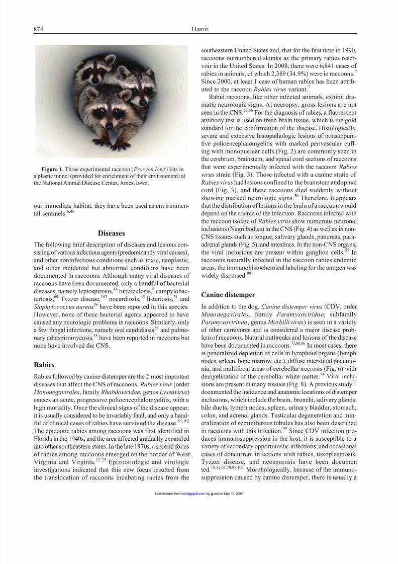
Raccoons are highly susceptible to a variety of infectious
diseases, both in free-ranging and under experimental con-
ditions (Fig. 1). Many of these diseases are common to
domestic dogs and cats. Raccoons can also transmit many
diseases to human beings and animals. Since raccoons share
by guest on May 15, 2016vdi.sagepub.comDownloaded from
8/16/2019 Raccoon SNC
http://slidepdf.com/reader/full/raccoon-snc 2/12
874 Hamir
our immediate habitat, they have been used as environmen-
tal sentinels.6,40
Diseases
The following brief description of diseases and lesions con-
sisting of various infectious agents (predominantly viral causes),
and other noninfectious conditions such as toxic, neoplastic,
and other incidental but abnormal conditions have been
documented in raccoons. Although many viral diseases of
raccoons have been documented, only a handful of bacterial
diseases, namely leptospirosis,44 tuberculosis,8 campylobac-
teriosis,69 Tyzzer disease,103 nocardiosis,65 listeriosis,51 and
Staphylococcus aureus36 have been reported in this species.
However, none of these bacterial agents appeared to have
caused any neurologic problems in raccoons. Similarly, only
a few fungal infections, namely oral candidiasis51
and pulmo-
nary adiaspiromycosis,35 have been reported in raccoons but
none have involved the CNS.
Rabies
Rabies followed by canine distemper are the 2 most important
diseases that affect the CNS of raccoons. Rabies virus (order
Mononegavirales, family Rhabdoviridae, genus Lyssavirus)causes an acute, progressive polioencephalomyelitis, with a
high mortality. Once the clinical signs of the disease appear,
it is usually considered to be invariably fatal, and only a hand-
ful of clinical cases of rabies have survived the disease.57,102
The epizootic rabies among raccoons was first identified in
Florida in the 1940s, and the area affected gradually expanded
into other southeastern states. In the late 1970s, a second focus
of rabies among raccoons emerged on the border of West
Virginia and Virginia.12,25 Epizootiologic and virologic
investigations indicated that this new focus resulted from
the translocation of raccoons incubating rabies from the
Figure 1. Three experimental raccoon ( Procyon lotor ) kits in
a plastic tunnel (provided for enrichment of their environment) at
the National Animal Disease Center, Ames, Iowa.
southeastern United States and, that for the first time in 1990,
raccoons outnumbered skunks as the primary rabies reser-
voir in the United States. In 2008, there were 6,841 cases of
rabies in animals, of which 2,389 (34.9%) were in raccoons.7
Since 2000, at least 1 case of human rabies has been attrib-
uted to the raccoon Rabies virus variant.7
Rabid raccoons, like other infected animals, exhibit dra-matic neurologic signs. At necropsy, gross lesions are not
seen in the CNS.55,56 For the diagnosis of rabies, a fluorescent
antibody test is used on fresh brain tissue, which is the gold
standard for the confirmation of the disease. Histologically,
severe and extensive histopathologic lesions of nonsuppura-
tive polioencephalomyelitis with marked perivascular cuff-
ing with mononuclear cells (Fig. 2) are commonly seen in
the cerebrum, brainstem, and spinal cord sections of raccoons
that were experimentally infected with the raccoon Rabies
virus strain (Fig. 3). Those infected with a canine strain of
Rabies virus had lesions confined to the brainstem and spinal
cord (Fig. 3), and these raccoons died suddenly without
showing marked neurologic signs.56 Therefore, it appearsthat the distribution of lesions in the brain of a raccoon would
depend on the source of the infection. Raccoons infected with
the raccoon isolate of Rabies virus show numerous neuronal
inclusions (Negri bodies) in the CNS (Fig. 4) as well as in non-
CNS tissues such as tongue, salivary glands, pancreas, para-
adrenal glands (Fig. 5), and intestines. In the non-CNS organs,
the viral inclusions are present within ganglion cells.55
In
raccoons naturally infected in the raccoon rabies endemic
areas, the immunohistochemical labeling for the antigen was
widely dispersed.99
Canine distemperIn addition to the dog, Canine distemper virus (CDV; order
Mononegavirales, family Paramyxoviridae, subfamily
Paramyxovirinae, genus Morbillivirus) is seen in a variety
of other carnivores and is considered a major disease prob-
lem of raccoons. Natural outbreaks and lesions of the disease
have been documented in raccoons.75,80,86 In most cases, there
is generalized depletion of cells in lymphoid organs (lymph
nodes, spleen, bone marrow, etc.), diffuse interstitial pneumo-
nia, and multifocal areas of cerebellar necrosis (Fig. 6) with
demyelination of the cerebellar white matter.94
Viral inclu-
sions are present in many tissues (Fig. 8). A previous study31
documented the incidence and anatomic locations of distemperinclusions, which include the brain, bronchi, salivary glands,
bile ducts, lymph nodes, spleen, urinary bladder, stomach,
colon, and adrenal glands. Testicular degeneration and min-
eralization of seminiferous tubules has also been described
in raccoons with this infection.59
Since CDV infection pro-
duces immunosuppression in the host, it is susceptible to a
variety of secondary opportunistic infections, and occasional
cases of concurrent infections with rabies, toxoplasmosis,
Tyzzer disease, and neosporosis have been documen
ted.16,32,61,70,87,103
Morphologically, because of the immuno-
suppression caused by canine distemper, there is usually a
by guest on May 15, 2016vdi.sagepub.comDownloaded from
8/16/2019 Raccoon SNC
http://slidepdf.com/reader/full/raccoon-snc 3/12
Pathology of neurologic disorders of raccoons 875
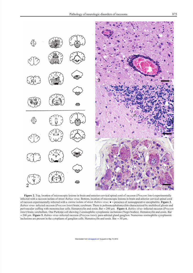
Figure 2. Top, location of microscopic lesions in brain and anterior cervical spinal cord of raccoon ( Procyon lotor ) experimentally
infected with a raccoon isolate of street Rabies virus. Bottom, location of microscopic lesions in brain and anterior cervical spinal cord
of raccoon experimentally infected with a canine isolate of street Rabies virus. ● = presence of nonsuppurative encephalitis. Figure 3.
Rabies virus –infected raccoon ( Procyon lotor ) brain; cerebrum. There is polioencephalomyelitis characterized by multifocal gliosis and
perivascular cuffing with mononuclear cells. Hematoxylin and eosin. Bar = 200 µm. Figure 4. Rabies virus –infected raccoon ( Procyon
lotor ) brain; cerebellum. One Purkinje cell showing 2 eosinophilic cytoplasmic inclusions (Negri bodies). Hematoxylin and eosin. Bar
= 200 µm. Figure 5. Rabies virus –infected raccoon ( Procyon lotor ); para-adrenal gland ganglion. Numerous eosinophilic cytoplasmic
inclusions are present in the cytoplasm of ganglion cells. Hematoxylin and eosin. Bar = 50 µm.
by guest on May 15, 2016vdi.sagepub.comDownloaded from
8/16/2019 Raccoon SNC
http://slidepdf.com/reader/full/raccoon-snc 4/12
876 Hamir
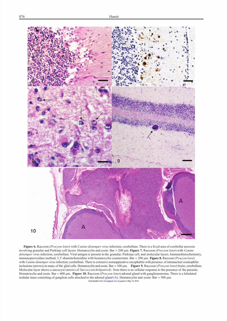
Figure 6. Raccoon ( Procyon lotor ) with Canine distemper virus infection; cerebellum. There is a focal area of cerebellar necrosis
involving granular and Purkinje cell layers. Hematoxylin and eosin. Bar = 200 µm. Figure 7. Raccoon ( Procyon lotor ) with Canine
distemper virus infection; cerebellum. Viral antigen is present in the granular, Purkinje cell, and molecular layers. Immunohistochemistry,
immunoperoxidase method, 3,3′-diaminobenzidine with hematoxylin counterstain. Bar = 200 µm. Figure 8. Raccoon ( Procyon lotor )
with Canine distemper virus infection; cerebellum. There is extensive nonsuppurative encephalitis with presence of intranuclear eosinophilic
inclusions (arrows) in many of the glial cells. Hematoxylin and eosin. Bar = 100 µm. Figure 9. Raccoon ( Procyon lotor ) brain; cerebellum.
Molecular layer shows a sarcocyst (arrow) of Sarcocystis kirkpatricki. Note there is no cellular response to the presence of the parasite.
Hematoxylin and eosin. Bar = 400 µm. Figure 10. Raccoon ( Procyon lotor ) adrenal gland with ganglioneuroma. There is a lobulated
nodular mass consisting of ganglion cells attached to the adrenal gland (A). Hematoxylin and eosin. Bar = 500 µm.
by guest on May 15, 2016vdi.sagepub.comDownloaded from
8/16/2019 Raccoon SNC
http://slidepdf.com/reader/full/raccoon-snc 5/12
Pathology of neurologic disorders of raccoons 877
paucity of host cellular inflammatory infiltrate that is directed
at the opportunistic pathogen. The CDV antigen (Fig. 7), how-
ever, can be demonstrated in the brains of these raccoons.61,70
Pseudorabies and herpesvirus encephalitis
Although since 2003 no commercially produced pig herdshave been found to be infected with Pseudorabies virus,
sporadic infections have been found in feral pigs. Prior to
1981, there had been 2 reports of herpesviral infection in
raccoons.91,97
Although the affected raccoon tissues were
examined by electron microscopy, the tissues were not tested
for the presence of pseudorabies. The disease can infect many
mammalian species and, on the basis of experimental
evidence, the raccoon is considered a major reservoir of
Pseudorabies virus.104 Microscopically, pseudorabies cases
involve the brain and show herpes-like viral lesions in neu-
ronal and non-neuronal tissues.30 A non-pseudorabies her-
pesviral disease has also been described in a raccoon with
lesions in non-neuronal tissues.60 The only documentedcase of herpesvirus encephalitis was described in 1981.91 The
encephalitis was characterized by perivascular cuffing,
choroiditis, and presence of magenta-colored intranuclear
inclusions in choroid plexus and adrenal medulla.91 As indi-
cated previously, the case was not differentiated from pseu-
dorabies.
Other viral diseases
Encephalomyocarditis virus (order Picornavirales, family
Picornaviridae, genus Cardiovirus) was isolated from a rac-
coon in Florida,26 and an experimental transmission study
has been documented.105 However, although some of the
raccoons in the study seroconverted, no clinical signs or
lesions were observed in the experimental raccoons.105 In
addition to the above-mentioned diseases that affect the ner-
vous system, raccoons have been shown to have evidence
(serology, polymerase chain reaction, Western blot, etc.) of
other viruses such as Venezuelan equine encephalitis virus5
and West Nile virus.29 However, clinical cases of these dis-
eases have not been documented in raccoons.
Prion diseases
Although naturally occurring transmissible spongiform enceph-alopathies (TSEs) have not been reported in the raccoon,
reports of successful experimental transmission of similar
spongiform diseases, transmissible encephalopathy of mink
(TME) and sheep scrapie, have been documented in this spe-
cies21,49,52,53
by intracerebral (IC) and oral inoculations.21
In
the IC-infected route, the raccoons were administered TME
and developed a rapidly progressive disease within 190 days,
whereas the orally infected raccoon developed neurologic
signs after 306 days, which included weakness, behavior
change, and incoordination. Scrapie-infected raccoons required
a 2-year period to develop the disease when inoculated by the
IC route.49,52,53 Microscopically, the lesions were identical in
both groups (TME and scrapie) and consisted of widespread
vacuolar changes, neuronal degeneration, and astrocytosis in
the brain (except in cerebellum). Neuronal vacuolar changes,
although described in scrapie and related diseases including
naturally occurring TME, were rare.49,52,53
According to some researchers, Spiroplasma mirum is
the causative agent of animal TSEs.3,4 However, when
this organism was inoculated intracerebrally into raccoons,
they did not succumb to any form of disease, and neither
lesions nor abnormal prion protein of TSE was detected in
the brains of these animals when necropsied at 30 months
postinoculation.42
Toxoplasmosis
Various surveys indicate a high prevalence of Toxoplasma
gondii antibodies in U.S. raccoon populatiotns,9,16,77 and, in
1992, a case of clinical toxoplasmosis with presence of theorganism in liver and brain was reported.16 In the 1992 case,
the raccoon was also found to have co-infection with CDV.16
Experimental transmission of the organism has also been
shown in the raccoon.18
Neospora caninum
Antibodies to Neospora caninum were demonstrated in approx-
imately 10% of raccoons from the eastern United States88 and,
to date, only 1 naturally occurring case in a juvenile rac-
coon has been documented.87 This raccoon also had con-
current CDV infection. Thus, the findings of necrotizing
encephalitis in the raccoon were largely due to the viral
infection.87
Sarcocystis neurona encephalitis
Sarcocystis neurona is the etiological agent of equine protozoal
myeloencephalitis (EPM), which has resulted in the deaths
of large numbers of horses in North America. Sarcocystis
neurona has also been reported in the raccoon, skunk, mink,
and cat.15,17,20 In a retrospective survey of 84 adult raccoons,
2 raccoons were found with S. neurona– associated granulo-
matous encephalitis.38 Both raccoons also had extensive non-
suppurative myocarditis and 1 raccoon revealed S. neurona in the myocardium by immunohistochemistry. The CNS
lesions were characterized by perivascular infiltration with
mononuclear cells and presence of randomly distributed
glial nodules. The organisms were observed in the inflamma-
tory foci.38
The life cycle of S. neurona is not fully under-
stood. The opossum is recognized as a definite host, and horses
and other susceptible animals are considered as aberrant
hosts. Susceptible animals become infected by ingesting S.
neurona sporocysts, which get excreted in feces of infected
opossums.19
by guest on May 15, 2016vdi.sagepub.comDownloaded from
8/16/2019 Raccoon SNC
http://slidepdf.com/reader/full/raccoon-snc 6/12
878 Hamir
Other Sarcocystis species in brain
Four species of Sarcocystis have been reported in striated
muscles of raccoons.81,98
However, in North America, only
Sarcocystis kirkpatricki has been documented in the muscles
of raccoons. Although the prevalence of infection of this para-
site appears to be variable, up to 50% of raccoons in a studyfrom the northeastern United States were found to be infected
in the heart, tongue, diaphragm, masseter muscle, or the
esophagus.81 A previous study98 designated this organism
S. kirkpatricki and reported a prevalence of 66% in raccoons
from Illinois. Neither of these studies reported Sarcocystis in
the brain of these raccoons. Subsequently, there has been a
single report of S. kirkpatricki in the cerebellum (Fig. 9)
of a raccoon with dual infections of T. gondii and CDV.16
Also, a retrospective survey of 760 raccoons revealed 9 ani-
mals with sarcocysts of S. kirkpatricki in the brains.39 Seven
of the raccoons had concurrent viral diseases (canine distem-
per or rabies), suggesting that concurrent viral infections
in raccoons may facilitate infection of brain tissue withS. kirkpatricki.39
Eosinophilic meningitis and encephalitis
In normal free-ranging raccoons, eosinophilia of cerebrospi-
nal fluid appears to be a common finding.71 A case of gener-
alized eosinophilic myositis with eosinophilia of blood and
cerebrospinal fluid has been described in a raccoon,66 which
was presumed to be due to immune-mediated causes. Also,
a raccoon with experimental canine Rabies virus infection
has been documented with eosinophilic polioencephalomy-
elitis.62 However, that raccoon did not show eosinophilia of
the blood.
Neural and visceral larva migrans
Neural and visceral larva migrans have been documented in
a large number of domestic and wild animal species (Snyder
DE: 1983, The prevalence, cross-transmissibility to domes-
tic animals and adult structure of Baylisascaris procyonis
(Nematoda) from Illinois raccoons ( Procyon lotor ). PhD diss.
University of Illinois, Urbana, IL).79 The common round
worm, Baylisascaris procyonis, found in raccoons, appears
to be a frequent etiologic agent that is responsible for severe
CNS disease in these parantenic hosts. However, viscerallarva migrans due to B. procyonis has not been documented
in raccoons. Other nematode species known to cause neu-
ral and visceral larva migrans include Toxocara spp.,
Angiostrongylus spp., and Strongyloides spp.
In raccoons, a form of visceral larva migrans associated
with a trematode ( Phagicola sp.) has been described.67
However, these parasites did not migrate into neuronal tissues.
A case of verminous encephalitis associated with an ascarid
larval nematode in a raccoon has been described.68 Although
the morphologic characteristics indicated that the nema-
tode was an ascarid larva, it was smaller than the larva of
Baylisascaris sp. This appears to be the first documented
case of cerebral larva migrans in a raccoon.68
Neoplasms
Neoplastic lesions have infrequently been documented in the
family Procyonidae. With the exception of thyroid adeno-
mas76,85 and adrenal gland adenomas,64 neoplasia appears to
be rather rare in raccoons, and only infrequent case reports
have been documented. In a retrospective survey of over
400 raccoon necropsies,13
only 1 astrocytoma of the brain
was found. Since that time, 1 additional astrocytoma involving
the brain, cervical spinal cord, and spinal nerves has been
documented.58 In both cases, the characteristics of the neo-
plasm were similar to that published in other species. Also,
a ganglioneuroma involving an adrenal gland has been seen
(Fig. 10; Hamir AN, personal observation).
Environmental toxins
Lead
Lead is an environmental toxin with multisystemic toxic
effects.31,34 A clinical case showing neurologic abnormalities
in a free-ranging raccoon has been documented,14
and also
high levels of tissue lead levels have been reported from dif-
ferent areas of the United States.14,39 In the reported case,14
the CNS lesions were subtle and consisted of mild endothe-
lial hyperplasia and scattered neuronophagia. In experimental
animals, administration of inorganic lead may lead to the
impairment of immune functions, resulting in increased sus-
ceptibility to infectious agents.74,84,90
Although neurologic
manifestations of lead toxicosis has been reported in a rac-
coon,14
experimental administration of oral lead acetate did
not succeed in producing neurologic signs when the com-
pound was administered over an 8-week period.50
Ethylene glycol
Neurologic signs of ethylene glycol (antifreeze) toxicity include
depression, ataxia, seizures, and coma,22
and since these signs
can be confused with other disorders in this species, clinical
differential diagnosis of this condition is rather difficult. Adiagnosis of ethylene glycol toxicosis is rarely reported in
free-ranging wildlife;2 however, morphologic lesions of this
toxicity are often seen in diagnostic cases. There has been
only 1 reported case of ethylene glycol poisoning in a free-
ranging raccoon.22
Since antifreeze is commonly found in
the environment, and cases of this toxicity are frequently
seen in domestic animals, it may be assumed that cases of
ethylene glycol toxicity are being overlooked in free-ranging
wildlife.
by guest on May 15, 2016vdi.sagepub.comDownloaded from
8/16/2019 Raccoon SNC
http://slidepdf.com/reader/full/raccoon-snc 7/12
Pathology of neurologic disorders of raccoons 879
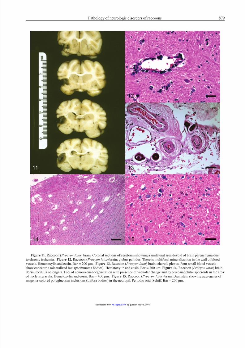
Figure 11. Raccoon ( Procyon lotor ) brain. Coronal sections of cerebrum showing a unilateral area devoid of brain parenchyma due
to chronic ischemia. Figure 12. Raccoon ( Procyon lotor ) brain; globus pallidus. There is multifocal mineralization in the wall of blood
vessels. Hematoxylin and eosin. Bar = 200 µm. Figure 13. Raccoon ( Procyon lotor ) brain; choroid plexus. Four small blood vessels
show concentric mineralized foci (psommoma bodies). Hematoxylin and eosin. Bar = 200 µm. Figure 14. Raccoon ( Procyon lotor ) brain;
dorsal medulla oblongata. Foci of neuroaxonal degeneration with presence of vacuolar change and hypereosinophilic spheroids in the area
of nucleus gracilis. Hematoxylin and eosin. Bar = 400 µm. Figure 15. Raccoon ( Procyon lotor ) brain. Brainstem showing aggregates of
magenta-colored polyglucosan inclusions (Lafora bodies) in the neuropil. Periodic acid–Schiff. Bar = 200 µm.
by guest on May 15, 2016vdi.sagepub.comDownloaded from
8/16/2019 Raccoon SNC
http://slidepdf.com/reader/full/raccoon-snc 8/12
880 Hamir
Trauma
It has been the author’s experience that during routine nec-
ropsy of free-ranging raccoons (>500) from the eastern and
northwestern United States,55 minor abrasions and/or trauma
to the skin was a common finding in these animals. Fighting
among males appeared to be the main reason for these skinlesions (Hamir AN, personal observation). Physical trauma
due to gunshot wounds, collisions with automobiles, and
falls from heights (in raccoons with neurologic abnormalities)
were frequently observed in field specimens. In the latter
cases, the animals had usually some preexisting neurological
abnormalities. One raccoon with neurologic signs was observed
to have a piece of porcupine quill logged in the brain tissue
(Hamir AN, personal observation).
Miscellaneous conditions of unknown etiology
Ischemic encephalopathy
One acute and 1 chronic case of cerebral infarction syndrome
has been described in a free-ranging and a laboratory-
confined raccoon, respectively.63 Both were mature adults.
The raccoon with the acute form of the condition had abnor-
mal neurologic signs whereas the raccoon with the chronic
lesion did not. In the chronic case, the lesion was present
unilaterally (Fig. 11) and since only one-half of the brain
was available for examination in the acute case, it was not
possible to determine if there were multiple lesions. In both
cases, the nature and distribution of the lesions was compat-
ible with an ischemic etiology, although vascular lesions
were not observed.63
Cerebrovascular mineralization
A case of cerebrovascular mineralization in an aged (>10
years old) female with uremia associated with polycystic
kidney disease has been reported in a raccoon.47 The vascu-
lar lesions were bilateral and consisted of multifocal miner-
alized foci in the walls of blood vessels in globus pallidus
(Fig. 12). The affected vessels were patent, and there was no
inflammatory cellular response to the mineralized foci.
Morphologically, this lesion appeared to be similar to a non-
symptomatic condition that has been documented in other
species.
27
Neuronal lipid vacuolation
Spongiform changes affecting the neuropil as well as neuro-
nal cell bodies have been described in a newborn calf,43
in
experimental and natural cases of rabies in skunks,11 foxes11
and in a heifer.23 Neuronal perikaryonic vacuolation is also
seen in scrapie and related spongiform encephalopathes,33
and in lysosomal and acquired storage diseases of various
animals.100
Naturally occurring TSE and storage diseases
have not been documented in raccoons. Rabies, on the other
hand, is commonly encountered in raccoons of the eastern
United States. However, vacuolation of either the neurons or
the neuropil has not been reported in raccoons with naturally
acquired rabies, or in raccoons experimentally infected with
either a dog or a raccoon isolate of Rabies virus.55
During a 2-year period, clear vacuoles were morphologi-cally detected in neurons of 42% of raccoon brains exam-
ined in Oregon.41,46 Neither age nor sex predisposition was
apparent in this population. Microscopically, the vacuoles
were variable in size, were in the perikarya, and were con-
sistently present in pontine nuclei. Brain tissues were neg-
ative for Rabies virus antigen by fluorescent antibody test
and for the protease-resistant protein (PrP) prion by immu-
nohistochemistry. Electron microscopic examination of the
brainstem of selected cases revealed accumulation of elec-
tron-dense material within neuronal perikarya.41 Based
on light and electron microscopic findings, the accumu-
lated intracellular material appeared to have a high lipid
content. These lesions indicate a form of neuronal storagecondition.
Mineralized foci (psammoma bodies) in meninges
and choroid plexus
With advancing age, the human brain shows increased pres-
ence of focal mineralized foci.10
Often referred to as psam-
moma bodies, such mineralized foci are small, hardened
bodies composed of concentric rings of calcium carbonate
and calcium and magnesium phosphate.10
Microscopic evidence of multifocal mineralizations (psam-
moma bodies) were seen in the brain of 62% of raccoons
necropsied on Parramore Island, Virginia.45 The mineralized
foci revealed concentric laminations and were present in
small capillaries of meninges of the brain, choroid plexus
(Fig. 13), or at both these sites. In 2 raccoons, the lesions
were confined to the meninges of the proximal cervical spi-
nal cord. In most cases, the affected vessels appeared to have
been completely occluded. However, no evidence of ischemic
changes in the brain parenchyma was seen, and none of the
raccoons showed abnormal neurologic signs prior to eutha-
nasia. The condition appears to be a common incidental his-
topathological finding in raccoons from the eastern United
States.45 It is speculated that mineralization and subsequent
occlusion of larger blood vessels may result in cerebral isch-emia. Although the exact cause of this condition is not known,
a primary vascular insult with resultant dystrophic mineral-
ization of the affected vessels is suspected.45
Neuroaxonal dystrophy
During a 12-month period, microscopic evidence of neuroax-
onal dystrophy was seen in 12 out of 29 raccoons from Iowa.54
In the affected animals, clinical signs were not apparent.
Three of the animals were kits (<3 months), and none
by guest on May 15, 2016vdi.sagepub.comDownloaded from
8/16/2019 Raccoon SNC
http://slidepdf.com/reader/full/raccoon-snc 9/12
Pathology of neurologic disorders of raccoons 881
revealed lesions. The brain did not show any gross lesions.
Microscopically, the lesions were confined to the dorsal
caudal medulla where certain nuclei (predominantly gracilis
and cuneate) were affected. The lesions consisted of eosino-
philic axonal spheroids (Fig. 14) with presence of ampho-
philic periodic acid–Schiff-positive granular material within
some of the degenerate neurons and axones. In a few cases,the latter had completely disappeared leaving behind clear
areas in the neuropil (vacuolar degeneration). None of the
affected cases revealed any appreciable numbers of inflam-
matory cells within the areas of degeneration. Neuroaxonal
dystrophy has been observed in human beings and in various
animals, and these changes have been associated with advanc-
ing age. Although none of the 3 raccoons examined in the
study were old, it was presumed that since the lesions were not
present in kits, the findings may be age-related. Subsequently,
it was shown that severe neuroaxonal dystrophy was present
in old (>7 years of age) raccoons.37
Lafora bodies (polyglucosan inclusions)
Polyglucosan inclusions or Lafora bodies are inclusions of
complex glycoprotein polymers in tissues.100 Although not
reported often in wild animals, Lafora bodies have frequently
been reported in dogs and in a few other domestic animals.
These inclusions are age-related and may or may not be
associated with abnormal neurologic signs.100
There has been
only 1 reported occurrence of Lafora bodies in the brain
(Fig. 15) of a raccoon, which did not show any overt abnormal
neurologic signs.37
AmyloidosisAmyloidosis has been documented in both free-ranging and
captive raccoons.48 It is predominantly seen in the pancreas
where it is deposited in the islets of Langerhans. Although
not reported in the brain, a single case of amyloidosis in cho-
roid plexus of an aged raccoon has been observed (Hamir AN
et al., unpublished data). Amyloid accumulation was associ-
ated with multifocal lymphocytic cell infiltrations. The rac-
coon did not show any neurologic signs.
Congenital conditions
Although albinism is not uncommon in raccoons,
89
only afew other congenital lesions have been reported in this species.
Individual case reports of congenital amelia of forelimbs,73
lack of tail,73
raccoon with 2 tails,101
congenital diaphragmatic
hernia,96 renal hypoplasia,92 dental anomalities,82 and other
skeletal anomalies93
have been reported.
Congenital abnormalities affecting the nervous system or
other body systems that can cause adverse effect on the ner-
vous system are rare, and only 1 documentation of a report of
ocular and nasomaxillary anomalies with neural defects has
been published.95
However, during a field safety trial of the
vaccinia-rabies glycoprotein (V-RG) vaccine,72 a raccoon
with a unilateral micro-ophthalmia was observed (Hamir AN
et al., unpublished data).
Conclusion
Raccoons are almost ubiquitous in North America. Since the1980s, a large number of diseases and lesions, many involv-
ing the nervous system, have been documented in this spe-
cies. As a fair number of these have been individual case
reports, it is difficult to access the general importance of
these conditions on the local populations of raccoons. Also,
some of the described conditions appear not to have been of
immediate severe consequence to the raccoons. However,
such reports are scientifically useful in recognizing patho-
logical conditions that exist at a given time and are also
useful for future disease investigations. Furthermore, since
raccoons are no longer confined to North America, it would
be of interest to compare disease prevalence and pathology
in different raccoon populations of the world with conditionsthat are reported in the current review.
Acknowledgements
The author would like to thank Dr. Charles E. Rupprecht,
now at Centers for Disease Control and Prevention,
Atlanta, GA, for the opportunity to examine and learn
from a large number of raccoon necropsies from his project
of vaccinia-rabies glycoprotein (V-RG) vaccine develop-
ment during the 1980s.
Declaration of conflicting interests
The authors declared no potential conflicts of interest with
respect to the research, authorship, and/or publication of
this article.
Funding
The authors received no financial support for the research,
authorship, and/or publication of this article.
References
1. Aliev FF, Sanderson GC: 1966, Distribution and status of the
raccoon in the Soviet Union. J Wildl Manage 30:497–502.
2. Amstrup CS, Gardner C, Meyers KC, Oehme FW: 1989,
Ethylene glycol (antifreeze) poisoning in a free-ranging
polar bear. Vet Hum Toxicol 31:317–319.
3. Bastian FO, Purnell DM, Tully JG: 1984, Neuropathology
of spiroplasma infection in the rat brain. Am J Pathol 114:
496–514.
4. Bastian FO, Sanders DE, Forbes WA, et al.: 2007, Spiro-
plasma spp. from transmissible spongiform encephalopa-
thy brains or ticks induce spongiform encephalopathy in
ruminants. J Med Microbiol 56:1235–1242.
5. Bigler WJ: 1971, Serologic evidence of Venezuelan equine
encephalitis virus infections in raccoons of south central Flor-
ida. J Wildl Dis 7:166–170.
by guest on May 15, 2016vdi.sagepub.comDownloaded from
8/16/2019 Raccoon SNC
http://slidepdf.com/reader/full/raccoon-snc 10/12
882 Hamir
6. Bigler WM, Jenkins JH, Cumbie PM, et al.: 1975, Wildlife
and environmental health: raccoons as indicators of zoo-
noses and pollutants in southeastern United States. J Am Vet
Med Assoc 167:592–597.
7. Blanton JD, Robertson K, Palmer D, Rupprecht CE: 2009,
Rabies surveillance in the United States during 2008. J Am
Vet Med Assoc 235:676–689. 8. Bruning-Fann CS, Schmitt SM, Fitzgerald SD, et al.: 2001,
Bovine tuberculosis in free-ranging carnivores from Michi-
gan. J Wildl Dis 37:58–64.
9. Burridge MJ, Bigler WJ, Forrester DJ, Hannemann JM: 1979,
Serologic survey for Toxoplasma gondii in wild mammals in
Florida. J Am Vet Med Assoc 175:964–967.
10. Carpenter MB, Sutin J: 1983, The diencephalon. In: Human
neuroanatomy 8th ed., pp. 134–154. Williams & Wilkins,
Baltimore, MD.
11. Charlton KM, Casey GA, Webster WA, Bundza A: 1987,
Experimental rabies in skunks and foxes. Pathogenesis of
the spongiform lesions. Lab Invest 57:634–645.
12. Childs JE, Curns AT, Dey ME, et al.: 2000, Predicting thelocal dynamics of epizootic rabies among raccoons in the
United States. Proc Natl Acad Sci U S A 97:13666–13671.
13. Diters RW, Kircher CH, Nielsen SW: 1978, Astrocytoma in a
raccoon. J Am Vet Med Assoc 173:1152–1153.
14. Diters RW, Nielsen SW: 1978, Lead poisoning of raccoons in
Connecticut. J Wildl Dis 14:187–192.
15. Dubey JP, Hamir AN: 2000, Immunohistochemical confirma-
tion of Sarcocystis neurona infections in raccoons, mink,
cat, skunk, and pony. J Parasitol 86:1150–1152.
16. Dubey JP, Hamir AN, Hanlon CA, Rupprecht CE: 1992,
Prevalence of Toxoplasma gondii infection in raccoons. J Am
Vet Med Assoc 200:534–536.
17. Dubey JP, Hamir AN, Hanlon CA, et al.: 1990, Fatal necrotiz-
ing encephalitis in a raccoon associated with a Sarcocystis-
like protozoon. J Vet Diagn Invest 2:345–347.
18. Dubey JP, Hamir AN, Shen SK, et al.: 1993, Experimental
Toxoplasma gondii infection in raccoons ( Procyon lotor ).
J Parasitol 79:548–552.
19. Dubey JP, Saville WJ, Stanek JF, et al.: 2001, Sarcocystis
neurona infections in raccoons ( Procyon lotor ): evidence for
natural infection with sarcocysts, transmission of infection
to opossums ( Didelphis virginiana), and experimental induc-
tion of neurologic disease in raccoons. Vet Parasitol 100:
117–129.
20. Dubey JP, Speer CA, Hamir AN, et al.: 1991, Development of a
Sarcocystis-like apicomplexan protozoan in the brain of a rac-
coon ( Procyon lotor ). J Helminthol Soc Washington 58:250–255.
21. Eckroade RJ, ZuRhein GM, Hanson RP: 1973, Transmis-
sible mink encephalopathy in carnivores: clinical, light and
electron microscopic studies in raccoons, skunks and ferrets.
J Wildl Dis 9:229–240.
22. Foley P, McBurney S: 2002, Ethylene glycol toxicosis in a
free-ranging raccoon ( Procyon lotor ) from Prince Edward
Island. Can Vet J 43:291–292.
23. Foley GL, Zachary JF: 1995, Rabies-induced spongiform
change and encephalitis in a heifer. Vet Pathol 32:309-311.
24. Frantz AC, Criacks P, Schley L: 2005, Spatial behavior of a
female raccoon ( Procyon lotor ) at the edge of the species’
European distribution range. Eur J Wildl Res 51:126–130.
25. Frost A, Hamir AN: 1998, The North American raccoon
rabies epizootic. Vet Bull 68:1343–1349.
26. Gainer JH, Bigler WI: 1967, Encephalomyocarditis (EMC)
virus recovered from two cotton rats and a raccoon. BullWildl Dis Assoc 3:47–49.
27. Gavier-Widen D, Wells GAH, Simmons MM, et al.: 2001,
Histological observations on the brains of symptomless
7-year-old cattle. J Comp Pathol 124:52–59.
28. Gehrt SD: 2003, Raccoon Procyon lotor and allies. In: Wild mam-
mals of North America: biology, management, and conserva-
tion, ed. Feldhamer GA, Thompson BC, Chapman JA, pp.
611–634. Johns Hopkins, Baltimore, MD.
29. Gómez A, Kilpatrick AM, Kramer LD, et al.: 2008, Land
use and West Nile virus seroprevalence in wild mammals.
Emerg Infect Dis 14:962–965.
30. Goyal SM, Drolet R, King P: 1986, Pseudorabies in free-
ranging raccoons. J Am Vet Med Assoc 189:1163–1164. 31. Goyer RA: 1990, Lead toxicity: from overt to subclinical to
subtle health effects. Environ Health Perspec 86:177–181.
32. Habermann RT, Herman CM, Williams FP Jr: 1958, Distem-
per in raccoons and foxes suspected of having rabies. J Am
Vet Med Assoc 132:31–35.
33. Hadlow WJ: 1995, Neuropathology and the scrapie-kuru con-
nection. Brain Pathol 5:27–31.
34. Hamir AN: 1986, Review of lead poisoning in dogs. Vet
Bull 56:1059–1070.
35. Hamir AN: 1999, Pulmonary adiaspiromycosis of raccoons
( Procyon lotor ) from Oregon. J Vet Diagn Invest 11:565–567.
36. Hamir AN: 2010, Systemic Staphylococcus aureus infection
in a free-ranging raccoon ( Procyon lotor ). J Wildl Dis 46:
665–668.
37. Hamir AN: 2011, Spontaneous lesions in aged captive rac-
coons ( Procyon lotor ). J Am Assoc Lab Anim Sci 50:322-325.
38. Hamir AN, Dubey JP: 2001, Myocarditis and encephalitis
associated with Sarcocystis neurona infection in raccoons
( Procyon lotor ). Vet Parasitol 95:335–340.
39. Hamir AN, Dubey JP, Rupprecht CE: 1999, Prevalence of
Sarcocystis kirkpatricki in the central nervous system and
striated muscles of raccoons from eastern United States. J
Parasitol 85:748–750.
40. Hamir AN, Ebel JG, Manzell KL, Rupprecht CE: 1994,
Blood lead levels of wild raccoons ( Procyon lotor ) from the
eastern United States. J Wildl Dis 30:115–118.
41. Hamir AN, Fischer KA: 1999, Neuronal vacuolations in rac-
coons from Oregon. J Vet Diagn Invest 11:303–307.
42. Hamir AN, Greenlee JJ, Stanton T, et al.: 2011, Experimental
inoculation of raccoons ( Procyon lotor ) with Spiroplasma
mirum and transmissible mink encephalopathy (TME). Can
J Vet Res 75:18–24.
43. Hamir AN, Habecker P, Jenny A, et al.: 2001, Idiopathic dis-
seminated intracytoplasmic neuronal vacuolation in a neo-
natal Holstein calf born in the USA. J Vet Diagn Invest 13:
349–351.
by guest on May 15, 2016vdi.sagepub.comDownloaded from
8/16/2019 Raccoon SNC
http://slidepdf.com/reader/full/raccoon-snc 11/12
Pathology of neurologic disorders of raccoons 883
44. Hamir AN, Hanlon CA, Niezgoda M, Rupprecht CE: 2001,
The prevalence of interstitial nephritis and leptospirosis in
283 raccoons ( Procyon lotor ) from 5 different sites in the
United States. Can Vet J 42:869–871.
45. Hamir AN, Hanlon CA, Rupprecht CE: 2001, Prevalence of
psammoma bodies in meninges and choroid plexuses of
raccoons ( Procyon lotor ) from Parramore Island, Virginia.J Vet Diagn Invest 13:76–79.
46. Hamir AN, Heidel JR, Picton RA, Rupprecht CE: 1997, Neu-
ronal vacuolation in raccoons ( Procyon lotor ). Vet Pathol 34:
250–252.
47. Hamir AN, Klein L: 1996, Polycystic kidney disease in a rac-
coon ( Procyon lotor ). J Wildl Dis 32:674–677.
48. Hamir AN, Kunkle RA, Miller MJ, et al.: 2007, Age-related
lesions in laboratory-confined raccoons ( Procyon lotor )
inoculated with the agent of chronic wasting disease of mule
deer. J Vet Diagn Invest 19:680–686.
49. Hamir AN, Kunkle RA, Miller JM, Richt J: 2005, Experi-
mental second raccoon passage of sheep scrapie and trans-
missible mink encephalopathy (TME) agents. Vet Pathol 42:844–851.
50. Hamir AN, Lehmann B, Raju N , et al.: 1999, Experimen-
tal lead toxicosis of raccoons ( Procyon lotor ). J Comp Pathol
120:147–154.
51. Hamir AN, Mattson DE, Sonn RJ, et al.: 2000, Concurrent
candidiasis, listeriosis and adenovirus infections in a rac-
coon ( Procyon lotor ). Vet Rec 146:320–322.
52. Hamir AN, Miller JM, Cutlip RC, et al.: 2003, Experimental
inoculation of scrapie and chronic wasting disease agents to
raccoons ( Procyon lotor ). Vet Rec 153:121–123.
53. Hamir AN, Miller JM, O’Rourke KI, et al.: 2004, Transmis-
sion of transmissible mink encephalopathy (TME) to rac-
coons ( Procyon lotor ) by intracerebral inoculation. J Vet
Diagn Invest 16:57–63.
54. Hamir AN, Miller JM, Stack MJ, et al.: 2002, Neuroaxonal
dystrophy in raccoons ( Procyon lotor ) from Iowa. J Vet Diagn
Invest 14:175–178.
55. Hamir AN, Moser G, Rupprecht CE: 1992, Morphologic
and immunoperoxidase study of neurologic lesions in
naturally acquired rabies of raccoons J Vet Diagn Invest
4:369–373.
56. Hamir AN, Moser G, Rupprecht CE: 1996, Clinicopathologi-
cal findings in raccoons experimentally infected with two
isolates of rabies virus. J Vet Diagn Invest 8:31–37.
57. Hamir AN, Niezgoda M, Rupprecht CE: 2011, Recovery from
and clearance of rabies virus in a domestic ferret. J Am Assoc
Lab Anim Sci 50:248-251.
58. Hamir AN, Picton R, Blythe LL, Heidel JR: 2008, Diagnostic
exercise: astrocytoma with involvement of medulla oblon-
gata, spinal cord and spinal nerves in a raccoon ( Procyon
lotor ). Vet Pathol 45:949–951.
59. Hamir AN, Raju N, Hable CP, Rupprecht CE: 1992, Retro-
spective study of testicular degeneration in raccoons with
canine distemper infection. J Vet Diagn Invest 4:159–163.
60. Hamir AN, Raju N, Kao M, Rupprecht CE: 1995, Herpesvi-
rus infection in a raccoon ( Procyon lotor ). J Wildl Dis 31:
420–423.
61. Hamir AN, Rupprecht CE: 1990, Absence of rabies encepha-
litis in a raccoon with concurrent rabies and canine distemper
infection. Cornell Vet 80:197–201.
62. Hamir AN, Rupprecht CE: 1990, Eosinophilic encephalomy-elitis in a raccoon experimentally infected with a dog isolate
of rabies virus. J Vet Diagn Invest 2:145–147.
63. Hamir AN, Rupprecht CE: 1994, Ischemic encephalopathy in
raccoons ( Procyon lotor ). J Wildl Dis 30:609–611.
64. Hamir AN, Rupprecht CE: 1995, Adrenal gland adenomas in
raccoons ( Procyon lotor ) from the eastern United States. J
Vet Diagn Invest 7:413–416.
65. Hamir AN, Rupprecht CE: 1996, Pyogranulomatous perito-
nitis associated with Nocardia-like organisms in a raccoon
( Procyon lotor ). J Wildl Dis 32:373–375.
66. Hamir AN, Rupprecht CE, Ziemer EL: 1989, Generalized
eosinophilic myositis with eosinophilia of blood and cerebro-
spinal fluid in a raccoon ( Procyon lotor ). J Vet Diagn Invest 1:192–194.
67. Hamir AN, Snyder DE, Hanlon CA, Rupprecht CE: 1993, A trem-
atode ( Phagicola sp.)-induced mesenteric lymphadenitis and
enteritis in raccoons ( Procyon lotor ). Vet Pathol 30:373–376.
68. Hamir AN, Snyder DE, Lichenfels JR: 1999, Cerebral larva
migrans in a raccoon ( Procyon lotor ). Vet Pathol 36:618–620.
69. Hamir AN, Sonn RJ, Franklin S, Wesley IV: Enteritis with
intestinal intussusceptions associated with Campylobacter jejuni
infection in a raccoon ( Procyon lotor ) kit and isolation of C.
jejuni and Arcobacter spp. from adult raccoons. Vet Rec 155:
338-340, 2004.
70. Hamir AN, Summers BA, Rupprecht CE: 1998, Concurrent
rabies and canine distemper encephalitis in a raccoon ( Procyon
lotor ). J Vet Diagn Invest 10:194–196.
71. Hanlon CA, Ziemer EL, Hamir AN, Rupprecht CE: 1989,
Cerebrospinal fluid analysis of rabid and vaccinia-rabies
glycoprotein recombinant, orally vaccinated raccoons ( Pro-
cyon lotor ). Am J Vet Res 50:364–367.
72. Hanlon CL, Hayes DE, Hamir AN, et al.: 1989, Proposed
field evaluation of a rabies recombinant vaccine for raccoons
( Procyon lotor ): site selection, target species characteristics,
and placebo baiting trials. J Wildl Dis 25:555–567.
73. Heidt GA: 1969, Congenital reduction of fore-legs and tail in
a raccoon ( Procyon lotor L). Am Midl Nat 82:280–281.
74. Hemphill FE, Kaeberle ML, Buck WB: 1971, Lead suppres-
sion of mouse resistance to Salmonella typhimurium. Sci-
ence 172:1031–1032.
75. Hoff GL, Bigler WJ, Proctor SJ, Stallings LP: 1974, Epizootic
of canine distemper virus infection among urban raccoons
and gray foxes. J Wildl Dis 10:423–428.
76. Ippen VR: 1987, Ein Beitrag zu den tumoren der nicht-do-
mestizierten landraubtiere (Fissipedia) [Tumors of undo-
mesticated beasts of prey (Fissipedia)]. Dtsch Tierarztl
Wochenschr 94:61–63. Article in German.
by guest on May 15, 2016vdi.sagepub.comDownloaded from
8/16/2019 Raccoon SNC
http://slidepdf.com/reader/full/raccoon-snc 12/12
884 Hamir
77. Jacobs JS, Stanley AM: 1962, Prevalence of Toxoplasma
antibodies in rabbits, squirrels, and raccoons collected in
and near the Patuxent Wildlife Research Center. J Parasitol
48:550.
78. Kaufmann JH: 1982, Raccoons and allies. In: Wild mammals
of North America, ed. Chapman JA, Feldhamer GA, pp.
567–585. Johns Hopkins, Baltimore, MD. 79. Kazacos KR, Boyce WM: 1989, Baylisascaris larva migrans.
J Am Vet Med Assoc 195:894–903.
80. Kilham L, Habermann RT, Herman CM: 1956, Jaundice and
bilirubinemia as manifestations of canine distemper in rac-
coons and ferrets. Am J Vet Res 17:144–148.
81. Kirkpatrick CE, Hamir AN, Dubey JP, Rupprecht CE: 1987,
Sarcocystis in muscles of raccoons ( Procyon lotor L.). J Pro-
tozool 34:445–447.
82. Knable A, Werner WE: 1964, Raccoon with unusual dental
complement. J Mammal 45:493.
83. Koizumi N, Uchida M, Makino T, et al.: 2009, Isolation and
characterization of Leptospira spp. from raccoon in Japan. J
Vet Med Sci 71:425–429. 84. Koller LD: 1973, Immunosuppression produced by lead,
cadmium, and mercury. Am J Vet Res 34:1457–1458.
85. Kronberger H: 1962, Geschwülste bei Zootieren [Tumors in
zoo animals]. Nord Vet Med 14:297–304.
86. Lednicky JA, Dubach J, Kinsel MJ, et al.: 2004, Geneti-
cally distant American Canine distemper virus lineages have
recently caused epizootics with somewhat different charac-
teristics in raccoons living around a large suburban zoo in
the USA. Virol J 1:2.
87. Lemberger KY, Gondim LF, Pessier AP, et al.: 2005, Neospora
caninum infection in a free-ranging raccoon ( Procyon lotor )
with concurrent canine distemper virus infection. J Parasitol 91:
960–961.
88. Lindsay DS, Spencer J, Rupprecht C, Blagbum BL: 2001,
Prevalence of agglutinating antibodies to Neospora cani-
num in raccoons, Procyon lotor . J Parasitol 87:1197–1198.
89. Long CA, Hogan A: 1988, Two independent loci for albinism
in raccoons, Procyon lotor . J Hered 79:387–389.
90. Luster MI, Faith RE, Kimmel CA: 1978, Depression of
humoral immunity in rats following chronic developmental
lead exposure. J Environm Pathol Toxicol 1:397–402.
91. Maurer KE, Nielsen SW: 1981, Neurologic disorders in
the raccoon in northeastern United States. J Am Vet Med
Assoc 179:1095–1098.
92. Mech LD, Anderson NV: 1966, Renal hypoplasia and com-
pensatory hypertrophy in a raccoon. J Mammal 47:128–129.
93. Michael ED: 1968, Unusual pelage and skeleton of a raccoon.
Southwest Nat 13:105. 94. Potgieter LN, Patton CS: 1984, Multifocal cerebellar cortical
necrosis caused by canine distemper virus infection in a raccoon.
J Am Vet Med Assoc 185:1397–1399.
95. Render JA, Kazacos EA, Kazakos KR, et al.: 1983, Ocular,
naso-maxillary, and neural anomalies in raccoons, Procyon
lotor (L). J Wildl Dis 19:234–243.
96. Sanderson GC: 1960, A congenital diaphragmatic hernia in
the raccoon. Am Midl Nat 64:500–501.
97. Sanger VL, Bicknell EJ, Trapp AL, et al.: 1978, Viral inclu-
sions in raccoon liver cells. J Wildl Dis 14:240–243.
98. Snyder DE, Sanderson GC, Toivio-Kinnucan M, Blagburn
BL: 1990, Sarcocystis kirkpatricki n. sp. (Apicomplexa: Sar-
cocystidae) in muscles of raccoons ( Procyon lotor ) fromIllinois. J Parasitol 76:495–500.
99. Stein LT, Rech RR, Harrison L, Brown CC: 2010, Immunohis-
tochemical study of rabies virus within the central nervous sys-
tem of domestic and wildlife species. Vet Pathol 47:630–636.
100. Summers BA, Cummings JF, deLahunta A: 1995, Lysosomal
storage diseases and neuronal glycoprotenosis. In: Veterinary
neuropathology, pp. 214–236, 326–327. Mosby, St. Louis, MO.
101. Vellard J, Penteado J: 1931, Une queue multiple de Procyon
[ Procyon with multiple tails]. Bull Soc Zoo Fr 56:255–260.
102. Willoughby RE Jr, Tieves KS, Hoffman GM, et al.: 2005,
Survival after treatment of rabies with induction of coma. N
Engl J Med 352:2508–25014.
103. Wojcinski ZW, Barker IK: 1986, Tyzzer’s disease as a com-
plication of canine distemper in a raccoon. J Wildl Dis 22:
55–59.
104. Wright JC, Thawley DG: 1980, Role of raccoon in the trans-
mission of pseudorabies: a field and laboratory investigation.
Am J Vet Res 41:581–583.
105. Zimmerman JJ, Hill RE Jr, Smith KE, et al.: 1994, Encepha-
lomyocarditis virus infection in raccoons ( Procyon lotor ). J
Zoo Wildl Med 25:233–239.
by guest on May 15 2016vdi sagepub comDownloaded from