Embed Size (px)
Citation preview

International Journal of Radiation Research, April 2016 Volume 14, No 2
Radiation attenuation properties of shields containing micro and Nano WO3 in diagnostic X-ray
energy range
INTRODUCTION
Over the past decades nanotechnology has
enabled us to create a wide variety of life-
changing products. This technology is also
currently used for production of multipurpose
radiation shields. Since the discovery of X-rays
and radioactivity, �lexible lead-based radiation
shields have been widely used in radiology
departments.Currently,theshieldingdesignofX
and g facilities, protective equipments and
clothing is mainly based on lead materials.
However,recentlytherehasbeenagreatdealof
concern expressed about the toxicity of lead (1).
Inadditiontheheavinessoftheleadwouldcause
backstrain,andorthopedic injuries inradiation
workers who wear the lead aprons for long
durations (2-5). Therefore production of
environmentally-friendly lead-free radiation
shields with less weight compared to
conventional lead-basedshields isa challenging
issue in diagnostic radiology and nuclear
medicine (6-11). Different investigations have
been performed to obtain the properties of
aprons made by combining elements with
different K absorption energies, i.e. copper, tin,
ABSTRACT
Background: It has recently been shown that the par�cle size of materials
used for radia�on shielding can affect the magnitude of radia�on a�enua�on.
Over the past years, applica�on of nano-structured materials in radia�on
shielding has a�racted a�en�on world-wide. The purpose of this study was to
inves�gate the shielding proper�es of the lead-free shields containing micro
and nano-sized WO3 against low energy x-rays. Materials and Methods: The
radia�on shields were constructed using nano and micro WO3 par�cles
incorporated into an EPVC polymer matrix. The a�enua�on coefficients of the
designed shields were evaluated for low energy x-rays (diagnos�c radiology
energy range). Results: The results indicate that nano-structured WO3/PVC
shields have higher photon a�enua�on proper�es compared to those of the
micro-sized samples. Conclusion: Our experiment clearly shows that the
smaller size of nano-structured WO3 par�cles can guarantee a be�er
radia�on shielding property. However, it is too early to draw any conclusion
on the possible mechanisms of enhanced a�enua�on of nano-sized WO3
par�cles.
Keywords: Radia�on, a�enua�on, micro, Nano, WO3, in diagnos�c
radiology, X-ray.
A. Aghaz1, R. Faghihi1, S.M.J. Mortazavi2*, A. Haghparast3,
S. Mehdizadeh4, S. Sina4
1NuclearEngineeringDepartment,ShirazUniversity,Shiraz,Iran2MedicalPhysicsDepartmentandTheCenterforResearchonRadiologicalSciences,ShirazUniversityof
MedicalSciences,Shiraz,Iran3MedicalPhysicsDepartment,KermanshahUniversityofMedicalSciences,Kermanshah,Iran
4RadiationResearchCenter,ShirazUniversity,Shiraz,Iran
*Correspondingauthor:
Dr.SMJMortazavi,
Fax:+987112349332
E-mail:
Revised: Sept. 2015
Accepted: Oct. 2015
Int. J. Radiat. Res., April 2016; 14(2): 127-131
► Original Article
DOI: 10.18869/acadpub.ijrr.14.2.127
barium,tungsten,antimony,yttrium,andlead(5,
6, 9, 12).Recently theuseofNanoparticles in the
design of lead-free radiation shields have been
investigated (13).ProtectivematerialswithNano
particles have shown to have good mechanical
properties to be used in developing radiation
shields (1, 13-15). The size and concentration of
Nano-particlesusedinmakingradiationshields
aretwoimportantfactorsaffectingtheradiation
attenuation properties of the protective
materials(1,13,16).Thepurposeofthisstudyisto
designnanoandmicro-structuredshieldsusing
WO3particles incorporated inEPVCpolymericmatrix, and to compare the radiation shielding
properties of nano and micro-sized shielding
materials.
MATERIALSANDMETHODS
Flexible sheets of WO3 were produced in a
polyvinyl chloride (EPVC)polymericmatrix. In
thisexperiment, thenanoandmicro-structured
shields with the purity of +99% were
constructedusingWO3particlesincorporatedin
EPVCpolymericmatrixwith20%,50%and60%
of mass proportions. The grain sizes of nano
particles used in construction of the shields
were 20 to 100 nm, while the average micro
WO3 particle size was less than 20 µm. The
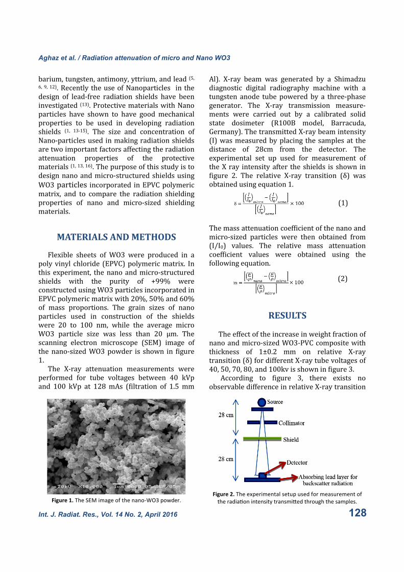
scanning electron microscope (SEM) image of
thenano-sizedWO3powder is shown in �igure
1.
The X-ray attenuation measurements were
performed for tube voltages between 40 kVp
and 100 kVp at 128mAs (�iltration of 1.5mm
Al). X-ray beam was generated by a Shimadzu
diagnostic digital radiography machine with a
tungsten anode tube powered by a three-phase
generator. The X-ray transmission measure-
ments were carried out by a calibrated solid
state dosimeter (R100B model, Barracuda,
Germany).ThetransmittedX-raybeamintensity
(I)wasmeasuredbyplacing thesamplesat the
distance of 28cm from the detector. The
experimental set up used for measurement of
theX ray intensityafter the shields is shown in
�igure 2. The relative X-ray transition (δ) was
obtainedusingequation1.
Themassattenuationcoef�icientofthenanoand
micro-sized particles were then obtained from
(I/I0) values. The relative mass attenuation
coef�icient values were obtained using the
followingequation.
RESULTS
Theeffectoftheincreaseinweightfractionof
nanoandmicro-sizedWO3-PVCcompositewith
thickness of 1±0.2 mm on relative X-ray
transition(δ)fordifferentX-raytubevoltagesof
40,50,70,80,and100kvisshownin�igure3.
According to �igure 3, there exists no
observabledifferenceinrelativeX-raytransition
Aghaz et al. / Radiation attenuation of micro and Nano WO3
Figure 1. The SEM image of the nano-WO3 powder. Figure 2. The experimental setup used for measurement of
the radia�on intensity transmi�ed through the samples.
(1)
(2)
128 Int. J. Radiat. Res., Vol. 14 No. 2, April 2016

(δ)for20%weightpercentageofbothnanoand
micro-sized WO3 incorporated to EPVC for
voltages of 80 kv. For voltage of 100 kv, all
samplesshowedthesamebehavior.
For 40, 50, and 70 kv, the X-ray transitions
(δ) for sampleswith 50%, and 60% nano-sized
WO3 are at least 21%of the δwithmicro-sized
partcles.
The maximum relative X-ray transitions (δ)
for samples with nano-sized WO3 relative to
micro-sized WO3 were observed in 70 kv for
60%weightpercentageofWO3.Involtageof70
kvp,whichisnearthekedgeofthetungsten,the
average X-ray transition of the X-ray are more
thanothervoltages,forallsampleswith20%and
60% weight percentage, this is because of the
fact that the X-ray attenuation increase in the
energiesequaltothek-edgeoftheelements.The
totalphotonattenuation in shieldsdecreasesby
increasingthephotonenergy.Thereforeitcanbe
concluded that the maximum differences in
photon attenuation between Nano and micro-
structuredsamplesareobserved for40,50,and
70 kvp. Such differences aremore observed for
samples containing 50 and 60% weight
percentageofWO3.
Figure 4 compares the relative values of the
mass attenuation coef�icient (m) for voltages of
40, 50, 70, 80, and 100 kv for sampleswith all
mass percentages. The �igure indicates that the
maximum increase in the mass attenuation
coef�icient is observed at 70kvp and for the
sampleswith60%masspercentageofWO3.The
resultsshowthat thesamplescontainingNano-
sized particles can attenuate the 70 kvp beam
betterthansampleswithmicro-sizedwo3,while
forhigherenergybeams,i.e.80and100kvp,the
rate of increase inmass attenuation coef�icient
decreaseandthusthemassattenuationbecome
almostsimilar.
The radiation dose measured after the
samples are compared for different photon
energiesin�igures5and6.Accordingto�igure5,
for tube voltage (kVp) of 40 kV, the Nano-
structuredshieldswith20,50,and60%weight
percentageofWO3,reducethedose12.15,27.64,
and 34.72% compared to micro-structured
shields. Figure6 indicates that for tubevoltage
of 100 Kv, the dose reductions due to Nano-
shieldswith20,50, and60%WO3contentwere
foundtobe0.79,2.11,and2.98%.Themaximum
dose reductions were found to be 41.27% at
70kVpforsampleswith60%weightpercentage
ofWO3.According to the �igures, for lowkVps,
the samples containing Nano-particles can
reduce the dose signi�icantly more than the
micro-structured samples, while for higher
energies, i.e. 80 and 100kvp, no considerable
difference is observed for Nano and micro-
structuredsamples.
Aghaz et al. / Radiation attenuation of micro and Nano WO3
Figure 3.The percentage difference between the X-ray
transmission of nano and micro-structured samples, (δ), as
func�on of WO3 weight percentage for different X-ray
voltages.
Figure 4. percentage difference between the mass
a�enua�on coefficients (m/ρ) of nano and micro-structured
samples, (m), as func�on of WO3 weight percentage for
different X-ray voltages.
Int. J. Radiat. Res., Vol. 14 No. 2, April 2016 129
Aghaz et al. / Radiation attenuation of micro and Nano WO3
DISCUSSION
The X-ray beam attenuation by
nanostructured and microstructured materials
has been investigated inthis study. The results of
this study show that the 80, and 100 kvp X-ray
beams lead to an almost unchanged dose after
passing through both nano and micro-structured
samples. While, for beams with 40, 50 and 70
kVp, the values of the dose after Nano-structured
samples are less than the dose after micro-
structured samples for all concentrations. The
results also indicate that the Nano-structured
WO3/PVC samples have greater absorption low
energy X-ray photons compared to the samples
produced with micro-structured WO3/PVC.
According to the results the nanostructure-
based shields can reduce the radiation dose
signi�icantly in comparison with the micro-
structured ones with the same proporsion of
WO3/PVC. Because of the smaller sizes of the
Nano particles, the crackle is blocked more
ef�iciently. Light, non-lead, and safer shields for
use in diagnostic radiology, can be constructed
using suitable weight percentage of Nano-WO3
powder.
Con�lictofInterest:Declarednone.
REFERENCES
1. Scuderi GJ, Brusovanik GV, Campbell DR, Henry RP, Kwone
B, Vaccaro AR (2006) Evalua�on of non–lead-based
protec�ve radiological material in spinal surgery. The Spine
Journa, 6: 577-582.
Figure 5. The dose measured a?er the micro and nano- structured shields with 1.1 cm thickness as a func�on of the weight
percentage (wt %) of WO3 for (a) 40 kV and (b) 50 Kv X-ray beam.
Figure 6.The dose measured a?er the micro and nano-
structured shields with 1.1mm thickness as a func�on of the
weight percentage (wt %) of WO3 for (a) 70 kV (b) 80 Kv, and
c) 100kV X-ray beam.
130 Int. J. Radiat. Res., Vol. 14 No. 2, April 2016

Aghaz et al. / Radiation attenuation of micro and Nano WO3
2. Moore B., van Sonnenberg E., Casola G., Novelline R.A.
(1992). The rela�onship between back pain and lead
apron use in radiologists. Am J Roentgenol, 158, 191-193.
3. Klein LW, Miller DL, Balter S, Laskey W, Haines D, Norbash
A, Mauro MA, Goldstein JA. (2009). Occupa�onal health
hazards in the interven�onal laboratory: Time for a safer
environment. Catheteriza�on and Cardiovascular
Interven�ons, 73(3): 432-438.
4. Ross AM, Segal J, Borenstein D, Jenkins E, Cho S. (1997).
Prevalence of spinal disc disease among interven�onal
cardiologists. Am J Cardiol, 79(1): 68-70.
5. Yaffe MJ, Mawdsley GE, Lilley M, Servant R, Reh G. (1991)
Composite Materials for X-ray Protec�on. Health Physics,
60(5): 661-664.
6. Takano Y, Okazaki K, Ono K, Kai M (2005) Experimental
and theore�cal studies on radia�on protec�ve effect of a
lighter non-lead protec�ve apron. Jpn J Radiol Technol,
61: 1027-1032.
7. Zuguchi M, Chida K, Taura M, Inaba Y, Ebata A, Yamada S .
(2008) Usefulness of non-lead aprons for radia�on
protec�on of physicians performing interven�onal
procedure. Radiat Prot Dosim, 131(4): 531–534.
8. Kumagai M, Shintani M, Kuranishi M (1999) Evalua�on of
X-ray shielding performance of protec�ve aprons. Nihon
Hoshasen Gijutsu Gakkai Zasshi, 55: 379–384.
9. Christodoulou EG, Goodsi� MM, Larson SC, Darner KL,
SaL J, Chan HP (2003) Evalua�on of the transmi�ed
exposure through lead equivalent aprons used in a
radiology department, including contribu�on from
backsca�er. Med Phys, 30: 1033-1038.
10. Webster EW. (1966). Experiments with medium Z-
materials for shielding against low-energy X-rays.
Radiology, 86(146).
11. Webster EW (1991) Addendum to ‘Composite materials
for X-ray protec�on. Health Phys, 61: 917–918.
12. Murphy PH, Wu Y, Glaze SA (1993) A�enua�on
proper�es of lead composite aprons. Radiology, 186: 269
–272.
13. Botelho MZ, Kunzel R, Okuno E, Levenhagen RS, Basegio
T, Bergmann CP (2011) X-ray transmission through
nanostructured and microstructured CuO materials.
Applied Radia�on and Isotopes, 69: 527-531.
14. Lines MG (2008) Nanomaterials for prac�cal func�onal
uses Journal of Alloys and Compounds., 449, 249-252.
15. Moriarty, P., 2001. Nanostructured materials. Rep Prog
Phys, 64: 297–381.
16. Taylor EW (2007) Organics, polymers and
nanotechnology for radia�on hardening and shielding
applica�ons. SPIE6713(671307): 1-10.
Int. J. Radiat. Res., Vol. 14 No. 2, April 2016 131