Embed Size (px)
Citation preview

This Provisional PDF corresponds to the article as it appeared upon acceptance. Fully formattedPDF and full text (HTML) versions will be made available soon.
DNMT (DNA methyltransferase) inhibitors radiosensitize human cancer cells bysuppressing DNA repair activity
Radiation Oncology 2012, 7:39 doi:10.1186/1748-717X-7-39
Hak Jae Kim ([email protected])Jin Ho Kim ([email protected])
Eui Kyu Chie ([email protected])Da Young Park ([email protected])In Ah Kim ([email protected])Il Han Kim ([email protected])
ISSN 1748-717X
Article type Research
Submission date 5 September 2011
Acceptance date 20 March 2012
Publication date 20 March 2012
Article URL http://www.ro-journal.com/content/7/1/39
This peer-reviewed article was published immediately upon acceptance. It can be downloaded,printed and distributed freely for any purposes (see copyright notice below).
Articles in Radiation Oncology are listed in PubMed and archived at PubMed Central.
For information about publishing your research in Radiation Oncology or any BioMed Central journal,go to
http://www.ro-journal.com/authors/instructions/
For information about other BioMed Central publications go to
http://www.biomedcentral.com/
Radiation Oncology
© 2012 Kim et al. ; licensee BioMed Central Ltd.This is an open access article distributed under the terms of the Creative Commons Attribution License (http://creativecommons.org/licenses/by/2.0),
which permits unrestricted use, distribution, and reproduction in any medium, provided the original work is properly cited.
DNMT (DNA methyltransferase) inhibitors radiosensitize
human cancer cells by suppressing DNA repair activity
Hak Jae Kim, Aff1
Email: [email protected]
Jin Ho Kim, Aff1
Email: [email protected]
Eui Kyu Chie, Aff1
Email: [email protected]
Da Young Park, Aff2
Email: [email protected]
In Ah Kim, Aff1 Aff2 Aff3
Email: [email protected]
Il Han Kim, Aff1 Aff2
Corresponding affiliation: Aff2
Email: [email protected]
Aff1 Department of Radiation Oncology, Seoul National University College of
Medicine, Seoul, Republic of Korea
Aff2 Cancer Research Institute, Seoul National University College of Medicine,
Seoul, Republic of Korea
Aff3 Department of Radiation Oncology, Seoul National University Bundang
Hospital, Seongnam, Republic of Korea
Aff4 Institute of Radiation Medicine, Medical Research Center, Seoul National
University, Seoul, Republic of Korea
Aff5 Department of Radiation Oncology, Seoul National University College of
Medicine, Seoul, Republic of Korea
Abstract
Background
Histone modifications and DNA methylation are two major factors in epigenetic
phenomenon. Unlike the histone deacetylase inhibitors, which are known to exert
radiosensitizing effects, there have only been a few studies thus far concerning the role of
DNA methyltransferase (DNMT) inhibitors as radiosensitizers. The principal objective of this
study was to evaluate the effects of DNMT inhibitors on the radiosensitivity of human cancer
cell lines, and to elucidate the mechanisms relevant to that process.

Methods
A549 (lung cancer) and U373MG (glioblastoma) cells were exposed to radiation with or
without six DNMT inhibitors (5-azacytidine, 5-aza-2'-deoxycytidine, zebularine, hydralazine,
epigallocatechin gallate, and psammaplin A) for 18 hours prior to radiation, after which cell
survival was evaluated via clonogenic assays. Cell cycle and apoptosis were analyzed via
flow cytometry. Expressions of DNMT1, 3A/3B, and cleaved caspase-3 were detected via
Western blotting. Expression of γH2AX, a marker of radiation-induced DNA double-strand
break, was examined by immunocytochemistry.
Results
Pretreatment with psammaplin A, 5-aza-2'-deoxycytidine, and zebularine radiosensitized both
A549 and U373MG cells. Pretreatment with psammaplin A increased the sub-G1 fraction of
A549 cells, as compared to cells exposed to radiation alone. Prolongation of γH2AX
expression was observed in the cells treated with DNMT inhibitors prior to radiation as
compared with those treated by radiation alone.
Conclusions
Psammaplin A, 5-aza-2'-deoxycytidine, and zebularine induce radiosensitivity in both A549
and U373MG cell lines, and suggest that this effect might be associated with the inhibition of
DNA repair.
Keywords
Cancer, Epigenetics, DNA methylation, DNA methyltransferase inhibitor, Radiosensitization
Background
Epigenetic alteration is one of the most important gene regulatory mechanisms. Unlike
genetic alterations, epigenetic events are not changes in gene function that occur in
conjunction with DNA sequence changes. Recently, epigenetic studies have been conducted
in many different aspects of biology, and particularly in the cancer field. DNA methylation
and histone modifications are two principal factors in epigenetic phenomena. These two
mechanisms perform a crucial function in carcinogenesis and tumor progression.
DNA methylation is controlled by DNA methyltransferase (DNMT), an enzyme that
catalyzes the transfer of a methyl moiety from S-adenosyl-l-methionine to the 5-postion of
cytosines in the CpG dinucleotide [1]. DNMT overexpression has been detected in a variety
of malignancies, including lung, prostate, and colorectal tumors [2-4].
Because DNA methylation is a reversible biochemical process, DNMT may be a viable target
for the treatment of cancer. Since two cytidine analogues, 5-azacytidine and 5-aza-2'-
deoxycytidine, have been reported in the 1980s, several DNMT inhibitors are currently under
investigation for their possible utility in treating a variety of tumors [5-7].

It has become widely accepted that histone modification and DNA methylation are intricately
interrelated in terms of affecting chromatin structure and gene expression [8]. Because these
two parameters have long been implicated in the regulation of cellular radioresponse, histone
deacetylase (HDAC) inhibitors and DNMT inhibitors might be considered potential targets
for radiosensitization. Actually, several studies have reported that HDAC inhibitors such as
trichostatin A induce radiosensitization [9-11]. However, relatively little information is
currently available concerning the use of DNMT inhibitors in this context [12,13]. This
allows us to evaluate the functions of DNMT inhibitors as radiosensitizing agents.
We tried to assess the influence of a variety of DNMT inhibitors on radiosensitivity in two
human cancer cell lines of different histologic origins, and to elucidate the mechanisms
relevant to those influences.
Methods
Cell culture and DNMT inhibitors
In this study, two different cancer cell lines were chosen: A549, a human lung cancer cell line
harboring wild-type p53, and U373MG, a human glioblastoma cell line harboring inactive
mutant p53. The A549 and U373MG cell lines were purchased from the Korean Cell Line
Bank. Cells were cultured at 37°C in water saturated with 5% CO2. The cultures were
maintained in RPMI media (Welgene, Daegu, Korea), supplemented with 10% fetal bovine
serum and 12.5 μg/ml of gentamicin.
5-azacytidine, 5-aza-2'-deoxycytidine, zebularine, hydralazine, epigallocatechin gallate
(EGCG), and psammaplin A were obtained from Sigma Chemical Co. (St. Louis, MO, USA),
and dissolved as concentrated stock solutions in DMSO, stored at −20°C, and diluted in the
respective culture media at the time of use. Control cells were treated with media containing
an equal concentration of the drug carrier, DMSO.
Clonogenic assay
Cells were trypsinized from the exponentially growing monolayer cultures. The appropriate
numbers of cells were seeded into T25 flasks, and then incubated for 24 hours prior to
treatment. To compare the combined cytotoxic effect of DNMT inhibitors and radiation with
that of radiation alone, radiation was administered with 6 MV of x-rays from a linear
accelerator (Clinac 2100 C or Clinac 21EX, Varian Medical systems, Palo Alto, CA, USA)
with graded doses of x-rays. In combined treatment, DNMT inhibitors (5-azacytidine, 5-aza-
2'-deoxycytidine, zebularine, hydralazine, EGCG and psammaplin A) were administered for
18 hours prior to radiation. Our previous studies for epigentics ,in which HDAC inhibitors
had been used, showed that the greatest degree of radiosensitization was observed when cells
are pretreated with epigenetic drugs before irradiation for 18 hours. Based on these results, all
subsequent experiments were done with DNMT inhibitors added before radiation for 18
hours [10]. To cancel out additive cytotoxicity of DNMT inhibitor and reveal synergism with
radiation, we normalized surviving fractions of irradiated cells by using corresponding
plating efficacies of unirradiated cells within respective groups. The cells were incubated for
14 to 21 days to allow for the formation of colonies. The colonies formed were fixed with
methanol and stained with 0.5% crystal violet; the number of colonies containing at least 50

cells was determined, and the surviving fraction (SF) was then calculated. Each point on the
survival curves represents the mean surviving fraction from at least three dishes.
Flow cytometric analysis
The effects of DNMT inhibitors on cell cycles were analyzed via flow cytometry. All
centrifugation procedures were conducted for 4 min at 1500 rpm. The cells were treated with
RPMI or an IC50s concentration of DNMT inhibitors for 18 hours. The cells were sham-
irradiated or irradiated with 6 Gy of 6 MV x-ray and were collected at 0, 2, 6, 12 and 24
hours after radiation. The cells were harvested at the indicated times and fixed in 2 mL of
80% ethanol for 24 hours for fixation. The fixed cells were then washed twice in PBS, and
suspended in PBS on ice for 5 min. After centrifugation, the cell pellets were washed and
resuspended in 5 μg/ml propidium iodide (Molecular Probes, Eugene, OR, USA) and 0.1%
RNase A (Sigma). At least 1 x 10⁴ events were counted.
Western blot for DNMT1, DNMT3A/3B, and cleaved caspase-3
Cell lysates were prepared in cell lysis buffer (iNtRON Biotechnology, Seoul, Korea). The
total cellular proteins (50 μg) were separated on SDS-PAGE and transferred to nitrocellulose
membranes (Millipore Corp., Bedford, MA, USA). The membranes were blocked with
blocking solution in 5% nonfat dry milk (25 mM Tris, pH 7.5; 0.15 M NaCl; 0.05% Tween)
for 1 hour and probed with primary rabbit polyclonal IgG antibody at a dilution of 1:1,000
overnight. The antibody for DNMT1 was obtained from Abcam (Cambridge, UK), and
DNMT3A, DNMT3B and cleaved caspase-3 antibody were purchased from Cell Signaling
Technology (Beverly, MA, USA). The membranes were incubated with blocking solution
containing a dilution of HRP-conjugated goat anti-rabbit IgG as a secondary antibody (Santa
Cruz, Biotechnology, CA, USA) at 1:2,000 for 2 hours. Western blot protein detection was
conducted using the ECL kit (Intron Biotechnology, Seongnam, Korea) according to the
manufacturers’ recommendations. As a control, monoclonal antibody against actin (Santa
Cruz) was utilized.
Immunocytochemistry
Cells were grown and treated in tissue culture chamber slides (Nalge Nunc International,
Naperville, IL, USA). At the specified times, the medium was aspirated and the cells were
fixed in 4% paraformaldehyde for 10 minutes at room temperature. The paraformaldehyde
was aspirated, and the cells were treated for 15 min with a 0.2% NP40/PBS solution. The
cells were then washed twice in PBS, and anti-γH2AX antibody (Cell Signaling Technology)
was added at a dilution of 1:200 in 1% bovine serum albumin and incubated overnight at 4°C.
The cells were again washed twice in PBS prior to 1 hour of incubation in the dark with an
FITC-labeled secondary antibody (Invitrogen, Camarillo, CA, USA) at a dilution of 1:50 in
1% bovine serum albumin. The secondary antibody solution was then aspirated and the cells
were washed twice in PBS, followed by 30 minutes of incubation in the dark with 4',6-
diamindino-2-phenylindole (1 μg/mL) in PBS and two subsequent washings. The coverslips
were then mounted with antifade solution (Vector Laboratories, Burlingame, CA, USA).
Slides were examined with a Leica DMRXA fluorescent microscope (Leica, Wetzlar,
Germany). The images were captured using a Photometrics Sensys CCD camera (Leica) and
imported into the IP Labs image analysis software package (Leica). For each treatment
condition, the numbers of γH2AX foci were counted in 50 cells. Cells were classified positive
(i.e., containing radiation-induced γH2AX foci) when more than five foci were detected [14].

Statistics
Kaleidagraph version 3.51 (Synergy Software, Reading, PA, USA) was utilized to fit the
survival data of irradiated cells into a linear quadratic (LQ) model and to estimate the α and β
values. The LQ model was defined as follows:
SF = e-(αd + βd2)
Differences in mean values between groups were compared using Student t-test. Probability
values of p < 0.05 were regarded as statistically significant.
Results
Cytotoxic effect and determination of IC50s of DNMT inhibitors
Increasing concentrations of six DNMT inhibitors reduced the viability of two cell lines. The
50% inhibitory concentrations (IC50s) were determined after an 18-hour exposure to six
DNMT inhibitors, and those concentration were used in subsequent experiments. The IC50s of
DNMT inhibitors are shown in Table 1. IC50 value of zebularine in A549 cells could not be
obtained because no significant inhibition of cell growth was observed at any dose. Thus,
IC50 value in U373MG, 800 uM, was used in subsequent experiments in A549 cells.
Table 1 IC50 values for six DNMT inhibitors
Drugs Cell lines
A549 U373MG
5-azacytine 3 uM 3 uM
5-aza-2'-deoxycytidine 300 nM 100 nM
Zebularine 800 uM ND*
Hydralazine 2 uM 20 uM
Epigallocatechin gallate 15 uM 5 uM
Psammaplin A 5 ug/ml 5 ug/ml
*ND; not determined, IC50 value in U373MG, 800 uM, was used in subsequent experiments
in A549 cells
Effects of DNMT inhibitors on radiosensitivity of A549 and U373MG cell lines
Survival curves of A549 and U373MG cells treated with DNMT inhibitors and radiation
were compared with those of cells treated with radiation alone. Among six DNMT inhibitors,
psammaplin A, 5-aza-2'-deoxycytidine, and zebularine pretreatment significantly enhanced
radiation cell killing in both A549 and U373MG cells lines (Figure 1A and 1B). Using the
dose required to generate a SF of 0.5 as a reference, the dose enhancement ratios (DER) were
estimated. The DERs of psammaplin A, 5-aza-2'-deoxycytidine, and zebularine for the A549
cell line were 1.29, 1.96, and 1.69, respectively and for U373MG were 1.29, 1.55, and 2.13,
respectively.
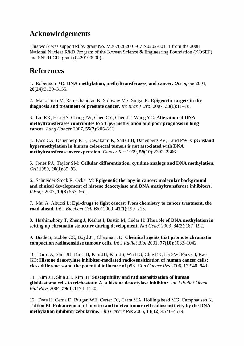
Figure 1 The effects of DNMT inhibitors on tumor cell radiosensitivity. Survival curves
of (A) A549 cells and (B) U373MG cells treated with the respective DNMT inhibitors prior
to radiation and radiation were compared with those of radiation alone. Points, mean for three
independent experiments; bars, SE
Effects of DNMT inhibitors on DNMT expression
DNMT1, DNMT3A, and DNMT3B are the three main functional methyltransferases
responsible for the establishment and maintenance of DNA methylation patterns in mammals.
The effects of DNMT inhibitors on the levels of DNMT expression were analyzed via
Western blotting using specific antibodies against DNMT1, DNMT3A, and DNMT3B.
Western blot analysis revealed a drastic depletion of DNMT1 and DNMT3A by psammaplin
A, 5-aza-2'-deoxycytidine, and zebularine in both A549 and U373MG cell lines. However, no
depletion of DNMT3B by the three DNMT inhibitors was observed in either of the cell lines
(Figure 2). These results indicate that DNMT inhibitors induce selective demethylation in
each of the evaluated tumor cell lines.
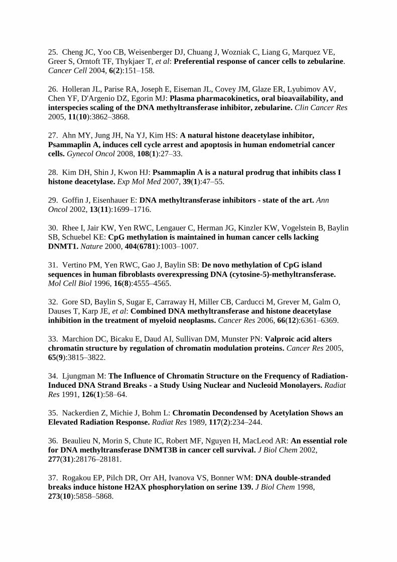
Figure 2 Methylation status was determined after exposure to DNMT inhibitors using
Western blot analysis of DNMT1, 3A/3B. The cells were treated with DNMT inhibitors for
18 hours. A drastic depletion of DNMT1 and DNMT3A by psammaplin A, 5-aza-2'-
deoxycytidine, and zebularine in both A549 and U373MG cell lines was observed. However,
there was no depletion of DNMT3B by the three DNMT inhibitors in either of the cell lines.
Each blot is representative of two independent experiments, with actin used as a loading
control
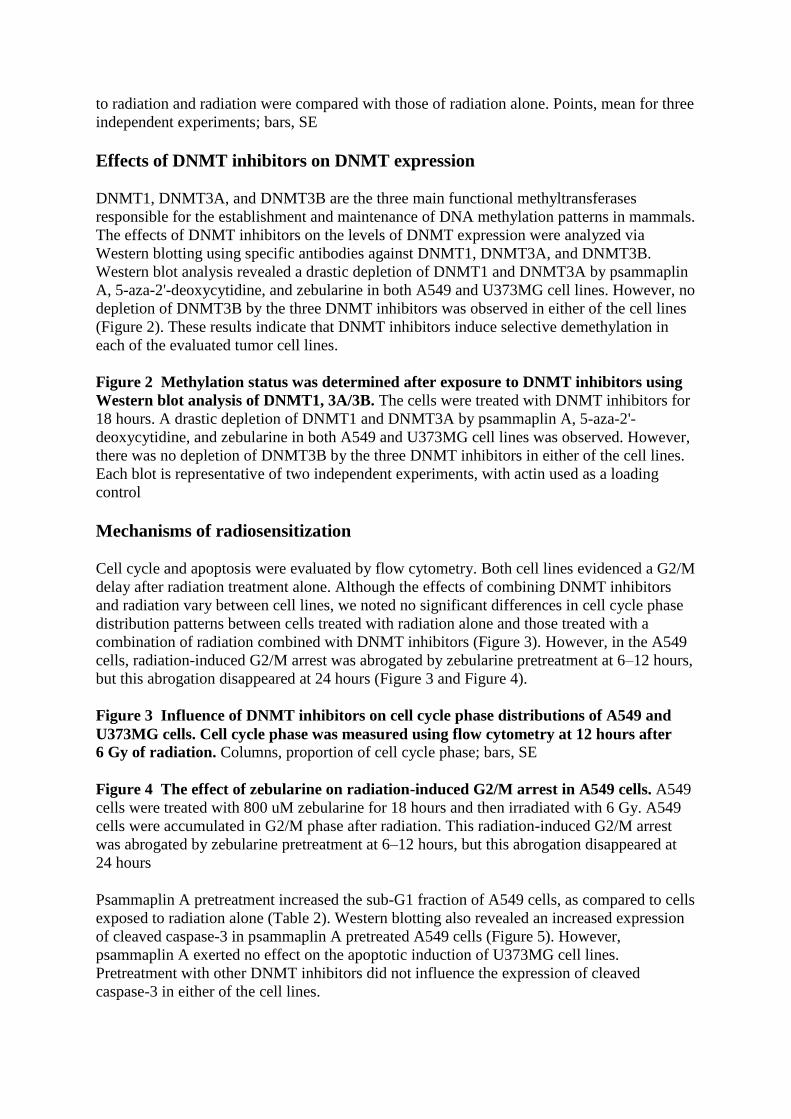
Mechanisms of radiosensitization
Cell cycle and apoptosis were evaluated by flow cytometry. Both cell lines evidenced a G2/M
delay after radiation treatment alone. Although the effects of combining DNMT inhibitors
and radiation vary between cell lines, we noted no significant differences in cell cycle phase
distribution patterns between cells treated with radiation alone and those treated with a
combination of radiation combined with DNMT inhibitors (Figure 3). However, in the A549
cells, radiation-induced G2/M arrest was abrogated by zebularine pretreatment at 6–12 hours,
but this abrogation disappeared at 24 hours (Figure 3 and Figure 4).
Figure 3 Influence of DNMT inhibitors on cell cycle phase distributions of A549 and
U373MG cells. Cell cycle phase was measured using flow cytometry at 12 hours after
6 Gy of radiation. Columns, proportion of cell cycle phase; bars, SE
Figure 4 The effect of zebularine on radiation-induced G2/M arrest in A549 cells. A549
cells were treated with 800 uM zebularine for 18 hours and then irradiated with 6 Gy. A549
cells were accumulated in G2/M phase after radiation. This radiation-induced G2/M arrest
was abrogated by zebularine pretreatment at 6–12 hours, but this abrogation disappeared at
24 hours
Psammaplin A pretreatment increased the sub-G1 fraction of A549 cells, as compared to cells
exposed to radiation alone (Table 2). Western blotting also revealed an increased expression
of cleaved caspase-3 in psammaplin A pretreated A549 cells (Figure 5). However,
psammaplin A exerted no effect on the apoptotic induction of U373MG cell lines.
Pretreatment with other DNMT inhibitors did not influence the expression of cleaved
caspase-3 in either of the cell lines.
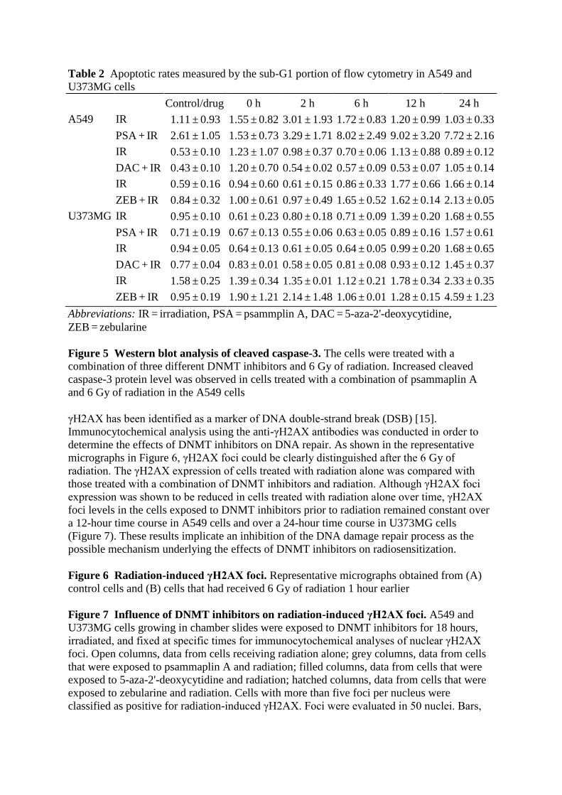
Table 2 Apoptotic rates measured by the sub-G1 portion of flow cytometry in A549 and
U373MG cells
Control/drug 0 h 2 h 6 h 12 h 24 h
A549 IR 1.11 ± 0.93 1.55 ± 0.82 3.01 ± 1.93 1.72 ± 0.83 1.20 ± 0.99 1.03 ± 0.33
PSA + IR 2.61 ± 1.05 1.53 ± 0.73 3.29 ± 1.71 8.02 ± 2.49 9.02 ± 3.20 7.72 ± 2.16
IR 0.53 ± 0.10 1.23 ± 1.07 0.98 ± 0.37 0.70 ± 0.06 1.13 ± 0.88 0.89 ± 0.12
DAC + IR 0.43 ± 0.10 1.20 ± 0.70 0.54 ± 0.02 0.57 ± 0.09 0.53 ± 0.07 1.05 ± 0.14
IR 0.59 ± 0.16 0.94 ± 0.60 0.61 ± 0.15 0.86 ± 0.33 1.77 ± 0.66 1.66 ± 0.14
ZEB + IR 0.84 ± 0.32 1.00 ± 0.61 0.97 ± 0.49 1.65 ± 0.52 1.62 ± 0.14 2.13 ± 0.05
U373MG IR 0.95 ± 0.10 0.61 ± 0.23 0.80 ± 0.18 0.71 ± 0.09 1.39 ± 0.20 1.68 ± 0.55
PSA + IR 0.71 ± 0.19 0.67 ± 0.13 0.55 ± 0.06 0.63 ± 0.05 0.89 ± 0.16 1.57 ± 0.61
IR 0.94 ± 0.05 0.64 ± 0.13 0.61 ± 0.05 0.64 ± 0.05 0.99 ± 0.20 1.68 ± 0.65
DAC + IR 0.77 ± 0.04 0.83 ± 0.01 0.58 ± 0.05 0.81 ± 0.08 0.93 ± 0.12 1.45 ± 0.37
IR 1.58 ± 0.25 1.39 ± 0.34 1.35 ± 0.01 1.12 ± 0.21 1.78 ± 0.34 2.33 ± 0.35
ZEB + IR 0.95 ± 0.19 1.90 ± 1.21 2.14 ± 1.48 1.06 ± 0.01 1.28 ± 0.15 4.59 ± 1.23
Abbreviations: IR = irradiation, PSA = psammplin A, DAC = 5-aza-2'-deoxycytidine,
ZEB = zebularine
Figure 5 Western blot analysis of cleaved caspase-3. The cells were treated with a
combination of three different DNMT inhibitors and 6 Gy of radiation. Increased cleaved
caspase-3 protein level was observed in cells treated with a combination of psammaplin A
and 6 Gy of radiation in the A549 cells
γH2AX has been identified as a marker of DNA double-strand break (DSB) [15].
Immunocytochemical analysis using the anti-γH2AX antibodies was conducted in order to
determine the effects of DNMT inhibitors on DNA repair. As shown in the representative
micrographs in Figure 6, γH2AX foci could be clearly distinguished after the 6 Gy of
radiation. The γH2AX expression of cells treated with radiation alone was compared with
those treated with a combination of DNMT inhibitors and radiation. Although γH2AX foci
expression was shown to be reduced in cells treated with radiation alone over time, γH2AX
foci levels in the cells exposed to DNMT inhibitors prior to radiation remained constant over
a 12-hour time course in A549 cells and over a 24-hour time course in U373MG cells
(Figure 7). These results implicate an inhibition of the DNA damage repair process as the
possible mechanism underlying the effects of DNMT inhibitors on radiosensitization.
Figure 6 Radiation-induced γH2AX foci. Representative micrographs obtained from (A)
control cells and (B) cells that had received 6 Gy of radiation 1 hour earlier
Figure 7 Influence of DNMT inhibitors on radiation-induced γH2AX foci. A549 and
U373MG cells growing in chamber slides were exposed to DNMT inhibitors for 18 hours,
irradiated, and fixed at specific times for immunocytochemical analyses of nuclear γH2AX
foci. Open columns, data from cells receiving radiation alone; grey columns, data from cells
that were exposed to psammaplin A and radiation; filled columns, data from cells that were
exposed to 5-aza-2'-deoxycytidine and radiation; hatched columns, data from cells that were
exposed to zebularine and radiation. Cells with more than five foci per nucleus were
classified as positive for radiation-induced γH2AX. Foci were evaluated in 50 nuclei. Bars,
SE. *p < 0.01 as determined by a logistic regression compared with radiation alone (6 Gy)
group
Discussion
Recently, histone modifications and DNA methylation, both of which are prominent
epigenetic mechanisms, have been evaluated with a view toward enhancing the
radiosensitivity of tumor cells via the regulation of chromatin structure modifications and the
expression of genes involved in cell cycle checkpoints, apoptosis, and DNA repair. A number
of investigators have previously reported that several HDAC inhibitors exert direct cytotoxic
effects, and can sensitize tumor cells to radiotherapy [14,16-19]. Kim et al. also demonstrated
that trichostatin A, which is the most potent HDAC inhibitor identified thus far, enhanced
radiosensitivity in a variety of human cancer cell lines [11,20]. Unlike the HDAC inhibitors,
however, little information is currently available regarding the effects of DNMT inhibitors on
radiosensitization [12,13].
There are two classes of DNMT inhibitors: the nucleoside analogues and the non-nucleoside
analogues. Several compounds are currently being evaluated in preclinical and clinical trials
for the treatment of solid and hematological malignancies. Contrary to their effect in
hematologic malignancies, the clinical effects of these compounds against solid tumors have
yet to be evaluated [21]. Therefore, the use of epigenetic therapies in combination with other
regimens, such as conventional chemotherapeutics or radiotherapy, should be considered. As
DNA methylation is intricately interrelated with chromatin structure and gene expression,
parameters long thought to be involved in the regulation of cellular radioresponse, it is
critically important to evaluate the effects of DNMT inhibitors on radiosensitization. In order
to assess this possibility, we employed a total of six DNMT inhibitors. Among them, 5-aza-
2'-deoxycytidine, zebularine, and psammaplin A were shown to exert a radiosensitizing effect
in both A549 and U373MG cells. Although some differences in the degrees of
radiosensitization were detected according to the different DNMT inhibitors and cell lines
used, these results demonstrate that some DNMT inhibitors may prove to be useful radiation
sensitizers in human cancer cells.
5-aza-2'-deoxycytidine, along with 5-azacytidine, is the first DNMT inhibitor reported, and
evidences clinical efficacy in cases of myelodysplastic syndrome and acute myelogenous
leukemia. Several previous studies have demonstrated that 5-aza-2'-deoxycytidine exerts a
radiosensitizing effect, with or without HDAC inhibitors, in a variety of cancer cell lines
[13,22,23]. Recently, the radiosensitization effect of 5-azacytidine was also referenced by
Hofstetter et al., who demonstrated that 5-azacytidine-induced genomic hypomethylation
induces enhanced radiation sensitivity in colorectal carcinoma [24]. These results are logical
consequences, considering that 5-azacytidine has a molecular structure and exerts clinical
effects similar to those of 5-aza-2'-deoxycytidine. However, in this study, 5-azacytidine did
not sensitize either of the tested cancer cell types to radiotherapy. 5-azacytidine may exert
differing effects on radiosensitivity according to cell type, and the IC50 of 5-azacytidine
varies among different cell lines. Although this may explain, at least in part, the different
results detected in our study, we did not successfully generate a definitive explanation for the
absence of any detectable radiosensitizing effect of 5-azacytidine. Because very few studies
have addressed the role of 5-azacytidine as a radiosensitizer, further studies will be required
to resolve this issue.

Zebularine, another 5-azacytidine derivative, was also shown in this study to induce
radiosensitivity. Zebularine has been demonstrated to have anticancer properties, which can
cause re-expression of the epigenetically silenced tumor suppressor p16 in solid
malignancies. However, its clinical development is limited by its higher dosage and poor
bioavailability, as observed previously in rats, mice, and monkeys [6,25,26]. Owing to the
relatively high toxicity of these nucleoside analogue compounds, non-nucleoside DNMT
inhibitors are currently being investigated for their possible use as anticancer agents.
The natural marine product, psammaplin A, was initially isolated from the Psammaplysilla
sponge in 1987. Since that time, psammaplin A has been shown to exert potent cytotoxicity
against several cancer cell lines, via the selective induction of genes associated with cell
cycle arrest and apoptosis [27,28]. Because psammaplin A has already been shown to exhibit
potent histone deacetylase inhibitory activity, this drug is a promising radiosensitizing agent
[28]. This is the first study in which the radiosensitizing effects of psammaplin A have been
confirmed. As psammaplin A has not yet been the subject of clinical trials, further clinical
research should be conducted to determine its efficacy when administered in combination
with radiotherapy.
Three different DNMTs exist, all of which are viable targets for DNMT inhibitors. DNMT1,
the most abundant of the three, is responsible for methylation during DNA replication
(maintenance methyltransferase). Other known methyltransferases include DNMT3A and
DNMT3B, which exhibit identical preferences for hemi-methylated and non-methylated
DNA, and have thus been classified as de novo methyltransferases. However, it has been
demonstrated that DNMT1 and DNMT3A/3B do not appear to be completely distinct in their
activities [29]. This supposition is based on two lines of evidence: first, despite being
DNMT1-deficient, a colon adenocarcinoma cell line was shown to be capable of retaining
80% of its methylation level while replicating, which implies that DNMT3 may perform a
function in the maintenance of methylation [30]; secondly, forced DNMT1 overexpression in
cancer cell lines does, indeed, induce de novo methylation [31]. Gene silencing of DNMT,
which functions as an oncogene, has been proposed as a good cancer treatment strategy [32].
In this study, three different DNMT inhibitors, all of which demonstrated radiosensitizing
effects, depleted DNMT1 and DNMT3A, but no such DNMT3B depletion was noted in the
A549 and U373MG cell lines. Depletion of DNMT, along with several members of the
structural maintenance of chromatin proteins (SMCs), SMC-associated protein, and
heterochromatin proteins, was reported to correlate with chromatin decondensation [33].
Considering that the effect of chromatin compaction on protection of DNA against radiation-
induced DSBs has been relatively well-established [34,35], the relaxation of chromatin
structure via the downregulation of DNMT with DNMT inhibitors may be a possible
mechanism for radiosensitization.
Selective inhibition of DNMT isoforms shown in this study may be attributable, in part, to
the doses of DNMT inhibitors. Beaulieu et al. previously reported that Western blot analysis
with anti-DNMT1, DNMT3A, or DNMT3B antibodies treated with increasing doses of
DNMT inhibitors effected a selective dose-dependent inhibition of the target isoform of
DNMT [36]. However, as only the IC50 values of the DNMT inhibitors were employed in this
study, we were unable to ascertain whether this selective inhibition of DNMT isoforms was a
function of the concentration of the drug.
In addition to the physical modification of chromatin structure, cellular processes that can
affect intrinsic radiosensitivity were evaluated in this study. Cell cycle, apoptosis, and DNA
repair have all been shown to influence radiosensitivity.
The proportion of cells in the G2/M phase detected in this study was not significantly altered
by pretreatment with DNMT inhibitors, and no abrogation of radiation-induced G2/M arrest
was noted except in the zebularine-treated A549 cell line. Radiation-induced G2/M arrest was
abrogated as the result of zebularine pretreatment at 6–12 hours, but this effect disappeared at
24 hours in the A549 cell line. In another study, zebularine was not found to result in the
abrogation of radiation-induced G2/M arrest in a U251 glioblastoma cell line [12]. Thus,
although there appears to be some relationship between cell lines and radiation-induced
G2/M arrest, it is less likely that the radiosensitization induced by the DNMT inhibitors
employed in this study is the result of a general inhibition of G2 checkpoint activation.
Apoptosis has previously been regarded as a potential mechanism for radiosensitization.
Different results have been reported regarding the role of apoptosis as a radiosensitizing
mechanism induced by DNMT inhibitors. Dote et al. previously reported that a combination
of radiation and zebularine did not significantly increase the sub-G1 population (apoptotic
cells) [12]. On the other hand, Qui et al. demonstrated that 5-aza-2'-deoxycytidine induces
radiosensitization in certain gastric cancer cell lines via induced increases in the apoptotic
rate, as evidenced by enhanced expression of the p53, RASSF1, and DAPK gene families
[23]. In this study, the addition of 5-aza-2'-deoxycytidine or zebularine to radiation did not
increase the apoptosis rate in either of the cell lines. However, exposure to psammaplin A in
the A549 cell line induced a significant increase in apoptotic death. Psammaplin A has been
reported to exert cytotoxic effects on cancer cells via the selective induction of apoptosis-
related genes [27]. This effect of psammalin A may result in an increase in radiation-induced
apoptosis. However, in this study, radiation-induced apoptosis was noted only in the A549
cell line, and not in the U373MG cell line. This finding may be attributable, at least in part, to
p53 expression status. The U373MG cell line, which contains mutated p53, might be
comparatively resistant to radiation-induced apoptosis, considering that apoptosis is mediated
by the p53 protein.
DNA repair is another process involved in the determination of cellular radiosensitivity. The
activation of DNA repair of cancer cells after sublethal DNA damage induced by radiation
might be one of the most important factors in resistance. The expression of γH2AX has been
recently identified as a sensitive indicator of radiation-induced DSBs [37]. In this study,
γH2AX expression in cells treated with a combination of DNMT inhibitors (5-aza-2'-
deoxycytidine and psammaplin A, as well as zebularine) was found to be similar to the results
achieved with radiation treatment at 1 hour after initial treatment, but was significantly
greater over time. This finding is generally consistent with the results obtained in other
previous studies, in which zebularine and 5-aza-2'-deoxycytidine were employed as
radiosensitizing agents [12,22]. Dote et al. evaluated the expression of γH2AX foci after
exposure to 2 Gy with and without zebularine pretreatment. Whereas zebularine had no effect
on radiation-induced γH2AX foci at 1 hour, the number of γH2AX per cell was significantly
greater in the zebularine-treated cells at 24 hours after irradiation, which suggested the
possible presence of unrepaired DNA damage [12]. DNA methylation is intimately associated
with histone deacetylases in terms of the epigenetic regulation of gene expression. Actually,
HDAC inhibitors such as LBH589 and MS-275 have been shown to enhance radiosensitivity
through similar mechanisms as those of the DNMT inhibitors. These HDAC inhibitors
prolonged γH2AX expression, suggesting an inhibition of DNA repair [14,38]. A previous
preclinical study demonstrated that an HDAC inhibitor downmodulated the expression of
DNA-PK and Rad51, which participated in the recovery of DSB, thereby abrogating key
cellular pathways involved in DNA DSB repair [17]. These results indicate that the
impairment of DNA DSB repair may be one of the most crucial mechanisms underlying
enhanced radiation responses in epigenetic phenomena. Based on this assumption, further
investigations are warranted to determine whether or not alterations in the methylation
patterns of a specific gene or set of genes involved in DNA repair might be modulated by
DNMT inhibitors, and that these changes might contribute to the observed enhancements of
radiosensitivity.
Several remain to be determined in future studies. First, a unified treatment schedule –
specifically, the administration of DNMT inhibitors 18 hours prior to radiation - was
employed in this study, and thus the optimal treatment schedule of DNMT inhibitors and
radiation remains to be established. In other studies, DNMT inhibitors were administered for
different treatment durations, i.e. 2 ~ 48 hours prior to radiation, considering several factors
such as drug half-lives and the expression of radiosensitivity-related genes [22,23]. Further
investigations into the optimal treatment schedule of DNMT inhibitors and radiation for
clinical applications will be necessary in the future.
Second, another important issue will involve assessments of the synergistic effects of DNMT
inhibitors and HDAC inhibitors on radiosensitivity. Although some previous studies have
reported that the combined gene silencing reversal effect was superior to that of treatment
with a single agent [32,39], only a few studies have thus far evaluated the influence of this
combined effect on increased radiosensitivity. Third, the mechanisms underlying DNMT
inhibitor-induced radiosensitzations need further investigation. We speculate that suppression
of DNMTs by the DNMT inhibitors was associated with enhanced radiosensitivity through
the change in DNA structure. However, the relationship between DNA methylation and
cellular radiosensitivity is to be elucidated in the future study.
Conclusions
Taken together, our study indicate that psammaplin A, 5-aza-2'-deoxycytidine, and zebularine
have the potential to increase radiosensitivity in lung cancer A549 and glioblastoma
U373MG cells, most probably by modulating the impairment of the DNA repair process.
Further investigations will be required to identify other additional mechanisms associated
with radiosensitivity, and to confirm the synergistic effects on radiosensitivity with other
epigenetic drugs, such as the HDAC inhibitors. Also, future studies should be conducted to
determine definitively whether the combination of DNMT inhibitors and radiation has real
potential as a clinical strategy for the treatment of cancer.
Competing interests
The authors declare that there are no conflicts of interest.
Author’s contributions
HJK, IHK, Idea and study design, JHK, IAK Analysis and development of methods. HJK,
Manuscript writing. DYP, Technique work. All authors, Review and final approval.
Acknowledgements
This work was supported by grant No. M2070202001-07 N0202-00111 from the 2008
National Nuclear R&D Program of the Korean Science & Engineering Foundation (KOSEF)
and SNUH CRI grant (0420100900).
References
1. Robertson KD: DNA methylation, methyltransferases, and cancer. Oncogene 2001,
20(24):3139–3155.
2. Manoharan M, Ramachandran K, Soloway MS, Singal R: Epigenetic targets in the
diagnosis and treatment of prostate cancer. Int Braz J Urol 2007, 33(1):11–18.
3. Lin RK, Hsu HS, Chang JW, Chen CY, Chen JT, Wang YC: Alteration of DNA
methyltransferases contributes to 5'CpG methylation and poor prognosis in lung
cancer. Lung Cancer 2007, 55(2):205–213.
4. Eads CA, Danenberg KD, Kawakami K, Saltz LB, Danenberg PV, Laird PW: CpG island
hypermethylation in human colorectal tumors is not associated with DNA
methyltransferase overexpression. Cancer Res 1999, 59(10):2302–2306.
5. Jones PA, Taylor SM: Cellular differentiation, cytidine analogs and DNA methylation.
Cell 1980, 20(1):85–93.
6. Schneider-Stock R, Ocker M: Epigenetic therapy in cancer: molecular background
and clinical development of histone deacetylase and DNA methyltransferase inhibitors.
IDrugs 2007, 10(8):557–561.
7. Mai A, Altucci L: Epi-drugs to fight cancer: from chemistry to cancer treatment, the
road ahead. Int J Biochem Cell Biol 2009, 41(1):199–213.
8. Hashimshony T, Zhang J, Keshet I, Bustin M, Cedar H: The role of DNA methylation in
setting up chromatin structure during development. Nat Genet 2003, 34(2):187–192.
9. Biade S, Stobbe CC, Boyd JT, Chapman JD: Chemical agents that promote chromatin
compaction radiosensitize tumour cells. Int J Radiat Biol 2001, 77(10):1033–1042.
10. Kim IA, Shin JH, Kim IH, Kim JH, Kim JS, Wu HG, Chie EK, Ha SW, Park CI, Kao
GD: Histone deacetylase inhibitor-mediated radiosensitization of human cancer cells:
class differences and the potential influence of p53. Clin Cancer Res 2006, 12:940–949.
11. Kim JH, Shin JH, Kim IH: Susceptibility and radiosensitization of human
glioblastoma cells to trichostatin A, a histone deacetylase inhibitor. Int J Radiat Oncol
Biol Phys 2004, 59(4):1174–1180.
12. Dote H, Cerna D, Burgan WE, Carter DJ, Cerra MA, Hollingshead MG, Camphausen K,
Tofilon PJ: Enhancement of in vitro and in vivo tumor cell radiosensitivity by the DNA
methylation inhibitor zebularine. Clin Cancer Res 2005, 11(12):4571–4579.

13. Cho HJ, Kim SY, Kim KH, Kang WK, Kim JI, Oh ST, Kim JS, An CH: The
combination effect of sodium butyrate and 5-Aza-2'-deoxycytidine on radiosensitivity in
RKO colorectal cancer and MCF-7 breast cancer cell lines. World J Surg Oncol 2009,
7:49.
14. Camphausen K, Burgan W, Cerra M, Oswald KA, Trepel JB, Lee MJ, Tofilon PJ:
Enhanced radiation-induced cell killing and prolongation of gammaH2AX foci
expression by the histone deacetylase inhibitor MS-275. Cancer Res 2004, 64(1):316–321.
15. Sedelnikova OA, Rogakou EP, Panyutin IG, Bonner WM: Quantitative detection of
(125)IdU-induced DNA double-strand breaks with gamma-H2AX antibody. Radiat Res
2002, 158(4):486–492.
16. Camphausen K, Cerna D, Scott T, Sproull M, Burgan WE, Cerra MA, Fine H, Tofilon
PJ: Enhancement of in vitro and in vivo tumor cell radiosensitivity by valproic acid. Int J
Cancer 2005, 114(3):380–386.
17. Chinnaiyan P, Vallabhaneni G, Armstrong E, Huang SM, Harari PM: Modulation of
radiation response by histone deacetylase inhibition. Int J Radiat Oncol Biol Phys 2005,
62(1):223–229.
18. Munshi A, Kurland JF, Nishikawa T, Tanaka T, Hobbs ML, Tucker SL, Ismail S,
Stevens C, Meyn RE: Histone deacetylase inhibitors radiosensitize human melanoma
cells by suppressing DNA repair activity. Clin Cancer Res 2005, 11(13):4912–4922.
19. Zhang Y, Jung M, Dritschilo A: Enhancement of radiation sensitivity of human
squamous carcinoma cells by histone deacetylase inhibitors. Radiat Res 2004,
161(6):667–674.
20. Kim IA, No M, Lee JM, Shin JH, Oh JS, Choi EJ, Kim IH, Atadja P, Bernhard EJ:
Epigenetic modulation of radiation response in human cancer cells with activated
EGFR or HER-2 signaling: potential role of histone deacetylase 6. Radiother Oncol 2009,
92(1):125–132.
21. Aparicio A, Eads CA, Leong LA, Laird PW, Newman EM, Synold TW, Baker SD, Zhao
M, Weber JS: Phase I trial of continuous infusion 5-aza-2'-deoxycytidine. Cancer
Chemother Pharmacol 2003, 51(3):231–239.
22. De Schutter H, Kimpe M, Isebaert S, Nuyts S: A systematic assessment of radiation
dose enhancement by 5-Aza-2'-deoxycytidine and histone deacetylase inhibitors in head-
and-neck squamous cell carcinoma. Int J Radiat Oncol Biol Phys 2009, 73(3):904–912.
23. Qiu H, Yashiro M, Shinto O, Matsuzaki T, Hirakawa K: DNA methyltransferase
inhibitor 5-aza-CdR enhances the radiosensitivity of gastric cancer cells. Cancer Sci
2009, 100(1):181–188.
24. Hofstetter B, Niemierko A, Forrer C, Benhattar J, Albertini V, Pruschy M, Bosman FT,
Catapano CV, Ciernik IF: Impact of genomic methylation on radiation sensitivity of
colorectal carcinoma. Int J Radiat Oncol Biol Phys 2010, 76(5):1512–1519.
25. Cheng JC, Yoo CB, Weisenberger DJ, Chuang J, Wozniak C, Liang G, Marquez VE,
Greer S, Orntoft TF, Thykjaer T, et al: Preferential response of cancer cells to zebularine.
Cancer Cell 2004, 6(2):151–158.
26. Holleran JL, Parise RA, Joseph E, Eiseman JL, Covey JM, Glaze ER, Lyubimov AV,
Chen YF, D'Argenio DZ, Egorin MJ: Plasma pharmacokinetics, oral bioavailability, and
interspecies scaling of the DNA methyltransferase inhibitor, zebularine. Clin Cancer Res
2005, 11(10):3862–3868.
27. Ahn MY, Jung JH, Na YJ, Kim HS: A natural histone deacetylase inhibitor,
Psammaplin A, induces cell cycle arrest and apoptosis in human endometrial cancer
cells. Gynecol Oncol 2008, 108(1):27–33.
28. Kim DH, Shin J, Kwon HJ: Psammaplin A is a natural prodrug that inhibits class I
histone deacetylase. Exp Mol Med 2007, 39(1):47–55.
29. Goffin J, Eisenhauer E: DNA methyltransferase inhibitors - state of the art. Ann
Oncol 2002, 13(11):1699–1716.
30. Rhee I, Jair KW, Yen RWC, Lengauer C, Herman JG, Kinzler KW, Vogelstein B, Baylin
SB, Schuebel KE: CpG methylation is maintained in human cancer cells lacking
DNMT1. Nature 2000, 404(6781):1003–1007.
31. Vertino PM, Yen RWC, Gao J, Baylin SB: De novo methylation of CpG island
sequences in human fibroblasts overexpressing DNA (cytosine-5)-methyltransferase.
Mol Cell Biol 1996, 16(8):4555–4565.
32. Gore SD, Baylin S, Sugar E, Carraway H, Miller CB, Carducci M, Grever M, Galm O,
Dauses T, Karp JE, et al: Combined DNA methyltransferase and histone deacetylase
inhibition in the treatment of myeloid neoplasms. Cancer Res 2006, 66(12):6361–6369.
33. Marchion DC, Bicaku E, Daud AI, Sullivan DM, Munster PN: Valproic acid alters
chromatin structure by regulation of chromatin modulation proteins. Cancer Res 2005,
65(9):3815–3822.
34. Ljungman M: The Influence of Chromatin Structure on the Frequency of Radiation-
Induced DNA Strand Breaks - a Study Using Nuclear and Nucleoid Monolayers. Radiat
Res 1991, 126(1):58–64.
35. Nackerdien Z, Michie J, Bohm L: Chromatin Decondensed by Acetylation Shows an
Elevated Radiation Response. Radiat Res 1989, 117(2):234–244.
36. Beaulieu N, Morin S, Chute IC, Robert MF, Nguyen H, MacLeod AR: An essential role
for DNA methyltransferase DNMT3B in cancer cell survival. J Biol Chem 2002,
277(31):28176–28181.
37. Rogakou EP, Pilch DR, Orr AH, Ivanova VS, Bonner WM: DNA double-stranded
breaks induce histone H2AX phosphorylation on serine 139. J Biol Chem 1998,
273(10):5858–5868.

38. Geng L, Cuneo KC, Fu A, Tu TX, Atadja PW, Hallahan DE: Histone deacetylase
(HDAC) inhibitor LBH589 increases duration of gamma-H2AX foci and confines
HDAC4 to the cytoplasm in irradiated non-small cell lung cancer. Cancer Res 2006,
66(23):11298–11304.
39. Cameron EE, Bachman KE, Myohanen S, Herman JG, Baylin SB: Synergy of
demethylation and histone deacetylase inhibition in the re-expression of genes silenced
in cancer. Nat Genet 1999, 21(1):103–107.
Figure 1
Figure 2

Figure 3
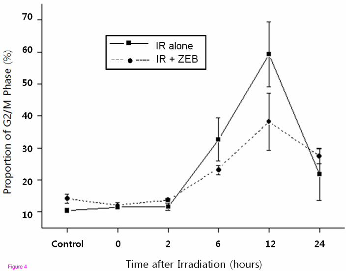
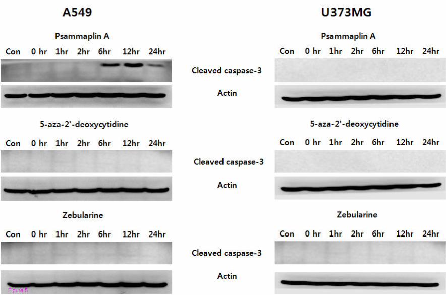
Figure 5

Figure 6
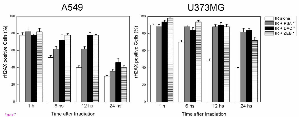
Figure 7