Embed Size (px)
Citation preview

RADIATION PROTECTION
Dr.P.P.MohananThrissur
2
Outline
• Basis for protection, radiation risk and recommendations
• Personal dosimetry• Protection tools
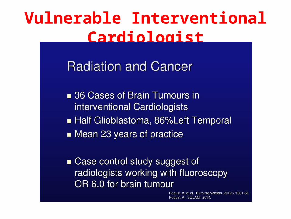
Vulnerable Interventional Cardiologist

POTENNTIAL HAZARDS

5
Coronary interventionalists must also have a thorough knowledge of specialized equipment, techniques, and devices used to perform PCI competently
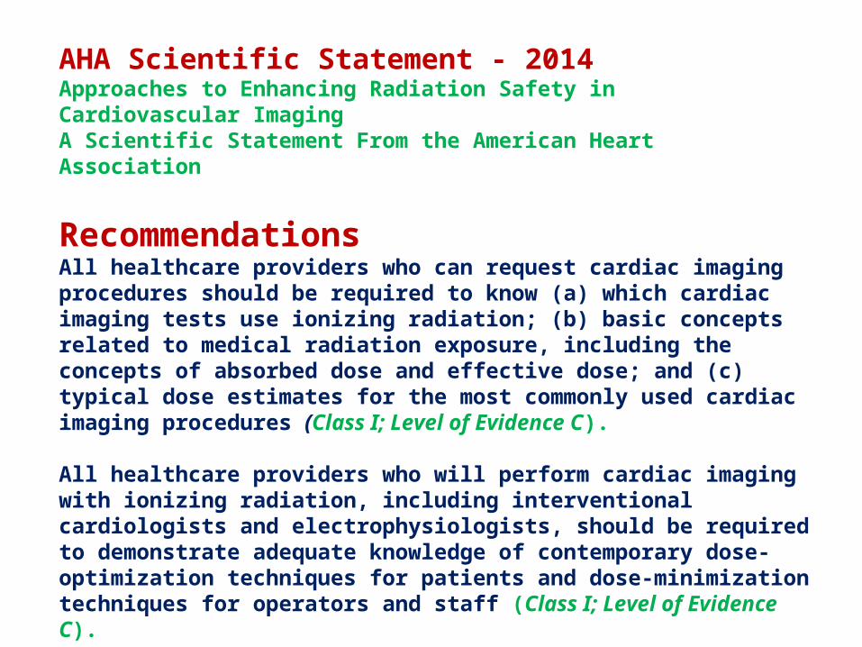
AHA Scientific Statement - 2014Approaches to Enhancing Radiation Safety in Cardiovascular ImagingA Scientific Statement From the American Heart Association
RecommendationsAll healthcare providers who can request cardiac imaging procedures should be required to know (a) which cardiac imaging tests use ionizing radiation; (b) basic concepts related to medical radiation exposure, including the concepts of absorbed dose and effective dose; and (c) typical dose estimates for the most commonly used cardiac imaging procedures (Class I; Level of Evidence C).
All healthcare providers who will perform cardiac imaging with ionizing radiation, including interventional cardiologists and electrophysiologists, should be required to demonstrate adequate knowledge of contemporary dose-optimization techniques for patients and dose-minimization techniques for operators and staff (Class I; Level of Evidence C).

Recommendations
1.Education. of clinicians and patients
2.Justification that a particular cardiac imaging test with radiation is needed
3.Optimization of radiation exposure (choosing the smallest dose that provides high-quality images).

Lecture 6: Standards and guidance 8
Importance of training
• European Guidelines published in 2000.
• Radiologists 30-50 hours• Cardiologists 20-30 hours• Other doctors using
fluoroscopy X rays systems 15-20 hoursAvailable at:
http://europa.eu.int/comm/environment/radprot

Lecture 6: Standards and guidance 9
Cardiologist Patient Protection
Responsibilities
Advice of qualified expert Training
criteria
Optimization
Equipment design and suppliers Quality
assurance
Justification

Lecture 6: Standards and guidance 10
Limits on Patient Doses?
• There are no regulatory limits on the radiation dose a patient may receive
– Question: do you think that the benefit outweighs the risk???

Time of onset of clinical signs Time of onset of clinical signs of skin injury depending onof skin injury depending on
dose receiveddose received
SymptomsSymptoms Dose range Dose range Time of onset Time of onset
(Gy) (Gy) (day) (day)
Erythema 3-10 14-21 Epilation >3 14-18 Dry desquamation 8-12 25-30 Moist desquamation 15-20 20-28 Blister formation 15-25 15-25 Ulceration >20 14-21 Necrosis >25 >21
Ref.: IAEA-WHO: Diagnosis and Treatment of Radiation Injuries.

12
X-Ray
Scatterradiation
Measures taken to reduce radiation exposure to patient will also benefit the operator/cath. lab. staff

Radiation risk
• Stochastic effect
• Deterministic effect


Deterministic vs. stochastic effects (representative, not scaled).
Picano E et al. Eur Heart J 2014;eurheartj.eht394

Radiation effectsRadiation effects
Early(deterministic only)
LocalRadiation injury ofindividual organs:functional and/or
morphologicalchanges withinhrs-days-weeks
CommonAcute radiation disease
Acute radiation syndrome
Late
DeterministicRadiation dermatitisRadiation cataractaTeratogenic effects
StochasticTumours LeukaemiaGenetic effects

Deterministic effect
• Gray is unit of exposure of radiation
• ONE CHEST X RAY 0.15 mGRAY• 10000 chest x ray • Or• 100 CT abdomen =• 30 mins to 1 hr fluoroscopy exposure
18
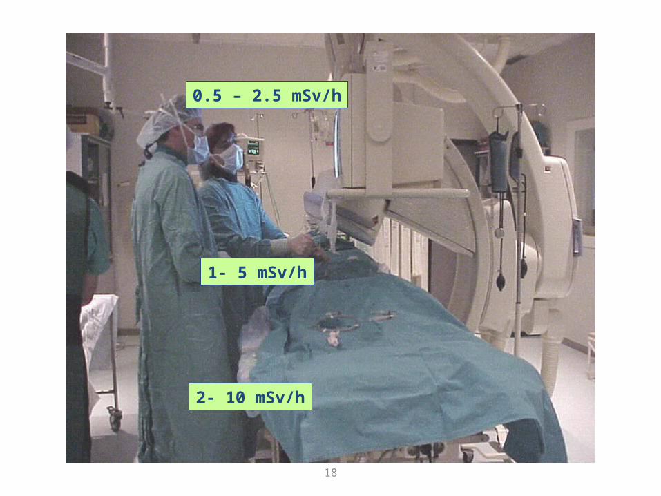
1- 5 mSv/h
0.5 – 2.5 mSv/h
2- 10 mSv/h

19
Limits on Occupational Doses (ICRP)*Annual Dose Limit
(mSv)
Effective dose, worker 20
Equivalent dose to lens of eye
150
Equivalent dose to skin 500
Equivalent dose to hands and feet
500
Effective dose to embryo or fetus
1
Effective dose, public 1*Please follow the recommendations as prescribed by your national authority
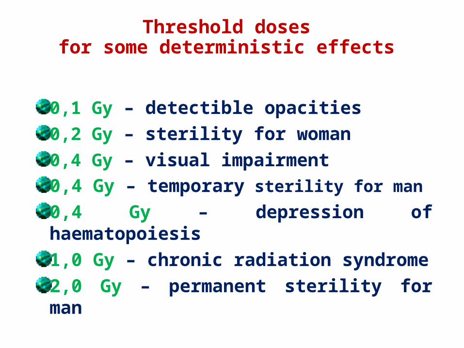
Threshold doses for some deterministic effects
0,1 Gy – detectible opacities 0,2 Gy – sterility for woman0,4 Gy – visual impairment 0,4 Gy – temporary sterility for man
0,4 Gy – depression of haematopoiesis1,0 Gy – chronic radiation syndrome2,0 Gy – permanent sterility for man

Radiation dose in coronary angiography and Intervention:
Journal of Medical Radiation Sciences

A Typical Fluoroscopic LabOverhead shields need to be positioned down next to the patient and adjacent to the image intensifier to seal scatteroff from below.
Table skirts shield the highest backscatter levels from the x-ray tube.

23
To obtain the images …
• Two technologies are used:– Image intensifier– Flat panel detector

The X-ray Imaging Process
Absorption and transmission of x-rays contribute to the imaging process and patient dose.
Scattered or partially absorbed x–rays contribute to occupational exposure, but are less than 1% of the primary beam intensity.
TubeFocalSpot
Absorption
Transmission
Scatterx-rays
Primaryx-ray beam
ImageReceptor


26
65.9
22.9
11.314.8
6.12.6 3.2
56.5
25.5
15.3
10.2
3.6 2.96.6
0
10
20
30
40
50
60
70
H 3000
Innova
Comparison of Philips H 3000 and Innova 2000 in PCI Characteristics of procedures & lesionsCharacteristics of procedures & lesions
%

Recommendations
1.Education. of clinicians and patients
2.Justification that a particular cardiac imaging test with radiation is needed
3.Optimization of radiation exposure (choosing the smallest dose that provides high-quality images).
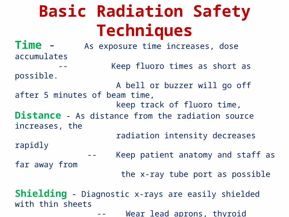
Basic Radiation Safety TechniquesTime - As exposure time increases, dose accumulates -- Keep fluoro times as short as possible. A bell or buzzer will go off after 5 minutes of beam time, keep track of fluoro time, Distance - As distance from the radiation source increases, the radiation intensity decreases rapidly -- Keep patient anatomy and staff as far away from the x-ray tube port as possible
Shielding - Diagnostic x-rays are easily shielded with thin sheets -- Wear lead aprons, thyroid shields, leaded glasses and use overhead leaded shields and table skirts

29
Optimization means ...
• To avoid acquiring more images than necessary:– Take care of the fluoroscopy time.– Take care of the number of series.– Take care of the number of frames per series.
• To avoid acquiring images with more quality (and more dose) than necessary:– It could be possible to accept sometimes some
noisy images in fluoroscopy and also in cine acquisitions.

ALARA rule
• As low as reasonably achievable
• Reduce number of exam • Reduce time of exam• Use alternaive

31
Minimize Exposure Time
• Everything you do to minimize exposure time reduces radiation dose!!– Minimize fluoro and cine times– Whenever possible, step out of room– Step behind barrier (or another person)
during fluoro or cine– Use pulsed fluoroscopy– minimizes time
X ray tube is producing X rays

32
Siemens Axiom ArtisCine normal mode20 cm PMMA177 Gy/fr (entrance PMMA)
Siemens Axiom Artis, Fluoro low dose 20 cm PMMA13 Gy/fr (entrance PMMA)

Lecture 9: Optimization of Radiation Protection in Cardiology
33
The proposed reference levels for Coronary Angiography and PTCA were DAP 45 Gy•cm2 and 75 Gy•cm2; fluoroscopy time 7.5 min and 17 min and number of frames 1250 and 1300, respectively.

34
Influence of operation modes: from low fluoroscopy to cine, scatter dose rate could increase in a factor of 10(from 2 to 20 mSv/h for normal size)

35
Distance between patient and detector

36
d
2d
Because the same energy is spread over a surface 4 times larger at a doubled distance, the same object will receive only a fourth of the dose when moved away from “d” to “2d”
Source
Doubling the distance from the source divides the dose by a factor of 4
The inverse square law

37
The inverse square law

38
Collimation

39
Collimators use in reduce exposure
FOV 15
dose reduction 25%

40
FOV 20
Collimators reduce exposure

41
Anti-scatter grid
Increase DAP and skin dose x 2 timesImprove image quality

42
proper filtering
improper filtering causes image deterioration
FILTERING
Filtering prevents image saturation in low absorption areas
43
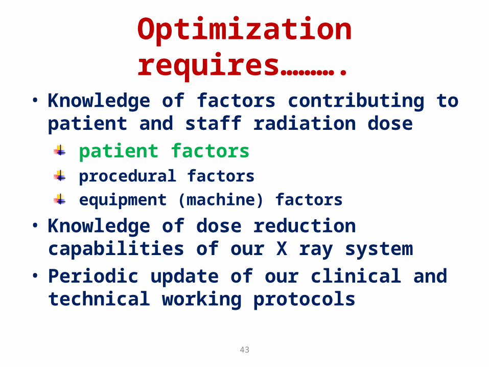
Optimization requires……….
• Knowledge of factors contributing to patient and staff radiation dose
patient factors procedural factors equipment (machine) factors
• Knowledge of dose reduction capabilities of our X ray system
• Periodic update of our clinical and technical working protocols

44
• Optimization is especially important in more complex PTCA procedures
chronic total occlusion bifurcation lesion degenerated saphenous
vein graft lesion lesion in severely
tortuous vessel ostial lesion

45
Exposure variation in exposure rate (DAP rate) with projection
Cusma JACC 1999
Projection Fluoroscopy entrance dose
rate
(mGy/min)
Cine
entrance dose rate
(mGy/min)
AP 31 388
RAO 30° 19 203
LAO 40° 20 216
LAO 40°, Cran 30° 80 991
LAO 40°, Cran 40° 99 1236
LAO 40°, Caud 20° 29 341


51
Optimisation
1. Use of the wedge filter on bright peripheral areas2. 2-3 sequences (except for difficult anatomic details)3. 12.5-15 frames/s (25-30 only if heart rate exceeds
90-100 bpm or in paediatric patients) 4. 60 images per sequence at average (12.5-15 fr/s)
except if collaterals have to be imaged or in case of slow flow

52
Basic Radiation Protection
• Time (T), Distance (D), and Shielding (S)
• Time– minimize exposure time• Distance– increasing distance• Shielding– use shielding effectively;
portable and pull-down shields, protective aprons; stand behind someone else

53
1- 5 mSv/h
0.5 – 2.5 mSv/h
2- 10 mSv/h

Lecture 7: Occupational exposure and protective devices
54
Radiation Monitoring Badge
Plastic filter Metal filters Open windows
Open window
Lecture 7: Occupational exposure and protective devices
55
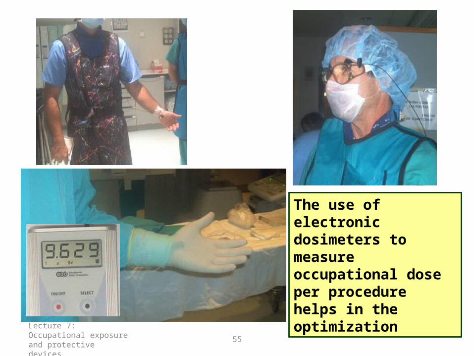
The use of electronic dosimeters to measure occupational dose per procedure helps in the optimization

Lecture 7: Occupational exposure and protective devices
56
Types of Personal Radiation Monitors• Film• Thermoluminescent dosimeters (TLDs)• Optically stimulated luminescence (OSL)
dosimeters• Electronic personal dosimeters

57
Advantages and Disadvantages of Personal Radiation Monitors
• Electronic dosimeters— insensitive to heat, no permanent record, minimum dose > 0.1 mSv, no imaging capability, calibration can be difficult, must rely on employee for care of device (somewhat delicate), employee must read-out dosimeter and record results, weekly or monthly readout
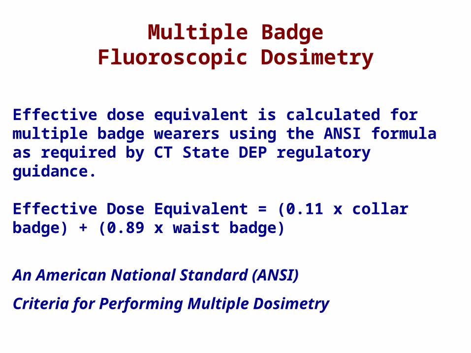
Multiple BadgeFluoroscopic Dosimetry
Effective dose equivalent is calculated for multiple badge wearers using the ANSI formula as required by CT State DEP regulatory guidance.
Effective Dose Equivalent = (0.11 x collar badge) + (0.89 x waist badge)
An American National Standard (ANSI)
Criteria for Performing Multiple Dosimetry

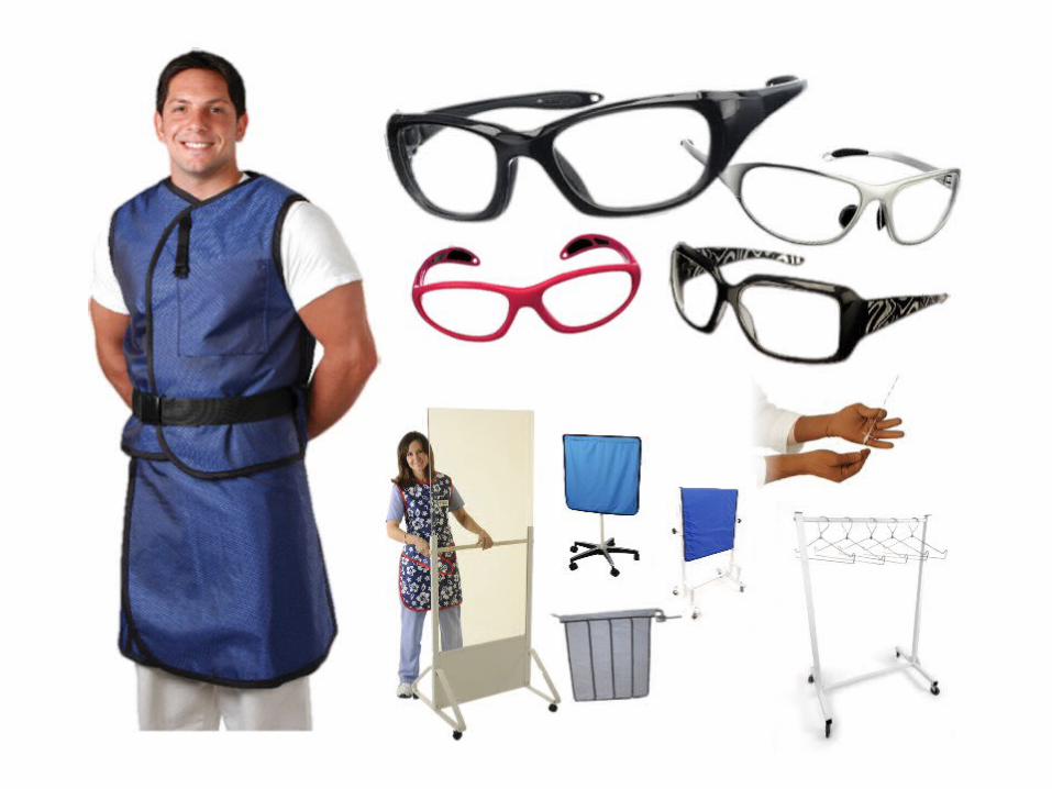
Protection tools

60
Personal protective equipment
• Registrants and licensees shall ensure that workers are provided with suitable and adequate personal protective equipment.
• Protective equipment includes
• lead aprons, thyroid protectors, protective eye-wear and gloves.
• The need for these protective devices should be established by the RPO.

61
Weight: 80 gramsLead equivalent: 0.75mm front and side shields leaded glass
Lead apron typically attenuates >90%Vest-Skirt Combination distributing 70% of the total weight onto the hips leaving only 30% of the total weight on the shoulders.
Option with light material reducing the weight by over 23% while still providing 0.5 mm Pb protection at 120 kVp

C. Radiation Protection and How does it Work?

64
0.25 mm lead
60 kV; 100% 2 - 3 %
100 kV; 100% 8 - 15 %
Attenuation measured with lead apronsAttenuation measured with lead aprons
X ray beam filtration has a great influence!!

Thyroid collar
• Standard 0.5mm lead apron• Protect you from 95%• FROM RADIATION EXPOSURE

Wear Protective Eyewear
“In the 59 interventional radiologists aged between 29 and 63 (median age 35), PSC cataracts were found in five participants and an additional 22 had evidence of PSC changes”
46% experienced changes in the eyes
G. Eyewear

67
DETERMINISTIC LENS THRESHOLD AS QUOTED BY ICRP
OPACITIES THRESHOLD
>0.1 Sv/year CONTINUOUS ANNUAL RATE
>0.15 Sv/year CONTINUOUS ANNUAL RATE
CATARACT

68
UP TO 2 mSv IN LENS COULD BE RECEIVED IN A SINGLE PROCEDURE
if protection tools are not used
WITH 3 PROCED./DAY IT IS POSSIBLE TO RECEIVE 1500 mSv/year
IN FOUR YEARS WILL BE POSSIBLE TO HAVE LENS OPACITIES


Lecture 7: Occupational exposure and protective devices
70
Radiation Protection of HandsBest way to minimize dose to fingers and hand:Keep your fingers out of the beam!!!
Dose rate outside of the beam and on side of patient opposite X ray tube:
Very low compared to in the beam!!!
71
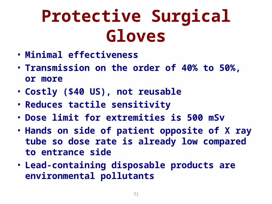
Protective Surgical Gloves
• Minimal effectiveness• Transmission on the order of 40% to 50%, or more• Costly ($40 US), not reusable• Reduces tactile sensitivity• Dose limit for extremities is 500 mSv• Hands on side of patient opposite of X ray tube so
dose rate is already low compared to entrance side• Lead-containing disposable products are
environmental pollutants

H. Gloves
ProteXProGuard
2 Thicknesses
Non-reactive Latex
Anatomically correct
Resterilize up to 4x
Come sterile
Sizes 6-9 w/ ½ sizes
Reduces Radiation
Powder Free
FDA 510(k)
Cost effective
Beaded Cuff
Thicker Feel
Premium, Higher $
Soft-Touch™ interior finish
Thinner Texture
ATTENUATION 60 KvP 80 KvP 100 KvP 120 KvPProGuard RR1, ProteX PX-10.0088” / 0.22mm
45% 35% 26% 23%
ProGuard RR2, ProteX PX-20.012” / 0.30mm
55% 43% 35% 31%
Protech Radiation Reducing Gloves


Rate of appropriateness for the four procedures.

79
Procedure optimization in the cath. lab.patients and staff share a lot……
– correct indications– fluoro time reduction– frame rate reduction (25 12,5/sec)– collimation/filtering– LAO cranial projection limitation – distance from X ray source – lead apron and thyroid protection– protective glasses and suspended screen
(staff)
(patient)