Embed Size (px)
Citation preview

Policy No: 2015-8005 - v3 Policy: Radiation Safety Management Plan - CHW
This document reflects what is currently regarded as safe practice. However, as in any clinical situation, there may be factors which cannot be covered by a single set of guidelines. This document does not replace the need for the application of clinical judgement to each individual presentation.
Approved by: Policy, Procedure and Guideline Committee Date Effective: 1st April 2019 Review Period: 3 years Team Leader: Radiation Safety Officer Area/Dept: Radiation Safety
Date of Publishing: 16 August 2019 11:09 AM Date of Printing: Page 1 of 53 K:\CHW P&P\ePolicy\Mar 19\Radiation Safety Management Plan - CHW.docx This Policy/Procedure may be varied, withdrawn or replaced at any time. Compliance with this Policy/Procedure is mandatory.
RADIATION SAFETY MANAGEMENT PLAN - CHW
POLICY©
DOCUMENT SUMMARY/KEY POINTS
• This Radiation Safety Management Plan provides information about ionising radiation, basic radiation safety, implementation of radiation safety, and how ionising radiation is utilised within The Children’s Hospital at Westmead (CHW).
CHANGE SUMMARY
• Updated to reflect updated Australian Standards, the use of EMR and clarification of sections to greater understanding for the reader.

Policy No: 2015-8005 - v3 Policy: Radiation Safety Management Plan - CHW
This document reflects what is currently regarded as safe practice. However, as in any clinical situation, there may be factors which cannot be covered by a single set of guidelines. This document does not replace the need for the application of clinical judgement to each individual presentation.
Approved by: Policy, Procedure and Guideline Committee Date Effective: 1st April 2019 Review Period: 3 years Team Leader: Radiation Safety Officer Area/Dept: Radiation Safety
Date of Publishing: 16 August 2019 11:09 AM Date of Printing: Page 2 of 53 K:\CHW P&P\ePolicy\Mar 19\Radiation Safety Management Plan - CHW.docx This Policy/Procedure may be varied, withdrawn or replaced at any time. Compliance with this Policy/Procedure is mandatory.
READ ACKNOWLEDGEMENT
Email notification of release of this document to the following, with their discretion to distribute to their staff:
• Chief Radiographer
• Chief Nuclear Medicine Scientist
• NUM – Camperdown Ward
• NUM – Commercial Travellers Ward
• NUM – Operating Suite
• HOD – Dentistry
• HOD – Haematology
• HOD – Gastroenterology
• Operations Manager – Kids Research (KR)
• Laboratory Manager – Endocrinology
• Members of the Radiation Safety Committee

Policy No: 2015-8005 - v3 Policy: Radiation Safety Management Plan - CHW
Date of Publishing: 16 August 2019 11:09 AM Date of Printing: Page 3 of 53 This Policy/Procedure may be varied, withdrawn or replaced at any time. Compliance with this Policy/Procedure is mandatory.
TABLE OF CONTENTS 1 Introduction .................................................................................................................. 5 2 General Information .................................................................................................... 5 2.1 Ionising Radiation .......................................................................................................... 5 2.2 Radiation Units and Quantities ...................................................................................... 6 2.3 Sources of Ionising Radiation ........................................................................................ 7 2.4 Radiation Protection Principles ...................................................................................... 7 2.5 Radiation Safety Structure at CHW ............................................................................... 8
2.5.1 Radiation Safety Officer (RSO) .............................................................................. 8 2.5.2 Radiation Safety Committee (RSC) ........................................................................ 8
3 The NSW Radiation Control Act and Regulation ...................................................... 9 3.1 Licences ........................................................................................................................ 9
3.1.1 Radiation User Licence Exemptions .................................................................... 10 3.2 Control of Radiation Exposure ..................................................................................... 11 4 Personnel Monitoring Services ................................................................................ 11 4.1 Optically Stimulated Luminescent Dosimetry (OSLD) ................................................. 12 4.2 Thermoluminescent Dosimetry (TLD) .......................................................................... 12 4.3 Electronic Personal Dosimeters .................................................................................. 12 5 Use of Radioactive Substances and Radiation Apparatus in Laboratories ......... 12 5.1 Classification of Laboratories ...................................................................................... 13 5.2 Requirements for Laboratories .................................................................................... 13 5.3 Handling Unsealed Radioactive Substances ............................................................... 14 5.4 Radiation Apparatus .................................................................................................... 15 5.5 Sealed Sources ........................................................................................................... 16 5.6 Radioactive Waste Management within Laboratories ................................................. 16
5.6.1 Low Activity Waste ............................................................................................... 16 5.6.2 High Activity Waste .............................................................................................. 17 5.6.3 Packaging and Containment of Radioactive Waste ............................................. 17
5.7 Record Keeping ........................................................................................................... 18 5.8 Decontamination Procedures ...................................................................................... 18 5.9 Decommissioning of a Radioisotope Laboratory ......................................................... 19 6 Procedures – Radionuclides in the Wards .............................................................. 19 6.1 Diagnostic Procedures ................................................................................................ 19
6.1.1 Commercial Travellers (CT) Ward ........................................................................ 20 6.2 Therapeutic Procedures .............................................................................................. 20
6.2.1 Camperdown Ward .............................................................................................. 21 7 Procedures – Nuclear Medicine Department .......................................................... 22 7.1 Licencing ..................................................................................................................... 22 7.2 Radiation Hazards ....................................................................................................... 23
7.2.1 Sources of External Exposure .............................................................................. 23 7.2.2 Sources of Internal Exposure ............................................................................... 23
7.3 Diagnostic Procedures ................................................................................................ 24 7.4 Therapeutic procedures ............................................................................................... 24
7.4.1 Medical Emergencies involving patients undergoing radionuclide therapy .......... 25

Policy No: 2015-8005 - v3 Policy: Radiation Safety Management Plan - CHW
Date of Publishing: 16 August 2019 11:09 AM Date of Printing: Page 4 of 53 This Policy/Procedure may be varied, withdrawn or replaced at any time. Compliance with this Policy/Procedure is mandatory.
7.5 Reporting of Maladministration or Equipment Malfunction .......................................... 26 7.6 Radioactive Waste Disposal ........................................................................................ 26 8 Procedures: Department of Medical Imaging ......................................................... 27 8.1 Licencing ..................................................................................................................... 27 8.2 Staff Protection ............................................................................................................ 27
8.2.1 Radiation Monitoring Badges ............................................................................... 27 8.2.2 Equipment ............................................................................................................ 28 8.2.3 X-ray Personal Protective Equipment .................................................................. 28
8.3 Pregnancy and patients ............................................................................................... 28 8.3.1 Identification ......................................................................................................... 28 8.3.2 Patient Not Pregnant ............................................................................................ 29 8.3.3 Procedure when Patient is Pregnant .................................................................... 29 8.3.4 Procedure when a Patient is found to be Pregnant AFTER an X-ray .................. 29
8.4 High Dose Fluoroscopy Procedures ............................................................................ 29 8.5 General ........................................................................................................................ 29 8.6 Use of Mobile X-ray within Wards ............................................................................... 30
8.6.1 Radiation Safety Guidelines: ................................................................................ 30 9 Procedures – Operating Suite .................................................................................. 32 9.1 Orthopaedic Surgeons ................................................................................................. 32 9.2 Sentinel Node Biopsy .................................................................................................. 33 10 Procedures – Haematology (Blood Bank) ............................................................... 33 10.1 Licencing ..................................................................................................................... 33 10.2 Threat Levels ............................................................................................................... 33 11 Procedures – Department of Dentistry .................................................................... 35 11.1 Licencing ..................................................................................................................... 35 11.2 X-ray Units ................................................................................................................... 36 12 Death Procedures – Bodies containing radioactive material ................................ 36 12.1 Post-mortem or embalming ......................................................................................... 37 12.2 Cremation .................................................................................................................... 37 12.3 Handling of the coffin ................................................................................................... 37 12.4 Direct burial or mausoleum entombment ..................................................................... 38 13 Radiation Dosimetry of Patient Procedures ........................................................... 38 13.1 Diagnostic Medical Imaging ......................................................................................... 38 13.2 Nuclear Medicine ......................................................................................................... 40 14 Radiation and Pregnancy ......................................................................................... 40 15 Radiation Incidents and Special Procedures .......................................................... 41 15.1 Internal Reporting of Radiation Incidents .................................................................... 41 15.2 Legislative Definition of a Radiation Accident .............................................................. 41 15.3 Reporting Radiation Accidents to the Environmental Protection Authority .................. 42 15.4 Radioactive Contamination Response ........................................................................ 43
15.4.1 Personnel Decontamination .............................................................................. 43 15.4.2 Work Surface Decontamination ........................................................................ 44 15.4.3 Equipment Decontamination ............................................................................. 44
16 Irradiation of volunteers for research purposes .................................................... 45 Appendix A: Disaster procedures ...................................................................................... 47 Appendix B – Glossary ........................................................................................................ 48
Policy No: 2015-8005 - v3 Policy: Radiation Safety Management Plan - CHW
Date of Publishing: 16 August 2019 11:09 AM Date of Printing: Page 5 of 53 This Policy/Procedure may be varied, withdrawn or replaced at any time. Compliance with this Policy/Procedure is mandatory.
Appendix C – Radiation Unit Conversion Table ................................................................ 50 Appendix D – Radioactive Waste Holding Tanks .............................................................. 51 Appendix E – Legislative Dose Limits ................................................................................ 52 Appendix F – Quality Management in Radiation Safety ................................................... 53
1 Introduction
This plan is a guide for workers involved, directly or indirectly, with the use of ionising radiation apparatus and / or radioactive materials. Further information may be obtained from the Radiation Safety Officer, Nuclear Medicine Department.
2 General Information
2.1 Ionising Radiation Ionising radiation is defined as radiation capable of producing ions in its passage through matter. Examples of ionising radiation are:
• Alpha (α) particles are identical with helium nuclei; consisting of two protons and two neutrons. Alpha particles are usually emitted by heavy radioactive atoms such as uranium and radium. Being large and relatively slow, they quickly dissipate their energy by colliding with the atoms of the material through which they travel causing ionisation to take place. Alpha particles thus have very little power of penetration and are stopped completely by a thin sheet of paper, the outer layer of human skin, or a few centimetres of air. Alpha emitters are most damaging when incorporated into the body, and are not normally used unless securely sealed.
• Beta (β) particles are high speed electrons emitted from the nuclei of radioactive atoms. Being light weight and emitted with a speed close to that of light, beta particles have greater penetrating ability than alpha particles of the same energy, but still will be stopped by a few millimetres of aluminium, a centimetre or so of human tissue, or a few metres of air, dependent on their energy. Beta emitters are also most hazardous when ingested, but can also be hazardous externally; especially to the cornea.
• Positrons (β+) have the same mass as an electron but carry a positive charge instead of a negative charge. They have the same properties as beta particles however they eventually combine with an electron which results in the emission of two gamma rays. Radioactive substances which emit positrons are used in positron emission tomography (PET scans).
• Gamma (γ) rays are electromagnetic radiations of the same family as visible light and travel at the same speed. They have a high penetration power and can pass through several hundred metres of air or many centimetres of dense materials such as iron or lead. Gamma emitters are hazardous internally and externally, although less damaging than the particles sources.

Policy No: 2015-8005 - v3 Policy: Radiation Safety Management Plan - CHW
Date of Publishing: 16 August 2019 11:09 AM Date of Printing: Page 6 of 53 This Policy/Procedure may be varied, withdrawn or replaced at any time. Compliance with this Policy/Procedure is mandatory.
• X-rays are physically identical to gamma rays and differ only in their means of production, which is usually by means of electrons striking a dense material as occurs in a common diagnostic x-ray machine.
• Neutrons are subatomic particles with no net energy charge and a mass slightly larger than that of a proton.
2.2 Radiation Units and Quantities • Energy (eV): the energy gained by an electron in passing through an electric potential
of 1 Volt. This is a very small amount of energy, we generally talk in terms of kilo (k) or mega (M) electron volts.
• Activity (Becquerel, Bq): the number of nuclear disintegrations occurring in a given quantity of material per unit time, scientific unit is the Becquerel (Bq); which is defined as one nuclear disintegration per second.
• Half-life
o The physical half-life is the time for half the amount of a substance to undergo radioactive decay
o The biological half-life is the time for half the amount of a substance to be eliminated from the body following absorption.
o The effective half-life is the time taken for the radiological effect of the substance absorbed into the body to be reduced by half by biological elimination and radioactive decay.
• Exposure (C/kg): the measurement of the amount of ionisation produced in air by a given radiation source. It is measured in coulombs per kilogram of air at normal temperature and pressure and is directly related to the number of radioactive particles or gamma rays per unit area incident on a given body of mass.
• Absorbed Dose (Gray, Gy): the measure of energy deposition in any medium by any type of ionising radiation. The SI unit is the Gray (Gy) and is defined as an energy deposition of 1 J/kg:
1 𝐺𝐺𝐺𝐺 = 1 𝐽𝐽 ∙ 𝑘𝑘𝑘𝑘−1
• Equivalent Dose (Sievert, Sv): Different ionising radiations have different radiobiological effectiveness. To determine the doses of different radiations and to obtain the total biologically effective dose, the absorbed dose of each type of radiation is multiplied by a radiation weighting factor, 𝑤𝑤𝑅𝑅, which reflects the ability of the particular type of radiation to cause damage.
𝐻𝐻𝑇𝑇 = �𝑤𝑤𝑅𝑅𝐷𝐷𝑇𝑇,𝑅𝑅𝑅𝑅
• Effective Dose (Sievert, Sv): Different organs and tissues have differing sensitivities to radiation. Effective dose is obtained by summing the equivalent doses to all tissues and organs of the body multiplied by a weighting factor, 𝑤𝑤𝑇𝑇 , for each tissue or organ.

Policy No: 2015-8005 - v3 Policy: Radiation Safety Management Plan - CHW
Date of Publishing: 16 August 2019 11:09 AM Date of Printing: Page 7 of 53 This Policy/Procedure may be varied, withdrawn or replaced at any time. Compliance with this Policy/Procedure is mandatory.
𝐸𝐸 = �𝑤𝑤𝑇𝑇𝐻𝐻𝑇𝑇𝑇𝑇
2.3 Sources of Ionising Radiation Radiation exposure may be experienced in the workplace (occupational exposure), by members of the public (general exposure), or by patients (medical exposure). Only occupational and general exposures are limited by regulations. The nature of the exposure may be intentional or accidental.
The majority of the average annual radiation dose to the population is from natural sources of radiation. In Australia the background radiation dose equivalent is of the order 1.5 mSv. The sources of this radiation are varied, refer to Figure 1 below, but the largest component is natural radon, which arises from the decay of trace amounts of uranium in the ground.
Medical sources are the largest man-made component of background. Cosmic radiation arises mainly from the sun, and increases quickly with altitude above sea level, as the earth’s atmosphere is a natural radiation shield. Ingestion of radiation from food and drink, world-wide, is largely natural and thus almost impossible to reduce.
Figure 1: UNSCEAR 2008 Global Background Radiation Dose by component
2.4 Radiation Protection Principles Radiation effects are divided into two groups and defined as follows:
• Stochastic Effects: malignant disease and heritable effects for which the probability of an effect occurring, but not its severity, is regarded as a function of dose without threshold.
• Tissue Reactions (previously called Deterministic Effects): injury in populations of cells, characterised by a threshold dose and an increase in the severity of the reaction as the dose is increased further.
The objective of radiation protection is to prevent harmful tissue reactions (deterministic effects), and to limit the occurrence of stochastic effects to acceptable levels.

Policy No: 2015-8005 - v3 Policy: Radiation Safety Management Plan - CHW
Date of Publishing: 16 August 2019 11:09 AM Date of Printing: Page 8 of 53 This Policy/Procedure may be varied, withdrawn or replaced at any time. Compliance with this Policy/Procedure is mandatory.
This objective is achieved by a philosophy based on:
• Justification: the process of determining whether either (1) a planned activity involving radiation is, overall, beneficial, i.e. whether the benefits to individuals and to society from introducing or continuing the activity outweigh the harm (including radiation detriment) resulting from the activity; or (2) a proposed remedial action in an emergency or existing exposure situation is likely, overall, to be beneficial, i.e., whether the benefits to individuals and society (including reduction in radiation detriment) from introducing or continuing the remedial action outweigh its cost and any harm or damage it causes.
• Optimisation: the process of determining what level of protection and safety makes exposures, and the probability and magnitude of potential exposures, as low as reasonably achievable, economic and societal factors being taken into account. (the ALARA principle)
• Dose and Risk Limitation: setting limits to the equivalent dose, not including natural or medical radiation, which can be received in any year by workers and the general public.
Dose limits are treated as just that, and not a permitted maximum. For patients, the lowest radiation dose which provides the diagnostic information or medical therapy should always be aimed for, utilising the ALARA principle.
The occupational and general dose limits are set by the International Commission on Radiological Protection (ICRP; Publication 103 (2007)) and have been incorporated into the NSW Radiation Control Regulation (2013).
2.5 Radiation Safety Structure at CHW Radiation safety at CHW is structured such that there is an appointed Radiation Safety Officer who reports to the Radiation Safety Committee. The overall responsibility for radiation safety at CHW rests with the Radiation Safety Committee.
2.5.1 Radiation Safety Officer (RSO) The NSW Radiation Control Regulation (2013) provides for the appointment of a Radiation Safety Officer to advise and assist an employer in fulfilling their responsibilities for radiation safety where ionising radiation is in routine use. The EPA has a guideline for RSOs and Radiation Safety Committees available on their website.
2.5.2 Radiation Safety Committee (RSC) The Radiation Safety Committee is comprised of members from the radiation-based specialities within CHW: including wards where radiation is used routinely, research facilities and the Radiation Safety Officer. The Committee reports to the CHW Safety and Quality Improvement Committee on a biannual basis. The Committee’s terms of reference cover ionising and non-ionising radiation safety, and its activities include receiving regular reports from the RSO, considering research proposals involving radiation or radioactive material, investigating incidents involving radiation, and approving procedures for uses of radiation.

Policy No: 2015-8005 - v3 Policy: Radiation Safety Management Plan - CHW
Date of Publishing: 16 August 2019 11:09 AM Date of Printing: Page 9 of 53 This Policy/Procedure may be varied, withdrawn or replaced at any time. Compliance with this Policy/Procedure is mandatory.
3 The NSW Radiation Control Act and Regulation
The NSW Environmental Protection Authority (EPA), through its Hazardous Materials, Chemicals and Radiation section is responsible for administering the NSW Radiation Control Act 1990 (the Act) and the NSW Radiation Control Regulation 2013 (the Regulation). The Act and Regulation control all uses of radiation apparatus and radioactive materials in NSW.
The objectives of the Act are:
• to secure the protection of persons and the environment from exposure to ionising and harmful non-ionising radiation to the maximum extent that is reasonably practicable, taking into account social and economic factors and recognising the need for the use of radiation for beneficial purposes
• to protect security-enhanced sources from misuse that may result in harm to people or the environment
• To promote the radiation protection principles. The Act allows for the adoption of documents forming part of the National Directory for Radiation Protection. The following have been gazetted, and are relevant to this Radiation Management Plan:
• RFS 1 - Fundamentals for Protection Against Ionising Radiation (2014)
• RPS C-1 - Code for Radiation Protection in Planned Exposure Situations (2016)
• RPS 8 – Code of Practice: Exposure of Humans to Ionising Radiation for Research Purposes
• RPS 11 – Code of Practice: Security of Radioactive Sources
• RPS 14 – Code of Practice: Radiation Protection in the Medical Applications of Ionising Radiation
• In addition, the following three Safety Guides are available to assist in meeting the requirements of RPS 14:
• RPS 14.1 Safety Guide for Radiation Protection in Diagnostic and Interventional Radiology
• RPS 14.2 Safety Guide for Radiation Protection in Nuclear Medicine
• RPS 14.3 Safety Guide for Radiation Protection in Radiotherapy
There are significant penalties for both the employer (CHW) and the individual for breaches of the Act and its associated subordinate legislative documents, in the form of fines, imprisonment or both.
3.1 Licences The EPA issues two different types of radiation licence, as per the Regulation (2013):
• Radiation User Licence

Policy No: 2015-8005 - v3 Policy: Radiation Safety Management Plan - CHW
Date of Publishing: 16 August 2019 11:09 AM Date of Printing: Page 10 of 53 This Policy/Procedure may be varied, withdrawn or replaced at any time. Compliance with this Policy/Procedure is mandatory.
A radiation user licence may have one or more conditions attached to it. These conditions are determined by the work proposed under the licence, as well as the qualifications and experience the applicant must have to be eligible to be granted a user licence. Licence conditions form part of the licence and must be adhered to by the licensee. It is the responsibility of each person using radiation to acquire and maintain their own radiation user licence. A copy must be given to their Department Head, and the RSO.
• Radiation Management Licence
CHW maintains a Radiation Management Licence (RML); the purpose is defined by the EPA as “to regulate, restrict or prohibit the possession, sale, storage, giving away, and disposal of regulated material to protect the community and the environment from exposure to radiation.”
The Radiation Safety Officer (RSO) is responsible for maintaining the Radiation Management Licence for CHW. The RSO will maintain a record of all regulated materials and premises to ensure that the RML is current, and of the radiation users and their licence conditions.
It is the responsibility of the regulated material owner (department, laboratory or research facility) to ensure that a valid EPA Certified Radiation Expert (CRE) Compliance Certificate, where applicable, is maintained and a copy provided to the RSO.
3.1.1 Radiation User Licence Exemptions The Regulation allows for a person to be exempt from having to hold a user licence for specific regulated material, provided that a defined level of supervision is maintained. Persons exempt from holding a radiation user licence include:
• A medical registrar in training in nuclear medicine, diagnostic radiology, ophthalmology, dermatology, rheumatology, or a discipline which uses fluoroscopy,
• Students in any subspecialty of medical radiation science,
• Undergraduate or postgraduate students whose coursework or research requires them to use radioactive substances or radiation apparatus,
• Registered nurses who are required to administer radiopharmaceuticals
Exemptions can only be granted by an appropriately licensed person who is entitled to grant exemptions by a condition of their licence, and all exempted persons are subjected to supervision by appropriately licenced persons. The valid exemption notice must be either displayed at each place in which the regulated material to which the approval relates are proposed to be, or given to each person to whom it relates. The notice must:
• specify the regulated material to which it relates
• set out any additional conditions to which it is subject
• identify each person, or class of persons, to whom it relates
• Identify the person or persons, or class or classes of persons who are to supervise each person, or class of persons, to whom it relates. For example, radiographers are to supervise student radiographers undertaking clinical experience or use the individuals' names.

Policy No: 2015-8005 - v3 Policy: Radiation Safety Management Plan - CHW
Date of Publishing: 16 August 2019 11:09 AM Date of Printing: Page 11 of 53 This Policy/Procedure may be varied, withdrawn or replaced at any time. Compliance with this Policy/Procedure is mandatory.
3.2 Control of Radiation Exposure All persons, including hospital staff, who are exposed to ionising radiation as part of their employment are deemed to be occupationally exposed, and therefore are subject to legal radiation dose limits. The limits are set out in Schedule 5 of the Regulation.
4 Personnel Monitoring Services
The Regulation (2013) imposes responsibilities on CHW to record and monitor all occupationally exposed persons in their employ who are involved in the use of ionising radiation for any one of the purposes listed in the following:
• Nuclear medicine
• Diagnostic or interventional radiology (other than dentistry, veterinary and chiropractic applications)
• Scientific research in laboratories classified as medium or high level laboratories where radioactive substances not contained in a sealed source device are used.
Other departments and/or research areas may be monitored as well, but it is not a mandatory requirement. Employees issued with a personal radiation monitoring device (OSLD or TLD) by their employer are required under the legislation to wear the personal monitoring device/dosimeter while at work. An employee may be fined for not wearing the dosimeter/device when working with ionising radiation.
If an employee is required to wear a lead gown during the course of their duties, the dosimeter/device should be worn under the gown. If an employee is issued with two badges, one is to be worn under the gown (labelled IN) and the other worn on the collar of the gown (labelled OUT).
• New Staff
When a new staff member is recruited to an area that requires personal radiation monitoring either the designated officer within that department or the Radiation Safety Officer can arrange for a personal badge. If the staff member previously worked in a facility that issued a radiation badge that staff member needs to provide their exit dosimetry letter to the Radiation Safety Officer and Department Head.
• Exiting Staff
Under the Radiation Control Regulation when a staff member leaves CHW, CHW must provide a copy of the radiation exposure record relating to their employment. These are provided by the Radiation Safety Officer, each department liaison should contact the Radiation Safety Officer prior to the staff member leaving.
Monitoring of radiation exposure is carried out by the relevant individual departments, with different measuring techniques used, dependent on the type of ionising radiation used. The results are held by a designated officer within each relevant department and held centrally with the Radiation Safety Officer in the Historion database. If any results are abnormally high, these are reported to the Radiation Safety Officer and the Department Head.

Policy No: 2015-8005 - v3 Policy: Radiation Safety Management Plan - CHW
Date of Publishing: 16 August 2019 11:09 AM Date of Printing: Page 12 of 53 This Policy/Procedure may be varied, withdrawn or replaced at any time. Compliance with this Policy/Procedure is mandatory.
An abnormally high result is as outlined below:
• Monthly Monitoring Period: 500 µSv or greater
• Quarterly Monitoring Period: 1 mSv or greater
4.1 Optically Stimulated Luminescent Dosimetry (OSLD) These dosimeters use aluminium oxide crystals dispersed in plastic wafers as the detector material, and behave in a similar way to TLDs. The amount of radiation exposure captured on the crystals is measured by applying a green light (from either a laser or a light-emitting diode (LED)) and measuring the amount of blue light emitted. The amount of blue light emitted is proportional to the radiation exposure
4.2 Thermoluminescent Dosimetry (TLD) Thermoluminescence dosimeters use the electron trapping process; when ionising radiation interacts with a thermoluminescent material, electrons are energised and caught in traps (imperfections/impurities within the crystal structure) in the forbidden band. To determine the dose received the thermoluminescent material is heated to a known temperature (usually 200°C) which enables the electrons to move back into the excitation band and then return to the valence band (ground state) by the emission of a light photon. This light output is measured using a photomultiplier tube and is proportional to the initial radiation dose captured by the dosimeter.
4.3 Electronic Personal Dosimeters An Electronic Personal Dosimeter (EPD) is a “real-time” dosimeter which provides a direct display of either accumulated dose or dose rate. Generally EPD’s use miniature Geiger-Müller as the detectors but new generations are implementing solid-state detectors. Generally these are used within the Nuclear Medicine department with therapy procedures but can be issued to other staff on an as required basis via the Radiation Safety Officer.
5 Use of Radioactive Substances and Radiation Apparatus
in Laboratories
The laboratory managers are responsible for ensuring that all procedures are performed safely, that staff are appropriately trained, including specific radiation safety training, and where necessary, that staff are issued with personal radiation monitors. All laboratory staff are responsible for performing all procedures in accordance with the written laboratory standard protocols, procedures and policies, and in compliance with this Radiation Safety Management Plan.
The Radiation Safety Officer will oversee and provide advice on radiation safety within laboratories using radiation apparatus and unsealed substances.

Policy No: 2015-8005 - v3 Policy: Radiation Safety Management Plan - CHW
Date of Publishing: 16 August 2019 11:09 AM Date of Printing: Page 13 of 53 This Policy/Procedure may be varied, withdrawn or replaced at any time. Compliance with this Policy/Procedure is mandatory.
5.1 Classification of Laboratories Schedule 2 of the Regulation (2013) outlines the guidelines for the classification of laboratory as low, medium or high; according to the radionuclides used, the operations conducted with them, and the activities handled. If more than one radionuclide is used within the laboratory, the total (combined) activities and procedures must be used to ensure the correct classification is selected. All laboratories that meet these classification levels must be registered as a premise under the Act (1990). Contact the Radiation Safety Officer for more information.
A substance is only considered radioactive if its specific activity exceeds 100 Bq/g and the total activity exceeds the thresholds listed in Schedule 1 of the Regulation (2013)
5.2 Requirements for Laboratories The Australian Standard, Safety in Laboratories; Part 4 Ionising Radiations (AS/NZS 2243.4: 2018); outlines the minimum requirements for a low level radioisotope laboratory. There are additional requirements for medium and high level laboratories.
• Have restricted access and minimum traffic
• A sign must be conspicuously placed either in or near the entry to all registered labs listing the Occupier (person in charge of the lab), the EPA registration number for the premises, the registration expiry date and the name and telephone number of the person to contact in the event of an emergency affecting the premises.
• Display certain signs:
o Radiation warning signs (including on doors or entryways)
o Eating/drinking prohibition sign
o Safe use procedures
o Accident procedures
• Be equipped with benches with surfaces which lend themselves to decontamination e.g. stainless steel, or covered with an easily decontaminated impervious and removable material.
• Have an easily decontaminated floor, preferably continuous vinyl with welded seams, covered to the wall.
• Have storage areas for both bulk radioisotopes and radioactive waste, shielded if necessary.
• Any sink where low activity radioactive material may be disposed of must be designated and labelled. These sinks may not be used for any other purpose.
• Have a log of radioactive materials received, used and disposed of.
• If required, any fume cupboard must comply with the relevant Australian Standard and have appropriate shielding for dispensing and handling high specific activity materials.
• Be able to be securely locked with entry only to authorised persons.

Policy No: 2015-8005 - v3 Policy: Radiation Safety Management Plan - CHW
Date of Publishing: 16 August 2019 11:09 AM Date of Printing: Page 14 of 53 This Policy/Procedure may be varied, withdrawn or replaced at any time. Compliance with this Policy/Procedure is mandatory.
• Protective clothing and basic decontamination equipment must be readily available, including a shower.
• Hand washing facilities, preferably with sensor taps, or foot or elbow operated taps, must be available.
• Radiation monitoring equipment suitable for the radionuclides used must be available.
• Walls and furniture surfaces must be easily decontaminated.
• There are specified limits to radiation dose rate levels around laboratories, especially in public areas.
5.3 Handling Unsealed Radioactive Substances • When using unsealed radioactive sources, care should be taken where possible to
minimise internal and external contamination. Internal contamination may result from inhalation, ingestion, skin wounds or skin penetration.
• No unsealed radioactive sources should be manipulated with unprotected hands. Gloves should always be worn.
• 14
C on the skin may be absorbed into the body at a rate of 0.3% per minute (18% per
hour).
• 3H (as tritiated water) may be absorbed through the skin at a rate of up to 23% per
minute.
• Volatile radionuclides must be handled in a fumehood only.It should be remembered that penetration of gloves may occur when handling some iodine compounds. A second pair of gloves is thus recommended.
• Gloves should be removed in the proper surgical manner (remove one glove, hold in the other hand and fold the second glove over the first) and disposed of correctly after use, to avoid contamination of hands.
• Mouth pipetting of any radioactive substance is totally prohibited.
• Precautions should be taken to avoid punctures, cuts and any open skin wounds.
• Cover all working surfaces with absorbent paper and clearly mark the area as a radiation working area. Plastic backed “bluey” is particularly suitable for this purpose.
• Wash hands thoroughly after using radionuclides.
• Pure beta emitters such as 32P and 35S should be handled whilst standing behind a protective barrier made of a low atomic number material such as Perspex.
• Radionuclides which emit gamma rays, such as 131I, will require shielding with lead.
• Food, beverages, smoking items, handbags, cosmetics, handkerchiefs and eating and drinking utensils are prohibited in laboratories where unsealed sources are used.
• Contain waste appropriately and immediately.
• Be familiar with decontamination and radiation monitoring procedures.
• Use only self-adhesive labels in radiation working areas.

Policy No: 2015-8005 - v3 Policy: Radiation Safety Management Plan - CHW
Date of Publishing: 16 August 2019 11:09 AM Date of Printing: Page 15 of 53 This Policy/Procedure may be varied, withdrawn or replaced at any time. Compliance with this Policy/Procedure is mandatory.
• Monitor radiation exposure by:
o wearing a body personal radiation monitor and/or finger monitors if appropriate
o periodic thyroid counting after working with radioactive iodine
o Self-monitoring after working with unsealed sources.
• At the end of each procedure the area should be completely cleaned and checked for any contamination.
• All containers must be clearly labelled with the name of the radionuclide, its chemical form and activity, along with the measurement time and date. If the material is sterile, this must be clearly indicated. The name of the responsible person should also appear on the label.
• Non-radioactive work, particularly record keeping, must not be performed in the area designated for radioactive work.
• Glassware, forceps, scissors and other instruments for use with radioactivity should be marked as such, and not removed from the area.
• Maintenance work to fixtures and plant should be carried out only after the Radiation Safety Officer has given clearance.
• No new procedures involving radioactive substances are to be commenced until the Radiation Safety Officer has been consulted with regard to radiation safety, approval by the laboratory manager or research manager will also need to be sought.
5.4 Radiation Apparatus Radiation Apparatus is defined as apparatus that produces ionising radiation when energised or that would, if assembled or repaired, be capable of producing ionising radiation when energised.
All radiation apparatus must adhere to the relevant Code of Practice and legislative requirements. Including the ARPANSA Code of practice for protection against ionizing radiation emitted from X-ray analysis equipment (1984) and AS2243.4
• X-ray analysis equipment: X-ray diffraction, absorption and fluorescence equipment are referred to as X-ray analysis equipment. The majority of these units do not require individual radiation user licences to be operated, including the Faxitron MX-20 and the Skyscan benchtop CT. Please contact the Radiation Safety Officer to ensure regulatory compliance for specific pieces of equipment.
• Bone Densitometry Equipment: for example and the pQCT unit require an EPA issued radiation user licence to operate this equipment.
• Mobile Fluoroscopy Unit: EPA issued radiation user licence required to operate this equipment.
• Apparatus that are not for direct human research do not require a compliance certificate. Apparatus must be registered with the EPA as they are Radiation Regulated Material (RRM). Contact the Radiation Safety Officer prior to purchasing, or disposing of these units.

Policy No: 2015-8005 - v3 Policy: Radiation Safety Management Plan - CHW
Date of Publishing: 16 August 2019 11:09 AM Date of Printing: Page 16 of 53 This Policy/Procedure may be varied, withdrawn or replaced at any time. Compliance with this Policy/Procedure is mandatory.
5.5 Sealed Sources A sealed source is a radioactive substance bonded within metals or sealed in a capsule or other container in such a way as to:
a) Minimise the possibility of escape or dispersion of the radioactive substance, and
b) Allow the emission of ionizing radiation for use as required.
Examples of sealed sources used at CHW include the Total Body Neutron unit used by the Gastroenterology Department, the Blood Irradiator used by Haematology and check sources used for instrument response and calibration.
Any sealed source with a total activity over 40 MBq must be registered as a RRM with the EPA. Contact the Radiation Safety Officer for assistance with the appropriate documentation for obtaining or maintaining regulatory compliance.
5.6 Radioactive Waste Management within Laboratories All waste should be streamlined such that only one radionuclide is present within any radioactive waste bag, to allow for appropriate storage and disposal once the activity level is below the exemption level for that radionuclide.
It is the responsibility of the laboratory staff to ensure that a waste log is maintained as per their individual and the overarching hospital radiation licence conditions.
5.6.1 Low Activity Waste Low activity radioactive waste is waste that has a total activity which does not exceed the exemption levels/discharge levels specified for that radionuclide (see Table 1). This can be disposed of via the normal waste stream (general, clinical, linen), after consulting with the Radiation Safety Officer. A log must be kept of disposal date, activity, radionuclide, waste form/contents and the responsible person.
Nuclide Activity Concentration (Bq/g) Activity (Bq)
H-3 (tritiated compounds, including OBT) 1 x 10⁶ 1 x 10⁹
H-3 (elemental) 1 x 10⁶ 1 x 10⁹
C-14 1 x 10⁴ 1 x 10⁷
F-18 1 x 101 1 x 10⁶
P-32 1 x 10³ 1 x 10⁵
P-33 1 x 10⁵ 1 x 10⁸
S-35 1 x 10⁵ 1 x 10⁸
Y-90 1 x 10³ 1 x 10⁵
Tc-99m 1 x 10² 1 x 10⁷
I-125 1 x 10³ 1 x 10⁶
I-131 1 x 10² 1 x 10⁶ Table 1: Exemption Levels of commonly used Radionuclides (Radiation Protection Series No 6 National Directive for Radiation Protection: Schedule 4)
Policy No: 2015-8005 - v3 Policy: Radiation Safety Management Plan - CHW
Date of Publishing: 16 August 2019 11:09 AM Date of Printing: Page 17 of 53 This Policy/Procedure may be varied, withdrawn or replaced at any time. Compliance with this Policy/Procedure is mandatory.
5.6.2 High Activity Waste High activity radioactive waste is that which has a total activity above the specific exemption level for that specific radionuclide.
5.6.3 Packaging and Containment of Radioactive Waste All waste materials must be placed initially into the appropriate waste classification bag
o General waste includes household waste, kitchen waste, all disposable nappies, disposable gloves
o Clinical waste includes syringes (no needles), disposable gloves (blood stained or laboratory use), IV cannulas, laboratory specimens, sharps (within a sealed sharps container)
o Linen
Once these bags are full they must be sealed and placed within a red radioactive waste bag (double bagged). Any sharps containers containing radioactive waste do not need to be placed within a red radioactive bag, but the lid must be sealed.
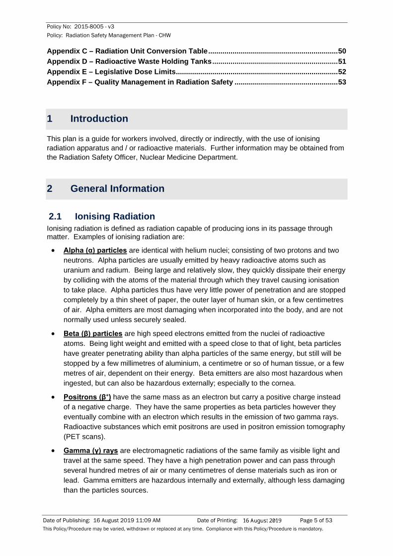
Once the red radioactive waste bag or sharps container has been sealed it must be labelled with the following information:
o Department of origin (laboratory name and room number)
o The radionuclide contained within the waste
o The total activity within the waste bag
o Date of entry into the Radioactive Waste Store (RWS) and also an estimated disposal date.
o Waste stream
Figure 2: Examples of appropriate labels for the Radioactive Waste Store
Access to the RWS is limited and you must contact the Radiation Safety Officer for transport of any radioactive waste. The Radiation Safety Officer can provide the red radioactive waste bags and any labels. All waste that enters the RWS must be sealed, labelled and the logbook completed. The logbook is located on the bench just inside the door of the RWS,

Policy No: 2015-8005 - v3 Policy: Radiation Safety Management Plan - CHW
Date of Publishing: 16 August 2019 11:09 AM Date of Printing: Page 18 of 53 This Policy/Procedure may be varied, withdrawn or replaced at any time. Compliance with this Policy/Procedure is mandatory.
and all fields must be completed by the person depositing the waste, this is to ensure accurate tracking of waste products.
5.7 Record Keeping It is a regulatory requirement that adequate records of receipts and usage of radioactive materials be kept.
• Receipt: full records of receipt of radioactive materials should be kept in the laboratory where the materials are to be held. Details should include radionuclide, activity, chemical form, date of receipt and place of storage
• Usage: it is necessary to keep records of usage of radioactive materials. For example, as a stock solution is used, a record should be kept of every fraction dispensed from that stock. Details to be recorded may include: identification of the stock solution, activity, volume, purpose, time and date.
• Disposal: it is essential to know what methods of disposal are used for radioactive materials. Methods of disposal include: flushing into the sewage system, disposal as dry or liquid waste and long stored waste.
5.8 Decontamination Procedures Decontamination is the removal of radioactive contamination from animate and inanimate surfaces. If contamination is found it must be removed at once. High specific activity isotopes can be held on a surface by ionic attachment, or by physical absorption or diffusion into cracks. Often a fresh spill on a clean and polished surface can be washed off without detectable residual contamination, whereas if it were allowed to react with the surface it might need drastic action to remove it. This is a very good reason for monitoring a working space immediately after using radioactive material, and for removing contamination associated with spills as quickly as possible.
Although special decontaminants involving the use of acids, alkalis, complexing agents and ion exchange materials are available, it is best to first try simple methods such as soap and water (preferably distilled) or detergent unless a special decontaminant is specifically indicated.
• Personnel Contamination
Wash with soap and water, avoiding spreading contamination to the eyes and mouth. Monitor with an appropriate radiation monitor until the count rate is less than 1000 cps (or the exposure is less than 10 μGy/hr, with the detector at a point close to (but not touching) the contaminated region of skin.
• Contamination of the Environment
Wearing gloves, cover spill with absorbent paper, and delineate contaminated area with "Radioactive" marking tape. Wash with detergent and water, monitor and repeat until the count rate can be reduced no further. If the count rate remains relatively high (>2000 cps), the surface should be covered, marked "contaminated", and allowed to decay. Dispose of plastic gloves, swabs and cleaning materials in a labelled plastic bag for proper disposal at a later stage. Where items of equipment have been contaminated with a short-lived isotope, it may be easier to store the equipment until all the activity has decayed than to attempt

Policy No: 2015-8005 - v3 Policy: Radiation Safety Management Plan - CHW
Date of Publishing: 16 August 2019 11:09 AM Date of Printing: Page 19 of 53 This Policy/Procedure may be varied, withdrawn or replaced at any time. Compliance with this Policy/Procedure is mandatory.
immediate decontamination. Contaminated clothing should be removed and placed in a plastic bag for assessment.
If ever in doubt contact the Radiation Safety Officer or Nuclear Medicine Department
5.9 Decommissioning of a Radioisotope Laboratory If a laboratory is no longer to be used for radioactive material use, it must be decommissioned as such. This involves:
o Removing all remaining stored radioactive material and waste
o Ensuring that there is no residual radiological contamination within the laboratory (the Radiation Safety Officer to complete this step)
o Removing all radiation warning signs from doors, storage locations and benches
o Notifying the Radiation Safety Officer of the decommissioning so that the lab, if registered as a premise on the Radiation Management Licence, can be removed from the licence.
6 Procedures – Radionuclides in the Wards
Nuclear medicine uses small quantities of radioactive materials (radiopharmaceuticals) to diagnose and treat disease. These studies are commonly performed on inpatients and these patients may be found in any ward. All inpatients will have their EMR (Electronic Medical Record) updated, within the Nuclear Medicine Progress Notes section, to include the time of injection and the type of Nuclear Medicine procedure performed. Any additional information required can be found in the completed report or by contacting the Nuclear Medicine department.
While paper request forms are still in use for outpatients, the sticker in Figure 3 will be used to document all the necessary information.
Figure 3: Radioactive Warning Label for Patient Notes
6.1 Diagnostic Procedures Many inpatients receive low doses of radioactive materials for diagnostic imaging investigations (nuclear medicine studies), and these patients may be accommodated anywhere within the Hospital. The external radiation hazard from such patients is small. The amount of radioactivity within the patient decreases rapidly with time according to the half-life
Policy No: 2015-8005 - v3 Policy: Radiation Safety Management Plan - CHW
Date of Publishing: 16 August 2019 11:09 AM Date of Printing: Page 20 of 53 This Policy/Procedure may be varied, withdrawn or replaced at any time. Compliance with this Policy/Procedure is mandatory.
of the radionuclide. In addition, the radionuclide is, in most cases, rapidly excreted from the body, mainly in the urine, so that the radioactivity in the patient is often not detectable after 1-2 days. It is therefore not necessary for ward staff to be issued with personal radiation monitors.
The patients are generally not a hazard to themselves, staff, visitors or other patients. If, in a particular situation, special handling or treatment of the patients is required, then advice will be given to ward staff by the Department of Nuclear Medicine
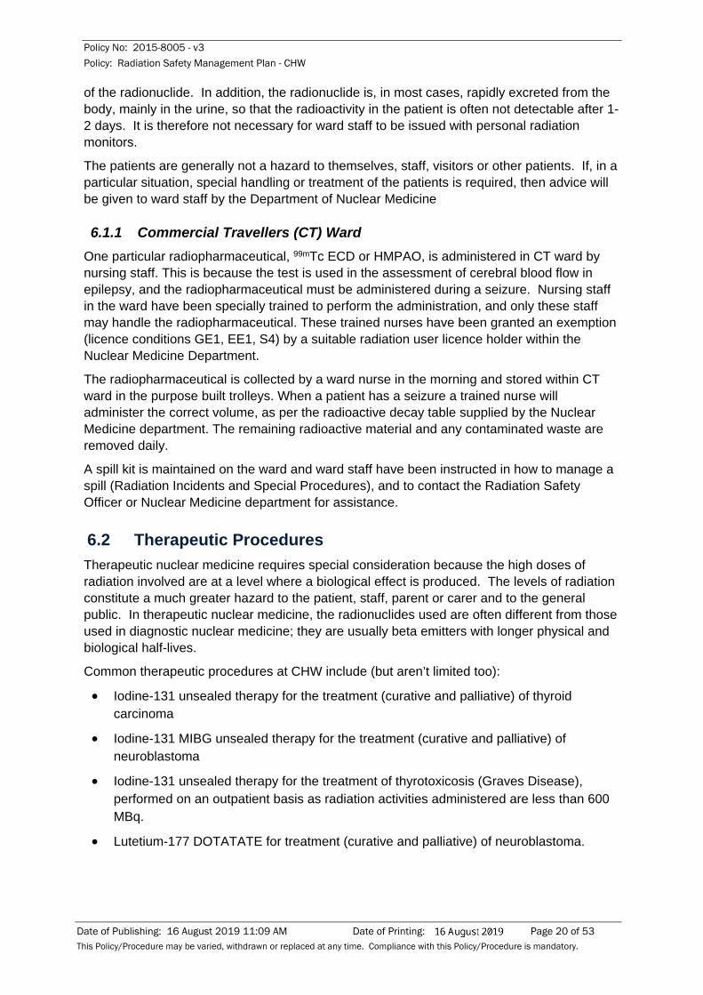
6.1.1 Commercial Travellers (CT) Ward One particular radiopharmaceutical, 99mTc ECD or HMPAO, is administered in CT ward by nursing staff. This is because the test is used in the assessment of cerebral blood flow in epilepsy, and the radiopharmaceutical must be administered during a seizure. Nursing staff in the ward have been specially trained to perform the administration, and only these staff may handle the radiopharmaceutical. These trained nurses have been granted an exemption (licence conditions GE1, EE1, S4) by a suitable radiation user licence holder within the Nuclear Medicine Department.
The radiopharmaceutical is collected by a ward nurse in the morning and stored within CT ward in the purpose built trolleys. When a patient has a seizure a trained nurse will administer the correct volume, as per the radioactive decay table supplied by the Nuclear Medicine department. The remaining radioactive material and any contaminated waste are removed daily.
A spill kit is maintained on the ward and ward staff have been instructed in how to manage a spill (Radiation Incidents and Special Procedures), and to contact the Radiation Safety Officer or Nuclear Medicine department for assistance.
6.2 Therapeutic Procedures Therapeutic nuclear medicine requires special consideration because the high doses of radiation involved are at a level where a biological effect is produced. The levels of radiation constitute a much greater hazard to the patient, staff, parent or carer and to the general public. In therapeutic nuclear medicine, the radionuclides used are often different from those used in diagnostic nuclear medicine; they are usually beta emitters with longer physical and biological half-lives.
Common therapeutic procedures at CHW include (but aren’t limited too):
• Iodine-131 unsealed therapy for the treatment (curative and palliative) of thyroid carcinoma
• Iodine-131 MIBG unsealed therapy for the treatment (curative and palliative) of neuroblastoma
• Iodine-131 unsealed therapy for the treatment of thyrotoxicosis (Graves Disease), performed on an outpatient basis as radiation activities administered are less than 600 MBq.
• Lutetium-177 DOTATATE for treatment (curative and palliative) of neuroblastoma.

Policy No: 2015-8005 - v3 Policy: Radiation Safety Management Plan - CHW
Date of Publishing: 16 August 2019 11:09 AM Date of Printing: Page 21 of 53 This Policy/Procedure may be varied, withdrawn or replaced at any time. Compliance with this Policy/Procedure is mandatory.
The following therapeutic procedures may also be conducted at CHW, but on a rare basis and generally do not require the use of the isolation room:
• Yttrium-90 Sir-spheres for the treatment of liver cancer (injection is performed under an interventional radiology procedure)
• Yttrium-90 injections to joints for radiation synovectomy (injection is performed under an interventional radiology procedure)
• Samarium-153 injections for pain relief of bone metastases
• Rhenium-186 injections to joints for radiation synovectomy (injection is under an interventional radiology procedure)
6.2.1 Camperdown Ward Therapeutic patients (administered activities greater than 600 MBq) are treated within room 14 in Camperdown Ward. This room is specifically designed for therapeutic patients; the toilet is connected to storage tanks located beneath the hospital, the room is shielded (door, leaded glass windows and walls), and a portable shield is also available within the room. Signage is erected outside the patient room, and access to the room is restricted.
Domestic services will prepare the room for patient arrival, including lining the floor and mattress with plastic. A Nuclear Medicine Scientist will prepare all the waste bins, linen bags and internal signage as per internal Nuclear Medicine procedure.
Prior to entry to the isolation room the following personal protective equipment shall be donned:
• Nurses performing minor procedures: disposable gloves and overshoes
• Nurses handling bodily fluids, washing the patient or dealing with spills: disposable gloves, gown, plastic apron and overshoes.
For those required to enter the isolation room, including nurses and the parent or carer, the following radiation safety principles must be followed to ensure that their radiation exposure is minimised:
• Maximise distance: radiation intensity decreases with increasing distance (inverse square law) from the source (in this case the patient is the radiation source). Nursing staff should maintain at least a 2 metre distance between themselves and the patient unless otherwise necessary. Parents or carers should stay at a distance of 2-3 metres from the patient.
• Reducing time with the patient: nursing staff should perform nursing duties quickly and efficiently, but without undue haste. These patients often require observation only, but it is important not to let them feel neglected. Staff may safely check on the patient many times a day.
• Shielding barrier: if prolonged entry into the isolation room is necessary, where feasible place the portable shielding barrier between you and the patient.

Policy No: 2015-8005 - v3 Policy: Radiation Safety Management Plan - CHW
Date of Publishing: 16 August 2019 11:09 AM Date of Printing: Page 22 of 53 This Policy/Procedure may be varied, withdrawn or replaced at any time. Compliance with this Policy/Procedure is mandatory.
A radiation dosimeter must be worn for every entry into the isolation room. Use the Personal Monitoring during Therapy log sheet to record name, entry time, displayed dose, exit time and displayed dose at that time.
The patient is to remain within the isolation room, except for any emergency reasons, or with the approval of the Nuclear Medicine Physician or Radiation Safety Officer. The isolation room bathroom is restricted to patient use only. The relevant admitting physician will instruct the patient and / or carers about the need for good personal hygiene, especially hand washing. Care should be taken to minimise contamination within the bathroom, especially around the toilet. The toilet should be flushed twice after each use.
For the patient to be discharged their residual activity must be less than 600 MBq, with a physical dose rate reading of less than 25 µSv/h at 1 metre, 9 µSv/h at 2 metres and 5 µSv/h at 3 metres, as per ARPANSA Radiation Protection Series Number 4 Recommendations for the Discharge of Patients Undergoing Treatment with Radioactive Substances (2002). The Nuclear Medicine Scientist will record all dose rate measurements within the patient’s records.
After the patient has been discharged from the room, a Nuclear Medicine Scientist will complete a radiological survey of the patient room, all waste bags and linen any waste with a dose rate measurement greater than 5 µSv/h (contaminated waste) must be labelled and transferred to the Radioactive Waste Store for decay. All non-contaminated waste will be released for normal disposal. Any contamination must be cleaned up by the Nuclear Medicine Scientist or the Radiation Safety Officer only. The ward staff will be notified once the room has been cleared for cleaning by Domestic Services.
7 Procedures – Nuclear Medicine Department
The Nuclear Medicine Department utilises radionuclides for the medical diagnosis, staging of disease, therapy and monitoring of the response of a disease process.
There are different area designations within the Nuclear Medicine department: controlled and supervised. Controlled areas are restricted access areas including the hot lab, injection rooms and the scanning rooms. There are specific procedures that employees are required to follow within these areas which aim to control the radiological hazard. These areas are labelled with the appropriate radiation hazard signs and also no eating or drinking is permitted in these areas. The supervised areas include the waiting room and the patient bathroom, and there are no specific procedures required to control the exposure to radiation.
7.1 Licencing Under the NSW Radiation Act (1990) and Regulation (2013) the Nuclear Medicine department is a registered “High Level Premise on which radioactive substances are kept or used”. Each piece of radiation apparatus owned and operated by the Nuclear Medicine department must hold a valid compliance certificate issued by a Certified Radiation Expert and must also be registered with the NSW EPA under the CHW Radiation Management Licence.
Each Nuclear Medicine Scientist, Nuclear Medicine Physician and Hospital Scientist must hold the appropriate NSW EPA issued Radiation User Licence conditions for their role. A
Policy No: 2015-8005 - v3 Policy: Radiation Safety Management Plan - CHW
Date of Publishing: 16 August 2019 11:09 AM Date of Printing: Page 23 of 53 This Policy/Procedure may be varied, withdrawn or replaced at any time. Compliance with this Policy/Procedure is mandatory.
valid exemption notice must be in sight on the Nuclear Medicine noticeboard for both Registrars and any visiting undergraduate student.
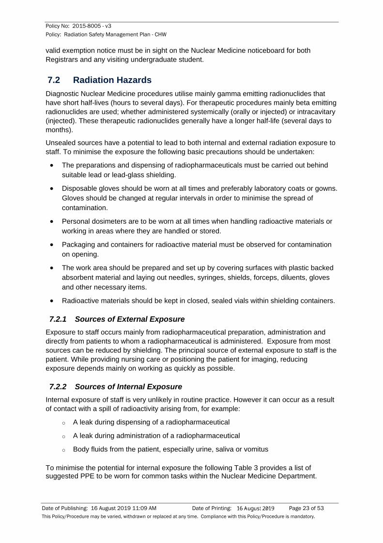
7.2 Radiation Hazards Diagnostic Nuclear Medicine procedures utilise mainly gamma emitting radionuclides that have short half-lives (hours to several days). For therapeutic procedures mainly beta emitting radionuclides are used; whether administered systemically (orally or injected) or intracavitary (injected). These therapeutic radionuclides generally have a longer half-life (several days to months).
Unsealed sources have a potential to lead to both internal and external radiation exposure to staff. To minimise the exposure the following basic precautions should be undertaken:
• The preparations and dispensing of radiopharmaceuticals must be carried out behind suitable lead or lead-glass shielding.
• Disposable gloves should be worn at all times and preferably laboratory coats or gowns. Gloves should be changed at regular intervals in order to minimise the spread of contamination.
• Personal dosimeters are to be worn at all times when handling radioactive materials or working in areas where they are handled or stored.
• Packaging and containers for radioactive material must be observed for contamination on opening.
• The work area should be prepared and set up by covering surfaces with plastic backed absorbent material and laying out needles, syringes, shields, forceps, diluents, gloves and other necessary items.
• Radioactive materials should be kept in closed, sealed vials within shielding containers.
7.2.1 Sources of External Exposure Exposure to staff occurs mainly from radiopharmaceutical preparation, administration and directly from patients to whom a radiopharmaceutical is administered. Exposure from most sources can be reduced by shielding. The principal source of external exposure to staff is the patient. While providing nursing care or positioning the patient for imaging, reducing exposure depends mainly on working as quickly as possible.
7.2.2 Sources of Internal Exposure Internal exposure of staff is very unlikely in routine practice. However it can occur as a result of contact with a spill of radioactivity arising from, for example:
o A leak during dispensing of a radiopharmaceutical
o A leak during administration of a radiopharmaceutical
o Body fluids from the patient, especially urine, saliva or vomitus To minimise the potential for internal exposure the following Table 3 provides a list of suggested PPE to be worn for common tasks within the Nuclear Medicine Department.

Policy No: 2015-8005 - v3 Policy: Radiation Safety Management Plan - CHW
Date of Publishing: 16 August 2019 11:09 AM Date of Printing: Page 24 of 53 This Policy/Procedure may be varied, withdrawn or replaced at any time. Compliance with this Policy/Procedure is mandatory.
PPE Task
Gloves
Unpacking radionuclide packages Administering diagnostic injections Handling closed waste containers Administering I-131 capsules Preparing low activity samples for counting
Gloves, gown
Milking Mo-99 generator Preparing radiopharmaceuticals Dispensing injections Nursing sweaty or incontinent patients Changing contaminated bed linen Administering lung ventilation radiopharmaceuticals Preparing Tc-99m sources for gamma camera QC
Gloves, gown, plastic gown Emptying bed pans, bottles or catheter bags
Gloves, gown, eye protection Giving therapy injections or oral liquids; Y-90, I-131 Labelling blood cells
Double gloves, gown, overshoes Cleaning up radiological spills Table 2: Suggested Personal Protective Equipment for use in the Nuclear Medicine Department
7.3 Diagnostic Procedures Each diagnostic procedure should be conducted so that the dose to the patient is the lowest necessary to achieve the clinical aim.
Local procedures for drawing up and injecting radiopharmaceuticals are covered in the Nuclear Medicine department procedures manual. However, it is emphasised that all such procedures should be carried out behind suitable lead or lead-glass shielding, wearing gloves, and the person involved must be wearing their issued personal radiation monitoring device. Gloves should be removed in the proper surgical manner and disposed of correctly after use.
Staff leaving designated areas should remove any protective clothing, wash their hands and monitor their hands, clothing and body as appropriate. All staff must be familiar with the contamination and decontamination procedures and the location of the nearest decontamination/spill kit.
7.4 Therapeutic procedures Therapeutic procedures require special consideration due to the high doses of radiation involved. The levels of radiation constitute a much greater hazard to the patient, staff, parent or carer and to the general public. In therapeutic nuclear medicine, the radionuclides used are often different from those used in diagnostic nuclear medicine; they are usually beta emitters with longer physical and biological half-lives.
The Nuclear Medicine department has local procedures for each therapeutic radionuclide administration, which contain the indications for therapy, type of radionuclide, general range of activity prescribed, the method of administration, the radiological hazard and any radiation safety procedures that are required.
For prescribed Iodine-131 at levels below 600 MBq are treated on an outpatient basis. Treatments involving Iodine-131 at activities above 600 MBq, or for other clinical reasons, are treated on an inpatient basis. Inpatient treatments are performed within the isolation room within Camperdown Ward. For further information refer to 6.2

Policy No: 2015-8005 - v3 Policy: Radiation Safety Management Plan - CHW
Date of Publishing: 16 August 2019 11:09 AM Date of Printing: Page 25 of 53 This Policy/Procedure may be varied, withdrawn or replaced at any time. Compliance with this Policy/Procedure is mandatory.
The following therapeutic procedures may also be conducted at CHW on a clinical needs basis only: Yttrium-90 Sir-spheres for targeted therapy, Yttrium-90 and Rhenium-186 injections to joints for radiation synovectomy and Samarium-153 injections for pain relief of bone metastases. These therapies may be administered in other areas (Operating Suite, Interventional Radiology Suite). Further information is available within the relevant local Nuclear Medicine procedures manual.
7.4.1 Medical Emergencies involving patients undergoing radionuclide
therapy In life-threatening situations, the patient’s medical management will always take precedence over radiation safety considerations.
• Patients Treated with Iodine-131
The majority of these patients have been treated with oral Iodine-131, but can also include intravenous Iodine-131 MIBG treatments. The range of prescribed activities varies, and patients may contain a wide range of radionuclide activities; dependent on the administered activity, the time elapsed since administration and the avidity of target tissue uptake.
Actions to be taken:
i. Initiate the CHW Medical Emergency Procedure (dial 2222)
ii. Staff involved in resuscitation should don examination gloves at the earliest possible opportunity.
iii. Materials which have come into direct contact with the patient should be, as far as is practicable, kept to one side for examination by the RSO or Nuclear Medicine Department staff.
iv. Notify the Nuclear Medicine Department immediately. Out of hours, the Nuclear Medicine Physician on call or the Nuclear Medicine Scientist on call
Transfer to PICU: if transfer is required, the fact that the patient may still contain radioactive material is not to override the patient’s emergency management.
i. As intubation, catherisation or nasogastric tube may be necessary, staff are to be gowned and wear gloves when handling the patient.
ii. Attempt to contain any urine, gastric contents or any other body fluids by means of absorbent pads, and hold the pads in a doubled bagged radioactive waste bag (with the appropriate inner bag for the waste) for examination by the RSO or Nuclear Medicine department staff.
iii. Any used suction bottles or urine bags must not be discarded until checked by the RSO or Nuclear Medicine Department staff.
iv. The RSO will advise staff as to what precautions are necessary, given the amount of radioactive material involved, and the elapsed time since administration.
• Patients requiring Surgery or Intensive Care
Whenever the medical condition of the patient deteriorates a Nuclear Medicine Specialist should be consulted. If surgery is not urgent, it should be postponed until the radioactivity in

Policy No: 2015-8005 - v3 Policy: Radiation Safety Management Plan - CHW
Date of Publishing: 16 August 2019 11:09 AM Date of Printing: Page 26 of 53 This Policy/Procedure may be varied, withdrawn or replaced at any time. Compliance with this Policy/Procedure is mandatory.
the patient has decreased to a suitable level. If urgent surgery or monitoring in an Intensive Care Unit is required, precautions against external radiation and possible contamination from body fluids should be considered.
If the patient requires surgery, the surgical team should plan the procedure in order to minimise any staff radiation exposure. This can be achieved by ensuring only essential staff are present in the operating theatre, and where possible, staff stand away from any organs containing high concentrations of radioactivity and that close contact with the patient is minimised. The wearing of two pairs of surgical gloves will give some basic protection to the hands against beta radiation.
On completion of the operation all equipment used or worn should be checked for contamination and, if necessary, decontaminated or stored until the radioactivity has decayed to negligible levels. All staff involved should be checked for any radioactive contamination and, if necessary, decontaminated before leaving the area.
Precautions for death of a patient after undergoing a Nuclear Medicine procedure are outlined in Death Procedures – Bodies containing radioactive material
7.5 Reporting of Maladministration or Equipment Malfunction In accordance with the NSW Radiation Act and the Regulation, the following must be reported to either the Chief Nuclear Medicine Scientist or the Department Chief, entered into the Incident Investigation Management System (IIMS) and the Radiation Safety Officer must be notified as soon as practicable. The Radiation Safety Officer has 48 hours to notify the NSW EPA, and 10 days to submit a written report, from the time that they are made aware.
o administration of a radioactive substance for diagnostic purposes in a quantity of more than 50% greater than prescribed
o administration of a radioactive substance for therapeutic purposes at an activity differing by more than 15% from that prescribed
o unintended administration of radiation as the result of a malfunction of radiation apparatus
o the administration of a radiation dose to the wrong patient or to the wrong part of a patient’s body
o administration of a radiopharmaceutical other than as prescribed.
7.6 Radioactive Waste Disposal Radioactive waste is generated in the Nuclear Medicine department during the drawing up of patient doses. Depending on the radionuclide, the waste can be segregated into two classes:
• Short Half-Life Waste (Tc-99m, I-123, others < 24 hours half-life)
Due to the short half-lives of Tc-99m and I-123, the waste should be placed in a short half-life sharps container in the shielded waste bin. Containers must not be overfilled. The waste can be disposed of via the hospitals waste stream after being stored for 96 hours.
• Long Half-Life Waste (I-131, Ga-67, TI-201, >24 hours half-life)
Any long half-life waste must be placed into a different container to that of the short-lived waste. Depending on the activity and external dose rate of the waste it may need to be

Policy No: 2015-8005 - v3 Policy: Radiation Safety Management Plan - CHW
Date of Publishing: 16 August 2019 11:09 AM Date of Printing: Page 27 of 53 This Policy/Procedure may be varied, withdrawn or replaced at any time. Compliance with this Policy/Procedure is mandatory.
moved to the Radioactive Waste Store, until it decays to below the exempt level for that specific radionuclide. This type of waste should be labelled with the radionuclide, date, activity level and type of waste.
When a waste bin is full (2/3 full for sharps containers) they must be sealed, labelled appropriately and recorded in the electronic Laboratory recording system. Any radioactive waste that has decayed sufficiently to below the relevant exemption level must have all radiation labels, symbols or tape removed, prior to disposal via the Hospital waste stream.
8 Procedures: Department of Medical Imaging
The Department of Medical Imaging provides paediatric digital imaging and interventional services utilising the following:
• 3 General radiography rooms
• 2 Fluoroscopy Rooms
• 1 Dental Room (Orthopantomograms (OPGs))
• 1 CT unit
• 5 Mobile Radiography units
• 1 Interventional Radiology Room
• 1 Cardiac Catheter Laboratory
• 3 Image Intensifiers (II)
• Ultrasonography units
• 2 MRI units (non-ionising radiation)
8.1 Licencing Under the NSW Radiation Act (1990) and Regulation (2013) the each piece of radiation apparatus owned and operated by the Department of Medical Imaging must hold a valid compliance certificate issued by a Certified Radiation Expert and must also be registered with the NSW EPA under the CHW Radiation Management Licence.
Each Radiographer and Radiologist must hold the appropriate NSW EPA issued Radiation User Licence conditions for their role. A valid exemption notice must be in sight on the Medical Imaging noticeboard for both Registrars and any visiting undergraduate student.
8.2 Staff Protection
8.2.1 Radiation Monitoring Badges OSL badges are issued on a 12 week basis.
Radiographers must wear OSL badges at chest level at all times.
When wearing a lead apron, badges must be worn underneath the apron.

Policy No: 2015-8005 - v3 Policy: Radiation Safety Management Plan - CHW
Date of Publishing: 16 August 2019 11:09 AM Date of Printing: Page 28 of 53 This Policy/Procedure may be varied, withdrawn or replaced at any time. Compliance with this Policy/Procedure is mandatory.
The radiation levels recorded on the processed badges will be returned to the department liaison and the Radiation Safety Officer.
8.2.2 Equipment The glass in front of some x-ray equipment operators consoles is 2.5 cm plate glass and is only sufficient to stop scattered radiation. At no time should the tube be rotated so that it is facing the console area.
The x-ray tube must not be used any closer than 1 m to the console area.
Lead aprons must be worn on all mobile work.
The CT scanner operator(s) areas are protected with lead glass. If it is necessary to be in the CT room during a scan, a lead apron must be worn, and the area adjacent to the scan bed should be avoided as this is the highest scatter dose area.
8.2.3 X-ray Personal Protective Equipment The NSW EPA released the Radiation Guideline 4: Compliance requirements for x-ray protective clothing in March 2018. This guideline provides advice on specific requirements for the use, inspection and testing of X-ray protective clothing to ensure minimum exposure to individuals using radiation apparatus. CHW requires compliance with this policy, and integrity testing of existing protective clothing is conducted annually by the Department of Medical Imaging. Integrity testing is also completed on all new protective clothing prior to first use.
An electronic record must be kept with the pass/fail results of all protective clothing, each individual piece of protective clothing must be labelled for referencing purposes. A copy of this is held by the Radiation Safety Officer and the Department Head. If a fault is found an image of the region must be taken and must be kept, and the apron marked as faulty. The apron must be immediately removed from use and returned to the Department Head or the Radiation Safety Officer.
A visual inspection of each article of x-ray protective clothing should be completed by the user/wearer at the time of each use. Clothing must not be used if the surface appears cracked or damaged.
8.3 Pregnancy and patients The following should be taken into account to protect all females of reproductive capacity, concentrating especially on those who are known to be pregnant or who feel they may be. The primary responsibility for identification of patients at risk rests with the referring doctor, while the Medical Imaging staff provide a secondary backup.
8.3.1 Identification To determine the pregnancy status, the question is asked: “Is the patient pregnant or suspected to be pregnant?” A positive answer to this is expected to include all those women who feel they may be pregnant, or are trying to fall pregnant as well as those who are sure they are pregnant.
Policy No: 2015-8005 - v3 Policy: Radiation Safety Management Plan - CHW
Date of Publishing: 16 August 2019 11:09 AM Date of Printing: Page 29 of 53 This Policy/Procedure may be varied, withdrawn or replaced at any time. Compliance with this Policy/Procedure is mandatory.
8.3.2 Patient Not Pregnant Upon performing the examination, if the answer to the above question is “no”, then caution should still be adopted in radiological procedures involving exposure of the lower abdominal and pelvic regions of women of reproductive capacity to ensure that the radiation dose received is as low as practicable.
8.3.3 Procedure when Patient is Pregnant If the patient is pregnant or suspected to be, then the case should be referred to a Radiologist to decide on the correct course of action.
If the foetus is to be irradiated, the radiographer should inform a senior radiographer so that all radiographic factors can be noted so that the foetal absorbed dose can be calculated and recorded at a later date. Ideally the following should be recorded: projection (view), field size, number of images per projection, kVp, mAs and the HVL of the x-ray unit. These factors are required to calculate the foetal absorbed dose.
8.3.4 Procedure when a Patient is found to be Pregnant AFTER an X-ray On the rare occurrence in which a patient will not be aware of a pregnancy at the time of an x-ray examination, and will naturally be concerned when the pregnancy becomes known.
In such cases an estimate of the radiation dose to the foetus should be performed by a Radiology Medical Physicist experienced in dosimetry. The patient can then be better informed of the risks (further information in Radiation and Pregnancy). In many cases the risk from the irradiation within the first 3 weeks following conception will be minimal. If the foetus is older, the dose involved may be considerable. It is however extremely rare for the dose to be large enough to warrant advising the patient to consider termination.
8.4 High Dose Fluoroscopy Procedures Several procedures are performed within the Interventional Suite and the Cardiac Catheter Laboratory that may utilise either the fluoroscopy and / or cine-mode acquisition for long periods of time. High doses to the patients skin (skin entrance dose) resulting in acute radiation burn damage are rare, but can occur if a procedure involves a long period of fluoroscopy/ cine acquisition. Extreme cases may require skin grafting.
Staff involved in such procedures should keep the Clinician aware of the length of time a procedure is taking, and ensure that dose-area product (DAP) information, if available, is recorded in the procedure notes, along with any other relevant data such as field size used. This information can be used to determine the actual dose to the patient and therefore aid clinicians in the appropriate treatments based on the skin dose.
Where a high dose is suspected, advice should be sought from an appropriate person (Medical Physicist or Radiation Safety Officer).
8.5 General Any member of the radiographic staff on becoming pregnant should notify the Chief Radiographer or Radiation Safety Officer to obtain a foetal radiation monitoring badge.
Any radiographer, upon resignation, will be provided with a copy of their radiation history from the Chief Radiographer.

Policy No: 2015-8005 - v3 Policy: Radiation Safety Management Plan - CHW
Date of Publishing: 16 August 2019 11:09 AM Date of Printing: Page 30 of 53 This Policy/Procedure may be varied, withdrawn or replaced at any time. Compliance with this Policy/Procedure is mandatory.
Radiographers using mobiles must make available a lead apron to other staff who are required to be less than 2 metres from the patient.
It is the radiographers responsibly to ensure the area is clear when they perform diagnostic examinations involving ionising radiation.
8.6 Use of Mobile X-ray within Wards Mobile x-ray units are utilised in various areas within CHW when it is impractical or not medically acceptable to transfer the patient to the Medical Imaging department. They are limited in their use as they offer a restricted choice of imaging techniques, and they must also be operated by a licenced Radiographer at all times. The reasons behind the limitations are the lower image quality compared to fixed imaging equipment within the Medical Imaging department, restricted imaging techniques and the potential need to repeat images.
8.6.1 Radiation Safety Guidelines: • Operator
o Stands at least 2 metres away from the x-ray unit, where feasible
o Stands outside the path of the primary beam
o Must wear a lead equivalent apron/gown (in compliance with 8.2)
o Provides radiation safety advice if the x-ray tube or head needs to be placed at an oblique angle, where the 2 metre safe distance doesn’t apply.
• Others in the vicinity
• When a person is required to be present during an x-ray procedure, that person should not remain any closer to the patient or the x-ray unit than is necessary. The radiographer should ensure that any person who is required to remain in the room/vicinity during the procedure is either:
o Wearing a lead equivalent apron/gown (in compliance with 8.2)
o Standing at least 2 metres from the x-ray tube when the tube is aimed towards the floor or solid wall,
o Stands outside the path of the primary beam
o Standing behind portable protective shields, if available.

Policy No: 2015-8005 - v3 Policy: Radiation Safety Management Plan - CHW
Date of Publishing: 16 August 2019 11:09 AM Date of Printing: Page 31 of 53 This Policy/Procedure may be varied, withdrawn or replaced at any time. Compliance with this Policy/Procedure is mandatory.
Figure 4: Example of the Safe Operation of a Mobile X-ray Unit (Courtesy of Westmead Hospital)

Policy No: 2015-8005 - v3 Policy: Radiation Safety Management Plan - CHW
Date of Publishing: 16 August 2019 11:09 AM Date of Printing: Page 32 of 53 This Policy/Procedure may be varied, withdrawn or replaced at any time. Compliance with this Policy/Procedure is mandatory.
9 Procedures – Operating Suite
The major hazard in the operating theatres arises from diagnostic x-rays. Staff should:
• Wear a lead or lead equivalent gown while in theatre where x-rays are being used. At least 0.3 mm lead equivalence at 100 kVp.
• Never place any part of their body in the x-ray beam
• Should stand as far back from the beam as possible to minimise scattered radiation exposure.
• When the x-ray beam is pointing horizontally (taking a lateral image of the patient), stand on the Image Intensifier side of the patient, rather than on the side of the x-ray tube. This is as the scattered radiation from the patient can be as much as ten times higher on the beam entrance side of the patient.
• Wear any radiation monitors that are issued to them.
All lead and lead equivalent aprons, thyroid shields and other personal protective devices throughout CHW meet the minimum design criteria as outlined in Australian Standard (AS/NZS 4543.3: 2000). The gowns are maintained by the Department of Medical Imaging, who perform annual integrity tests as per the Radiation Guideline 4: Compliance requirements for x-ray protective clothing.
Licenced Radiographers
The licenced radiographer on duty within the Operating Suite is responsible for performing the mobile fluoroscopic procedures in accordance with the Medical Imaging department’s standard protocols and procedures.
This includes:
• Following imaging protocols to ensure optimal data acquisition and analysis
• Performing quality assurance procedures for instrumentation and image quality
• Ensuring that no staff member receives a radiation exposure
Licenced radiographers are only to operate the mobile x-ray equipment as per the Chief Radiographer’s instructions.
9.1 Orthopaedic Surgeons Any Orthopaedic surgeon who operates any piece of ionising apparatus within the Operating Suite must hold a valid EPA issued Radiation User licence with the appropriate condition (IA22: Use radiation apparatus for medical fluoroscopy – Specialists other than radiologists).
Any operator must comply with the Australian Orthopaedic Association policy Radiation Safety for Orthopaedic Surgeons (2004). When a review of this policy has been conducted, the new version will replace this currently endorsed (May 2015) version. This is not expected to occur in the foreseeable future as the principles of personnel radiation protection should remain constant.

Policy No: 2015-8005 - v3 Policy: Radiation Safety Management Plan - CHW
Date of Publishing: 16 August 2019 11:09 AM Date of Printing: Page 33 of 53 This Policy/Procedure may be varied, withdrawn or replaced at any time. Compliance with this Policy/Procedure is mandatory.
9.2 Sentinel Node Biopsy If and when a Sentinel node biopsy procedure is to be performed within the Operating Suite, advice and the necessary equipment will be sourced from Westmead Adults Hospital. The Sentinel node is the first lymph node to receive drainage from a primary tumour. Lymphatic mapping using 99mTc colloid allows the Sentinel node and other nodes which have taken up the radioactive substance to be identified and biopsied. Low activities of 99mTc are administered and staff do not need to wear lead aprons. The tissue samples sent for pathology tests may be handled in the usual manner.
10 Procedures – Haematology (Blood Bank)
The Haematology Department owns and operates a Blood Irradiator; classified as a Security Enhanced Sealed Source, which is used to kill lymphocytes which cause transfusion-associated graft-versus-host disease within donated blood. In accordance with the Code of Practice for the Security of Radioactive Sources (2007) CHW has an endorsed source security plan in place, and the designated responsible person must ensure that it is:
o Implemented and complied with,
o Made available to the EPA at such times as the EPA may require,
o Is reviewed at least every 12 months
Access to the blood irradiator room is controlled with restricted access to Irradiator staff and Hospital Security Staff only. Those requiring access to the room must contact the Radiation Safety Officer, and / or the responsible person, and complete the required NSW EPA identity checks.
10.1 Licencing Under the Regulation there is no requirement for users of the blood irradiator to hold an individual radiation user licence. The unit itself must be registered with the NSW EPA under the CHW Radiation Management Licence, and serviced by an approved service provider on an annual basis.
10.2 Threat Levels The Australian Government’s National Threat Assessment Centre sets the threat level for the radiological sector, and these levels are used to determine the procedural and security requirements, as set out in Schedule D of the Code. The responsible person and the Radiation Safety Officer must ensure that if the threat level changes, that Schedule D is followed accordingly.

Policy No: 2015-8005 - v3 Policy: Radiation Safety Management Plan - CHW
Date of Publishing: 16 August 2019 11:09 AM Date of Printing: Page 34 of 53 This Policy/Procedure may be varied, withdrawn or replaced at any time. Compliance with this Policy/Procedure is mandatory.
Figure 5: RPS: 11 Security of Radioactive Sources Schedule D

Policy No: 2015-8005 - v3 Policy: Radiation Safety Management Plan - CHW
Date of Publishing: 16 August 2019 11:09 AM Date of Printing: Page 35 of 53 This Policy/Procedure may be varied, withdrawn or replaced at any time. Compliance with this Policy/Procedure is mandatory.
11 Procedures – Department of Dentistry
The Department of Dentistry owns and operates 4 dental x-ray units:
• two fixed units within the Dental Clinic, Surgery 1 and Surgery 2
• one portable unit within the Operating Suite/ Middleton Day Surgery
11.1 Licencing Under the NSW Radiation Act (1990) and Regulation (2013) the each piece of radiation apparatus owned and operated by the Department of Dentistry must hold a valid compliance certificate issued by a Certified Radiation Expert and must also be registered with the NSW EPA under the CHW Radiation Management Licence.
In accordance with the Regulation:
1. A person is exempt from the requirement to hold a radiation user licence in relation to the use, for dental diagnostic purposes, of extra-oral x-ray apparatus used with intra-oral image receptors if the person:
i. is registered under the Health Practitioner Regulation National Law to practise in the dental profession (other than as a student) as a dentist, a dental therapist, a dental hygienist or an oral health therapist, and is registered in the corresponding division of that profession, and
ii. meets all applicable requirements of the Code of Practice and Safety Guide for Radiation Protection in Dentistry in relation to the use of the apparatus.
2. A person is exempt from the requirement to hold a radiation user licence in relation to the use, for dental diagnostic purposes, of extra-oral x-ray apparatus used with intra-oral image receptors if the person:
i. is registered as a student in the dental profession under the Health Practitioner Regulation National Law, and
ii. is subject to:
1. immediate supervision at all times while the person is using the apparatus during clinical experience in the course of training, and
2. general supervision at all other times.
iii. In this clause:
o Code of Practice and Safety Guide for Radiation Protection in Dentistry means the Code so entitled, published by the Chief Executive Officer of the Australian Radiation Protection and Nuclear Safety Agency, as in force from time to time.
o general supervision means supervision by a qualified person who oversees the person being supervised and ensures that the person follows safe radiation work practices in relation to the use of the apparatus in respect of which the supervision is required.

Policy No: 2015-8005 - v3 Policy: Radiation Safety Management Plan - CHW
Date of Publishing: 16 August 2019 11:09 AM Date of Printing: Page 36 of 53 This Policy/Procedure may be varied, withdrawn or replaced at any time. Compliance with this Policy/Procedure is mandatory.
o immediate supervision means supervision by a qualified person who is present at all times during, and is observing and directing, the use by the person being supervised of the apparatus in respect of which the supervision is required.
o qualified person means a person who satisfies the requirements of subclause (1) (i) and (ii).
11.2 X-ray Units In accordance with the Regulation, all dental x-ray units (acquired since 2005) must be of a type registered by the Therapeutic Goods Administration (TGA), and comply with the specifications of Australian Standard AS/NZS 3200.2.201:2000, which applies to the following X-ray equipment used for general and specialist dental practice:
1. X-ray equipment for use with intra-oral image receptors;
2. X-ray equipment for panoramic radiography; and
3. dedicated cephalometric X-ray equipment.
If they were acquired prior to 2005 (implementation of the Code) which does not comply with the relevant requirements of AS/NZS 3200.2.201:2000, must be modified to comply with the requirements specified in Schedule 2 of the Code, except where otherwise approved by the regulatory authority, or phased out of use on a time scale approved by the regulatory authority.
12 Death Procedures – Bodies containing radioactive
material
If a patient dies during treatment with radioactive materials, the nuclear medicine specialist managing the patient’s care should ensure, after consultation with the radiation safety officer, that exposure to radiation of any persons handling the body is minimised. At the time of death, the body should have a label attached clearly documenting the radionuclide, form and estimated residual activity. The body should be handled as little as possible. The Radiation Safety Officer or delegated person should be consulted before any procedures, such as laying out or post-mortem are commenced and before the body is released for embalming, burial or cremation. If a patient dies shortly after undergoing a diagnostic nuclear medicine procedure, no special precautions are required.
Half-life (days)
Indicative maximum activity administered (MBq)
Autopsy / Embalming (MBq)
Burial (MBq) Cremation (MBq)
P-32 14.3 200 100 2 000 30 Sr-89 50.7 200 50 2 000 20 Y-90 2.7 2 000 200 2 000 70 I-131 8.0 10 000 10 400 400
Table 3: Maximum activities proposed for autopsy, embalming, burial or cremation of the body of a patient who has died during treatment with unsealed radioactive substances (IAEA 2007)

Policy No: 2015-8005 - v3 Policy: Radiation Safety Management Plan - CHW
Date of Publishing: 16 August 2019 11:09 AM Date of Printing: Page 37 of 53 This Policy/Procedure may be varied, withdrawn or replaced at any time. Compliance with this Policy/Procedure is mandatory.
All corpses released for autopsy, embalming, cremation or burial above the limits stated in Table 4 should have a label attached, identifying the radionuclide and its activity at the time of release, together with a release statement signed by the radiation safety officer or qualified expert.
12.1 Post-mortem or embalming Radioactive material remaining in the corpse can be a hazard through two main pathways; external radiation exposure and radioactive contamination. External exposure to penetrating radiation emanating from a radioactive source occurs at a distance from the source, and can be partially shielded by the corpse itself. Radioactive contamination is associated with actual contact with the radioactive material and spread of the material, similar to chemical contamination.
• Prevention of Contamination: for unsealed radioactive material, standard precautions similar to those used for infection control should be used, including gloves, mask, goggles and gown. Most unsealed radioactive material is either taken up by the target organ and/or eliminated by excretion in the first few days of treatment. The most significant post-death risk of contamination from radioactive material may remain in the body fluids, tissues or organs. Further advice concerning precautionary actions can be obtained from the Nuclear Medicine Physician or the Radiation Safety Officer where the patient was treated.
It is highly unlikely that a patient at CHW will be treated with sealed sources, but if they have been the following will be relevant:
• Reduction of external exposure: the working distance from the sealed source implant site should be maximised while dealing with the corpse. If an organ containing radioactive material needs to be handled, suitable tools such as tongs or forceps should be used to maximise the distance of the hands from the radioactive material, and the time spent carrying out the procedure kept as short as possible.
12.2 Cremation When the radioactive treatment involves unsealed radioactive material that has been incorporated into bone, or permanently implanted sealed sources (rarely used with paediatric patients) encapsulated in metal that survives the combustion process, some radioactivity will remain in or amongst the skeletal remains. Contact the Radiation Safety Officer for advice in regard to this.
Where the radioactive treatment involves radioactive materials which have not been incorporated into the bone of the deceased and which are expelled into the cremation furnace and flue during combustion, the remains will not be radioactive and so do not require handling and storage precautions.
12.3 Handling of the coffin No restrictions are normally needed in dealing with the closed coffin following the death of a patient that has been recently released from a hospital after treatment with radioactive material.

Policy No: 2015-8005 - v3 Policy: Radiation Safety Management Plan - CHW
Date of Publishing: 16 August 2019 11:09 AM Date of Printing: Page 38 of 53 This Policy/Procedure may be varied, withdrawn or replaced at any time. Compliance with this Policy/Procedure is mandatory.
12.4 Direct burial or mausoleum entombment No restrictions are normally needed for direct burial or mausoleum entombment following death of a patient recently treated with radioactive material.
13 Radiation Dosimetry of Patient Procedures
The radiation dosimetry of diagnostic x-ray procedures differs from that of Nuclear Medicine procedures as the dose received is mainly a function of the imaging equipment characteristics and the number of views/projections that are taken. For medical imaging procedures the radiation dose is limited mainly to the field of view being imaged, whereas in Nuclear Medicine procedures an appreciable dose may be received by organs other than those being imaged, due to the uptake of the radiopharmaceutical.
13.1 Diagnostic Medical Imaging The main parameters which affect patient radiation exposure, and hence radiation absorbed dose are:
i. X-ray tube operating voltage (kV)
ii. X-ray tube operating current (mA)
iii. X-ray tube operating wave form
iv. Radiographic field size
v. Patient shape and size
vi. Number of projections per examination
vii. Source to image distance
viii. Beam quality (HVL)
For CT scans and fluoroscopic procedures there are further variable machine operating characteristics which affect radiation dose. The radiation dose from any given radiological procedure may vary greatly, depending on the technique used.
The skin receives the greatest radiation dose in radiological procedures and is thus the critical organ. In some cases, other, more radiosensitive tissues in the field of view may receive an appreciable dose and so become the critical organ.
Estimation of radiation dose for radiological procedures may be done directly using radiation monitoring devices. Most publicised radiation doses are average values only, and any one individual examination the dose to the patient may be considerably greater or smaller than the publicised value. Below is a table of average effective doses for common radiological procedures at CHW. Values were calculated from random samples of examinations from CHW DAPs and DLPs and converted using approximate conversion coefficients. For patient specific dosimetry please contact the Radiation Safety Officer to provide a estimation of their dose.

Policy No: 2015-8005 - v3 Policy: Radiation Safety Management Plan - CHW
Date of Publishing: 16 August 2019 11:09 AM Date of Printing: Page 39 of 53 This Policy/Procedure may be varied, withdrawn or replaced at any time. Compliance with this Policy/Procedure is mandatory.
Examination Age Group Dose (mSv)
Chest X-ray Neonate 0.002
Toddler 0.002
Child 0.02
Extremity Neonate 0.002
Toddler 0.001
Child 0.002
Spine Neonate 0.002
Toddler 0.03
Child 0.14
Hip/Pelvis Neonate 0.004
Toddler 0.01
Child 0.05
MBS Neonate 0.02
Toddler 0.04
Child 0.2
MCU Neonate 0.05
Toddler 0.04
Child 0.1
Head CT Neonate 1
Toddler 2.5
Child 2.5
Teenager 3
Chest CT Neonate 0.8
Toddler 0.8
Child 1
Teenager 1.5 Table 4: Estimated Effective Dose for common radiological procedures.
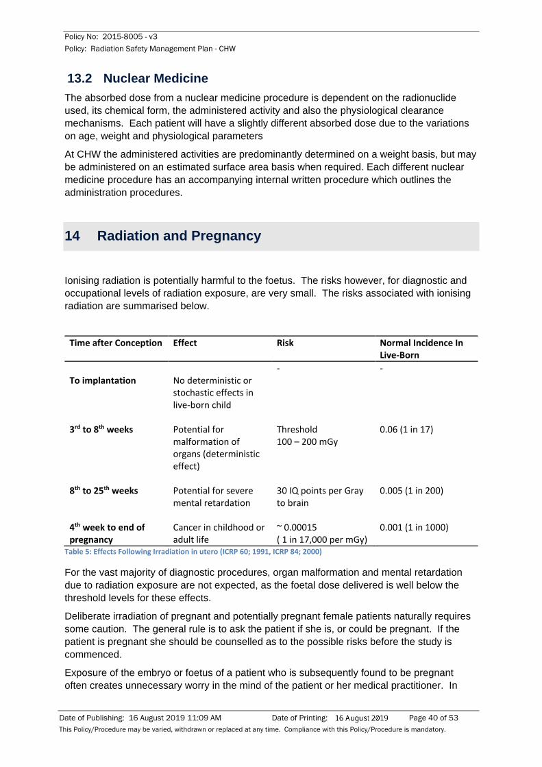
Policy No: 2015-8005 - v3 Policy: Radiation Safety Management Plan - CHW
Date of Publishing: 16 August 2019 11:09 AM Date of Printing: Page 40 of 53 This Policy/Procedure may be varied, withdrawn or replaced at any time. Compliance with this Policy/Procedure is mandatory.
13.2 Nuclear Medicine The absorbed dose from a nuclear medicine procedure is dependent on the radionuclide used, its chemical form, the administered activity and also the physiological clearance mechanisms. Each patient will have a slightly different absorbed dose due to the variations on age, weight and physiological parameters
At CHW the administered activities are predominantly determined on a weight basis, but may be administered on an estimated surface area basis when required. Each different nuclear medicine procedure has an accompanying internal written procedure which outlines the administration procedures.
14 Radiation and Pregnancy
Ionising radiation is potentially harmful to the foetus. The risks however, for diagnostic and occupational levels of radiation exposure, are very small. The risks associated with ionising radiation are summarised below.
Time after Conception Effect Risk Normal Incidence In Live-Born
To implantation
No deterministic or stochastic effects in live-born child
- -
3rd to 8th weeks Potential for malformation of organs (deterministic effect)
Threshold 100 – 200 mGy
0.06 (1 in 17)
8th to 25th weeks Potential for severe mental retardation
30 IQ points per Gray to brain
0.005 (1 in 200)
4th week to end of pregnancy
Cancer in childhood or adult life
~ 0.00015 ( 1 in 17,000 per mGy)
0.001 (1 in 1000)
Table 5: Effects Following Irradiation in utero (ICRP 60; 1991, ICRP 84; 2000)
For the vast majority of diagnostic procedures, organ malformation and mental retardation due to radiation exposure are not expected, as the foetal dose delivered is well below the threshold levels for these effects.
Deliberate irradiation of pregnant and potentially pregnant female patients naturally requires some caution. The general rule is to ask the patient if she is, or could be pregnant. If the patient is pregnant she should be counselled as to the possible risks before the study is commenced.
Exposure of the embryo or foetus of a patient who is subsequently found to be pregnant often creates unnecessary worry in the mind of the patient or her medical practitioner. In

Policy No: 2015-8005 - v3 Policy: Radiation Safety Management Plan - CHW
Date of Publishing: 16 August 2019 11:09 AM Date of Printing: Page 41 of 53 This Policy/Procedure may be varied, withdrawn or replaced at any time. Compliance with this Policy/Procedure is mandatory.
fact, the risks of radiation exposure, even at relatively large levels, is very small compared to the normal risks of pregnancy (Table 6: Effects Following Irradiation in utero (ICRP 60; 1991, ICRP 84; 2000)
All cases of accidental or unintentional irradiation of a foetus or embryo must, for the sake of all concerned, be referred to the Radiation Safety Officer for investigation and assessment.
Female staff working with radiation or radioactive material are sometimes concerned as to the well-being of their foetus if they fall pregnant and continue to work in the same situation until the pregnancy is recognised. When a pregnancy is confirmed arrangements should be made to ensure that the woman works only under such conditions that the foetus is accorded the same protection as for a member of the public (1 mSv annual effective dose, as defined in Schedule 5 of the NSW Radiation Control Regulation 2013). Staff working in areas where radiation is used routinely may request special radiation exposure monitoring (usually a TLD or OSLD badge) whilst they remain at work. (Contact the Radiation Safety Officer for details)
For more specific information please contact your supervisor/NUM, and /or the Radiation Safety Officer.
15 Radiation Incidents and Special Procedures
Emergency procedures for persons arriving at the Emergency Department with, or suspected to be, radioactive material contamination or radiation exposure are contained in the Disaster Response Plan - CHW Healthplan
15.1 Internal Reporting of Radiation Incidents If a near miss, incident or accident occurs that involves ionising radiation (for example a spill, personal contamination, exposure), as soon as practicable after the event it must be entered into the Incident Information Management System (IIMS), preferably by the end of the notifier’s work day. The supervisor should be informed as soon as possible, with notification sent to the Radiation Safety Officer.
15.2 Legislative Definition of a Radiation Accident As defined by the Act (1990) and the Regulation (2013) a radiation accident is to be treated as having occurred if there is an occurrence that involves:
a) the unplanned or unexpected emission of radiation where it is likely that:
• that one or more persons have, or could have, received an effective dose of radiation equal to or in excess of:
o 5 millisieverts, in the case of an occupationally exposed person, or
o 1 millisievert, in any other case, or
• that the premises or the environment may have become contaminated within the meaning of section 21 of the Act.
b) the misuse of radiation apparatus or maladministration of a radioactive substance used for medical purposes and that involves any of the following:
Policy No: 2015-8005 - v3 Policy: Radiation Safety Management Plan - CHW
Date of Publishing: 16 August 2019 11:09 AM Date of Printing: Page 42 of 53 This Policy/Procedure may be varied, withdrawn or replaced at any time. Compliance with this Policy/Procedure is mandatory.
• the administration of a radioactive substance for diagnostic purposes in a quantity of more than 50 per cent more than that prescribed,
• the administration of a radioactive substance for therapeutic purposes at an activity differing by more than 15 per cent from that prescribed,
• the administration of a therapeutic dose of radiation from radiation apparatus or a sealed source device which differs from the total prescribed treatment dose by more than 10 per cent,
• the administration of a dose of radiation for diagnostic and interventional purposes from a radiation apparatus that results in one or more persons receiving an effective dose of radiation equal to or in excess of 1 millisievert,
• the unintended administration of radiation as a result of a malfunction of radiation apparatus,
• the administration of a radiation dose to the wrong patient or to the wrong part of a patient’s body,
• the administration of a radiopharmaceutical otherwise than as prescribed.
15.3 Reporting Radiation Accidents to the Environmental Protection Authority
The Act (1990) and the Regulation (2013) impose certain responsibilities on employers (CHW) regarding radiation accidents.
Employers must:
1. Report and investigate apparent radiation accidents within the specified time frames
2. Maintain a record of radiation accidents
3. Rectify faults or defects in radiation apparatus and inform anyone who may have been exposed to radiation as a result of these faults.
All such incidents must be reported to the Radiation Control section of the EPA by the RSO or designated person in their absence:
• within 48 hours of becoming aware of an apparent radiation accident, (initial notification):
o the place where it occurred,
o the period during which the emission of radiation was uncontrolled,
o the area over which any radioactive substances may have been dispersed,
o any steps taken to rectify the accident,
o Any personal injury or exposure that may have resulted.
• within 10 days of becoming aware of an apparent radiation accident:
o assessment of the radiation dose that any person may have received as a result of the accident,

Policy No: 2015-8005 - v3 Policy: Radiation Safety Management Plan - CHW
Date of Publishing: 16 August 2019 11:09 AM Date of Printing: Page 43 of 53 This Policy/Procedure may be varied, withdrawn or replaced at any time. Compliance with this Policy/Procedure is mandatory.
o Any steps taken to reduce the risk of a similar accident occurring in the future.
A record of all radiation accidents must be maintained by the person responsible for the regulated material. At CHW IIMS is used as the accident recording tool. The following information must be included within the appropriate record:
• particulars of the accident indicating, as far as is possible, the place where it occurred and the period during which emission of radiation was uncontrolled,
• the name of any occupationally exposed person or other person who was there during that period,
• an estimate of the radiation dose to which any person may have been exposed,
• details and results of any medical examinations undertaken as a result of the accident,
• particulars of the area over which any radioactive substances may have been dispersed,
• particulars of any steps taken to rectify the accident,
• the time at which the accident was reported to the employer,
• the probable cause of the accident,
• particulars of any investigations conducted into the accident, together with the results of the investigations,
• particulars of any steps taken to reduce the risk of a similar accident occurring in the future.
If a fault or defect may exist in any radiation apparatus or sealed source device that is regulated material, the Radiation Safety Officer or designated person must:
• must immediately investigate the apparent fault and, if necessary, cause the apparatus or sealed source device to be removed, replaced or repaired, and
• must, as soon as practicable (but, in any case, within 7 days), inform all persons who may have been exposed to radiation in quantities in excess of those that would normally be received from the apparatus or sealed source device in faultless condition that they may have been so exposed.
15.4 Radioactive Contamination Response Radioactive contamination occurs when radioactive material is deposited on or in an object, surface or person. A person can be contaminated internally, externally or both. People who are externally contaminated can become internally contaminated if radioactive material is transferred from the external surface, via inhalation, ingestion or absorption through open wounds.
15.4.1 Personnel Decontamination • Eyes: Irrigate gently for 5 minutes, preferably with sterile saline to avoid irritation of
conjunctiva.
• Skin: Wash gently with warm soap and water, do not scrub the skin as this will remove natural oils and may abrade skin making it more permeable to surface contamination.

Policy No: 2015-8005 - v3 Policy: Radiation Safety Management Plan - CHW
Date of Publishing: 16 August 2019 11:09 AM Date of Printing: Page 44 of 53 This Policy/Procedure may be varied, withdrawn or replaced at any time. Compliance with this Policy/Procedure is mandatory.
Avoid spreading surface contamination, especially to nose, mouth and eyes. Use water only on mucous membranes.
If contamination on hands persists, moisten skin lightly with emollient skin cream and wear cotton gloves, inside disposable gloves for an hour or two, remove gloves and wash hands again. Monitor for level of contamination.
• Hair: Wash thoroughly with mild shampoo, taking care not to spread contamination to eyes, ears, nose or mouth.
• Nails: Scrub with soft nail brush, warm water and soap. Clip nails if necessary, being careful not to cut skin.
15.4.2 Work Surface Decontamination If a radioactive spill was to occur it is imperative to minimise the spread of contamination and the potential cross-contamination of those working in the vicinity of the spill.
• Restrict traffic, marking off the “dirty” area from the “clean” area with CAUTION RADIATION tape/sign or similar
• Using a contamination monitor survey and mark the contaminated areas, check for presence of sharps. Note the initial contamination level so to determine the effectiveness of the decontamination attempt.
• Moisten with detergent and wipe up with absorbent paper or cloths, always working inwards from the edge of the contaminated area (to reduce the potential to spread the contamination)
• Place used cleaning materials immediately into a plastic waste bag. Double bag this within a RED radioactive waste bag.
• Monitor the marked area, and repeat decontamination technique as necessary
• Seal and label bag of waste with: time, date, place and isotope involved and known activity of isotope (Bq)
• All sharps must be placed into a yellow sharps bin and then placed into a labelled RED radioactive waste bag.
15.4.3 Equipment Decontamination Many of the radioisotopes used in hospitals have relatively short half-lives. In many cases it will be preferable to store or isolate the contaminated item until the level of radioactivity is reduced to an acceptable level rather than to attempt decontamination. If the decision is made to decontaminate the item, advice should be sought from the RSO on appropriate methods.
It is usually desirable to initially attempt decontamination with detergents, such as a customised commercially available detergent. Specialised cleaning methods such as the use of ultrasonic cleaning baths may also be appropriate. The use of chelating agents such as a 10 percent solution of sodium citrate may prove effective. If the contamination is due to iodine radioisotopes, the affected area should not be treated with any material that contains oxidising agents or acids as these can result in the production of volatile molecular

Policy No: 2015-8005 - v3 Policy: Radiation Safety Management Plan - CHW
Date of Publishing: 16 August 2019 11:09 AM Date of Printing: Page 45 of 53 This Policy/Procedure may be varied, withdrawn or replaced at any time. Compliance with this Policy/Procedure is mandatory.
radioiodine and the risk of inhalation. The use of acid on metal surfaces may also cause unnecessary corrosion and result in greater difficulty in future decontamination procedures.
16 Irradiation of volunteers for research purposes
Under NSW legislation, the Act and the Regulation, CHW has to comply with the ARPANSA Code of Practice “Exposure of Humans to Ionizing Radiation for Research Purposes” (RPS8) for any research proposals involving irradiation of volunteers.
All research proposals involving the irradiation of volunteers (as part of Clinical Care/Standard of Care for specific patients, novel uses or purely research), by external radiation or administration of radionuclides, MUST be referred to the Radiation Safety Officer for consideration via the submission of the CHW Ionising Radiation Declaration Form. The RSO is required to prepare a report for the Ethics Committee. No work may take place without written approval. The RSO may refer any research proposals to the Radiation Safety Committee for advice and approval.
Exposures should only be performed when the volunteers understand the risks involved and willingly participate. A distinction is also drawn between those volunteers (generally patients) who may benefit from the proposed exposures and those who may not (generally normal volunteers).

Policy No: 2015-8005 - v3 Policy: Radiation Safety Management Plan - CHW
Date of Publishing: 16 August 2019 11:09 AM Date of Printing: Page 46 of 53 This Policy/Procedure may be varied, withdrawn or replaced at any time. Compliance with this Policy/Procedure is mandatory.
Participant Category Dose Constraintb Adults Total effective dose - In any year
- over 5 years - 5 mSvc - 10 mSv
Total effective dose in adult with life expectancy less than five years
- in any year 50 mSv
Equivalent dose to skin averaged over 1 cm2 - in any year 200 mSvd
Equivalent dose to any other organ or tissue - in any year 100 mSve
Children and foetuses Total effective dose to age 18 years, - Subject to:
• Effective dose from conception to birth; and • Effective dose in any year from birth to 18 years.
Total equivalent dose to age 18 years to any organ or tissue
5 mSv
0.1 mSv 0.5 mSv
100 mSv
Table 6: Dose Constraints for Participants in Researcha
a
A dose constraint for research participants specifies a maximum dose with which it should be possible to comply in normal circumstances it is intended to apply to radiation which is in addition to that received as part of normal clinical management. Dose constraints apply to diagnostic investigations not radiation therapy.
b
The dose constraint applies to the sum, over the relevant period, of doses received from external exposure and the 50 year committed dose (to age 70 years for children) from intakes over the same period.
c
When all the research participants are within the following specified age limits, the following total effective dose constrains apply:
- For adult 60 years or more – in any year - 8 mSv and - For adult 70 years or more – in any year – 12 mSv
d Derived from Table 3.1 of ICRP85 – factor of 10 below the threshold of 2 Sv for early transient erythema
e Derived from Table 3.1 ICRP85 - factor of 10 below the threshold of 1 Sv for detectable lens opacity.

Policy No: 2015-8005 - v3 Policy: Radiation Safety Management Plan - CHW
Date of Publishing: 16 August 2019 11:09 AM Date of Printing: Page 47 of 53 This Policy/Procedure may be varied, withdrawn or replaced at any time. Compliance with this Policy/Procedure is mandatory.
Appendix A: Disaster procedures
Please refer to the Disaster Response Plan CHW Healthplan

Policy No: 2015-8005 - v3 Policy: Radiation Safety Management Plan - CHW
Date of Publishing: 16 August 2019 11:09 AM Date of Printing: Page 48 of 53 This Policy/Procedure may be varied, withdrawn or replaced at any time. Compliance with this Policy/Procedure is mandatory.
Appendix B – Glossary
Definition Absorbed dose The measure of the energy deposited in any medium by any type of
ionising radiation. SI unit is the Gray.
Absorption When radiation imparts some or all of its energy to any material through which it passes.
Activity The number of nuclear transformations occurring in a given quantity of material per unit time. The SI unit is the Becquerel.
Alpha Particles
A strongly ionising particle emitted from the nucleus during radioactive decay having a mass and charge equal in magnitude to the helium nucleus consisting of 2 protons and 2 neutrons with a double positive charge.
Background Radiation
Ionizing radiation from natural sources, such as terrestrial radiation due to radionuclides in the soil or cosmic radiation originating in outer space.
Becquerel The SI unit for radioactivity defines as the amount of radionuclide decaying at a rate of one transformation per second. (1 Bq = 1 transformation/ second)
Beta Particles Charged particle emitted from the nucleus of an atom having a mass and charge equal in magnitude to that of the electron.
Bremsstrahlung X-rays that are produced when charged particles, usually electrons, moving with a very high velocity are slowed down rapidly by striking a target.
Curie The former unit of radioactivity, equal to 3.7 x 10¹⁰Bq
Decay Transformation of an unstable atomic nucleus into a more stable one, usually accompanied by the emission of charged particles and gamma rays.
Dose Limit The value of effective dose or equivalent dose to individuals from planned exposure situations that shall not be exceeded.
Equivalent Dose
A quantity used in radiation protection expressing all radiation on a common scale for calculating the effective absorbed dose. The unit is the Sievert, which is numerically equal to the absorbed dose in Grays multiplied by the radiation weighting factor.
Electron Volt Energy gained by an electron in passing through an electric potential of 1 volt.
Exposure A measure of the ionisation produced in air by x-ray or gamma radiation. The unit of exposure is Coulomb per kilogram.
Gamma Ray Gamma photons are the most energetic photons in the electromagnetic spectrum. Gamma rays (gamma photons) are emitted from the nucleus of some unstable (radioactive) atoms.
Gray The SI unit of absorbed dose, equal to one joule per kilogram.
Half-life, biological The time required, in the absence of further input, for a biological system or compartment to eliminate, by biological processes, half the amount of a substance that has entered it.
Half-life, effective The time required, as a result of the combined action of radioactive decay and biological elimination, for a system to reduce by half the amount of the substance that has entered it.
Half-life, physical The time required for a radioactive substance to reduce by half its activity by decay. Each radionuclide has a specific half-life.
Half Value Layer The thickness of a particular shielding material required to reduce the intensity to one-half its incident value.

Policy No: 2015-8005 - v3 Policy: Radiation Safety Management Plan - CHW
Date of Publishing: 16 August 2019 11:09 AM Date of Printing: Page 49 of 53 This Policy/Procedure may be varied, withdrawn or replaced at any time. Compliance with this Policy/Procedure is mandatory.
ICRP International Commission on Radiological Protection
Ionising Radiation Any electromagnetic or particulate radiation capable of producing ions, directly or indirectly, in its passage through matter.
Monitoring Periodic or continuous determination of the amount of ionising radiation or radioactive contamination present or incident on any part of an individual or his/her clothing.
Neutron Elementary particle with mass approximately the same as that of a hydrogen atom and electrically neutral.
Non-Ionising Radiation
Radiation that has lower energy levels and longer wavelengths than ionising radiation. It is not strong enough to affect the structure of atoms it contacts but is strong enough to heat tissue and can cause harmful biological effects. Examples include radio waves, microwaves, visible light, and infrared from a heat lamp.
OSLD Optically Stimulated Luminescence Dosimeter
Radiation Encompasses the entire Electromagnetic Spectrum; containing both ionising and non-ionising radiation.
Radiological Survey
The evaluation of radiation hazards incident to the production, use or existence of radioactive materials or other sources of radiation under a specific set of conditions.
Radionuclide
A nuclide with an unstable ratio of neutrons to protons placing the nucleus in a state of stress. It may undergo various types of rearrangement that involve the release of radiation, in order to reorganise into a more stable state.
Sievert The name of the SI unit of equivalent dose, effective dose. The unit is joule per kilogram (J kg-1).
Specific Activity The total radioactivity of a given nuclide per gram of a compound, element or radioactive nuclide.
TLD Thermoluminescence Dosimeter;
X-rays Penetrating electromagnetic radiations having wavelengths shorter than those of visible light. They are generally produced by bombarding a metallic target with fast electrons in a high vacuum.

Policy No: 2015-8005 - v3 Policy: Radiation Safety Management Plan - CHW
Date of Publishing: 16 August 2019 11:09 AM Date of Printing: Page 50 of 53 This Policy/Procedure may be varied, withdrawn or replaced at any time. Compliance with this Policy/Procedure is mandatory.
Appendix C – Radiation Unit Conversion Table
The International System (SI) units are a consistent set of units for use in all branches of science. The General Conference on Weights and Measures acting on the recommendation of the International commission on Radiation Units and Measurements (ICRU) has adopted special unit names for SI units in connection with radioactivity.
Factor Prefix Symbol Factor Prefix Symbol 1018 Exa E 10-3 Milli m 1015 Peta P 10-6 Micro µ 1012 Tera T 10-9 Nano n 109 Giga G 10-12 Pico p 106 Mega M 10-15 Femto f 103 Kilo k 10-18 Atto a
Table 7: Prefixes for SI Units
Radiation Units Physical Quantity SI Unit Non SI Unit Relationship
Activity Becquerel (Bq) Curie (Ci)
1 Bq = 2.7 x10⁻¹¹ Ci = 27.0 pCi 1 Ci = 3.7 x 10¹⁰ Bq =37 GBq
Absorbed Dose Gray (Gy) Rad
1 Gy = 100 rads 1 Gy = 1 J/kg 1 rad = 0.01 Gy = 10 mGy
Equivalent Dose Sievert (Sv) Rem
1 Sv -= 100 rem 1 Sv = 1 J/kg 1 rem = 0.01 Sv = 10 mSv
Exposure Coulomb/kilogram Roentgen (R)
1 C/kg = 3876 R =3.876 kR 1 R = 2.58 x 10⁴ C/kg =258 C/kg
Table 8: Relationship between SI and non SI Units for Radiation Quantities

Policy No: 2015-8005 - v3 Policy: Radiation Safety Management Plan - CHW
Date of Publishing: 16 August 2019 11:09 AM Date of Printing: Page 51 of 53 This Policy/Procedure may be varied, withdrawn or replaced at any time. Compliance with this Policy/Procedure is mandatory.
Appendix D – Radioactive Waste Holding Tanks
Within the undercroft of the Main Building CHW owns and operates two 9000 litre concrete holding tanks. These are connected to the toilet in room 14 within Camperdown Ward only. These tanks are used to allow for delay and decay of radionuclide waste from patients undergoing therapeutic nuclear medicine prior to discharge through the hospital sewage system. The two tanks are connected in series, with only one tank in operation at any one time. There are recirculation and out-flow pumps and pipes connected to the tanks. The other tank is used as a back-up only in the event of a discharge pump failure. The purpose of this system is to prevent the sudden discharge of large quantities of radioactive material into the hospitals sewage system, and to accumulate and dilute radioactive material, allow it to decay to a safe level and only then to discharge the waste over a reasonable period into the sewage system. The tank system is connected to an alarm panel which activates at both low level and high level filling. At low level, water is added to the tank to remove the alarm and to dilute any waste within the tank. At high level the contents of the decay tank are discharged into the normal sewage system. Access to the holding tank area is restricted, but the tanks are adequately shielded and monitored by the Radiation Safety Officer on a regular basis. A radiological survey is performed by the Radiation Safety Officer prior to entry, and prior to the removal of the observation lid. This observation lid can be removed using a custom tool held by the maintenance department. This allows for a visual water/waste level check. If the tank is greater than half full, and sufficient decay has occurred since the last therapeutic patient, the tank can be discharged into the normal sewage system. The Radiation Safety Officer maintains a record of Room 14 usage, radiological surveys of the holding tank area, and also the date of discharge of the tanks to the normal sewage system. A risk assessment and SWP are available for reference from the Nuclear Medicine Department.

Policy No: 2015-8005 - v3 Policy: Radiation Safety Management Plan - CHW
Date of Publishing: 16 August 2019 11:09 AM Date of Printing: Page 52 of 53 This Policy/Procedure may be varied, withdrawn or replaced at any time. Compliance with this Policy/Procedure is mandatory.
Appendix E – Legislative Dose Limits
As taken from Schedule 5 of the Regulation (2013).
Application Dose limit Dose limit Occupationally exposed
persons Members of public (other than
patients)
Effective dose 20 mSv per year averaged over a period of 5 consecutive calendar years4,
5, 6
1 mSv in a year7
Equivalent dose to:
(a) lens of the eye 20 mSv per year averaged over a period of 5 consecutive calendar years4,
5, 6
15 mSv in a year
(b)skin8 500 mSv in a year 50 mSv in a year
(c)the hands and feet 500 mSv in a year No limit specified
Table 9: Dose Limits for Exposure to Ionising Radiation
Note 1.The limits apply to the sum of the relevant doses from external exposure in the specified period and the committed dose
from intakes in the same period. In this Note, committed dose means the dose of radiation, arising from the intake of
radioactive material, accumulated by the body over 50 years following the intake (except in the case of intakes by children,
where it is the dose accumulated until the age of 70).
Note 2.Any dose resulting from medical diagnosis or treatment should not be taken into account.
Note 3.Any dose attributable to normal naturally occurring background levels of radiation should not be taken into account.
Note 4.With the further provision that the effective dose must not exceed 50mSv in any single year.
Note 5.When a female employee declares a pregnancy, the embryo or foetus should be afforded the same level of protection as
required for members of the public.
Note 6.When, in exceptional circumstances, a temporary change in the dose limitation requirements is approved by the
Authority, one only of the following conditions applies:
(a) The effective dose limit must not exceed 50mSv per year for the period, that must not exceed 5 years, for which
the temporary change is approved,
(b) The period for which the 20mSv per year average applies must not exceed 10 consecutive years and the effective
dose must not exceed 50mSv in any single year.
Note 7.In special circumstances, a higher value of effective dose could be allowed in a single year, provided that the average
over 5 years does not exceed 1mSv per year.
Note 8.The equivalent dose limit for the skin applies to the dose averaged over any 1 square centimetre of skin, regardless of
the total area exposed.

Policy No: 2015-8005 - v3 Policy: Radiation Safety Management Plan - CHW
Date of Publishing: 16 August 2019 11:09 AM Date of Printing: Page 53 of 53 This Policy/Procedure may be varied, withdrawn or replaced at any time. Compliance with this Policy/Procedure is mandatory.
Appendix F – Quality Management in Radiation Safety
1. Research involving Ionising Radiation Declaration
2. Radioactive Spill Clean Up Instructions
3. Radioactive Spill Kit contents
4. NSW EPA Radiation Accident Report Template
Copyright notice and disclaimer:
The use of this document outside Sydney Children's Hospitals Network (SCHN), or its reproduction in whole or in part, is subject to acknowledgement that it is the property of SCHN. SCHN has done everything practicable to make this document accurate, up-to-date and in accordance with accepted legislation and standards at the date of publication. SCHN is not responsible for consequences arising from the use of this document outside SCHN. A current version of this document is only available electronically from the Hospitals. If this document is printed, it is only valid to the date of printing.