Embed Size (px)
Citation preview
Contact Person Michael McGuigan Revision 6
Document Guide 10202.001 Effective Date 04/01/17
Review Date 04/01/20
1
RWII Radiation Generating Device User’s Study Guide
RADIATION SAFETY STUDY GUIDE
FOR USERS OF RADIATION GENERATING DEVICES
Environment, Safety, Health and Assurance
Ames Laboratory-USDOE
APPROVAL RECORD
Reviewed by: Document Control Coordinator (Hiliary Burns)
Approved by: ESH&A, Health Physicist (Mike McGuigan)
Approved by: Manager, Environment, Safety, Health & Assurance (Sean Whalen)

Contact Person Michael McGuigan Revision 6
Document Guide 10202.001 Effective Date 04/01/17
Review Date 04/01/20
2
RWII Radiation Generating Device User’s Study Guide
TABLE OF CONTENTS
TOPIC PAGE
1.0 INTRODUCTION 4
1.1 Training Requirement 4
1.2 On the Job Training 5
1.3 Course Objective 5
1.4 Course Overview 5
1.5 RW II Retraining 5
1.6 Regulations and Standards 5
Section Assessment #1 6
2.0 RADIATION PROTECTION BASICS 6
2.1 Background Radiation 6
2.2 ALARA 7
2.3 Exposure Reduction Techniques 7
Section Assessment #2 7
3.0 FUNDAMENTALS OF X-RAY PHYSICS 7
3.1 Nature of X-rays 7
3.2 Production of X-rays 8
3.3 Photon Energy and Power 12
3.4 Interaction of X-rays with Matter 13
Section Assessment #3 13
4.0 RADIATION QUANTITIES AND TERMS 13
4.1 Exposure 13
4.2 Absorbed Dose 14
4.3 Dose Equivalent 14
4.4 Shielding 14
4.5 Half-value Layer 14
Section Assessment #4 14
5.0 BIOLOGICAL EFFECTS OF IONIZING RADIATION 14
5.1 Mechanisms of Radiation Damage 14
5.2 Radio-sensitivity 15
5.3 Human Health Effects 15
5.4 Factors Influencing the Occurrence of Health Effects 16
Section Assessment #5 17
6.0 EXPOSURE LIMITS 17
6.1 Basis of Recent Guidelines 18
6.2 Regulatory Limits for (Radiological Workers) Occupational Exposure 18
6.3 Recommended Exposure Limits for Pregnant Workers 18
6.4 Regulatory Limits for Non-Occupational Exposure 18
Section Assessment #6 19
Contact Person Michael McGuigan Revision 6
Document Guide 10202.001 Effective Date 04/01/17
Review Date 04/01/20
3
RWII Radiation Generating Device User’s Study Guide
7.0 TYPES AND CHARACTERISTICS OF ANALYTICAL X-RAY SYSTEMS 19
7.1 X-ray Diffraction 19
7.2 X-ray Fluorescence 20
Section Assessment #7 20
8.0 RADIATION HAZARDS OF ANALYTICAL X-RAY SYSTEMS 20
8.1 Primary X-ray Beam 20
8.2 Diffracted X-ray Beam 21
8.3 Scattered and Secondary X-rays 21
Section Assessment #8 21
9.0 HAZARD CONTROL MEASURES FOR ANALYTICAL X-RAY SYSTEMS 21
9.1 Equipment Requirements 21
9.2 Area Requirements 23
9.3 Operating Requirements 23
9.4 Personnel Monitoring Requirements 24
Section Assessment #9 26
10.0 RESPONSIBIITY FOR X-RAY SAFETY 26
10.1 Ames Laboratory Leadership 26
10.2 ESH&A, Health Physics Group 26
10.3 Group Leader/Activity Supervisor 27
10.4 X-ray Device Operators 27
Section Assessment #10 28
11.0 RADIATION SAFETY REFERENCES 28
Contact Person Michael McGuigan Revision 6
Document Guide 10202.001 Effective Date 04/01/17
Review Date 04/01/20
4
RWII Radiation Generating Device User’s Study Guide
1.0 INTRODUCTION
Radiation generating devices are important tools in various areas of modern research. However,
the x-rays produced by such equipment can pose a hazard to human health. For this reason,
special precautions must be observed when these devices are used.
Ames Laboratory, in conjunction with the Department of Energy, is firmly committed to having a
radiological control program of the highest quality. This program as outlined in the Ames
Laboratory Radiological Protection Program (RPP), as mandated by Title 10, Federal Code of
Regulations, Part 835 (10 CFR 835), requires that managers and supervisors at all levels be
involved in the planning, scheduling and conduct of radiological work. This directive also requires
that adequate radiological safety shall not be compromised to achieve production or research
objectives.
At Ames Laboratory each new project which involves the use of a radiation generating device
must be approved by the Division/Institute/Program Director via Readiness Review. This
approval includes specific authorization by Environment, Safety, Health & Assurance (ESH&A)
and by the ALARA (As Low As Reasonably Achievable) Committee. Minor changes to previously
approved projects, such as the addition of new personnel, require the approval of ESH&A. No
student or member of the staff or faculty at Ames Laboratory may use X-ray equipment without
this written approval. Additional details concerning application for initial authorization of a project
are contained in the Ames Laboratory Radiation Protection Program Plan. Requirements for the
authorization of an individual to work on a previously approved project are described in the
following subsections.
This study guide should be used as a companion to the Laboratory’s Radiation Protection
Program Plan (RPP). The RPP describes the radiation protection program to the radiation
workers at Ames Laboratory. All individuals who wish to use x-ray equipment at Ames Laboratory
must review this guide and complete the associated examination before they can be authorized to
use the equipment. Additional authorization requirements are also addressed in this guide.
Questions concerning this guide or the authorization process should be directed to ESH&A at
294-2153.
1.1 Training Requirement
Each individual who wishes to be authorized to use radiation generating devices (RGD)
at Ames Laboratory must first receive training concerning the radiation hazards
associated with the use of the equipment, the function and importance of the
equipment's safety devices, proper operating procedures, and procedures to follow in
the event of a suspected radiation exposure. The training is provided, in part, through
this guide, written examination and radiation survey instrument training. Instructions
concerning dosimetry rings, usage, handling and exchange periods are also part of the
training process. Dosimetry is issued by ESH&A, Health Physics group.
1.2 On the Job Training
This written guide does not replace the requirement that the Group Leader or an
appropriate alternate provide practical, hands-on training for the respective radiation
generating device to be used.
Contact Person Michael McGuigan Revision 6
Document Guide 10202.001 Effective Date 04/01/17
Review Date 04/01/20
5
RWII Radiation Generating Device User’s Study Guide
After receiving authorization from the ESH&A Health Physics Group and receiving your
dosimetry you may begin using the RGD under the supervision of your Group Leader or
designee.
1.3 Course Objective
This study guide illustrates and reinforces the skills and knowledge needed for RGD
operators in the basics of safe operation of RGDs. Upon completion of this training
course, the participant will have the knowledge to work safely in areas controlled for
radiation generating device use by employing appropriate radiological safety practices.
1.4 Course Overview
Radiological Worker II (RW II) training for RGD usage is required for the worker whose
job assignment involves entry into the Radiological Buffer Areas or Controlled Areas,
which are the physical barriers surrounding the X-ray systems, and/or the areas
designated as high or very high radiation areas near the X-ray tubes. This course is
designed to prepare the worker to work safely around RGDs; and present methods to
use to ensure individual radiation exposure is maintained ALARA.
Successful Completion (Evaluation Criteria) – At the completion of the course the
participant must obtain a score of 80% or higher on a written exam. Section
assessments are periodically placed throughout this guide to highlight topics of interest.
Completing the questions in these parts will help prepare you for the final exam.
1.5 RWII Retraining
All individuals who use radiation generating devices at Ames Laboratory must possess a
basic understanding of ionizing radiation and its potential hazards as well as knowledge
of the particular rules and regulations governing radiation generating devices. Federal
regulations, 10 CFR 835 requires that retraining be conducted every 24 months. At
Ames Laboratory this requires the completion of a written exam and Radiation Survey
Instrument Refresher Training.
1.6 Regulations and Guidance
The prime compliance document for occupational radiation protection at Department of
Energy (DOE) sites is the Code of Federal Regulations, 10 CFR 835, “Occupational
Radiation Protection”. To implement this regulation, the Ames Laboratory Radiological
Protection Program (RPP) has been written to state how each of the safety
requirements will be accomplished at the Laboratory. The Radiation Safety Manual
presents the information and procedures that must be understood and practiced in order
to ensure all users of ionizing radiation at Ames Laboratory are in compliance with
existing regulatory requirements. Any resultant radiation exposures must be maintained
ALARA.
The American National Standards Institute (ANSI) details safety guidelines for x-ray
devices in two standards: one on analytical (x-ray diffraction and fluorescence) x-ray
equipment and another on industrial (nonmedical) x-ray installations;
ANSI N43.2-2001, Radiation Safety for X-Ray Diffraction and Fluorescence
Analysis Equipment and
Contact Person Michael McGuigan Revision 6
Document Guide 10202.001 Effective Date 04/01/17
Review Date 04/01/20
6
RWII Radiation Generating Device User’s Study Guide
ANSI N43.3-2008, American National Standard for General Radiation Safety for
Installations Using Non-Medical X-ray and Sealed Gamma-Ray Sources,
Energies up to 10 MeV).
Guidance for Radiological Worker training is also provided by the Nuclear Regulatory
Commission in 10 CFR 19.12 and 10 CFR 34.31.
Section Assessment #1
1) What is the primary Federal Regulation that requires Ames Laboratory to have an
occupational radiation protection program?
2) What is the name of the process by which the Ames Laboratory Division/Institute/Program
Directors give approval to research activities?
3) What is the frequency of refresher training for individuals who use radiation generating
devices?
2.0 RADIATION PROTECTION BASICS
Radiation as used in this study guide means alpha particles, beta particles, gamma rays, x-
rays, neutrons, high-speed electrons, high speed protons, and other particles capable of
producing ions. Radiation that has enough energy to cause ionization is called ionizing
radiation. Radiation, as used in this guide, does not include non-ionizing radiation, such as
radio waves or microwaves, or visible, infrared, or ultraviolet light.
An atom usually has a number of electrons equal to the number of protons in its nucleus so that
the atom is electrically neutral. A charged atom, called an ion, can have a positive or negative
charge. Free electrons also are called ions. An ion is formed when ionizing radiation interacts
with an orbiting electron and causes it to be ejected from its orbit, a process called ionization.
This leaves a positively charged atom (or molecule) and a free electron. Simply put ionizing
radiation is radiation that can cause ionization of the material it passes through. That is, it
removes electrons from atoms as it moves through matter.
2.1 Background Radiation
Background radiation, to which everyone is exposed, comes from both natural and
manmade sources. Natural background radiation can be categorized as cosmic and
terrestrial. Radon is the major contributor to terrestrial background. The most common
sources of manmade background radiation are medical procedures and consumer
products.
The average background dose to the general population from both natural and
manmade sources is about 360 mrem per year to the whole body. Naturally occurring
sources contribute an average of 200 mrem per year from radon daughters, about 40
mrem per year from internal emitters such as potassium-40, about 30 mrem per year
from cosmic sources, and about 30 mrem per year from terrestrial sources such as
naturally occurring uranium and thorium. Manmade sources contribute an average of
about 60 mrem per year to the whole body from medical procedures such as chest X-
rays.
Contact Person Michael McGuigan Revision 6
Document Guide 10202.001 Effective Date 04/01/17
Review Date 04/01/20
7
RWII Radiation Generating Device User’s Study Guide
2.2 ALARA
The effects of chronic exposure to low levels of radiation are not exactly known. It is
assumed there is a small risk to any dose. With this in mind the principle of keeping
doses to radiation As Low As Reasonably Achievable (ALARA) is a major part of
radiation safety policy at Ames Laboratory.
2.3 Exposure Reduction Techniques
How can exposure to radiation be minimized? There are four physical factors that affect
the exposure level in a radiation field: 1) distance, 2) time, 3) shielding, and 4) output
factors (kV, mA).
Exposure varies as the inverse square of the distance (assuming a point source,
as from scattering). Maximize the distance by moving away from the source of
the radiation. (Inverse square law, I2 = I1 ((D1 )2/(D2)
2))
Exposure increases linearly with time. Decrease the time spent in a radiation
area by planning ahead.
Exposure is inversely exponential with the thickness of shielding. Use shielding
appropriate for the type of radiation. Lead, concrete, and steel are effective in
shielding against x-rays and gamma ray sources.
Exposure increases linearly with current (mA) and as the square of the potential
(kV). Decrease the output factors to minimum levels while conducting
alignments or sample area adjustments.
Section Assessment #2
1) Name five types of ionizing radiation and give characteristics of each.
2) List the four methods for reducing radiation exposure.
3) What does ALARA mean relative to occupational radiation exposures?
3.0 FUNDAMENTALS OF X-RAY PHYSICS
In 1895, the German professor, Wilhelm Roentgen was performing experiments with high
speed electrons generated by an electron tube. During the course of his experiments, he
discovered the interaction of these electrons with matter produced a form of highly penetrating
radiation which he termed x-rays.
The ability of x-rays to penetrate matter differentially, as a function of density and elemental
composition, was recognized almost immediately after their discovery and led to widespread
applications for observing the internal structure of various objects, including the human body.
Progressive improvements in x-ray technology throughout the 1900s have greatly expanded the
uses of x-ray devices as research tools.
3.1 Nature of X-Rays
X-rays and gamma rays are a form of electromagnetic radiation. X-rays differ from
gamma rays in their point of origin. Gamma rays originate from within the atomic
nucleus, whereas x-rays originate from the electrons outside the nucleus and from free
electrons decelerating in the vicinity of atoms (i.e., bremsstrahlung).
Contact Person Michael McGuigan Revision 6
Document Guide 10202.001 Effective Date 04/01/17
Review Date 04/01/20
8
RWII Radiation Generating Device User’s Study Guide
X-rays consist of photons of electromagnetic radiation and are distinguished from
gamma rays only by their origin. Whereas gamma rays arise from transitions in the
nuclei of radioactive atoms, x-rays are produced from electron shifts around the nucleus
of the atom. Other types of electromagnetic radiation include radio waves, microwaves,
infrared, visible light, and ultraviolet. The types of radiation are distinguished by the
amount of energy carried by the individual photons.
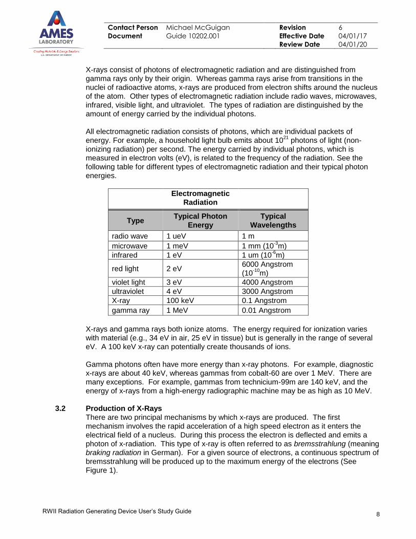
All electromagnetic radiation consists of photons, which are individual packets of
energy. For example, a household light bulb emits about 1021
photons of light (non-
ionizing radiation) per second. The energy carried by individual photons, which is
measured in electron volts (eV), is related to the frequency of the radiation. See the
following table for different types of electromagnetic radiation and their typical photon
energies.
Electromagnetic
Radiation
Type Typical Photon
Energy
Typical
Wavelengths
radio wave 1 ueV 1 m
microwave 1 meV 1 mm (10-3m)
infrared 1 eV 1 um (10-6m)
red light 2 eV 6000 Angstrom
(10-10
m)
violet light 3 eV 4000 Angstrom
ultraviolet 4 eV 3000 Angstrom
X-ray 100 keV 0.1 Angstrom
gamma ray 1 MeV 0.01 Angstrom
X-rays and gamma rays both ionize atoms. The energy required for ionization varies
with material (e.g., 34 eV in air, 25 eV in tissue) but is generally in the range of several
eV. A 100 keV x-ray can potentially create thousands of ions.
Gamma photons often have more energy than x-ray photons. For example, diagnostic
x-rays are about 40 keV, whereas gammas from cobalt-60 are over 1 MeV. There are
many exceptions. For example, gammas from technicium-99m are 140 keV, and the
energy of x-rays from a high-energy radiographic machine may be as high as 10 MeV.
3.2 Production of X-Rays
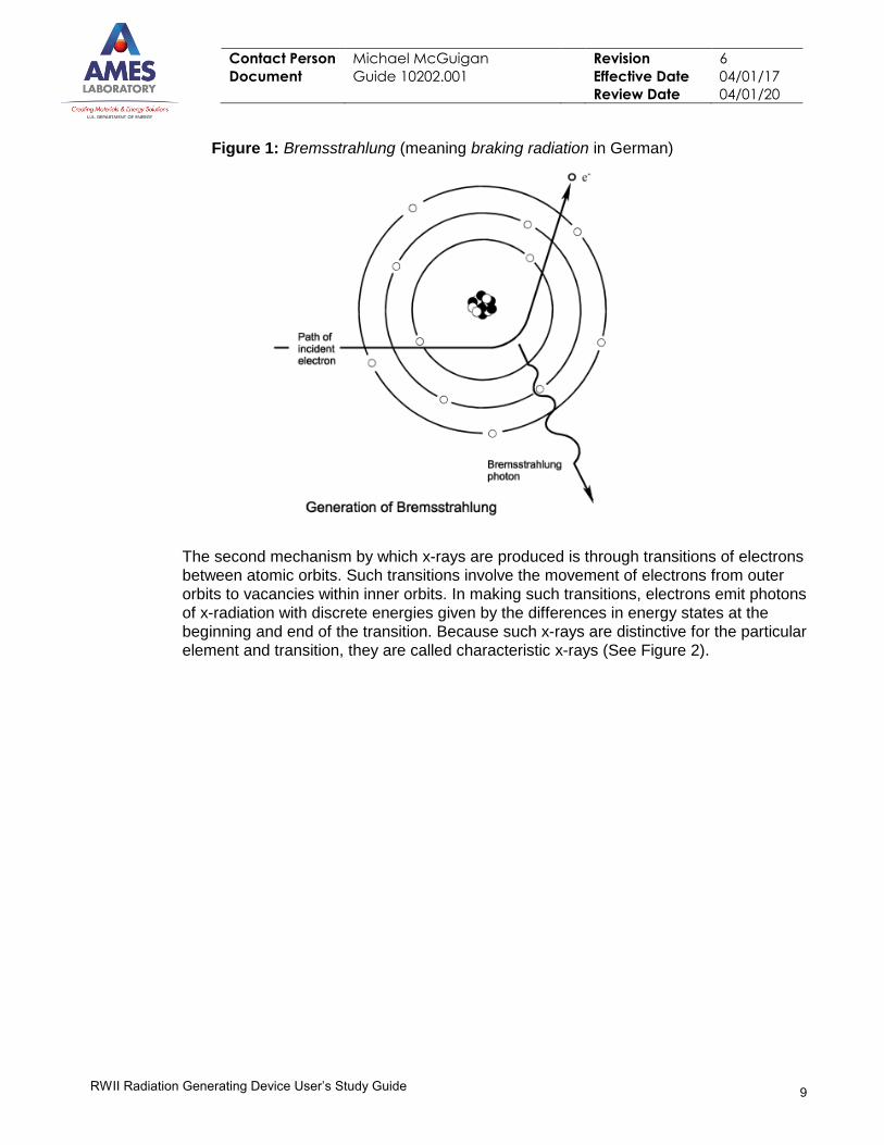
There are two principal mechanisms by which x-rays are produced. The first
mechanism involves the rapid acceleration of a high speed electron as it enters the
electrical field of a nucleus. During this process the electron is deflected and emits a
photon of x-radiation. This type of x-ray is often referred to as bremsstrahlung (meaning
braking radiation in German). For a given source of electrons, a continuous spectrum of
bremsstrahlung will be produced up to the maximum energy of the electrons (See
Figure 1).
Contact Person Michael McGuigan Revision 6
Document Guide 10202.001 Effective Date 04/01/17
Review Date 04/01/20
9
RWII Radiation Generating Device User’s Study Guide
Figure 1: Bremsstrahlung (meaning braking radiation in German)
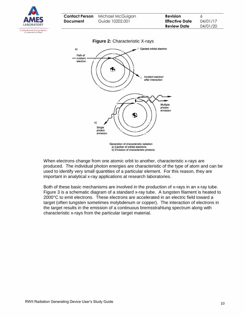
The second mechanism by which x-rays are produced is through transitions of electrons
between atomic orbits. Such transitions involve the movement of electrons from outer
orbits to vacancies within inner orbits. In making such transitions, electrons emit photons
of x-radiation with discrete energies given by the differences in energy states at the
beginning and end of the transition. Because such x-rays are distinctive for the particular
element and transition, they are called characteristic x-rays (See Figure 2).
Contact Person Michael McGuigan Revision 6
Document Guide 10202.001 Effective Date 04/01/17
Review Date 04/01/20
10
RWII Radiation Generating Device User’s Study Guide
Figure 2: Characteristic X-rays
When electrons change from one atomic orbit to another, characteristic x-rays are
produced. The individual photon energies are characteristic of the type of atom and can be
used to identify very small quantities of a particular element. For this reason, they are
important in analytical x-ray applications at research laboratories.
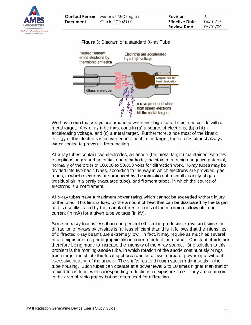
Both of these basic mechanisms are involved in the production of x-rays in an x-ray tube.
Figure 3 is a schematic diagram of a standard x-ray tube. A tungsten filament is heated to
2000°C to emit electrons. These electrons are accelerated in an electric field toward a
target (often tungsten sometimes molybdenum or copper). The interaction of electrons in
the target results in the emission of a continuous bremsstrahlung spectrum along with
characteristic x-rays from the particular target material.
Contact Person Michael McGuigan Revision 6
Document Guide 10202.001 Effective Date 04/01/17
Review Date 04/01/20
11
RWII Radiation Generating Device User’s Study Guide
Figure 3: Diagram of a standard X-ray Tube
We have seen that x-rays are produced whenever high-speed electrons collide with a
metal target. Any x-ray tube must contain (a) a source of electrons, (b) a high
accelerating voltage, and (c) a metal target. Furthermore, since most of the kinetic
energy of the electrons is converted into heat in the target, the latter is almost always
water-cooled to prevent it from melting.
All x-ray tubes contain two electrodes, an anode (the metal target) maintained, with few
exceptions, at ground potential, and a cathode, maintained at a high negative potential,
normally of the order of 30,000 to 50,000 volts for diffraction work. X-ray tubes may be
divided into two basic types, according to the way in which electrons are provided: gas
tubes, in which electrons are produced by the ionization of a small quantity of gas
(residual air in a partly evacuated tube), and filament tubes, in which the source of
electrons is a hot filament.
All x-ray tubes have a maximum power rating which cannot be exceeded without injury
to the tube. This limit is fixed by the amount of heat that can be dissipated by the target
and is usually stated by the manufacturer in terms of the maximum allowable tube
current (in mA) for a given tube voltage (in kV).
Since an x-ray tube is less than one percent efficient in producing x-rays and since the
diffraction of x-rays by crystals is far less efficient than this, it follows that the intensities
of diffracted x-ray beams are extremely low. In fact, it may require as much as several
hours exposure to a photographic film in order to detect them at all. Constant efforts are
therefore being made to increase the intensity of the x-ray source. One solution to this
problem is the rotating-anode tube, in which rotation of the anode continuously brings
fresh target metal into the focal-spot area and so allows a greater power input without
excessive heating of the anode. The shafts rotate through vacuum-tight seals in the
tube housing. Such tubes can operate at a power level 5 to 10 times higher than that of
a fixed-focus tube, with corresponding reductions in exposure time. They are common
in the area of radiography but not often used for diffraction.
Contact Person Michael McGuigan Revision 6
Document Guide 10202.001 Effective Date 04/01/17
Review Date 04/01/20
12
RWII Radiation Generating Device User’s Study Guide
Some diffraction methods require extremely fine x-ray beams. Such beams are most
efficiently produced by special demountable x-ray tubes, called microfocus tubes, in
which special attention is paid to achieving a very small focal spot. The design problem
(fine focusing of the electron beam) is similar to that of the electron microscope or the x-
ray microprobe. One focusing method is electrostatic and maintains the focusing cup
around the filament at a potential of a few hundred volts more negative than the
filament, thus concentrating the electrons into a narrower beam.
The focal spots of these tubes have areas of less than one percent of those of
conventional tubes. Typical sizes are 0.1 X 1 mm for a line focus and 0.05 mm (= 50
mm) diameter for a circular focus.
The maximum power at which an x-ray tube can operate continuously is limited by the
rate at which the target can be cooled. But if the tube is operated for only a small
fraction of a second, a pulse of x-rays can be obtained at a very high power level without
any cooling. This can be done by slowly charging a bank of capacitors and then
abruptly discharging them across a special x-ray tube. In this way an x-ray pulse lasting
about 30 nanoseconds at a peak voltage of 300 kV and a peak current of 5000 amperes
has been produced. (Such a brief flash of x-rays is useful only if its results can be
recorded.)
If increased attention is given, during the design of an x-ray tube, to focusing of the
electron beam and to the shape and placement of the target, the intensity of the beam
issued from the tube can be made about as large as that from a conventional tube, but
with a power input of one-tenth or less. As a result, water cooling is not needed; air
cooling is sufficient. This feature is important for portable apparatus. These tubes are
small, only about 4 to 8 inches in length, and operate typically at a voltage of about 50
kV and a tube current of the order of 1 mA, as compared to 10 mA or more in
conventional tubes.
3.3 Photon Energy and Power
It is important to distinguish between the energy of individual photons in an x-ray beam
and the total energy of all the photons in the beam. It is also important to distinguish
between average power and peak power in a pulsed X-ray device. Typically, the
individual photon energy is given in electron volts (eV), whereas the power of a beam is
given in watts (W). An individual 100 keV photon has more energy than an individual 10
keV photon. However, an x-ray beam consists of a spectrum (a distribution) of photon
energies and the rate at which energy is delivered by a beam is determined by the
number of photons of each energy. If there are many more low energy photons, it is
possible for the low energy component to deliver more energy.
The photon energy distribution may be varied by changing the voltage. The number of
photons emitted may be varied by changing the current.
The power supplies for many x-ray devices do not produce a constant potential (D.C.)
high voltage but instead energize the x-ray tube with a time varying or pulsating high
voltage. Since the bremsstrahlung x-rays produced are a spectrum of energies up to a
maximum equal to the electron accelerating maximum voltage, the accelerating voltage
Contact Person Michael McGuigan Revision 6
Document Guide 10202.001 Effective Date 04/01/17
Review Date 04/01/20
13
RWII Radiation Generating Device User’s Study Guide
of the x-ray device is often described in terms of the peak kilovoltage or kVp.
A voltage of 50 kVp will produce a spectrum of x-ray energies with the theoretical
maximum being 50 keV. The spectrum of energies is continuous from the maximum to
zero. X-ray beams are typically filtered to minimize the low-energy component. Low-
energy x-rays are not useful in radiography, but can deliver a significant dose.
The total number of photons produced by an x-ray device depends on the current, which
is measured in amperes, or amps (A). The current is controlled by increasing or
decreasing the number of electrons emitted from the cathode. The higher the electron
current, the more x-ray photons are emitted from the anode. Many x-ray devices have
meters to measure current. However, as mentioned above, x-rays can be produced by
voltage even if the current is too low to read on the meter. This is sometimes called
dark current. This situation can cause unnecessary exposure and should be addressed
in SOPs or work documents.
3.4 Interaction of X-Rays with Matter
X-rays transfer their energy to matter through chance encounters with bound electrons
or atomic nuclei. These chance encounters result in the ejection of energetic electrons
from the atom. Each of the electrons liberated goes on to transfer its energy to matter
through thousands of direct ionization events (i.e. events involving collisions between
charged particles). Since x-rays and gamma rays transfer energy in this "indirect"
manner, they are referred to as "indirectly ionizing radiations".
Because the encounters of photons with atoms are by chance, a given x-ray has a finite
probability of passing completely through the medium it is traversing. The probability
that an x-ray will pass completely through a medium depends upon numerous factors
including the energy of the x-ray and the medium's composition and thickness.
Section Assessment #3
1) Name the two basic types of electromagnetic radiation and give characteristics of each.
2) Define the term photon.
3) Briefly describe the mechanisms by which X-rays are produced.
4.0 RADIATION QUANTITIES AND TERMS
The following quantities and terms are essential to the description and measurement of various
forms of ionizing radiation.
4.1 Exposure
Exposure is a measure of the strength of a radiation field at some point. It is usually
defined as the amount of charge (i.e., sum of all ions of one sign) produced in a unit
mass of air when the interacting photons are completely absorbed in that mass. The
most commonly used unit of exposure is the "roentgen" (R) which is defined as the
amount of X or gamma radiation which produces 2.58 x 10⁻⁴ coulombs of charge per
kilogram of dry air. In cases where exposure is to be expressed as a rate, the unit
would be R/hr or, more commonly, milli-roentgen per hour (mR/hr).
Contact Person Michael McGuigan Revision 6
Document Guide 10202.001 Effective Date 04/01/17
Review Date 04/01/20
14
RWII Radiation Generating Device User’s Study Guide
4.2 Absorbed Dose
Whereas exposure is defined for air, the absorbed dose is the amount of energy
imparted by radiation to a given mass of any material. The most common unit of
absorbed dose is the "rad" which is defined as 100 ergs of energy per gram of the
material in question. Absorbed dose may also be expressed as a rate with units of
rad/hr or milli-rad/hr.
4.3 Dose Equivalent
Although the biological effects of radiation are dependent upon the absorbed dose,
some types of particles produce greater effects than others for the same amount of
energy imparted. In order to account for these variations when describing human health
risk from radiation exposure, the quantity "dose equivalent" is used. This is the
absorbed dose multiplied by certain "quality" and "modifying" factors indicative of the
relative biological-damage potential of the particular type of radiation. The unit of dose
equivalent is the "rem" or "milli-rem." Dose equivalent may likewise be expressed as a
rate with units of rem/hr or milli-rem/hr. For gamma or x-ray exposures, the numerical
value of the rem is essentially equal to that of the rad.
4.4 Shielding
Shielding is any material, which is placed around or adjacent to a source of penetrating
radiation for the purpose of attenuating the exposure rate from the source. For shielding
X-rays, materials composed of high atomic number elements such as lead are highly
effective.
4.5 Half-Value Layer
The half-value layer is that thickness of a given material (i.e. shielding) required to
reduce the exposure rate from a source of gamma or x-rays to one-half its unshielded
value.
Section Assessment #4
Define the following terms:
Exposure, Absorbed Dose, Dose Equivalent, Shielding and Half-Value Layer
5.0 BIOLOGICAL EFFECTS OF IONIZING RADIATION
The energy deposited by ionizing radiation as it interacts with matter may result in the breaking
of chemical bonds. It is called ionizing radiation because it contains enough energy to displace
an electron from its orbit around a nucleus. If the irradiated matter is living tissue, such
chemical changes may result in altered structure or function of constituent cells. These
changes may, in turn, be manifested as one or more possible effects at the level of the
organism.
5.1 Mechanisms of Radiation Damage
Because the cell is composed mostly of water, less than 20% of the energy deposited
by ionizing radiation is absorbed directly by macromolecules. More than 80% of the
energy deposited in the cell is absorbed by water molecules with the resultant formation
of highly reactive free radicals: H20 ----> H+ + OH-
Contact Person Michael McGuigan Revision 6
Document Guide 10202.001 Effective Date 04/01/17
Review Date 04/01/20
15
RWII Radiation Generating Device User’s Study Guide
These radicals and their products (e.g., hydrogen peroxide) may initiate numerous
chemical reactions, which result in damage to macromolecules and a corresponding
alteration of structure or function. Damage produced within a cell by the radiation-
induced formation of free radicals is described as being an indirect action of radiation.
5.2 Radio-sensitivity
Radio-sensitivity is the relative susceptibility of cells, tissues, organs, organisms and
other substances to the energetic actions of radiation. The cell nucleus is the major site
of radiation damage leading to cell death. This is due to the importance of the DNA
within the nucleus in controlling all cellular function. Damage to the DNA molecule may
prevent it from providing the proper template for the production of additional DNA or
RNA. This hypothesis is supported by research, which has shown that cells are most
sensitive to radiation damage during reproductive phases (i.e., during DNA replication).
In general, it has been found that cell radio-sensitivity is directly proportional to
reproductive capacity and to the degree of cell differentiation. Table 1 presents a list of
cells, which generally follow this principle.
TABLE 1
List of Cells in Order of Decreasing Radio-sensitivity
Very Radio-sensitive Moderately Radio-Sensitive Relatively Radio-Sensitive
Mature lymphocytes
Erythroblasts and
Spermatagonia
Basal cells
Endothelial cells
Osteoblasts
Granulocytes and Osteocytes
Sperm
Erythocytes
Fibrocytes
Chondrocytes
Muscle and Nerve Cells
The considerable variation in the radio-sensitivities of various tissues is due, in part, to
the differences in the sensitivities of the cells that compose the tissues. Also important
in determining tissue sensitivity are such factors as the state of nourishment of the cells,
interactions between various cell types within the tissue, and the ability of the tissue to
repair itself.
The relatively high radio-sensitivity of tissues consisting of undifferentiated, rapidly
dividing cells suggest that, at the level of the human organism, a greater potential exists
for damage to the fetus or young child than to an adult for a given dose. This has, in
fact, been observed in the form of increased birth defects following irradiation of the
fetus and an increased incidence of certain cancers in individuals who were irradiated as
children.
5.3 Human Health Effects
The effects of ionizing radiation described at the level of the human organism can be
divided broadly into two categories: stochastic or non-stochastic effects.
5.3.1 Stochastic Effects
As implied from the name "stochastic", these are effects that occur by chance.
Stochastic effects caused by ionizing radiation consist primarily of genetic effects and
cancer; as the dose to an individual increases, the probability that cancer or a genetic
Contact Person Michael McGuigan Revision 6
Document Guide 10202.001 Effective Date 04/01/17
Review Date 04/01/20
16
RWII Radiation Generating Device User’s Study Guide
effect will occur also increases. At no time, even for high doses, is it certain that cancer
or genetic damage will result. Similarly, for stochastic effects, there is no threshold dose
below which it is relatively certain that an adverse effect cannot occur. In addition,
because stochastic effects can occur in unexposed individuals, one can never be certain
that the occurrence of cancer or genetic damage in an exposed individual is due to
radiation.
5.3.2 Non-stochastic Effects
Unlike stochastic effects, non-stochastic effects are characterized by a threshold dose
below which they do not occur. In addition, the magnitude of the effect is directly
proportional to the size of the dose. Furthermore, for non-stochastic effects, there is a
clear causal relationship between radiation exposure and the effect. Examples of non-
stochastic effects include sterility, erythema (skin reddening), ulceration, and cataract
formation. Each of these effects differs from the other in both its threshold dose and in
the time over which this dose must be received to cause the effect (i.e., acute vs.
chronic exposure).
5.4 Factors Influencing the Occurrence of Health Effects
The type and probability of occurrence of health effects resulting from exposure to X-
rays or other ionizing radiations is a function of several interrelated factors.
5.4.1 Dose and Dose Rate
The effect of ionizing radiation upon humans or other organisms is directly dependent
upon the size of the dose received. The dose, in turn, is dependent upon a number of
factors including the strength of the source, the distance from the source to the affected
tissue, and the time over which the tissue is irradiated.
The ability of a given dose of ionizing radiation to cause health effects is influenced by
the rate at which the dose is imparted. Because of various repair process which occur
within the human body, a given dose of radiation in excess of that needed to produce a
particular non-stochastic effect may, in fact, not produce such an effect if the dose is
imparted over a relatively long period of time (i.e. chronic exposure). It is conservatively
assumed that dose rate is not a critical factor in controlling the probable occurrence of
stochastic effects such as cancer or genetic damage.
5.4.2 Chronic vs. Acute Exposures
Long-term exposures to relatively low levels of ionizing radiation are referred to as
chronic exposures. As indicated by the previous section, such exposures have little
potential for inducing non-stochastic health effects and are of concern because of their
potential for initiating cancer or genetic damage. Short-term exposures to relatively high
levels of ionizing radiation are referred to as acute exposures. Such exposures have the
potential for producing both non-stochastic effects (e.g. erythema, epilation, and
ulceration) as well stochastic effects (e.g. cancer) in the irradiated tissue.
5.4.3 Whole Body vs. Localized Exposure
The effectiveness of a given dose of external radiation in causing biological damage is
dependent upon the portion of the body irradiated. For example, because of differences
in the radio-sensitivity of constituent tissues, the hand is far less likely to suffer biological

Contact Person Michael McGuigan Revision 6
Document Guide 10202.001 Effective Date 04/01/17
Review Date 04/01/20
17
RWII Radiation Generating Device User’s Study Guide
damage from a given dose of radiation than are the gonads. Similarly, a given dose to
the whole body has a greater potential for causing adverse health effects than does the
same dose to only a portion of the body.
Section Assessment #5
1) Describe how free radicals are formed and how they damage the body.
2) Define the term radio-sensitivity and give two examples of highly radio-sensitive cells.
3) What is the difference between a stochastic effect and non-stochastic effect?
4) What are the three factors that influence the occurrence of health effects after an
exposure to radiation?
6.0 EXPOSURE LIMITS
Concern over the biological effects of ionizing radiation began shortly after the discovery of x-
rays in 1895. From that time to the present, numerous recommendations regarding
occupational exposure limits have been proposed and modified by various radiation protection
groups, the most important being the International Commission on Radiological Protection
(ICRP). These guidelines have, in turn, been incorporated into regulatory requirements for
controlling the use of materials and devices emitting ionizing radiation.
Table 2 Summary of Dose Limits [10 CFR 835.202, 1003(a)(1), (a)(2)]
Type Of Exposure Limit
General Employee: Whole Body (internal + external) (TEDE) 5 rem/year
General Employee: Lens of Eye (external) 15 rem/year
General Employee: Extremity (hands and arms below the elbow;
feet and legs below the knees)
50 rem/year
General Employee: Any organ or tissue (other than lens of eye)
and skin
50 rem/year
Declared Pregnant Worker: Embryo/Fetus (internal and external) 0.5 rem/gestation period
Minors: Whole body (internal + external) 0.1 rem/year
Minors: Lens of the eye, skin, and extremities 10% of General Employee
limits Notes:
1. The weighting factors in Appendix 2B, 10 CFR 835 shall be used in converting organ dose equivalent to effective dose equivalent
for the whole body dose.
2. The annual limit of dose to "any organ or tissue" is based on the committed dose equivalent to that organ or tissue resulting from
internally deposited radionuclides over a 50-year period after intake plus any deep dose equivalent to that organ from external
exposures during the year.
3. Exposures due to background radiation, as a patient undergoing therapeutic and diagnostic medical procedures, and participation
as a subject in medical research programs shall not be included in either personnel radiation dose records or assessment of dose
against the limits in this Table.
4. See Appendix 2C for guidance on non-uniform exposure of the skin.
5. Whole body dose (TEDE) = effective dose equivalent from external exposures + committed
effective dose equivalent from internal exposures.
6. Lens of the eye dose equivalent = dose equivalent from external exposure determined at a tissue depth of 0.3 cm.
7. Shallow dose equivalent = dose equivalent from external exposure determined at a tissue depth of 0.007 cm.
Contact Person Michael McGuigan Revision 6
Document Guide 10202.001 Effective Date 04/01/17
Review Date 04/01/20
18
RWII Radiation Generating Device User’s Study Guide
6.1 Basis of Recent Guidelines
In general, the guidelines established for radiation exposure have had as their principle
objectives: (1) the prevention of acute radiation effects (e.g., erythema, sterility), and (2)
the limiting of the risks of late, stochastic effects (e.g., cancer, genetic damage) to
"acceptable" levels. Numerous revisions of standards and guidelines have been made
over the years to reflect both changes in the understanding of the risk associated with
various levels of exposure and changes in the perception of what constitutes an
"acceptable" level of risk.
Current guidelines for radiation exposure are based upon the conservative assumption
that there is no safe level of exposure. In other words, even the smallest exposure has
some probability of causing a late effect such as cancer or genetic damage. This
assumption has led to the general philosophy of not only keeping exposures below
recommended levels or regulatory limits but of also maintaining all exposures ALARA.
This is a fundamental tenet of current radiation safety practice.
6.2 Regulatory Limits for Occupational Exposure
Many of the recommendations of the ICRP and other radiation protection groups
regarding radiation exposure have been incorporated into regulatory requirements by
various countries. For Department of Energy facilities, radiation exposure limits are
found in Title 10, Part 835 of the Code of Federal Regulations (l0 CFR 835). Table 2
provides a summary of the dose limits for occupational external exposures, for those
designated at radiological workers.
6.3 Recommended Exposure Limits for Pregnant Workers
Because of the increased susceptibility of the human embryo and fetus to damage from
ionizing radiation, established dose limits to the embryo/fetus are much lower than for
adults. 10 CFR 835.206(a) requires the dose equivalent to the embryo/fetus from the
period of conception to birth, as a result of occupational exposure of a declared
pregnant worker be limited to 500 mrem. Pregnant workers are encouraged to declare
their pregnancy in writing to their supervisors; the Laboratory will then take steps to
keep exposures within the stated limit. Forms for declaring pregnancy are available
from ESH&A (G40 TASF).
6.4 Regulatory Limits for Non-occupational Exposure
For whole body exposure, those not designated as radiological workers, the non-
occupational exposure limit is 100 mrem/yr. This is the limit for persons not designated
as radiological workers. This is in addition to the 360 mrem/yr received, on average, by
individuals in the U.S. from natural background radiation and manmade radiation
sources. The 100 mrem/yr limit also applies to individuals under age 18 who work in the
vicinity of radiation sources.
Contact Person Michael McGuigan Revision 6
Document Guide 10202.001 Effective Date 04/01/17
Review Date 04/01/20
19
RWII Radiation Generating Device User’s Study Guide
Section Assessment #6
1) What is the primary radiation protection organization that sets occupational exposure
limits?
2) What is the amount of exposure received annually by naturally occurring background
radiation?
3) Allowable exposure to a declared pregnant work is (greater, less) than the allowable
whole body exposure to a general employee.
7.0 TYPES AND CHARACTERISTICS OF ANALYTICAL X-RAY SYSTEMS
Analytical x-ray systems generally consist of three basic components: an x-ray source, a
specimen support or holder, and a detector. In a given experimental setup, these three
components may be enclosed in one integral, commercially available unit or they may be three
distinct systems assembled by the user. The way in which the basic components of the x-ray
system are assembled depends largely upon the mode of operation. Analytical x-ray systems
have two principal modes of operation: diffraction and fluorescence.
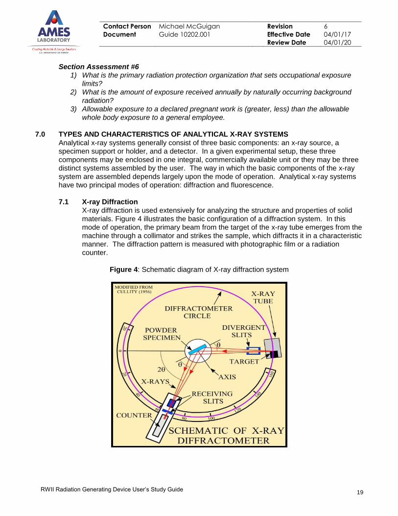
7.1 X-ray Diffraction
X-ray diffraction is used extensively for analyzing the structure and properties of solid
materials. Figure 4 illustrates the basic configuration of a diffraction system. In this
mode of operation, the primary beam from the target of the x-ray tube emerges from the
machine through a collimator and strikes the sample, which diffracts it in a characteristic
manner. The diffraction pattern is measured with photographic film or a radiation
counter.
Figure 4: Schematic diagram of X-ray diffraction system
Contact Person Michael McGuigan Revision 6
Document Guide 10202.001 Effective Date 04/01/17
Review Date 04/01/20
20
RWII Radiation Generating Device User’s Study Guide
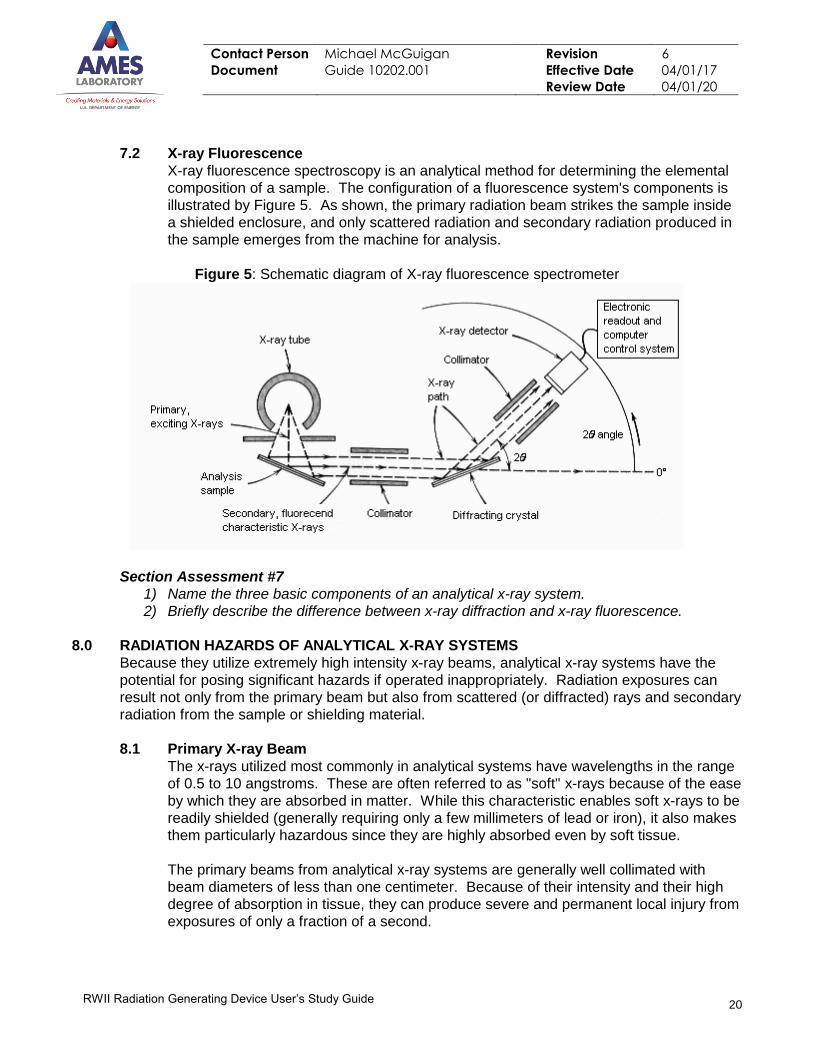
7.2 X-ray Fluorescence
X-ray fluorescence spectroscopy is an analytical method for determining the elemental
composition of a sample. The configuration of a fluorescence system's components is
illustrated by Figure 5. As shown, the primary radiation beam strikes the sample inside
a shielded enclosure, and only scattered radiation and secondary radiation produced in
the sample emerges from the machine for analysis.
Figure 5: Schematic diagram of X-ray fluorescence spectrometer
Section Assessment #7
1) Name the three basic components of an analytical x-ray system.
2) Briefly describe the difference between x-ray diffraction and x-ray fluorescence.
8.0 RADIATION HAZARDS OF ANALYTICAL X-RAY SYSTEMS
Because they utilize extremely high intensity x-ray beams, analytical x-ray systems have the
potential for posing significant hazards if operated inappropriately. Radiation exposures can
result not only from the primary beam but also from scattered (or diffracted) rays and secondary
radiation from the sample or shielding material.
8.1 Primary X-ray Beam
The x-rays utilized most commonly in analytical systems have wavelengths in the range
of 0.5 to 10 angstroms. These are often referred to as "soft" x-rays because of the ease
by which they are absorbed in matter. While this characteristic enables soft x-rays to be
readily shielded (generally requiring only a few millimeters of lead or iron), it also makes
them particularly hazardous since they are highly absorbed even by soft tissue.
The primary beams from analytical x-ray systems are generally well collimated with
beam diameters of less than one centimeter. Because of their intensity and their high
degree of absorption in tissue, they can produce severe and permanent local injury from
exposures of only a fraction of a second.
Contact Person Michael McGuigan Revision 6
Document Guide 10202.001 Effective Date 04/01/17
Review Date 04/01/20
21
RWII Radiation Generating Device User’s Study Guide
Potential exposure to the primary X-ray beam is generally not a major concern for
analytical systems operating in the fluorescence mode since the beam in these systems
are contained within a shielded enclosure. The possibility of leakage of the primary
beam through the shield, however, cannot be discounted.
Exposure to the primary beam of diffraction units is a major concern. In fact, the
greatest risk of acute accidental exposures from analytical systems occurs in
manipulations of the sample to be irradiated by the direct beam in diffraction studies.
Exposure rates on the order of 10,000 R/sec can exist at the tube housing port. At
these levels, erythema would be produced from exposures of only 0.03 second and
permanent injury could be inflicted in only 0.1 second. The fingers, of course, are the
part of the body most at risk from such high exposures.
8.2 Diffracted X-ray Beam
For x-ray diffraction systems, the diffracted beam is also small and well collimated with
an intensity of up to 80 R/hr. Prolonged or repeated exposures to a beam of this
intensity could result in an individual exceeding the annual dose limit for the particular
tissue irradiated.
8.3 Scattered and Secondary X-rays
Through interactions with the sample and shielding material, the primary beam in
diffraction systems often produces diffuse patterns of scattered and secondary x-rays in
the environment around the equipment. Exposure rates of 150 mR/hr near the shielded
sample are not uncommon. In contrast, scattered and secondary radiation levels in the
vicinity of a fluorescence system (where only secondary radiation emerges from the
shielded target-sample assembly) is generally an order of magnitude less.
Section Assessment #8
1) Define the term “soft X-rays.”
2) The greatest risk of exposures from analytical systems occurs during what activity?
9.0 HAZARD CONTROL MEASURES FOR X-RAY SYSTEMS
Requirements for controlling potential hazards associated with analytical x-ray systems are
specified in most states by law. At Ames Lab these requirements are detailed in the Radiation
Safety Manual. Portions of these hazard control and safety measures are summarized here.
9.1 Equipment Requirements
9.1.1 Beam Entry Shut-off Device
A device which prevents the entry of any portion of an individual's body into the primary
beam path or which causes the beam to be shut off upon entry into its path shall be
provided on all open-beam configurations.
9.1.2 Shutters
Shutters open and close a path for the X-ray beam. When a shutter is properly
functioning and closed, no X-rays can pass beyond the shutter. On open-beam
configurations, each port on the source housing shall be equipped with a shutter that
cannot be opened unless a collimator or coupling has been connected to the port.

Contact Person Michael McGuigan Revision 6
Document Guide 10202.001 Effective Date 04/01/17
Review Date 04/01/20
22
RWII Radiation Generating Device User’s Study Guide
Figure 6: Figure 7: Interlocked enclosure with shutter and enclosure status lights Interlocked enclosure with x-ray status light on the top of
the unit
9.1.3 Housing Interlock
Each x-ray tube housing shall be equipped with an interlock that shuts off the tube if the
tube is removed from the housing or if the housing is disassembled.
9.1.4 Shielding Provided by Housing
Each x-ray tube housing shall be constructed so that, with all shutters closed, the
radiation, measured at a distance of five centimeters from its surface, is not capable of
producing a dose in excess of 2.5 millirem in one hour.
9.1.5 Warning Lights
An easily visible warning light with the words "X-RAY ON" shall be located near any
switch that energizes an x-ray tube and shall be illuminated only when the tube is
energized (see Figures 4 & 5).
9.1.6 Labeling
All X-ray equipment shall be labeled with a readily discernible sign bearing the radiation
symbol and the words (see Figures 4 & 5):
(1)"CAUTION - HIGH INTENSITY X-RAY BEAM" on the X-ray source housing; and
(2)"CAUTION RADIATION - THIS EQUIPMENT PRODUCES RADIATION WHEN
ENERGIZED" near any switch that energizes the X-ray tube.
Contact Person Michael McGuigan Revision 6
Document Guide 10202.001 Effective Date 04/01/17
Review Date 04/01/20
23
RWII Radiation Generating Device User’s Study Guide
9.2 Area Requirements
9.2.1 Radiation Levels
The components of an analytical x-ray system shall be arranged and shall be sufficiently
shielded to ensure that radiation levels near the controls cannot result in an individual
receiving a dose in excess of 2.5 millirem whole body exposure in any one hour.
9.2.2 Surveys
Radiation surveys of all x-ray systems sufficient to show compliance with the radiation
levels established in 9.2.1 must be performed:
1. Upon installation of the equipment and at least every twelve months thereafter.
2. Following any change in the number, type, or arrangement of components in the
system.
3. Following any maintenance requiring the disassembly or removal of a system
component.
These surveys shall be conducted by ESH&A and it is the responsibility of the x-ray
system users to notify ESH&A if situation 2 or 3 should arise.
9.2.2.1 Survey requirement for X-ray User
The x-ray user shall conduct an x-ray survey when the system interlock has been
placed into an off normal state or bypassed. X-ray systems with the capability of
bypassing the interlock are required to have a Radiological Work Permit (RWP).
Only persons on the system’s RWP are authorized to bypass the system
interlock.
9.2.3 Posting
Laboratory space containing x-ray equipment is labeled at the entry way(s) as an x-ray
equipment laboratory. The interlocked Plexiglas barriers surrounding the x-ray systems
shall be posted as a “CONTROLLED AREA” or “BUFFER AREA” as necessary, and the
area immediately adjacent to all x-ray beam ports, whether open beam or closed beam,
shall be posted with a sign which reads “CAUTION [OR DANGER] - HIGH RADIATION
AREA” as appropriate.
9.3 Operating Requirements
9.3.1 Procedures
Standard operating procedures shall be written and available to all x-ray equipment
workers.
9.3.2 Bypassing Safety Devices
No individual shall bypass a safety device or interlock unless the individual has obtained
written approval from the Radiation Safety Officer. X-ray systems with the capability of
bypassing the interlock are required to have a Radiological Work Permit (RWP). Only
persons on the system’s RWP are authorized to bypass the system interlock.
Contact Person Michael McGuigan Revision 6
Document Guide 10202.001 Effective Date 04/01/17
Review Date 04/01/20
24
RWII Radiation Generating Device User’s Study Guide
9.3.3 Altering System Components
No operation involving the removal or alteration of shielding materials, tube housing,
shutter, collimators, or beam stops shall be performed until it has been determined that
the beam is off and will remain off until safe conditions have been restored.
9.4 Personnel Monitoring Requirements
As a minimum, finger or wrist dosimeters shall be provided to and shall be used by all
radiation generating device users. At Ames Laboratory, the Health Physics Group
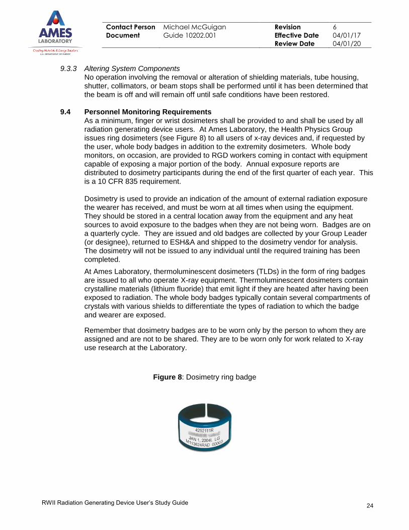
issues ring dosimeters (see Figure 8) to all users of x-ray devices and, if requested by
the user, whole body badges in addition to the extremity dosimeters. Whole body
monitors, on occasion, are provided to RGD workers coming in contact with equipment
capable of exposing a major portion of the body. Annual exposure reports are
distributed to dosimetry participants during the end of the first quarter of each year. This
is a 10 CFR 835 requirement.
Dosimetry is used to provide an indication of the amount of external radiation exposure
the wearer has received, and must be worn at all times when using the equipment.
They should be stored in a central location away from the equipment and any heat
sources to avoid exposure to the badges when they are not being worn. Badges are on
a quarterly cycle. They are issued and old badges are collected by your Group Leader
(or designee), returned to ESH&A and shipped to the dosimetry vendor for analysis.
The dosimetry will not be issued to any individual until the required training has been
completed.
At Ames Laboratory, thermoluminescent dosimeters (TLDs) in the form of ring badges
are issued to all who operate X-ray equipment. Thermoluminescent dosimeters contain
crystalline materials (lithium fluoride) that emit light if they are heated after having been
exposed to radiation. The whole body badges typically contain several compartments of
crystals with various shields to differentiate the types of radiation to which the badge
and wearer are exposed.
Remember that dosimetry badges are to be worn only by the person to whom they are
assigned and are not to be shared. They are to be worn only for work related to X-ray
use research at the Laboratory.
Figure 8: Dosimetry ring badge
Contact Person Michael McGuigan Revision 6
Document Guide 10202.001 Effective Date 04/01/17
Review Date 04/01/20
25
RWII Radiation Generating Device User’s Study Guide
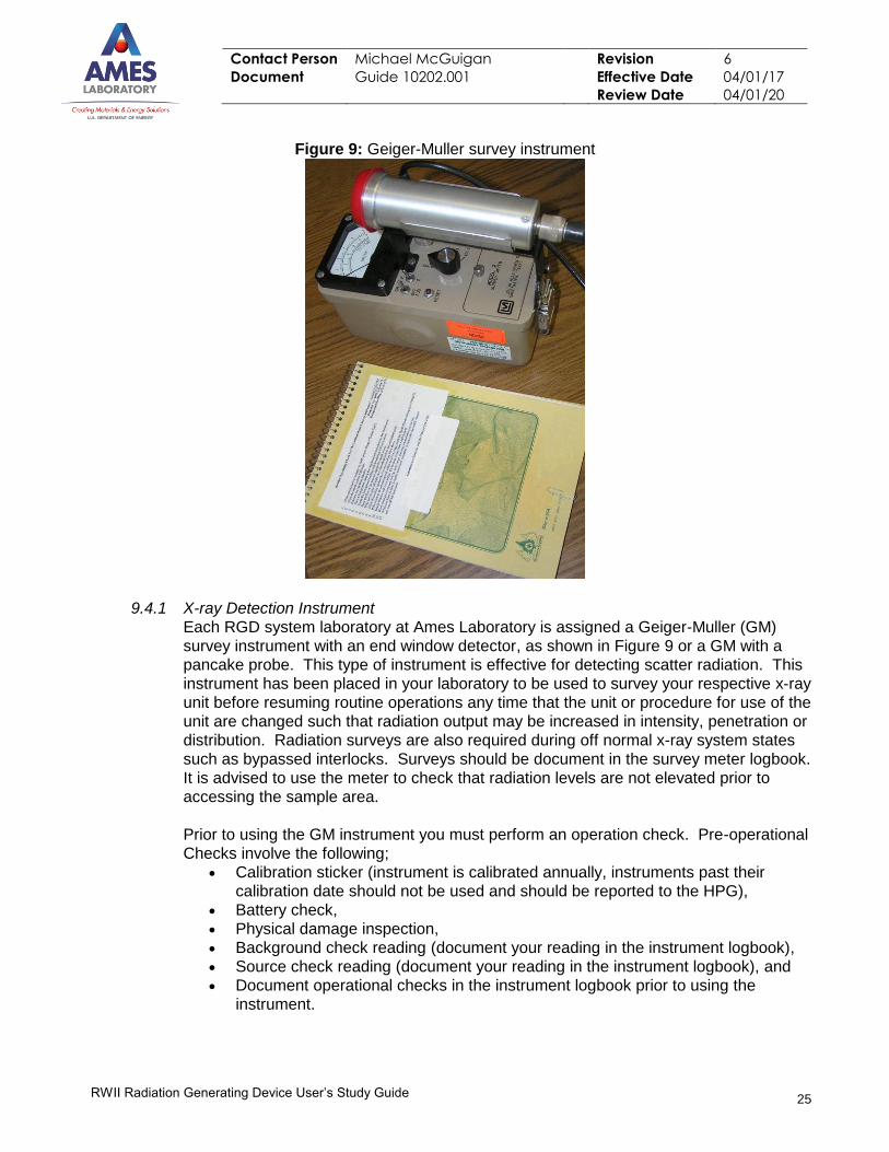
Figure 9: Geiger-Muller survey instrument
9.4.1 X-ray Detection Instrument
Each RGD system laboratory at Ames Laboratory is assigned a Geiger-Muller (GM)
survey instrument with an end window detector, as shown in Figure 9 or a GM with a
pancake probe. This type of instrument is effective for detecting scatter radiation. This
instrument has been placed in your laboratory to be used to survey your respective x-ray
unit before resuming routine operations any time that the unit or procedure for use of the
unit are changed such that radiation output may be increased in intensity, penetration or
distribution. Radiation surveys are also required during off normal x-ray system states
such as bypassed interlocks. Surveys should be document in the survey meter logbook.
It is advised to use the meter to check that radiation levels are not elevated prior to
accessing the sample area.
Prior to using the GM instrument you must perform an operation check. Pre-operational
Checks involve the following;
Calibration sticker (instrument is calibrated annually, instruments past their
calibration date should not be used and should be reported to the HPG),
Battery check,
Physical damage inspection,
Background check reading (document your reading in the instrument logbook),
Source check reading (document your reading in the instrument logbook), and
Document operational checks in the instrument logbook prior to using the
instrument.
Contact Person Michael McGuigan Revision 6
Document Guide 10202.001 Effective Date 04/01/17
Review Date 04/01/20
26
RWII Radiation Generating Device User’s Study Guide
Note that readings taken during the pre-operational check should be within +/-10 % of
previous readings already recorded in the instrument logbook. If a GM is outside +/-
10% of previous readings report this to the HPG. If the GM fails any of the pre-
operational checks it should not be used and your results should be reported to the HPG
so a properly operating meter can be supplied to you.
Section Assessment #9
1) Define the following control devices and how they work: 1) Beam entry shut-off device,
2) Shutters, and 3) Housing interlock
2) When are radiation surveys of X-ray systems required?
3) Define the term dosimetry.
4) Describe the primary type of dosimeter used at Ames Laboratory and how it works.
5) Name three things done during a pre-operational check of a Geiger-Muller survey
instrument.
10.0 RESPONSIBIITY FOR X-RAY SAFETY
The responsibility for maintaining radiation exposures from radiation generating devices ALARA
is shared among the Ames Laboratory Line Management, the ESH&A office, the x-ray device
Group Leader (activity supervisor), and x-ray device operators.
10.1 Ames Laboratory Leadership
In accordance with the specific requirements of Ames Laboratory’s RPP, an ALARA
Committee has been established that oversees the implementation of Laboratory
policies and procedures for the safe use of radioactive materials and radiation
generating devices. The Committee consists of members of Ames Laboratory’s staff
and faculty appointed by the Director for terms of three years. In addition, the ALARA
Committee reviews all requests for use of radioactive materials and radiation generating
devices, reviews records of personnel dosimetry, and decides whether or not
authorization for use is to be granted.
10.2 ESH&A, Health Physics Group
ESH&A Health Physics Group is responsible for:
Establishing requirements and standards,
Offering consulting services and institutional training,
Approving all purchases, moves, transfers, and alterations of x-ray equipment,
Conducting laboratory inspections, surveying x-ray equipment, verifying the
appropriate safety program requirements have been met, and affixing
compliance labels to devices,
Administering the personnel dosimetry program, and
Coordinating with the Engineering Services Group for preventive maintenance
and repair of the engineered x-ray barrier safety systems in Ames Laboratory
spaces or owned by Ames Laboratory programs in ISU spaces. (Safety
interlocks are checked every six months to ensure proper functionality.)
Contact Person Michael McGuigan Revision 6
Document Guide 10202.001 Effective Date 04/01/17
Review Date 04/01/20
27
RWII Radiation Generating Device User’s Study Guide
10.3 Group Leader/Activity Supervisor
The individual authorized by the ALARA Committee as the activity supervisor on an
activity involving the use of radioactive material or a radiation generating device is
responsible for all activities conducted under the scope of that authorization. This
includes responsibility for ensuring that:
All individuals working on the activity are appropriately trained and supervised.
The ALARA Committee has formally authorized all individuals working on the
activity.
All rules, regulations, and procedures for the safe use of radiation generating
devices are observed during the activity.
An accurate record of radiation generating devices in his or her possession is
maintained.
The ESH&A Office is notified of any proposed changes in the storage or use of
the radioactive material or a radiation generating device prior to the
implementation of such changes.
All uses of radiation are evaluated to further reduce exposures to individuals
(ALARA).
All radioactive sources or source material are protected from unauthorized
access or removal.
All SOPs are current and distributed appropriately.
Maintain a list of qualified operators authorized for particular machines in each
respective RGD laboratory as well as a system user logbook and maintenance
logbook.
It is recommended that the system logbook contain the following information:
Name of person performing maintenance on system and employer if
outside contractor;
The reason the maintenance was performed;
Specific problems noted during maintenance;
Repairs performed (if a modification to the system is performed, reference
the date of approval from Group Leader and the ESH&A Office);
Time and date the maintenance was performed; and
Signature of the person who did the maintenance on the system
10.4 X-ray device Operators
Authorized X-ray-device operators are responsible for:
Keeping his or her personal exposure as low as reasonably achievable,
Wearing your ring dosimeters appropriately,
Knowing and following the standard operating procedure for each machine
operated,
Knowing and following the operator safety protocols,
Notifying their supervisors of any unsafe or hazardous work situations,
Before reaching into the primary beam location, verifying that the beam shutter is
closed or that machine power is off, and
Completing all required training, and assist the laboratory supervisor in
maintaining the required records (x-ray system logbooks).
Contact Person Michael McGuigan Revision 6
Document Guide 10202.001 Effective Date 04/01/17
Review Date 04/01/20
28
RWII Radiation Generating Device User’s Study Guide
Section Assessment #10
1) Who oversees implementation of the Laboratory’s RPP?
2) Name three things a Group Leader is responsible for relative to use of analytical x-ray
systems at Ames Laboratory.
3) Name three things an x-ray device operator is responsible for relative to use of
analytical x-ray systems at Ames Laboratory.
11.0 RADIATION SAFETY REFERENCES
Radiation Protection, 3rd edition, Jacob Shapiro, Harvard University Press, Cambridge,
Massachusetts, 1990.
Introduction to Health Physics, 3rd edition, Herman Cember, Pergamon Press, New York, NY
1996.
Radiologic Science for Technologists, 2nd edition, Stewart Bushong, The C.V. Mosby
Company, St. Louis, Missouri, 1980.
The Effects on Populations of Exposure to Low Levels of Ionizing Radiation: 1980, National
Academy Press, Washington, D.C., 1980.
Atoms, Radiation, and Radiation Protection, J.E. Turner, Pergamon Press, Oxford, England,
1995.