Embed Size (px)
Citation preview
RESEARCH ARTICLE Open Access
Radiation therapy is an important factor toimprove survival in pediatric patients withhead and neck rhabdomyosarcoma byenhancing local control: a historical cohortstudy from a single centerYuan\ Wen, Dongsheng Huang* , Weiling Zhang, Yi Zhang, Huimin Hu and Jing Li
Abstract
Background: The purpose of this study is to analyze the influence of radiation therapy on survival in a historicalcohort of 56 pediatric patients with head and neck rhabdomyosarcoma.
Methods: A historical cohort of 56 pediatric patients with head and neck rhabdomyosarcoma from June 1st, 2013to June 30th, 2019 was chosen. Clinical data and follow up results were collected including all diagnosis, treatmentand prognosis information. Overall survival (OS) and event free survival (EFS) as time-to-event distributions wereestimated with Kaplan-Meier method, and univariate analysis was performed with log rank test to detect differencesbetween groups. Multivariate analysis was performed to explore the risk factors for survival with Cox proportionalhazard model.
Results: The media follow up time of all 56 patients was 31.8 months (range 3.5–74.6 months). There were 26events during follow up, including 14 disease progressions and 12 relapses. The estimated 5-year OS of all patientswas 69.9%, and the estimated 5-year EFS was 48.8%. Patients with radiation therapy as a component of the initialtreatment plan had better 5-year OS and EFS compared with those without radiation therapy (OS 80.3% vs. 49.7%,p = 0.003 and EFS 63.9% vs. 21.9%, p < 0.001). In patients with events, those who received salvage radiation therapyhad better 5-year OS compared with those who didn’t (OS 66.0% vs. 31.2%, p = 0.033). On multivariate analysis,tumor size > 5 cm and non-initial radiation therapy were independent risk factors for OS in all patients, non-initialradiation therapy was an independent risk factor for EFS in all patients, and tumor size > 5 cm was an independentrisk factor for OS in patients with events.
Conclusions: Radiation therapy as a component of initial treatment can improve the OS and EFS in pediatric headand neck rhabdomyosarcoma patients by enhancing local control, and non-initial radiation therapy is an independentrisk factor for OS and EFS. Salvage radiation therapy still can improve OS in patients with disease progression andrelapse. Tumor size > 5 cm is an independent risk factor for OS in pediatric HNRMS patients with or without diseaseprogression/relapse.
Keywords: Rhabdomyosarcoma, Pediatric, Radiation therapy, Prognosis, Head and neck, Tumor size
© The Author(s). 2020 Open Access This article is licensed under a Creative Commons Attribution 4.0 International License,which permits use, sharing, adaptation, distribution and reproduction in any medium or format, as long as you giveappropriate credit to the original author(s) and the source, provide a link to the Creative Commons licence, and indicate ifchanges were made. The images or other third party material in this article are included in the article's Creative Commonslicence, unless indicated otherwise in a credit line to the material. If material is not included in the article's Creative Commonslicence and your intended use is not permitted by statutory regulation or exceeds the permitted use, you will need to obtainpermission directly from the copyright holder. To view a copy of this licence, visit http://creativecommons.org/licenses/by/4.0/.The Creative Commons Public Domain Dedication waiver (http://creativecommons.org/publicdomain/zero/1.0/) applies to thedata made available in this article, unless otherwise stated in a credit line to the data.
* Correspondence: [email protected] Department of Beijing Tongren Hospital, Capital Medical University,100730, 1# Dong Jiao Min Xiang, Dongcheng District, Beijing, China
Wen et al. BMC Pediatrics (2020) 20:265 https://doi.org/10.1186/s12887-020-02165-y
BackgroundRhabdomyosarcoma (RMS) is the most commonchildhood soft tissue sarcoma, accounting for about50% of all patients [1, 2]. It comprises about 4.5% ofall childhood cancer with an annual incidence of 4.5cases per 1 million children and young adults agedunder 20 years [3, 4]. This aggressive malignant tumorcan develop in any part of the body, and is thoughtto have a primitive mesenchymal cell origin, with apropensity for striated muscle differentiation [3–5].The treatment of RMS is a multimodal strategy, refer-ring to the combination of chemotherapy, surgery,and radiation therapy (RT), as well as recent biologic-ally targeted agents [2, 3, 6]. Over the last 3 decadesthe survival of RMS patients has improved substan-tially with 5-year OS exceeding 70% [2, 3]. But theprognosis of relapsed and metastatic patientsremained poor with 5-year OS about 30% [2, 3, 6].The prognosis of all RMS is strongly determined bythe ability of achieving local control (control of theprimary tumor site) [2, 7]. The main pattern of treat-ment failure including disease progression and relapseis local failure, and maintaining local control is ofcrucial importance throughout the treatment plan [7,8]. The two major modalities of local control are sur-gery and RT, which could be used separately or com-bined [2, 9].Head and neck is the most common region of pres-
entation, comprising 40% of all RMS [10, 11]. Theprimary sites of head and neck rhabdomyosarcoma(HNRMS) include orbit, parameningeal and nonpara-meningeal nonorbit head and neck. About 50% ofHNRMS cases are parameningeal type (unfavorablesite), arising in the middle ear/mastoid, nasopharynx/nasal cavity, paranasal sinus, parapharyngeal region,or pterygopalatine/infratemporal fossa [10, 11]. Localcontrol is a significant challenge for HNRMS, espe-cially for parameningeal type [12, 13]. Considering thecomplicated anatomy of this region, radical surgerywould usually cause severe functional and/or cosmeticsequelae, and in most patients there would be grossresidual or only a biopsy could be performed [11].Under this circumstance RT becomes the only appro-priate local control method before a second look ordelayed primary excision [14].We found in some of our pediatric HNRMS pa-
tients RT was not included as a component of thetreatment plan, which was mainly attributed to paren-tal refusal due to different personal considerations.This provided us with the possibility to compare theprognosis of these patients with others. Based on theabove we tried to analyze whether RT could improvethe survival in pediatric HNRMS patients, and to addevidence to it.
MethodsStudy designThis is a historical observational cohort study, based on allHNRMS patients diagnosed and treated in our pediatricdepartment from June 1st, 2013 to June 30th, 2019.
Diagnostic evaluation and risk stratificationThe pretreatment diagnostic workup options includedhead and neck computed tomography (CT) scan and/ormagnetic resonance imaging (MRI) with contrast, posi-tron emission tomography-computed tomography (PET-CT) scan, chest CT, radionuclide bone scan, bone mar-row aspirates and/or trephine biopsies, cerebrospinalfluid (CSF) test (parameningeal patients). The site, size(widest dimension) and invasiveness of the primarytumor, regional nodal involvement, and metastatic statuswere determined. Stage was assigned according to thepretreatment staging system of Soft Tissue SarcomaCommittee of Children’s Oncology Group (COG) [15].(Table 1).Surgical plan was determined based on pretreatment
workup results. Excision was attempted on condition ofno severe functional and/or cosmetic consequences.Otherwise only biopsy was done. Group was assigned ac-cording to intergroup rhabdomyosarcoma study (IRS)surgical-pathologic group system [15]. (Table 2) Patho-logic subtype was classified according to the fourth edi-tion of the World Health Organization classification oftumors of soft tissue and bone, which comprise 4 sub-types including embryonal, alveolar, spindle cell/scleros-ing and pleomorphic (only seen in adults) subtypes [16].At last the risk group was assigned with comprehen-
sive consideration of stage, group, and pathologic sub-type results, according to the risk group classification ofSoft Tissue Sarcoma Committee of Children’s OncologyGroup [15]. (Table 3) And chemotherapy was generallyguided by the risk group classification.
Table 1 TNM pretreatment staging system
Stage Sitea Tb Size Nc Md
1 Nonparameningeal T1 or T2 Any Any M0
2 Parameningeal T1 or T2 ≤5 cm N0 or Nx M0
3 Parameningeal T1 or T2 ≤5 cm N1 M0
>5 cm Any M0
4 Any T1 or T2 Any Any M1
a. For HNRMS favorable site refers to nonparameningeal site (orbit andnonorbit nonparameningeal head and neck); unfavorable site refers toparameningeal siteb. T1, confined to primary site; T2, surrounding tissue invasionc. Regional nodes N0, not involved; N1, involved; Nx, status unknownd. M0, no distant metastasis; M1 distant metastasis (includes positive cytologyin CSF, pleural, or peritoneal fluid)
Wen et al. BMC Pediatrics (2020) 20:265 Page 2 of 10
Treatment protocolThe chemotherapy we used for low risk group patientswas COG D9602 subgroup B VAC (vincristine, dactino-mycin, cyclophosphamide) regimen [17]. For intermedi-ate risk group patients, the COG D9803 standard VACregimen was used [18]. For high risk group patients, aswell as patients with disease progression or disease re-lapse, based on the standard VAC regimen, an optionalcombination with anthracyclines, platinum drugs, etopo-side or irinotecan was frequently used.RT was recommended to all patients except for Group
I embryonal patients. Generally, according to the recom-mendation of Soft Tissue Sarcoma Committee of Chil-dren’s Oncology Group, the RT dose was 36Gy forGroup I alveolar patients, 36Gy or 41.4Gy for Group IIpatients according to nodal involvement status, 45Gy forGroup III orbit patients, and 50.4Gy for other Group IIIpatients. Group IV patients were irradiated as for othergroups, including metastatic sites if possible. RT is initi-ated within 12 weeks after chemotherapy, and radiosen-sitizing agents were omitted during RT.Second-look surgery, delayed primary excision, or sal-
vage excision was considered only if no severe functionaland/or cosmetic consequences were anticipated.
Follow upAll patients were closely followed up since diagnosis.Clinical data during and after treatment were recorded.Frequency of off-therapy surveillance was every 3months for the first year, every 4 months for the secondand third year, and once a year for the fourth and fifth
year [8]. Clinical physical examination, blood routineand biochemical tests, head and neck CT or MRI withor without contrast, and chest CT or chest X-ray wererequired for surveillance, and PET-CT was optional toreplace all imaging examinations.Overall survival (OS) was defined as survival from
diagnosis to death of any cause. Disease progression(PD) was defined as primary tumor enlargement, and/ornew lesions, and/or metastasis during primary treatmentcourse. Disease relapse (RD) was defined as recurrenceof RMS in any form after last treatment. Event free sur-vival (EFS) was defined as survival from diagnosis to thefirst event of PD, RD, second tumor or death of anycause [7, 19, 20]. In this study only the first PD and RDwere discussed and analyzed. The patterns of PD andRD included local (primary site), regional (regionallymph node), metastatic, and any combinations.
Grouping and statistical methodsAccording to whether RT was included as a componentof initial treatment plan, patients were divided into ini-tial RT group (Group IRT) and non-initial RT group(Group NIRT). According to whether RT was includedas a component of salvage treatment plan, patients withevents, including all PD and RD patients, were dividedinto salvage RT group (Group SRT) and non-salvage RTgroup (Group NSRT).OS and EFS as time-to-event distributions were esti-
mated with Kaplan-Meier method, and survival rates wereestimated. Univariate analysis was performed with logrank test to detect differences between groups and Bonfer-roni adjustment was used to control type I error if morethan two groups were compared. Multivariate analysis wasperformed using Cox proportional hazard model to ex-plore risk factors and adjust confounding factors, and haz-ard ratios (HR) with 95% confidence intervals (CIs) werecalculated. Categorical variables were compared using chi-squared test or Fisher’s exact test between groups. A pvalue < 0.05 was considered statistically significant. Datawere analyzed with IBM SPSS Statistics 26.0.
ResultsPatients’ clinical characteristicsFrom June 1st, 2013 to June 30th, 2019, 56 pediatric pa-tients were admitted into our pediatric department, whowere diagnosed as HNRMS with pathological confirm-ation. These patients formed our cohort and were diag-nosed, stratified, treated and followed up by uniformprotocol. The median follow up time was 31.8 (range3.5–74.6) months for all patients, 37.6 (range 6.1–74.6)months for Group IRT, 20.6 (range 3.5–71.6) monthsfor Group NIRT, 35.7 (range 6.1–74.6) months forGroup SRT, and 20.9 (range 4.8–58.4) months forGroup NSRT. The specific clinical characteristics of all
Table 2 IRS surgical-pathologic group system
Group Definition
I Localized disease, completely resected
II Total gross resection, with evidence of regional spread
A Grossly resected tumor with microscopic residual disease
B Involved regional nodes completely resected with nomicroscopic residual disease
C Involved regional nodes grossly resected with evidence ofmicroscopic residual disease
III Biopsy only or incomplete resection with gross residual disease
IV Distant metastatic disease (excludes regional nodes andadjacent organ infiltration)
Table 3 COG risk group classification
Risk Group Histology Stage Group
Low Embryonal 1 I, II, III
Embryonal 2, 3 I, II
Intermediate Embryonal 2, 3 III
Alveolar 1, 2, 3 I, II, III
High Embryonal or Alveolar 4 IV
Wen et al. BMC Pediatrics (2020) 20:265 Page 3 of 10
patients and patients of different groups are showed inTable 4.
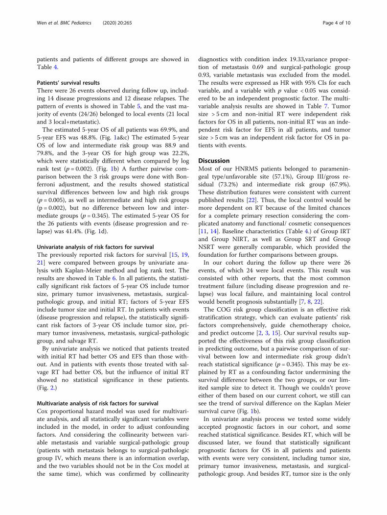
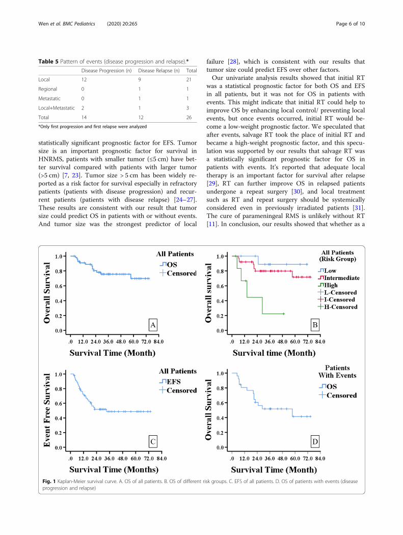
Patients’ survival resultsThere were 26 events observed during follow up, includ-ing 14 disease progressions and 12 disease relapses. Thepattern of events is showed in Table 5, and the vast ma-jority of events (24/26) belonged to local events (21 localand 3 local+metastatic).The estimated 5-year OS of all patients was 69.9%, and
5-year EFS was 48.8%. (Fig. 1a&c) The estimated 5-yearOS of low and intermediate risk group was 88.9 and79.8%, and the 3-year OS for high group was 22.2%,which were statistically different when compared by logrank test (p = 0.002). (Fig. 1b) A further pairwise com-parison between the 3 risk groups were done with Bon-ferroni adjustment, and the results showed statisticalsurvival differences between low and high risk groups(p = 0.005), as well as intermediate and high risk groups(p = 0.002), but no difference between low and inter-mediate groups (p = 0.345). The estimated 5-year OS forthe 26 patients with events (disease progression and re-lapse) was 41.4%. (Fig. 1d).
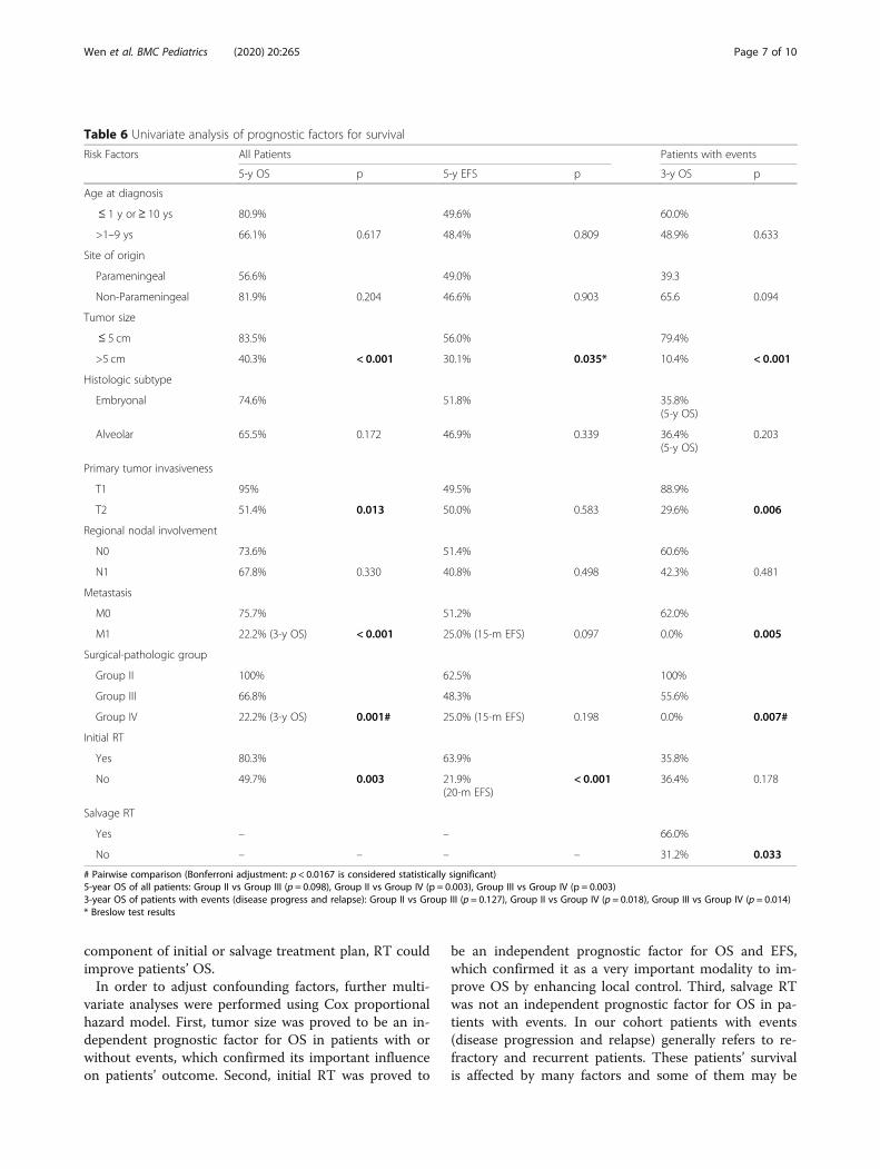
Univariate analysis of risk factors for survivalThe previously reported risk factors for survival [15, 19,21] were compared between groups by univariate ana-lysis with Kaplan-Meier method and log rank test. Theresults are showed in Table 6. In all patients, the statisti-cally significant risk factors of 5-year OS include tumorsize, primary tumor invasiveness, metastasis, surgical-pathologic group, and initial RT; factors of 5-year EFSinclude tumor size and initial RT. In patients with events(disease progression and relapse), the statistically signifi-cant risk factors of 3-year OS include tumor size, pri-mary tumor invasiveness, metastasis, surgical-pathologicgroup, and salvage RT.By univariate analysis we noticed that patients treated
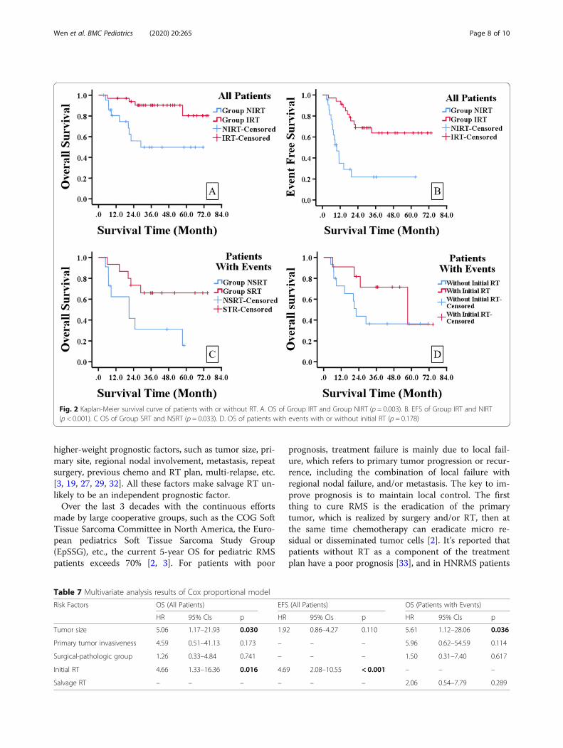
with initial RT had better OS and EFS than those with-out. And in patients with events those treated with sal-vage RT had better OS, but the influence of initial RTshowed no statistical significance in these patients.(Fig. 2.)
Multivariate analysis of risk factors for survivalCox proportional hazard model was used for multivari-ate analysis, and all statistically significant variables wereincluded in the model, in order to adjust confoundingfactors. And considering the collinearity between vari-able metastasis and variable surgical-pathologic group(patients with metastasis belongs to surgical-pathologicgroup IV, which means there is an information overlap,and the two variables should not be in the Cox model atthe same time), which was confirmed by collinearity
diagnostics with condition index 19.33,variance propor-tion of metastasis 0.69 and surgical-pathologic group0.93, variable metastasis was excluded from the model.The results were expressed as HR with 95% CIs for eachvariable, and a variable with p value < 0.05 was consid-ered to be an independent prognostic factor. The multi-variable analysis results are showed in Table 7. Tumorsize > 5 cm and non-initial RT were independent riskfactors for OS in all patients, non-initial RT was an inde-pendent risk factor for EFS in all patients, and tumorsize > 5 cm was an independent risk factor for OS in pa-tients with events.
DiscussionMost of our HNRMS patients belonged to paramenin-geal type/unfavorable site (57.1%), Group III/gross re-sidual (73.2%) and intermediate risk group (67.9%).These distribution features were consistent with currentpublished results [22]. Thus, the local control would bemore dependent on RT because of the limited chancesfor a complete primary resection considering the com-plicated anatomy and functional/ cosmetic consequences[11, 14]. Baseline characteristics (Table 4.) of Group IRTand Group NIRT, as well as Group SRT and GroupNSRT were generally comparable, which provided thefoundation for further comparisons between groups.In our cohort during the follow up there were 26
events, of which 24 were local events. This result wasconsisted with other reports, that the most commontreatment failure (including disease progression and re-lapse) was local failure, and maintaining local controlwould benefit prognosis substantially [7, 8, 22].The COG risk group classification is an effective risk
stratification strategy, which can evaluate patients’ riskfactors comprehensively, guide chemotherapy choice,and predict outcome [2, 3, 15]. Our survival results sup-ported the effectiveness of this risk group classificationin predicting outcome, but a pairwise comparison of sur-vival between low and intermediate risk group didn’treach statistical significance (p = 0.345). This may be ex-plained by RT as a confounding factor undermining thesurvival difference between the two groups, or our lim-ited sample size to detect it. Though we couldn’t proveeither of them based on our current cohort, we still cansee the trend of survival difference on the Kaplan Meiersurvival curve (Fig. 1b).In univariate analysis process we tested some widely
accepted prognostic factors in our cohort, and somereached statistical significance. Besides RT, which will bediscussed later, we found that statistically significantprognostic factors for OS in all patients and patientswith events were very consistent, including tumor size,primary tumor invasiveness, metastasis, and surgical-pathologic group. And besides RT, tumor size is the only
Wen et al. BMC Pediatrics (2020) 20:265 Page 4 of 10
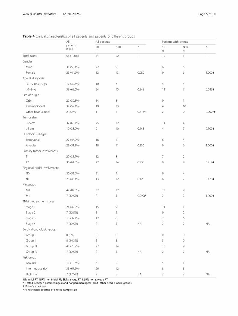
Table 4 Clinical characteristics of all patients and patients of different groups
Allpatientsn (%)
All patients Patients with events
IRTn
NIRTn
p SRTn
NSRTn
p
Total cases 56 (100%) 34 22 – 15 11 –
Gender
Male 31 (55.4%) 22 9 6 5
Female 25 (44.6%) 12 13 0.080 9 6 1.000#
Age at diagnosis
≤ 1 y or ≥ 10 ys 17 (30.4%) 10 7 4 4
>1–9 ys 39 (69.6%) 24 15 0.848 11 7 0.683#
Site of origin
Orbit 22 (39.3%) 14 8 9 1
Parameningeal 32 (57.1%) 19 13 4 10
Other head & neck 2 (3.6%) 1 1 0.813* 2 0 0.002*#
Tumor size
≤ 5 cm 37 (66.1%) 25 12 11 4
>5 cm 19 (33.9%) 9 10 0.143 4 7 0.109#
Histologic subtype
Embryonal 27 (48.2%) 16 11 6 5
Alveolar 29 (51.8%) 18 11 0.830 9 6 1.000#
Primary tumor invasiveness
T1 20 (35.7%) 12 8 7 2
T2 36 (64.3%) 22 14 0.935 8 9 0.217#
Regional nodal involvement
N0 30 (53.6%) 21 9 9 4
N1 26 (46.4%) 13 12 0.126 6 7 0.428#
Metastasis
M0 49 (87.5%) 32 17 13 9
M1 7 (12.5%) 2 5 0.099# 2 2 1.000#
TNM pretreatment stage
Stage 1 24 (42.9%) 15 9 11 1
Stage 2 7 (12.5%) 5 2 0 2
Stage 3 18 (32.1%) 12 6 2 6
Stage 4 7 (12.5%) 2 5 NA 2 2 NA
Surgical-pathologic group
Group I 0 (0%) 0 0 0 0
Group II 8 (14.3%) 5 3 3 0
Group III 41 (73.2%) 27 14 10 9
Group IV 7 (12.5%) 2 5 NA 2 2 NA
Risk group
Low risk 11 (19.6%) 6 5 5 1
Intermediate risk 38 (67.9%) 26 12 8 8
High risk 7 (12.5%) 2 5 NA 2 2 NA
IRT: initial RT; NIRT: non-initial RT; SRT: salvage RT; NSRT: non-salvage RT.*: Tested between parameningeal and nonparameningeal (orbit+other head & neck) groups#: Fisher’s exact testNA: not tested because of limited sample size
Wen et al. BMC Pediatrics (2020) 20:265 Page 5 of 10
statistically significant prognostic factor for EFS. Tumorsize is an important prognostic factor for survival inHNRMS, patients with smaller tumor (≤5 cm) have bet-ter survival compared with patients with larger tumor(>5 cm) [7, 23]. Tumor size > 5 cm has been widely re-ported as a risk factor for survival especially in refractorypatients (patients with disease progression) and recur-rent patients (patients with disease relapse) [24–27].These results are consistent with our result that tumorsize could predict OS in patients with or without events.And tumor size was the strongest predictor of local
failure [28], which is consistent with our results thattumor size could predict EFS over other factors.Our univariate analysis results showed that initial RT
was a statistical prognostic factor for both OS and EFSin all patients, but it was not for OS in patients withevents. This might indicate that initial RT could help toimprove OS by enhancing local control/ preventing localevents, but once events occurred, initial RT would be-come a low-weight prognostic factor. We speculated thatafter events, salvage RT took the place of initial RT andbecame a high-weight prognostic factor, and this specu-lation was supported by our results that salvage RT wasa statistically significant prognostic factor for OS inpatients with events. It’s reported that adequate localtherapy is an important factor for survival after relapse[29], RT can further improve OS in relapsed patientsundergone a repeat surgery [30], and local treatmentsuch as RT and repeat surgery should be systemicallyconsidered even in previously irradiated patients [31].The cure of parameningeal RMS is unlikely without RT[11]. In conclusion, our results showed that whether as a
Table 5 Pattern of events (disease progression and relapse).*
Disease Progression (n) Disease Relapse (n) Total
Local 12 9 21
Regional 0 1 1
Metastatic 0 1 1
Local+Metastatic 2 1 3
Total 14 12 26
*Only first progression and first relapse were analyzed
Fig. 1 Kaplan-Meier survival curve. A. OS of all patients. B. OS of different risk groups. C. EFS of all patients. D. OS of patients with events (diseaseprogression and relapse)
Wen et al. BMC Pediatrics (2020) 20:265 Page 6 of 10
component of initial or salvage treatment plan, RT couldimprove patients’ OS.In order to adjust confounding factors, further multi-
variate analyses were performed using Cox proportionalhazard model. First, tumor size was proved to be an in-dependent prognostic factor for OS in patients with orwithout events, which confirmed its important influenceon patients’ outcome. Second, initial RT was proved to
be an independent prognostic factor for OS and EFS,which confirmed it as a very important modality to im-prove OS by enhancing local control. Third, salvage RTwas not an independent prognostic factor for OS in pa-tients with events. In our cohort patients with events(disease progression and relapse) generally refers to re-fractory and recurrent patients. These patients’ survivalis affected by many factors and some of them may be
Table 6 Univariate analysis of prognostic factors for survival
Risk Factors All Patients Patients with events
5-y OS p 5-y EFS p 3-y OS p
Age at diagnosis
≤ 1 y or ≥ 10 ys 80.9% 49.6% 60.0%
>1–9 ys 66.1% 0.617 48.4% 0.809 48.9% 0.633
Site of origin
Parameningeal 56.6% 49.0% 39.3
Non-Parameningeal 81.9% 0.204 46.6% 0.903 65.6 0.094
Tumor size
≤ 5 cm 83.5% 56.0% 79.4%
>5 cm 40.3% < 0.001 30.1% 0.035* 10.4% < 0.001
Histologic subtype
Embryonal 74.6% 51.8% 35.8%(5-y OS)
Alveolar 65.5% 0.172 46.9% 0.339 36.4%(5-y OS)
0.203
Primary tumor invasiveness
T1 95% 49.5% 88.9%
T2 51.4% 0.013 50.0% 0.583 29.6% 0.006
Regional nodal involvement
N0 73.6% 51.4% 60.6%
N1 67.8% 0.330 40.8% 0.498 42.3% 0.481
Metastasis
M0 75.7% 51.2% 62.0%
M1 22.2% (3-y OS) < 0.001 25.0% (15-m EFS) 0.097 0.0% 0.005
Surgical-pathologic group
Group II 100% 62.5% 100%
Group III 66.8% 48.3% 55.6%
Group IV 22.2% (3-y OS) 0.001# 25.0% (15-m EFS) 0.198 0.0% 0.007#
Initial RT
Yes 80.3% 63.9% 35.8%
No 49.7% 0.003 21.9%(20-m EFS)
< 0.001 36.4% 0.178
Salvage RT
Yes – – 66.0%
No – – – – 31.2% 0.033
# Pairwise comparison (Bonferroni adjustment: p < 0.0167 is considered statistically significant)5-year OS of all patients: Group II vs Group III (p = 0.098), Group II vs Group IV (p = 0.003), Group III vs Group IV (p = 0.003)3-year OS of patients with events (disease progress and relapse): Group II vs Group III (p = 0.127), Group II vs Group IV (p = 0.018), Group III vs Group IV (p = 0.014)* Breslow test results
Wen et al. BMC Pediatrics (2020) 20:265 Page 7 of 10
higher-weight prognostic factors, such as tumor size, pri-mary site, regional nodal involvement, metastasis, repeatsurgery, previous chemo and RT plan, multi-relapse, etc.[3, 19, 27, 29, 32]. All these factors make salvage RT un-likely to be an independent prognostic factor.Over the last 3 decades with the continuous efforts
made by large cooperative groups, such as the COG SoftTissue Sarcoma Committee in North America, the Euro-pean pediatrics Soft Tissue Sarcoma Study Group(EpSSG), etc., the current 5-year OS for pediatric RMSpatients exceeds 70% [2, 3]. For patients with poor
prognosis, treatment failure is mainly due to local fail-ure, which refers to primary tumor progression or recur-rence, including the combination of local failure withregional nodal failure, and/or metastasis. The key to im-prove prognosis is to maintain local control. The firstthing to cure RMS is the eradication of the primarytumor, which is realized by surgery and/or RT, then atthe same time chemotherapy can eradicate micro re-sidual or disseminated tumor cells [2]. It’s reported thatpatients without RT as a component of the treatmentplan have a poor prognosis [33], and in HNRMS patients
Fig. 2 Kaplan-Meier survival curve of patients with or without RT. A. OS of Group IRT and Group NIRT (p = 0.003). B. EFS of Group IRT and NIRT(p < 0.001). C OS of Group SRT and NSRT (p = 0.033). D. OS of patients with events with or without initial RT (p = 0.178)
Table 7 Multivariate analysis results of Cox proportional model
Risk Factors OS (All Patients) EFS (All Patients) OS (Patients with Events)
HR 95% CIs p HR 95% CIs p HR 95% CIs p
Tumor size 5.06 1.17–21.93 0.030 1.92 0.86–4.27 0.110 5.61 1.12–28.06 0.036
Primary tumor invasiveness 4.59 0.51–41.13 0.173 – – – 5.96 0.62–54.59 0.114
Surgical-pathologic group 1.26 0.33–4.84 0.741 – – – 1.50 0.31–7.40 0.617
Initial RT 4.66 1.33–16.36 0.016 4.69 2.08–10.55 < 0.001 – – –
Salvage RT – – – – – – 2.06 0.54–7.79 0.289
Wen et al. BMC Pediatrics (2020) 20:265 Page 8 of 10

if the primary tumor is unresectable, RT and chemother-apy are the mainstay of initial treatment [11]. Here wecannot overemphasize the importance of RT in treatingpediatric HNRMS patients, and omitting RT may lead topoor prognosis.Despite the benefit of RT, about 40% (22/56) of our
patients’ parents initially rejected the adoption of RT fortheir children, which are generally due to two reasons:one is the concern about long-term morbidity relatedwith RT such as orbital hypoplasia, eye problems, andpituitary dysfunction, etc., the other is the fact that somekids show very good/complete response to initial chemoregiments, which enhanced parents’ confidence thatchemotherapy is reliable and capable of cure. Regardingthe two situations, we may consider introducing themless toxic RT modalities, such as proton radiotherapy,brachytherapy, etc., as well as adequately explaining thenecessity of RT, the risk of refusing it, and the limitedpredictive value of initial response to chemotherapy, toease their concern and enhance their confidence for RT.
LimitationsThis is a single-center historical cohort study with asmall sample size, but our uniform diagnostic and thera-peutic protocol could also be a strength. Our hospital isa tertiary center with domestically high-ranking ophthal-mology and otorhinolaryngology head & neck surgerydepartment, also the fact that we didn’t identify surgical-pathologic group I patients may indicate a selection bias.These factors may limit the generalizability of this study.We can’t acquire the PAX-FOXO1 fusion gene status innearly half of our alveolar patients, in order not to causefalse interpretations it was not analyzed in this study.But PAX-FOXO1 fusion gene status is absolutely a veryimportant prognostic factor and is widely reported [2,19, 20], not being able to analyze it could be a flaw ofthis study.
ConclusionsIn conclusion, RT as a component of initial treatmentcan improve the OS and EFS in pediatric HNRMS pa-tients by enhancing local control, and non-initial RT isan independent risk factor for OS and EFS. Salvage RTstill can improve OS in patients with disease progressionand relapse. Tumor size > 5 cm is an independent riskfactor for OS in pediatric HNRMS patients with or with-out disease progression/relapse.
AbbreviationsOS: Overall Survival; EFS: Event Free Survival; RMS: Rhabdomyosarcoma;RT: Radiation Therapy; HNRMS: Head and Neck Rhabdomyosarcoma;CT: Computed Tomography; PET-CT: Positron Emission Tomography-Computed Tomography; CSF: Cerebrospinal Fluid; COG: Children’s OncologyGroup; IRS: Intergroup Rhabdomyosarcoma Study; VAC: Vincristine,Dactinomycin, Cyclophosphamide.; PD: Disease Progression; RD: DiseaseRelapse; HR: Hazard Ratio; CI: Confidence Interval; Group IRT: Initial Radiation
Therapy Group; Group NIRT: Non-initial Radiation Therapy Group; GroupSRT: Salvage Radiation Therapy Group; Group NSRT: Non-salvage RadiationTherapy Group
AcknowledgementsNot applicable.
Authors’ ContubutionsYW designed the study, collected, analyzed and interpreted all the data, andcompleted the manuscript writing. DH initiated the study and participated indesigning, did the critical revision of the manuscript, and provided thefunding. WZ, YZ, HH, JL contributed in clinical data accumulation andcollection, and participated in study design. All authors read and approvedthe final manuscript.
FundingThis study was funded by the Special Fund of the Pediatric MedicalCoordinated Development Center of Beijing Hospitals Authority (No.XTZD20180203), and Beijing Hospitals Authority Mission Plan (Code:DFL20180201).
Availability of data and materialsNot applicable.
Ethics approval and consent to participateThis study was conducted in accordance with the declaration of Helsinki andwith approval from the Ethics Committee of Capital Medical University,Beijing Tongren Hospital. Written informed consent was obtained from allparticipants’ guardians.
Consent for publicationNot applicable.
Competing interestsThe authors declare that they have no competing interests.
Received: 25 April 2020 Accepted: 20 May 2020
References1. Amer KM, Thomson JE, Congiusta D, Dobitsch A, Chaudhry A, Li M,
Chaudhry A, Bozzo A, Siracuse B, Aytekin MN, et al. Epidemiology, incidence,and survival of Rhabdomyosarcoma subtypes: SEER and ICES databaseanalysis. J Orthop Res. 2019;37(10):2226–30.
2. Skapek SX, Ferrari A, Gupta AA, Lupo PJ, Butler E, Shipley J, Barr FG, HawkinsDS. Rhabdomyosarcoma. Nat Rev Dis Primers. 2019;5(1):1.
3. Chen C, Dorado Garcia H, Scheer M, Henssen AG. Current and futuretreatment strategies for Rhabdomyosarcoma. Front Oncol. 2019;9:1458.
4. Bisogno G, De Salvo GL, Bergeron C, Gallego Melcon S, Merks JH, Kelsey A,Martelli H, Minard-Colin V, Orbach D, Glosli H, et al. Vinorelbine andcontinuous low-dose cyclophosphamide as maintenance chemotherapy inpatients with high-risk rhabdomyosarcoma (RMS 2005): a multicentre, open-label, randomised, phase 3 trial. Lancet Oncol. 2019;20(11):1566–75.
5. Bisogno G, Jenney M, Bergeron C, Gallego Melcon S, Ferrari A, Oberlin O,Carli M, Stevens M, Kelsey A, De Paoli A, et al. Addition of dose-intensifieddoxorubicin to standard chemotherapy for rhabdomyosarcoma (EpSSG RMS2005): a multicentre, open-label, randomised controlled, phase 3 trial.Lancet Oncol. 2018;19(8):1061–71.
6. Yohe ME, Heske CM, Stewart E, Adamson PC, Ahmed N, Antonescu CR,Chen E, Collins N, Ehrlich A, Galindo RL, et al. Insights into pediatricrhabdomyosarcoma research: challenges and goals. Pediatr Blood Cancer.2019;66(10):e27869.
7. Casey DL, Chi YY, Donaldson SS, Hawkins DS, Tian J, Arndt CA, RodebergDA, Routh JC, Lautz TB, Gupta AA, et al. Increased local failure for patientswith intermediate-risk rhabdomyosarcoma on ARST0531: a report from theChildren's oncology group. Cancer. 2019;125(18):3242–8.
8. Vaarwerk B, Mallebranche C, Affinita MC, van der Lee JH, Ferrari A, ChisholmJC, Defachelles AS, De Salvo GL, Corradini N, Minard-Colin V, et al. Issurveillance imaging in pediatric patients treated for localizedrhabdomyosarcoma useful? The European experience. Cancer. 2020;126(4):823–31.
Wen et al. BMC Pediatrics (2020) 20:265 Page 9 of 10
9. Ermoian RP, Breneman J, Walterhouse DO, Chi YY, Meza J, Anderson J,Hawkins DS, Hayes-Jordan AA, Parham DM, Yock TI et al: 45 Gy is notsufficient radiotherapy dose for Group III orbital embryonalrhabdomyosarcoma after less than complete response to 12 weeks ofARST0331 chemotherapy: A report from the Soft Tissue Sarcoma Committeeof the Children's Oncology Group. Pediatr Blood Cancer 2017, 64(9).
10. Owosho AABCD, Huang SCM, Chen SM, Kashikar SD, Estilo CLD, WoldenSLM, Wexler LHM, Huryn JMD, Antonescu CRM. A clinicopathologic study ofhead and neck rhabdomyosarcomas showing FOXO1 fusion-positivealveolar and MYOD1-mutant sclerosing are associated with unfavorableoutcome. Oral Oncol. 2016;61:89–97.
11. Defachelles AS, Rey A, Oberlin O, Spooner D, Stevens MC. Treatment ofnonmetastatic cranial parameningeal rhabdomyosarcoma in childrenyounger than 3 years old: results from international society of pediatriconcology studies MMT 89 and 95. J Clin Oncol. 2009;27(8):1310–5.
12. Turner JH, Richmon JD. Head and neck rhabdomyosarcoma: a criticalanalysis of population-based incidence and survival data. Otolaryngol HeadNeck Surg. 2011;145(6):967–73.
13. Yang JC, Wexler LH, Meyers PA, Wolden SL. Parameningealrhabdomyosarcoma: outcomes and opportunities. Int J Radiat Oncol BiolPhys. 2013;85(1):e61–6.
14. Spalding AC, Hawkins DS, Donaldson SS, Anderson JR, Lyden E, LaurieF, Wolden SL, Arndt CA, Michalski JM. The effect of radiation timing onpatients with high-risk features of parameningeal rhabdomyosarcoma:an analysis of IRS-IV and D9803. Int J Radiat Oncol Biol Phys. 2013;87(3):512–6.
15. Malempati S, Hawkins DS. Rhabdomyosarcoma: review of the Children'soncology group (COG) soft-tissue sarcoma committee experience andrationale for current COG studies. Pediatr Blood Cancer. 2012;59(1):5–10.
16. Rudzinski ER, Anderson JR, Hawkins DS, Skapek SX, Parham DM, Teot LA.The World Health Organization classification of skeletal muscle tumors inpediatric Rhabdomyosarcoma: a report from the Children's oncology group.Arch Pathol Lab Med. 2015;139(10):1281–7.
17. Raney RB, Walterhouse DO, Meza JL, Andrassy RJ, Breneman JC, Crist WM,Maurer HM, Meyer WH, Parham DM, Anderson JR. Results of the intergroupRhabdomyosarcoma study group D9602 protocol, using vincristine anddactinomycin with or without cyclophosphamide and radiation therapy, fornewly diagnosed patients with low-risk embryonal rhabdomyosarcoma: areport from the soft tissue sarcoma Committee of the Children's oncologygroup. J Clin Oncol. 2011;29(10):1312–8.
18. Arndt CA, Stoner JA, Hawkins DS, Rodeberg DA, Hayes-Jordan AA, PaidasCN, Parham DM, Teot LA, Wharam MD, Breneman JC, et al. Vincristine,actinomycin, and cyclophosphamide compared with vincristine,actinomycin, and cyclophosphamide alternating with vincristine, topotecan,and cyclophosphamide for intermediate-risk rhabdomyosarcoma: children'soncology group study D9803. J Clin Oncol. 2009;27(31):5182–8.
19. Hibbitts E, Chi YY, Hawkins DS, Barr FG, Bradley JA, Dasgupta R, Meyer WH,Rodeberg DA, Rudzinski ER, Spunt SL, et al. Refinement of risk stratificationfor childhood rhabdomyosarcoma using FOXO1 fusion status in addition toestablished clinical outcome predictors: a report from the Children'soncology group. Cancer Med. 2019;8(14):6437–48.
20. Skapek SX, Anderson J, Barr FG, Bridge JA, Gastier-Foster JM, Parham DM,Rudzinski ER, Triche T, Hawkins DS. PAX-FOXO1 fusion status drivesunfavorable outcome for children with rhabdomyosarcoma: a children'soncology group report. Pediatr Blood Cancer. 2013;60(9):1411–7.
21. Gallego S, Zanetti I, Orbach D, Ranchere D, Shipley J, Zin A, Bergeron C, deSalvo GL, Chisholm J, Ferrari A, et al. Fusion status in patients with lymphnode-positive (N1) alveolar rhabdomyosarcoma is a powerful predictor ofprognosis: experience of the European Paediatric soft tissue sarcoma studygroup (EpSSG). Cancer. 2018;124(15):3201–9.
22. Lautz TB, Chi YY, Tian J, Gupta AA, Wolden SL, Routh JC, Casey DL,Dasgupta R, Hawkins DS, Rodeberg DA. Relationship between tumorresponse at therapy completion and prognosis in patients with group IIIrhabdomyosarcoma: a report from the Children's oncology group. Int JCancer. 2020.
23. Crist W, Gehan EA, Ragab AH, Dickman PS, Donaldson SS, Fryer C,Hammond D, Hays DM, Herrmann J, Heyn R, et al. The third intergroupRhabdomyosarcoma study. J Clin Oncol. 1995;13(3):610–30.
24. Mattke AC, Bailey EJ, Schuck A, Dantonello T, Leuschner I, Klingebiel T,Treuner J, Koscielniak E. Does the time-point of relapse influence outcomein pediatric rhabdomyosarcomas? Pediatr Blood Cancer. 2009;52(7):772–6.
25. Dantonello TM, Int-Veen C, Winkler P, Leuschner I, Schuck A, Schmidt BF,Lochbuehler H, Kirsch S, Hallmen E, Veit-Friedrich I, et al. Initial patientcharacteristics can predict pattern and risk of relapse in localizedrhabdomyosarcoma. J Clin Oncol. 2008;26(3):406–13.
26. Mazzoleni S, Bisogno G, Garaventa A, Cecchetto G, Ferrari A, Sotti G,Donfrancesco A, Madon E, Casula L, Carli M, et al. Outcomes and prognosticfactors after recurrence in children and adolescents with nonmetastaticrhabdomyosarcoma. Cancer. 2005;104(1):183–90.
27. Chisholm JC, Marandet J, Rey A, Scopinaro M, de Toledo JS, Merks JH, O'MearaA, Stevens MC, Oberlin O. Prognostic factors after relapse in nonmetastaticrhabdomyosarcoma: a nomogram to better define patients who can besalvaged with further therapy. J Clin Oncol. 2011;29(10):1319–25.
28. Wolden SL, Lyden ER, Arndt CA, Hawkins DS, Anderson JR, Rodeberg DA,Morris CD, Donaldson SS. Local control for intermediate-riskRhabdomyosarcoma: results from D9803 according to histology, group, site,and size: a report from the Children's oncology group. Int J Radiat OncolBiol Phys. 2015;93(5):1071–6.
29. Dantonello TM, Int-Veen C, Schuck A, Seitz G, Leuschner I, Nathrath M,Schlegel PG, Kontny U, Behnisch W, Veit-Friedrich I, et al. Survival followingdisease recurrence of primary localized alveolar rhabdomyosarcoma. PediatrBlood Cancer. 2013;60(8):1267–73.
30. De Corti F, Bisogno G, Dall'Igna P, Ferrari A, Buffa P, De Paoli A, Cecchetto G.Does surgery have a role in the treatment of local relapses of non-metastatic rhabdomyosarcoma? Pediatr Blood Cancer. 2011;57(7):1261–5.
31. Winter S, Fasola S, Brisse H, Mosseri V, Orbach D. Relapse after localizedrhabdomyosarcoma: evaluation of the efficacy of second-linechemotherapy. Pediatr Blood Cancer. 2015;62(11):1935–41.
32. Oberlin O, Rey A, Lyden E, Bisogno G, Stevens MC, Meyer WH, Carli M,Anderson JR. Prognostic factors in metastatic rhabdomyosarcomas: resultsof a pooled analysis from United States and European cooperative groups. JClin Oncol. 2008;26(14):2384–9.
33. Merks JH, De Salvo GL, Bergeron C, Bisogno G, De Paoli A, Ferrari A, Rey A,Oberlin O, Stevens MC, Kelsey A, et al. Parameningeal rhabdomyosarcomain pediatric age: results of a pooled analysis from north American andEuropean cooperative groups. Ann Oncol. 2014;25(1):231–6.
Publisher’s NoteSpringer Nature remains neutral with regard to jurisdictional claims inpublished maps and institutional affiliations.
Wen et al. BMC Pediatrics (2020) 20:265 Page 10 of 10