Embed Size (px)
Citation preview
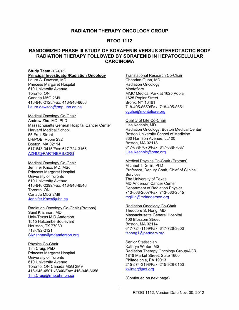
1 RTOG 1112, Version Date Nov. 30, 2012
RADIATION THERAPY ONCOLOGY GROUP
RTOG 1112 RANDOMIZED PHASE III STUDY OF SORAFENIB VERSUS STEREOTACTIC BODY
RADIATION THERAPY FOLLOWED BY SORAFENIB IN HEPATOCELLULAR CARCINOMA
Study Team (4/24/13) Principal Investigator/Radiation Oncology Laura A. Dawson, MD Princess Margaret Hospital 610 University Avenue Toronto, ON Canada M5G 2M9 416-946-2125/Fax: 416-946-6656 [email protected] Medical Oncology Co-Chair Andrew Zhu, MD, PhD Massachusetts General Hospital Cancer Center Harvard Medical School 55 Fruit Street LH/POB, Room 232 Boston, MA 02114 617-643-3415/Fax: 617-724-3166 [email protected] Medical Oncology Co-Chair Jennifer Knox, MD, MSc Princess Margaret Hospital University of Toronto 610 University Avenue 416-946-2399/Fax: 416-946-6546 Toronto, ON Canada M5G 2M9 [email protected] Radiation Oncology Co-Chair (Protons) Sunil Krishnan, MD Univ-Texas M D Anderson 1515 Holcombe Boulevard Houston, TX 77030 713-792-2121 [email protected] Physics Co-Chair Tim Craig, PhD Princess Margaret Hospital University of Toronto 610 University Avenue Toronto, ON Canada M5G 2M9 416-946-4501 x3340/Fax: 416-946-6656 [email protected]
Translational Research Co-Chair Chandan Guha, MD Radiation Oncology Montefiore MMC Medical Park at 1625 Poplar 1625 Poplar Street Bronx, NY 10461 718-405-8550/Fax: 718-405-8551 [email protected] Quality of Life Co-Chair Lisa Kachnic, MD Radiation Oncology, Boston Medical Center Boston University School of Medicine 830 Harrison Avenue, LL100 Boston, MA 02118 617-638-7070/Fax: 617-638-7037 [email protected] Medical Physics Co-Chair (Protons) Michael T. Gillin, PhD Professor, Deputy Chair, Chief of Clinical Services The University of Texas MD Anderson Cancer Center Department of Radiation Physics 713-563-2507/Fax: 713-563-2545 [email protected] Radiation Oncology Co-Chair Theodore S. Hong, MD Massachusetts General Hospital 100 Blossom Street Boston, MA 02114 617-724-1159/Fax: 617-726-3603 [email protected] Senior Statistician Kathryn Winter, MS Radiation Therapy Oncology Group/ACR 1818 Market Street, Suite 1600 Philadelphia, PA 19013 215-574-3198/Fax: 215-928-0153 [email protected] (Continued on next page)
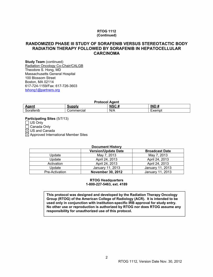
2 RTOG 1112, Version Date Nov. 30, 2012
RTOG 1112 (Continued)
RANDOMIZED PHASE III STUDY OF SORAFENIB VERSUS STEREOTACTIC BODY
RADIATION THERAPY FOLLOWED BY SORAFENIB IN HEPATOCELLULAR CARCINOMA
Study Team (continued) Radiation Oncology Co-Chair/CALGB Theodore S. Hong, MD Massachusetts General Hospital 100 Blossom Street Boston, MA 02114 617-724-1159/Fax: 617-726-3603 [email protected]
Protocol Agent Agent Supply NSC # IND # Sorafenib Commercial N/A Exempt Participating Sites (5/7/13)
US Only Canada Only US and Canada Approved International Member Sites
Document History Version/Update Date Broadcast Date
Update May 7, 2013 May 7, 2013 Update April 24, 2013 April 24, 2013
Activation April 24, 2013 April 24, 2013 Update January 11, 2013 January 11, 2013
Pre-Activation November 30, 2012 January 11, 2013
RTOG Headquarters 1-800-227-5463, ext. 4189
This protocol was designed and developed by the Radiation Therapy Oncology Group (RTOG) of the American College of Radiology (ACR). It is intended to be used only in conjunction with institution-specific IRB approval for study entry. No other use or reproduction is authorized by RTOG nor does RTOG assume any responsibility for unauthorized use of this protocol.
3 RTOG 1112, Version Date Nov. 30, 2012
(4/24/13)
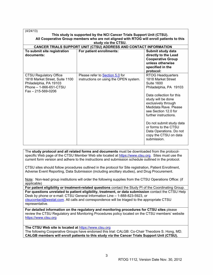
This study is supported by the NCI Cancer Trials Support Unit (CTSU). All Cooperative Group members who are not aligned with RTOG will enroll patients to this
study via the CTSU. CANCER TRIALS SUPPORT UNIT (CTSU) ADDRESS AND CONTACT INFORMATION
To submit site registration documents:
For patient enrollments: Submit study data directly to the Lead Cooperative Group unless otherwise specified in the protocol:
CTSU Regulatory Office 1818 Market Street, Suite 1100 Philadelphia, PA 19103 Phone – 1-866-651-CTSU Fax – 215-569-0206
Please refer to Section 5.0 for instructions on using the OPEN system.
RTOG Headquarters 1818 Market Street Suite 1600 Philadelphia, PA 19103 Data collection for this study will be done exclusively through Medidata Rave. Please see Section 12.0 for further instructions. Do not submit study data or forms to the CTSU Data Operations. Do not copy the CTSU on data submission.
The study protocol and all related forms and documents must be downloaded from the protocol-specific Web page of the CTSU Member Web site located at https://www.ctsu.org. Sites must use the current form version and adhere to the instructions and submission schedule outlined in the protocol. CTSU sites should follow procedures outlined in the protocol for Site registration, Patient Enrollment, Adverse Event Reporting, Data Submission (including ancillary studies), and Drug Procurement. Note: Non-lead group institutions will order the following supplies from the CTSU Operations Office: (if applicable) For patient eligibility or treatment-related questions contact the Study PI of the Coordinating Group. For questions unrelated to patient eligibility, treatment, or data submission contact the CTSU Help Desk by phone or e-mail: CTSU General Information Line – 1-888-823-5923, or [email protected]. All calls and correspondence will be triaged to the appropriate CTSU representative.
For detailed information on the regulatory and monitoring procedures for CTSU sites please review the CTSU Regulatory and Monitoring Procedures policy located on the CTSU members’ website https://www.ctsu.org
The CTSU Web site is located at https://www.ctsu.org The following Cooperative Groups have endorsed this trial: CALGB: Co-Chair Theodore S. Hong, MD. CALGB members will enroll patients to this study via the Cancer Trials Support Unit (CTSU).
4 RTOG 1112, Version Date Nov. 30, 2012
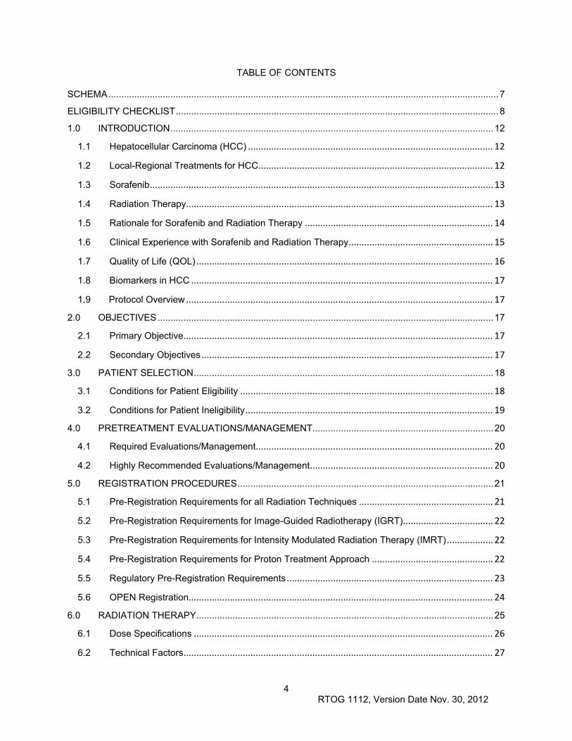
TABLE OF CONTENTS
SCHEMA ....................................................................................................................................................... 7
ELIGIBILITY CHECKLIST ............................................................................................................................. 8
1.0 INTRODUCTION ............................................................................................................................. 12
1.1 Hepatocellular Carcinoma (HCC) ............................................................................................... 12
1.2 Local-Regional Treatments for HCC ........................................................................................... 12
1.3 Sorafenib ..................................................................................................................................... 13
1.4 Radiation Therapy....................................................................................................................... 13
1.5 Rationale for Sorafenib and Radiation Therapy ......................................................................... 14
1.6 Clinical Experience with Sorafenib and Radiation Therapy ........................................................ 15
1.7 Quality of Life (QOL) ................................................................................................................... 16
1.8 Biomarkers in HCC ..................................................................................................................... 17
1.9 Protocol Overview ....................................................................................................................... 17
2.0 OBJECTIVES .................................................................................................................................. 17
2.1 Primary Objective ........................................................................................................................ 17
2.2 Secondary Objectives ................................................................................................................. 17
3.0 PATIENT SELECTION .................................................................................................................... 18
3.1 Conditions for Patient Eligibility .................................................................................................. 18
3.2 Conditions for Patient Ineligibility ................................................................................................ 19
4.0 PRETREATMENT EVALUATIONS/MANAGEMENT...................................................................... 20
4.1 Required Evaluations/Management............................................................................................ 20
4.2 Highly Recommended Evaluations/Management ....................................................................... 20
5.0 REGISTRATION PROCEDURES ................................................................................................... 21
5.1 Pre-Registration Requirements for all Radiation Techniques .................................................... 21
5.2 Pre-Registration Requirements for Image-Guided Radiotherapy (IGRT) ................................... 22
5.3 Pre-Registration Requirements for Intensity Modulated Radiation Therapy (IMRT) .................. 22
5.4 Pre-Registration Requirements for Proton Treatment Approach ............................................... 22
5.5 Regulatory Pre-Registration Requirements ................................................................................ 23
5.6 OPEN Registration...................................................................................................................... 24
6.0 RADIATION THERAPY ................................................................................................................... 25
6.1 Dose Specifications .................................................................................................................... 26
6.2 Technical Factors ........................................................................................................................ 27
5 RTOG 1112, Version Date Nov. 30, 2012
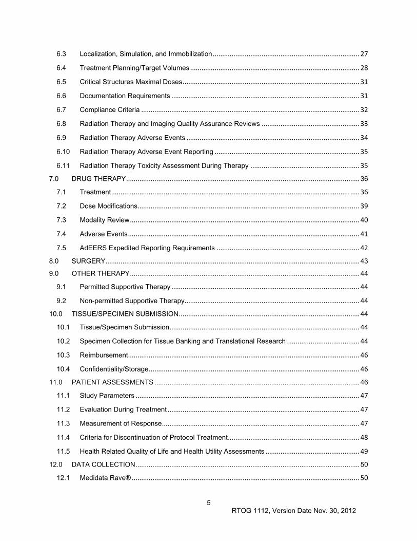
6.3 Localization, Simulation, and Immobilization .............................................................................. 27
6.4 Treatment Planning/Target Volumes .......................................................................................... 28
6.5 Critical Structures Maximal Doses .............................................................................................. 31
6.6 Documentation Requirements .................................................................................................... 31
6.7 Compliance Criteria .................................................................................................................... 32
6.8 Radiation Therapy and Imaging Quality Assurance Reviews .................................................... 33
6.9 Radiation Therapy Adverse Events ............................................................................................ 34
6.10 Radiation Therapy Adverse Event Reporting ............................................................................. 35
6.11 Radiation Therapy Toxicity Assessment During Therapy .......................................................... 35
7.0 DRUG THERAPY ............................................................................................................................ 36
7.1 Treatment .................................................................................................................................... 36
7.2 Dose Modifications...................................................................................................................... 39
7.3 Modality Review .......................................................................................................................... 40
7.4 Adverse Events ........................................................................................................................... 41
7.5 AdEERS Expedited Reporting Requirements ............................................................................ 42
8.0 SURGERY ....................................................................................................................................... 43
9.0 OTHER THERAPY .......................................................................................................................... 44
9.1 Permitted Supportive Therapy .................................................................................................... 44
9.2 Non-permitted Supportive Therapy ............................................................................................. 44
10.0 TISSUE/SPECIMEN SUBMISSION ................................................................................................ 44
10.1 Tissue/Specimen Submission ..................................................................................................... 44
10.2 Specimen Collection for Tissue Banking and Translational Research ....................................... 44
10.3 Reimbursement ........................................................................................................................... 46
10.4 Confidentiality/Storage ................................................................................................................ 46
11.0 PATIENT ASSESSMENTS ............................................................................................................. 46
11.1 Study Parameters ....................................................................................................................... 47
11.2 Evaluation During Treatment ...................................................................................................... 47
11.3 Measurement of Response ......................................................................................................... 47
11.4 Criteria for Discontinuation of Protocol Treatment ...................................................................... 48
11.5 Health Related Quality of Life and Health Utility Assessments .................................................. 49
12.0 DATA COLLECTION ....................................................................................................................... 50
12.1 Medidata Rave® ......................................................................................................................... 50
6 RTOG 1112, Version Date Nov. 30, 2012
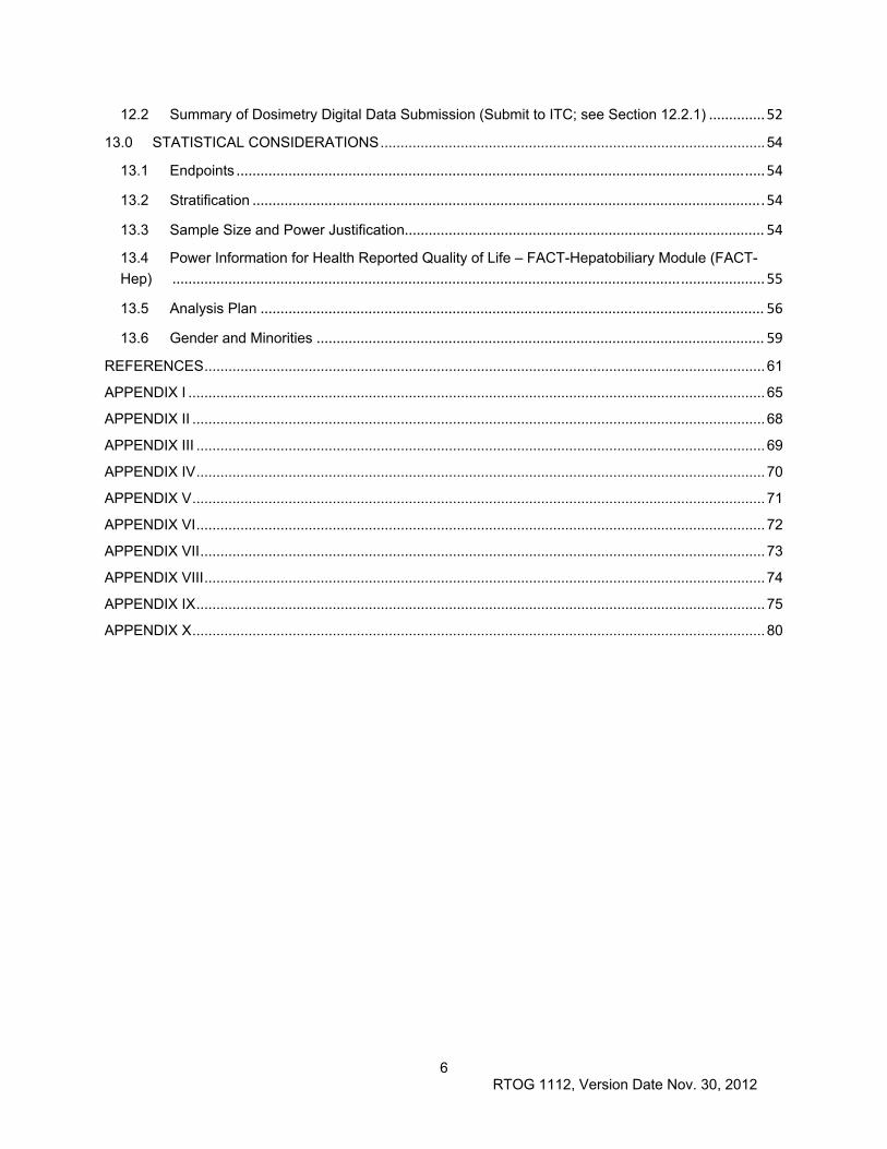
12.2 Summary of Dosimetry Digital Data Submission (Submit to ITC; see Section 12.2.1) .............. 52
13.0 STATISTICAL CONSIDERATIONS ................................................................................................ 54
13.1 Endpoints .................................................................................................................................... 54
13.2 Stratification ................................................................................................................................ 54
13.3 Sample Size and Power Justification.......................................................................................... 54
13.4 Power Information for Health Reported Quality of Life – FACT-Hepatobiliary Module (FACT-
Hep) .................................................................................................................................................... 55
13.5 Analysis Plan .............................................................................................................................. 56
13.6 Gender and Minorities ................................................................................................................ 59
REFERENCES ............................................................................................................................................ 61
APPENDIX I ................................................................................................................................................ 65
APPENDIX II ............................................................................................................................................... 68
APPENDIX III .............................................................................................................................................. 69
APPENDIX IV .............................................................................................................................................. 70
APPENDIX V ............................................................................................................................................... 71
APPENDIX VI .............................................................................................................................................. 72
APPENDIX VII ............................................................................................................................................. 73
APPENDIX VIII ............................................................................................................................................ 74
APPENDIX IX .............................................................................................................................................. 75
APPENDIX X ............................................................................................................................................... 80
7 RTOG 1112, Version Date Nov. 30, 2012
RADIATION THERAPY ONCOLOGY GROUP
RTOG 1112
Randomized Phase III Study of Sorafenib versus Stereotactic Body Radiation Therapy followed by Sorafenib in Hepatocellular Carcinoma
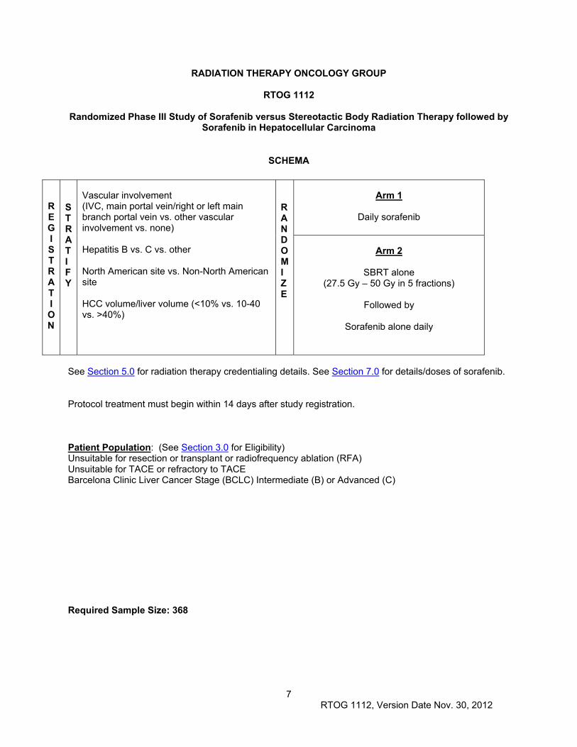
SCHEMA
R E G I S T R A T I O N
S T R A T I F Y
Vascular involvement (IVC, main portal vein/right or left main branch portal vein vs. other vascular involvement vs. none) Hepatitis B vs. C vs. other North American site vs. Non-North American site HCC volume/liver volume (<10% vs. 10-40 vs. >40%)
R A N D O MI Z E
Arm 1
Daily sorafenib
Arm 2
SBRT alone (27.5 Gy – 50 Gy in 5 fractions)
Followed by
Sorafenib alone daily
See Section 5.0 for radiation therapy credentialing details. See Section 7.0 for details/doses of sorafenib. Protocol treatment must begin within 14 days after study registration. Patient Population: (See Section 3.0 for Eligibility) Unsuitable for resection or transplant or radiofrequency ablation (RFA) Unsuitable for TACE or refractory to TACE Barcelona Clinic Liver Cancer Stage (BCLC) Intermediate (B) or Advanced (C) Required Sample Size: 368
8 RTOG 1112, Version Date Nov. 30, 2012
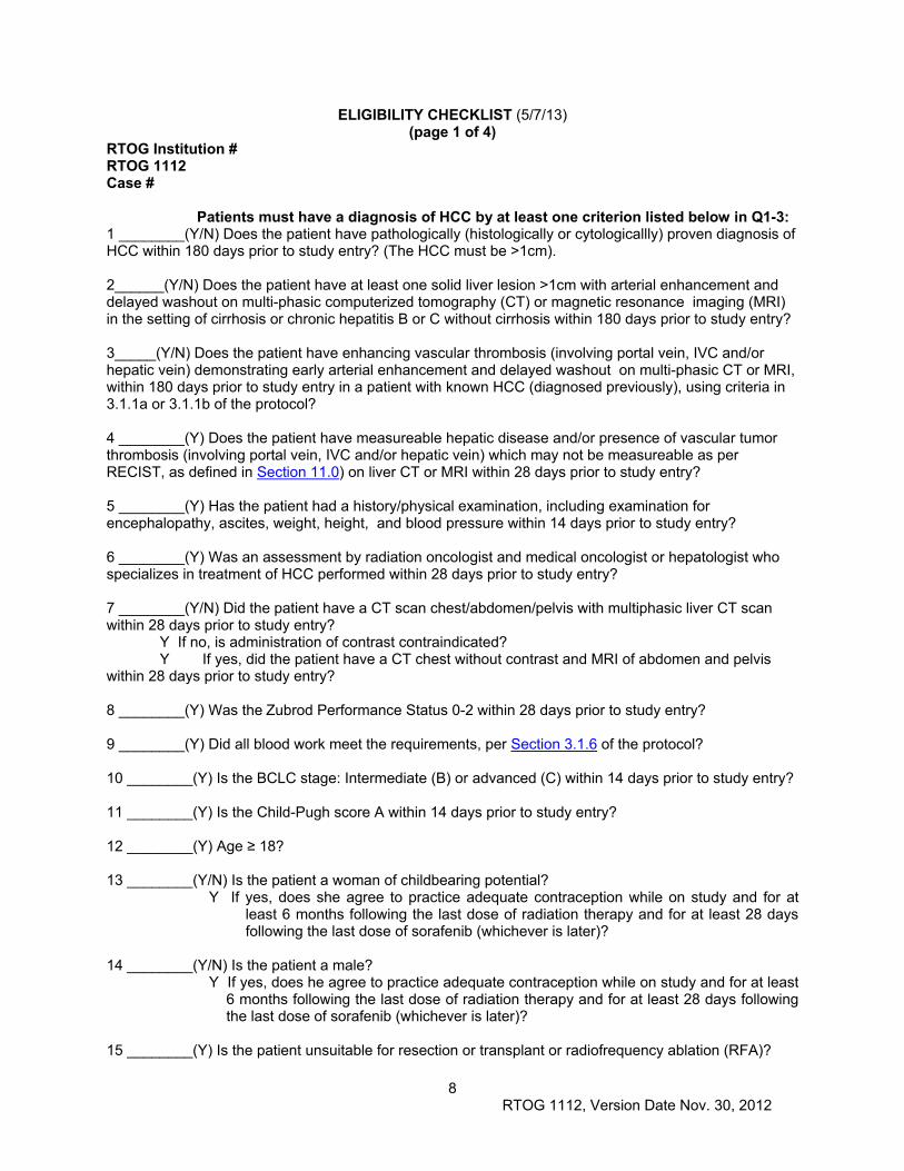
ELIGIBILITY CHECKLIST (5/7/13) (page 1 of 4)
RTOG Institution # RTOG 1112 Case # Patients must have a diagnosis of HCC by at least one criterion listed below in Q1-3: 1 ________(Y/N) Does the patient have pathologically (histologically or cytologicallly) proven diagnosis of HCC within 180 days prior to study entry? (The HCC must be >1cm). 2______(Y/N) Does the patient have at least one solid liver lesion >1cm with arterial enhancement and delayed washout on multi-phasic computerized tomography (CT) or magnetic resonance imaging (MRI) in the setting of cirrhosis or chronic hepatitis B or C without cirrhosis within 180 days prior to study entry? 3_____(Y/N) Does the patient have enhancing vascular thrombosis (involving portal vein, IVC and/or hepatic vein) demonstrating early arterial enhancement and delayed washout on multi-phasic CT or MRI, within 180 days prior to study entry in a patient with known HCC (diagnosed previously), using criteria in 3.1.1a or 3.1.1b of the protocol?
4 ________(Y) Does the patient have measureable hepatic disease and/or presence of vascular tumor thrombosis (involving portal vein, IVC and/or hepatic vein) which may not be measureable as per RECIST, as defined in Section 11.0) on liver CT or MRI within 28 days prior to study entry?
5 ________(Y) Has the patient had a history/physical examination, including examination for encephalopathy, ascites, weight, height, and blood pressure within 14 days prior to study entry?
6 ________(Y) Was an assessment by radiation oncologist and medical oncologist or hepatologist who specializes in treatment of HCC performed within 28 days prior to study entry? 7 ________(Y/N) Did the patient have a CT scan chest/abdomen/pelvis with multiphasic liver CT scan within 28 days prior to study entry? Y If no, is administration of contrast contraindicated? Y If yes, did the patient have a CT chest without contrast and MRI of abdomen and pelvis within 28 days prior to study entry?
8 ________(Y) Was the Zubrod Performance Status 0-2 within 28 days prior to study entry?
9 ________(Y) Did all blood work meet the requirements, per Section 3.1.6 of the protocol?
10 ________(Y) Is the BCLC stage: Intermediate (B) or advanced (C) within 14 days prior to study entry?
11 ________(Y) Is the Child-Pugh score A within 14 days prior to study entry?
12 ________(Y) Age ≥ 18?
13 ________(Y/N) Is the patient a woman of childbearing potential?
Y If yes, does she agree to practice adequate contraception while on study and for at least 6 months following the last dose of radiation therapy and for at least 28 days following the last dose of sorafenib (whichever is later)?
14 ________(Y/N) Is the patient a male?
Y If yes, does he agree to practice adequate contraception while on study and for at least 6 months following the last dose of radiation therapy and for at least 28 days following the last dose of sorafenib (whichever is later)?
15 ________(Y) Is the patient unsuitable for resection or transplant or radiofrequency ablation (RFA)?
9 RTOG 1112, Version Date Nov. 30, 2012
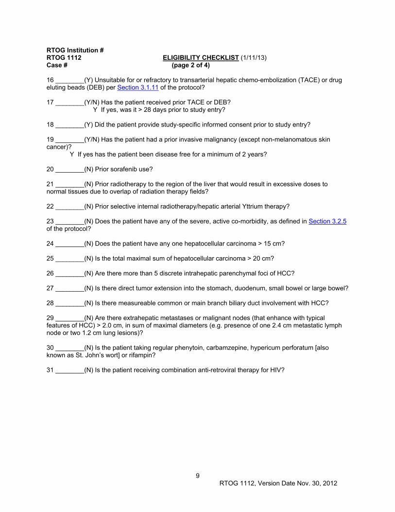
RTOG Institution # RTOG 1112 ELIGIBILITY CHECKLIST (1/11/13) Case # (page 2 of 4) 16 ________(Y) Unsuitable for or refractory to transarterial hepatic chemo-embolization (TACE) or drug eluting beads (DEB) per Section 3.1.11 of the protocol?
17 ________(Y/N) Has the patient received prior TACE or DEB?
Y If yes, was it > 28 days prior to study entry?
18 ________(Y) Did the patient provide study-specific informed consent prior to study entry?
19 ________(Y/N) Has the patient had a prior invasive malignancy (except non-melanomatous skin cancer)?
Y If yes has the patient been disease free for a minimum of 2 years?
20 ________(N) Prior sorafenib use?
21 ________(N) Prior radiotherapy to the region of the liver that would result in excessive doses to normal tissues due to overlap of radiation therapy fields?
22 ________(N) Prior selective internal radiotherapy/hepatic arterial Yttrium therapy?
23 ________(N) Does the patient have any of the severe, active co-morbidity, as defined in Section 3.2.5 of the protocol?
24 ________(N) Does the patient have any one hepatocellular carcinoma > 15 cm?
25 ________(N) Is the total maximal sum of hepatocellular carcinoma > 20 cm?
26 ________(N) Are there more than 5 discrete intrahepatic parenchymal foci of HCC?
27 ________(N) Is there direct tumor extension into the stomach, duodenum, small bowel or large bowel?
28 ________(N) Is there measureable common or main branch biliary duct involvement with HCC?
29 ________(N) Are there extrahepatic metastases or malignant nodes (that enhance with typical features of HCC) > 2.0 cm, in sum of maximal diameters (e.g. presence of one 2.4 cm metastatic lymph node or two 1.2 cm lung lesions)?
30 ________(N) Is the patient taking regular phenytoin, carbamzepine, hypericum perforatum [also known as St. John’s wort] or rifampin? 31 ________(N) Is the patient receiving combination anti-retroviral therapy for HIV?
10 RTOG 1112, Version Date Nov. 30, 2012
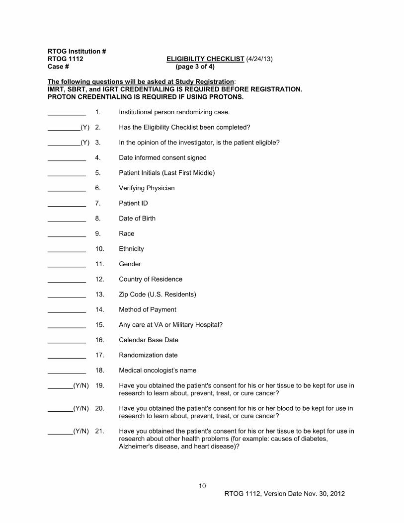
RTOG Institution # RTOG 1112 ELIGIBILITY CHECKLIST (4/24/13) Case # (page 3 of 4) The following questions will be asked at Study Registration: IMRT, SBRT, and IGRT CREDENTIALING IS REQUIRED BEFORE REGISTRATION. PROTON CREDENTIALING IS REQUIRED IF USING PROTONS. 1. Institutional person randomizing case. (Y) 2. Has the Eligibility Checklist been completed? (Y) 3. In the opinion of the investigator, is the patient eligible? 4. Date informed consent signed 5. Patient Initials (Last First Middle) 6. Verifying Physician 7. Patient ID 8. Date of Birth 9. Race 10. Ethnicity 11. Gender 12. Country of Residence 13. Zip Code (U.S. Residents) 14. Method of Payment 15. Any care at VA or Military Hospital? 16. Calendar Base Date 17. Randomization date 18. Medical oncologist’s name (Y/N) 19. Have you obtained the patient's consent for his or her tissue to be kept for use in
research to learn about, prevent, treat, or cure cancer? (Y/N) 20. Have you obtained the patient's consent for his or her blood to be kept for use in
research to learn about, prevent, treat, or cure cancer? (Y/N) 21. Have you obtained the patient's consent for his or her tissue to be kept for use in
research about other health problems (for example: causes of diabetes, Alzheimer's disease, and heart disease)?
11 RTOG 1112, Version Date Nov. 30, 2012
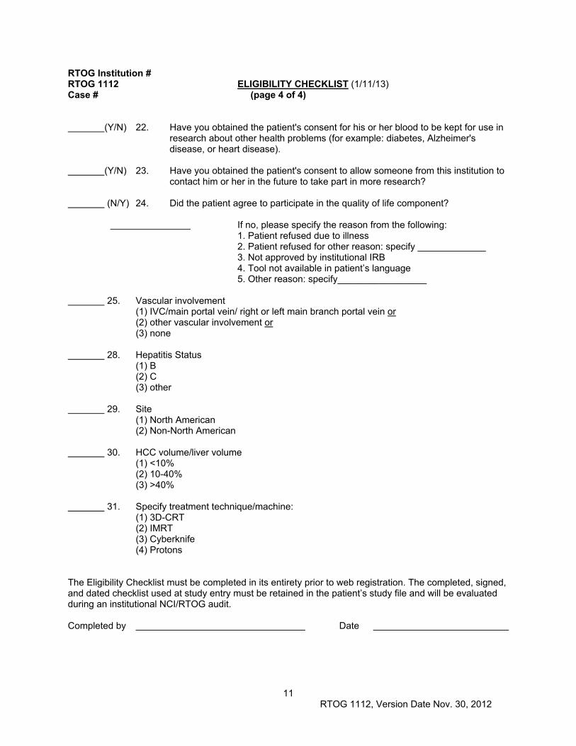
RTOG Institution # RTOG 1112 ELIGIBILITY CHECKLIST (1/11/13) Case # (page 4 of 4) (Y/N) 22. Have you obtained the patient's consent for his or her blood to be kept for use in
research about other health problems (for example: diabetes, Alzheimer's disease, or heart disease).
(Y/N) 23. Have you obtained the patient's consent to allow someone from this institution to contact him or her in the future to take part in more research? (N/Y) 24. Did the patient agree to participate in the quality of life component? If no, please specify the reason from the following: 1. Patient refused due to illness 2. Patient refused for other reason: specify _____________ 3. Not approved by institutional IRB 4. Tool not available in patient’s language 5. Other reason: specify_________________ 25. Vascular involvement (1) IVC/main portal vein/ right or left main branch portal vein or (2) other vascular involvement or (3) none 28. Hepatitis Status (1) B (2) C (3) other 29. Site (1) North American (2) Non-North American 30. HCC volume/liver volume (1) <10% (2) 10-40% (3) >40% 31. Specify treatment technique/machine: (1) 3D-CRT (2) IMRT (3) Cyberknife (4) Protons The Eligibility Checklist must be completed in its entirety prior to web registration. The completed, signed, and dated checklist used at study entry must be retained in the patient’s study file and will be evaluated during an institutional NCI/RTOG audit. Completed by Date
12 RTOG 1112, Version Date Nov. 30, 2012
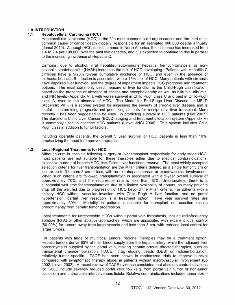
1.0 INTRODUCTION 1.1 Hepatocellular Carcinoma (HCC)
Hepatocellular carcinoma (HCC) is the fifth most common solid organ cancer and the third most common cause of cancer death globally, responsible for an estimated 600,000 deaths annually (Jemal 2010). Although HCC is less common in North America, the incidence has increased from 1.4 to 2.4 per 100,000 over the past two decades, and it is expected to continue to rise in parallel to the increasing incidence of Hepatitis C.
Cirrhosis, due to alcohol, viral hepatitis, autoimmune hepatitis, hemochromatosis, or non-alcoholic steatohepatitis (NASH) increases the risk of HCC developing. Patients with Hepatitis C cirrhosis have a 5-20% 5-year cumulative incidence of HCC, and even in the absence of cirrhosis, hepatitis B infection is associated with a 15% risk of HCC. Many patients with cirrhosis have impaired liver function, and the degree of impairment impacts HCC prognosis and treatment options. The most commonly used measure of liver function is the Child-Pugh classification, based on the presence or absence of ascites and encephalopathy as well as bilirubin, albumin, and INR levels (Appendix VII), with worse survival in Child Pugh class C and best in Child-Pugh class A, even in the absence of HCC. The Model for End-Stage Liver Disease, or MELD (Appendix VIII), is a scoring system for assessing the severity of chronic liver disease and is useful in determining prognosis and prioritizing patients for receipt of a liver transplant. More recently it has been suggested to be useful in predicting survival in HCC patients (Huo 2007). The Barcelona Clinic Liver Cancer (BCLC) staging and treatment allocation system (Appendix V) is commonly used to describe HCC patients (Llovet JNCI 2008). This system includes Child Pugh class in addition to tumor factors. Including operable patients, the overall 5 year survival of HCC patients is less than 10%, emphasizing the need for improved therapies.
1.2 Local-Regional Treatments for HCC Although cure is possible following surgery or liver transplant respectively for early stage HCC, most patients are not suitable for these therapies either due to medical contraindications, excessive burden of hepatic HCC, insufficient liver functional reserve. The most widely accepted selection criteria for liver transplantation are the Milan criteria defined as a single tumor 5 cm or less or up to 3 tumors 3 cm or less, with no extrahepatic spread or macrovascular involvement. When such criteria are followed, transplantation is associated with a 5-year overall survival of approximately 70%, and the recurrence rate is less than 15%. Unfortunately, there is a substantial wait time for transplantation due to a limited availability of donors, so many patients drop off the wait list due to progression of HCC beyond the Milan criteria. For patients with a solitary HCC without vascular invasion, with Child Pugh A liver function, and no portal hypertension, partial liver resection is a treatment option. Five year survival rates are approximately 50%. Mortality in patients unsuitable for transplant or resection results predominantly from hepatic tumor progression. Local treatments for unresectable HCCs without portal vein thrombosis, include radiofrequency ablation (RFA) or other ablative approaches, which are associated with excellent local control (80-90%) for tumors away from large vessels and less than 3 cm, with reduced local control for larger tumors. For patients with large or multifocal tumors, regional therapies may be a treatment option. Hepatic tumors derive 80% of their blood supply from the hepatic artery, while the adjacent liver parenchyma is supplied by the portal vein, making hepatic arterial directed therapies, such as transarterial chemoembolization (TACE), drug eluding beads (DEB) or radioembolization, relatively tumor specific. TACE has been shown in randomized trials to improve survival compared with symptomatic therapy alone, in patients without macrovascular involvement (Lo 2002, Llovet 2002). A recent review of TACE evidence concluded that absolute contraindications for TACE include severely reduced portal vein flow (e.g. from portal vein tumor or non-tumor occlusion) and untreatable arterial venous fistula. Relative contraindications included tumor size >
13 RTOG 1112, Version Date Nov. 30, 2012
10 cm. Patients with main portal vein thrombosis are not recommended to be treated with TACE (Raoul 2011). There is more controversy in patients with segmental portal vein invasion. The patients not suitable for TACE and/or with recurrent or refractory disease following TACE are the target HCC population for this study.
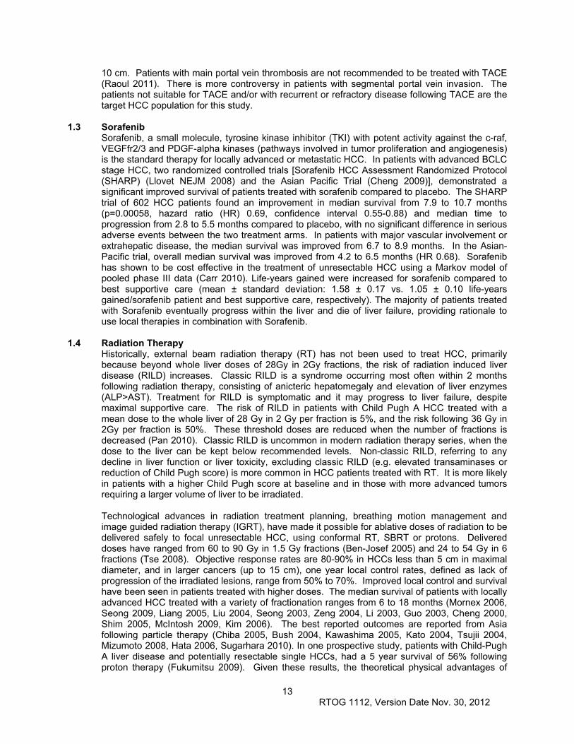
1.3 Sorafenib Sorafenib, a small molecule, tyrosine kinase inhibitor (TKI) with potent activity against the c-raf, VEGFfr2/3 and PDGF-alpha kinases (pathways involved in tumor proliferation and angiogenesis) is the standard therapy for locally advanced or metastatic HCC. In patients with advanced BCLC stage HCC, two randomized controlled trials [Sorafenib HCC Assessment Randomized Protocol (SHARP) (Llovet NEJM 2008) and the Asian Pacific Trial (Cheng 2009)], demonstrated a significant improved survival of patients treated with sorafenib compared to placebo. The SHARP trial of 602 HCC patients found an improvement in median survival from 7.9 to 10.7 months (p=0.00058, hazard ratio (HR) 0.69, confidence interval 0.55-0.88) and median time to progression from 2.8 to 5.5 months compared to placebo, with no significant difference in serious adverse events between the two treatment arms. In patients with major vascular involvement or extrahepatic disease, the median survival was improved from 6.7 to 8.9 months. In the Asian-Pacific trial, overall median survival was improved from 4.2 to 6.5 months (HR 0.68). Sorafenib has shown to be cost effective in the treatment of unresectable HCC using a Markov model of pooled phase III data (Carr 2010). Life-years gained were increased for sorafenib compared to best supportive care (mean ± standard deviation: 1.58 ± 0.17 vs. 1.05 ± 0.10 life-years gained/sorafenib patient and best supportive care, respectively). The majority of patients treated with Sorafenib eventually progress within the liver and die of liver failure, providing rationale to use local therapies in combination with Sorafenib.
1.4 Radiation Therapy Historically, external beam radiation therapy (RT) has not been used to treat HCC, primarily because beyond whole liver doses of 28Gy in 2Gy fractions, the risk of radiation induced liver disease (RILD) increases. Classic RILD is a syndrome occurring most often within 2 months following radiation therapy, consisting of anicteric hepatomegaly and elevation of liver enzymes (ALP>AST). Treatment for RILD is symptomatic and it may progress to liver failure, despite maximal supportive care. The risk of RILD in patients with Child Pugh A HCC treated with a mean dose to the whole liver of 28 Gy in 2 Gy per fraction is 5%, and the risk following 36 Gy in 2Gy per fraction is 50%. These threshold doses are reduced when the number of fractions is decreased (Pan 2010). Classic RILD is uncommon in modern radiation therapy series, when the dose to the liver can be kept below recommended levels. Non-classic RILD, referring to any decline in liver function or liver toxicity, excluding classic RILD (e.g. elevated transaminases or reduction of Child Pugh score) is more common in HCC patients treated with RT. It is more likely in patients with a higher Child Pugh score at baseline and in those with more advanced tumors requiring a larger volume of liver to be irradiated.
Technological advances in radiation treatment planning, breathing motion management and image guided radiation therapy (IGRT), have made it possible for ablative doses of radiation to be delivered safely to focal unresectable HCC, using conformal RT, SBRT or protons. Delivered doses have ranged from 60 to 90 Gy in 1.5 Gy fractions (Ben-Josef 2005) and 24 to 54 Gy in 6 fractions (Tse 2008). Objective response rates are 80-90% in HCCs less than 5 cm in maximal diameter, and in larger cancers (up to 15 cm), one year local control rates, defined as lack of progression of the irradiated lesions, range from 50% to 70%. Improved local control and survival have been seen in patients treated with higher doses. The median survival of patients with locally advanced HCC treated with a variety of fractionation ranges from 6 to 18 months (Mornex 2006, Seong 2009, Liang 2005, Liu 2004, Seong 2003, Zeng 2004, Li 2003, Guo 2003, Cheng 2000, Shim 2005, McIntosh 2009, Kim 2006). The best reported outcomes are reported from Asia following particle therapy (Chiba 2005, Bush 2004, Kawashima 2005, Kato 2004, Tsujii 2004, Mizumoto 2008, Hata 2006, Sugarhara 2010). In one prospective study, patients with Child-Pugh A liver disease and potentially resectable single HCCs, had a 5 year survival of 56% following proton therapy (Fukumitsu 2009). Given these results, the theoretical physical advantages of
14 RTOG 1112, Version Date Nov. 30, 2012
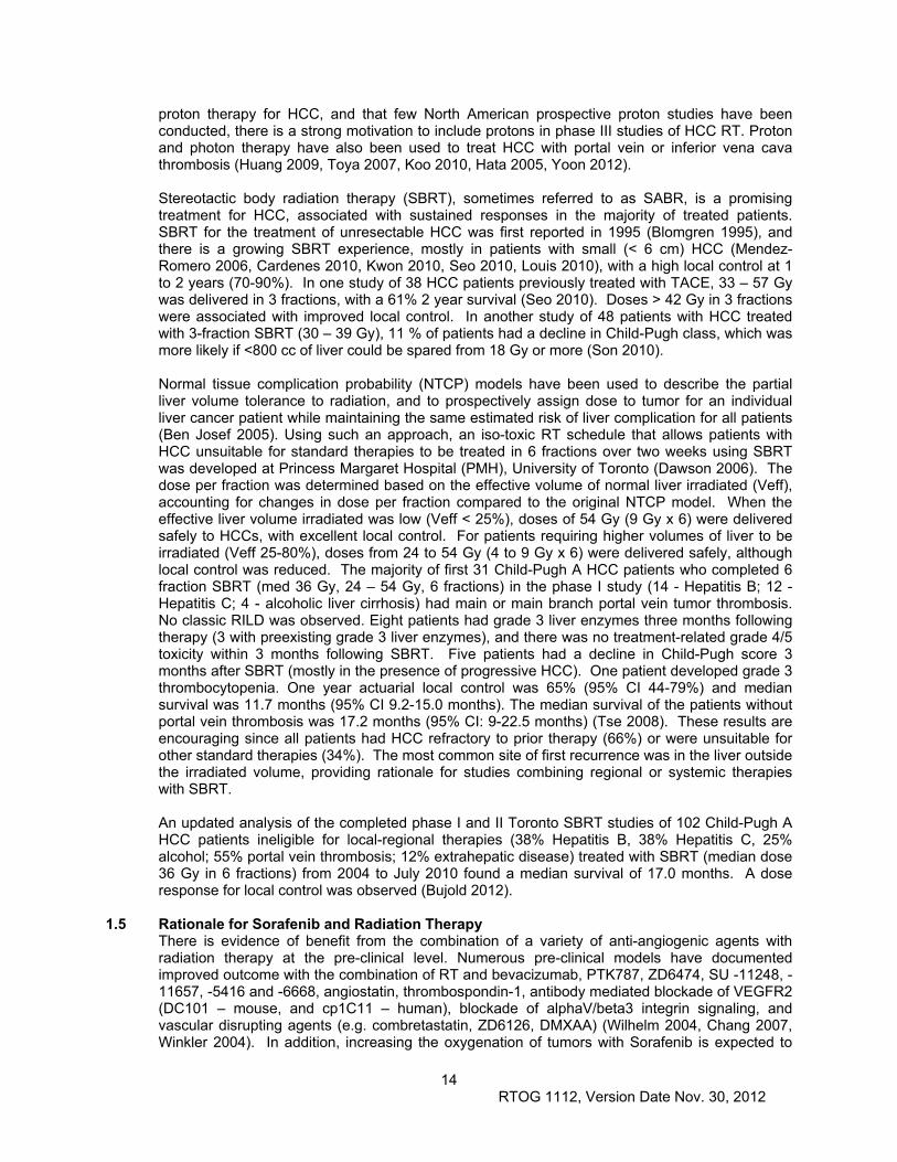
proton therapy for HCC, and that few North American prospective proton studies have been conducted, there is a strong motivation to include protons in phase III studies of HCC RT. Proton and photon therapy have also been used to treat HCC with portal vein or inferior vena cava thrombosis (Huang 2009, Toya 2007, Koo 2010, Hata 2005, Yoon 2012).
Stereotactic body radiation therapy (SBRT), sometimes referred to as SABR, is a promising treatment for HCC, associated with sustained responses in the majority of treated patients. SBRT for the treatment of unresectable HCC was first reported in 1995 (Blomgren 1995), and there is a growing SBRT experience, mostly in patients with small (< 6 cm) HCC (Mendez-Romero 2006, Cardenes 2010, Kwon 2010, Seo 2010, Louis 2010), with a high local control at 1 to 2 years (70-90%). In one study of 38 HCC patients previously treated with TACE, 33 – 57 Gy was delivered in 3 fractions, with a 61% 2 year survival (Seo 2010). Doses > 42 Gy in 3 fractions were associated with improved local control. In another study of 48 patients with HCC treated with 3-fraction SBRT (30 – 39 Gy), 11 % of patients had a decline in Child-Pugh class, which was more likely if <800 cc of liver could be spared from 18 Gy or more (Son 2010).
Normal tissue complication probability (NTCP) models have been used to describe the partial liver volume tolerance to radiation, and to prospectively assign dose to tumor for an individual liver cancer patient while maintaining the same estimated risk of liver complication for all patients (Ben Josef 2005). Using such an approach, an iso-toxic RT schedule that allows patients with HCC unsuitable for standard therapies to be treated in 6 fractions over two weeks using SBRT was developed at Princess Margaret Hospital (PMH), University of Toronto (Dawson 2006). The dose per fraction was determined based on the effective volume of normal liver irradiated (Veff), accounting for changes in dose per fraction compared to the original NTCP model. When the effective liver volume irradiated was low (Veff < 25%), doses of 54 Gy (9 Gy x 6) were delivered safely to HCCs, with excellent local control. For patients requiring higher volumes of liver to be irradiated (Veff 25-80%), doses from 24 to 54 Gy (4 to 9 Gy x 6) were delivered safely, although local control was reduced. The majority of first 31 Child-Pugh A HCC patients who completed 6 fraction SBRT (med 36 Gy, 24 – 54 Gy, 6 fractions) in the phase I study (14 - Hepatitis B; 12 - Hepatitis C; 4 - alcoholic liver cirrhosis) had main or main branch portal vein tumor thrombosis. No classic RILD was observed. Eight patients had grade 3 liver enzymes three months following therapy (3 with preexisting grade 3 liver enzymes), and there was no treatment-related grade 4/5 toxicity within 3 months following SBRT. Five patients had a decline in Child-Pugh score 3 months after SBRT (mostly in the presence of progressive HCC). One patient developed grade 3 thrombocytopenia. One year actuarial local control was 65% (95% CI 44-79%) and median survival was 11.7 months (95% CI 9.2-15.0 months). The median survival of the patients without portal vein thrombosis was 17.2 months (95% CI: 9-22.5 months) (Tse 2008). These results are encouraging since all patients had HCC refractory to prior therapy (66%) or were unsuitable for other standard therapies (34%). The most common site of first recurrence was in the liver outside the irradiated volume, providing rationale for studies combining regional or systemic therapies with SBRT.
An updated analysis of the completed phase I and II Toronto SBRT studies of 102 Child-Pugh A HCC patients ineligible for local-regional therapies (38% Hepatitis B, 38% Hepatitis C, 25% alcohol; 55% portal vein thrombosis; 12% extrahepatic disease) treated with SBRT (median dose 36 Gy in 6 fractions) from 2004 to July 2010 found a median survival of 17.0 months. A dose response for local control was observed (Bujold 2012).
1.5 Rationale for Sorafenib and Radiation Therapy There is evidence of benefit from the combination of a variety of anti-angiogenic agents with radiation therapy at the pre-clinical level. Numerous pre-clinical models have documented improved outcome with the combination of RT and bevacizumab, PTK787, ZD6474, SU -11248, -11657, -5416 and -6668, angiostatin, thrombospondin-1, antibody mediated blockade of VEGFR2 (DC101 – mouse, and cp1C11 – human), blockade of alphaV/beta3 integrin signaling, and vascular disrupting agents (e.g. combretastatin, ZD6126, DMXAA) (Wilhelm 2004, Chang 2007, Winkler 2004). In addition, increasing the oxygenation of tumors with Sorafenib is expected to
15 RTOG 1112, Version Date Nov. 30, 2012
improve the therapeutic ratio of radiation therapy to HCC. Sorafenib possesses dual antitumor activity by inhibiting the MAPK/ERK pathway and inhibiting neovascularization (Jain 2000). Sorafenib has been shown to inhibit proliferation and induce apoptosis in two HCC lines in vitro while also inhibiting tumor growth in an in vivo model (Liu 2006).
Another publication assessed combination treatment in a number of cell lines in vitro and HCT116 human colorectal xenografts in a subcutaneous flank model in nude mice (Plastaras 2007). Their data show that radiation followed by sorafenib appears to result in optimal anti-cancer effect compared to the concurrent administration of pre-treatment with sorafenib.
1.6 Clinical Experience with Sorafenib and Radiation Therapy Although there is rationale to combine local therapies with sorafenib in HCC, there are few clinical publications on the combination of Sorafenib or similar agents with RT. One retrospective review of 23 patients from Taiwan with advanced HCC treated with RT and sunitinib (a TKI with similar mechanisms as sorafenib) has been published (Chi 2010). Sixty percent of patients had two or more lesions and 22% had extrahepatic disease. All were unresectable and unsuitable for transhepatic chemo-embolization (TACE). Five patients had major portal vein thrombosis. Fifteen patients had Child-Pugh score A; 8 were Child-Pugh B. All patients received sunitinib (25 mg) at least 1 week before, during, and 2 weeks after radiation therapy. Thirteen patients continued maintenance sunitinib after RT until disease progression. The median radiation dose was 52.5 Gy in 15 fractions. The objective response rate was 74%. The 1-year survival rate was 70%, with a median survival of 16 months. Maintenance sunitinib was the most significant factor for survival. The time to progression was 10 months in the maintenance group compared with 4 months in the control group. There were three episodes of upper gastrointestinal bleeding and one episode of pancreatitis. Ten patients had grade 2 or more elevation of liver enzymes, and 15 developed grade 2 or more thrombocytopenia. The authors concluded that conformal hypofractionated RT and sunitinib can be delivered safely in HCC patients.
Another phase I study investigated concurrent sunitinib (25 – 35.7 mg) and 10 fraction conformal radiation therapy (40 – 50 Gy in 10 fractions) in 21 patients with 36 sites of oligometastases in various locations, including the liver (n=9). No dose limiting toxicity was seen when sunitinib was delivered prior to, during and following RT (Chi 2010).
Phase I studies of sorafenib and RT for liver cancer have been conducted at PMH, Toronto. In one phase I study of 30 Gy in 10 fractions combined with escalating dose sorafenib prior to, during, and following RT, no dose limiting toxicity (DLT) was observed in patients with locally advanced HCC, several with massive portal vein thrombosis (verbal communication, A Brade, PMH Toronto, January 2012). Two other phase I studies of six-fraction SBRT plus escalating dose sorafenib were conducted at PMH (Dawson, 2012; Brade 2012). One study was for patients with liver metastases, and the other was for patients with HCC. Both studies combined SBRT (6 fraction) with sorafenib delivered 7 days pre-RT, during RT and post RT (1 week for metastases and continuous for HCC), to maximize RT sensitization by increasing tumor oxygenation, to increase the antitumor activity via the MAPK/ERK pathway and by inhibiting neovascularization that may occur post RT. Fifteen patients with focal liver metastases were evaluable for toxicity (3 at dose level 200 mg po bid, 6 at dose level 600 mg po od and 6 at 800 mg po od for 4 weeks), with no DLT. Twelve evaluable patients with HCC were treated on study, with continued sorafenib post SBRT. There was no DLT in three evaluable HCC patients treated with SBRT with a low effective liver volume (Veff 30%) combined with 400 mg sorafenib po od. In patients with a liver Veff of 30-60%, 2 of 3 evaluable patients treated with sorafenib 400 mg po daily developed DLT (grade 3 small bowel obstruction and grade 3 GI bleed); thus sorafenib was de-escalated to 200 mg po daily. One of 6 evaluable patients at this dose level developed DLT (tumor rupture). For the present study, the maximal permitted RT doses to the normal tissues have been reduced, compared to the above studies, and sorafenib will be delivered following RT (rather than concurrently with RT), to reduce the risk of toxicity.
16 RTOG 1112, Version Date Nov. 30, 2012
1.7 Quality of Life (QOL) 1.7.1 QOL Overview
Quality of life (QOL) in HCC is understudied, but clearly of importance due to the expected poor overall survival in patients with advanced HCC, the co-morbidities that exist in these patients, the near universal presence of underlying liver disease and the potential for serious toxicity to occur from treatment. There are few published prospective studies using validated questionnaires to assess longitudinal QOL in patients with HCC receiving local or systemic therapies. Ringash et al reported (in abstract) on prospective QOL assessment in liver metastases and HCC patients receiving SBRT using the FACT-Hep (Ringash 2008). In this phase I/II study of SBRT for unresectable liver cancers (35% HCC), QOL using the Functional Assessment of Cancer Therapy–Hepatobiliary (FACT-Hep) and European Organization for Research and Treatment of Cancer Core Quality of Life Questionnaire (EORTC QLQ-C30) was collected at baseline, and at 1, 3 and 6 months post-treatment. Following SBRT, there was a trend for a decrease in QOL at 3 months; however, QOL at 6 months recovered in patients who were alive at that timepoint, suggesting a possible beneficial effect. Due to the paucity of QOL data in HCC and the potential benefit of localized SBRT on QOL, it will be important to measure differences in health-related QOL in HCC patients treated with Sorafenib as compared to SBRT followed by Sorafenib on this trial. If SBRT is associated with a sustained reduction in the burden of HCC compared to sorafenib alone, it may lead to improved QOL compared to sorafenib alone.
1.7.2 Functional Assessment of Cancer Therapy–Hepatobiliary (FACT-Hep) The FACT-Hep version 4 questionnaire will be used to measure QOL. The FACT-Hep is a 45-item self-report instrument designed to measure health-related quality of life (HRQL) in patients with hepatobiliary cancers. The FACT-Hep is validated and presents good internal consistency, test–retest reliability, and convergent and discriminate validity in patients with hepatobiliary cancer and HCC (Heffernan 2002, Steel 2006, Wang 2007, Steel 2004). The validity of FACT-Hep has recently been examined in a randomized controlled trial of an EGFRi or placebo (Cella 2012) In this study, FACT-Hep scores showed significant decline for progressive disease versus stable disease (e.g. difference in FACT-Hep total score -12.58; p = 0.004).
1.7.3 EuroQol (EQ-5D) Patient-reported outcomes (PROs) are increasingly being incorporated into clinical trials for documentation of effects of treatment not measured by traditional endpoints, such as overall survival. This is important with interventions that may increase treatment-related side effects without positively impacting survival. Quality-adjusted survival is an endpoint that incorporates a patient’s utility or preference of the health state that is combined with the time spent in that health state (Glasziou 1990).
The resultant is a quality-adjusted life-year (QALY). Utility can be measured by different methods including Standard Gamble, Time Trade-Off, and Health Utilities Index III. The EuroQol (EQ-5D) is another instrument for measuring utilities. It is a 2-part questionnaire that takes the patient approximately 5 minutes to complete (Schultz 2002). The first part consists of 5 items covering 5 dimensions, including: mobility, self-care, usual activities, pain/discomfort, and anxiety/depression. Each dimension can be graded on 3 levels including: 1-no problem, 2-moderate problems, and 3-extreme problems. There are 243 potential health states. The second part is a visual analogue scale (VAS) valuing current health state, measured on a 20 cm, 10 point-interval scale. Either the index score or the VAS score can be used in the quality-adjusted survival analysis (Wu 2002). The benefit of measuring quality-adjusted survival is that it can be compared to the outcomes of other interventions across disease sites and can be used by health policy makers to rank interventions.
The EQ-5D will be used to evaluate the effect of the addition of SBRT to sorafenib on quality-adjusted survival in this trial.
17 RTOG 1112, Version Date Nov. 30, 2012
1.8 Biomarkers in HCC 1.8.1 Liver Toxicity
The possibility for liver toxicity to occur following therapy for HCC limits the effectiveness of therapies for HCC, especially for patients with locally advanced HCC. Sinusoidal obstructive syndrome is thought to be an important component of radiation induced liver disease; however the exact pathophysiology has not been clearly elucidated. As children who develop veno-occlusive disease (VOD) following transplant develop significant increases in plasminogen activator inhibitor type I, tissue plasminogen activator, and D-dimer and significant decreases in prothrombin time, antithrombin, and α2-antiplasmin at the time of their clinical diagnosis of veno-occlusive disease (VOD), such factors may be useful for better understanding radiation (or sorafenib) induced liver sinusoidal obstructive syndrome related toxicity. In addition, transforming growth factor- ß is an important cytokine associated with tissue injury and wound healing and may be associated with non-specific liver disease, including cirrhosis, chronic hepatitis, or toxicity, from sorafenib or radiation. Other cytokines participate in the response to tissue injury, including proinflammatory cytokines IL-1-beta, IL-6, and tumor necrosis factor alpha. Baseline levels and temporal variations in levels of these cytokines may provide insight to liver toxicity pathogenesis, and may also be related to patient reported fatigue and decline in QOL.
1.8.2 Prognostic Factors HCC specimen microvessel density (MVD), pERK (marker of signal transduction), VEGFR-2 (marker of angiogenesis) and Ki-67 and MIB-1 (markers for proliferation) are potential prognostic markers in HCC. Circulating VEGF, soluble sVEGFR-s, Ang-1, Ang-2, PDGF, and sc-Kit have been correlated with sorafenib treatment response. Investigating changes in such potential biomarkers in a randomized trial may help to validate which biomarkers are most treatment predictive and/or prognostic.
1.9 Protocol Overview A randomized phase III study of sorafenib versus SBRT followed by sorafenib for locally advanced HCC (unsuitable for or refractory to surgery, RFA or TACE) is proposed. It is expected that the primary patient population will have BCLC stage C HCC, due primarily to tumor vascular thrombosis. The sequential timing of treatments in the experimental arm (SBRT followed by sorafenib), rather than concurrent sorafenib and SBRT, should reduce the risk of toxicity. The dose of sorafenib during the first 28 days following SBRT is half standard dose (200 mg po bid) based on the Toronto phase I experience to reduce potential increase in toxicity due to radiation sensitization that may occur during that time period following SBRT. The primary endpoint is overall survival, and the hypothesis is that SBRT followed by sorafenib will improve survival in HCC patients by improving hepatic and vascular control of HCC, compared to sorafenib alone.
2.0 OBJECTIVES 2.1 Primary Objective 2.1.1 To determine if SBRT improves overall survival in HCC patients treated with Sorafenib 2.2 Secondary Objectives 2.2.1 To determine the difference in time to progression (TTP) and progression-free survival (PFS) in
HCC patients treated with Sorafenib compared to SBRT followed by Sorafenib 2.2.2 To measure differences in toxicity in HCC patients treated with Sorafenib versus SBRT followed
by Sorafenib 2.2.3 To measure vascular thrombosis response post Sorafenib versus SBRT followed by Sorafenib 2.2.4 To measure differences in Health Related QOL and quality-adjusted survival in HCC patients
treated with Sorafenib compared to SBRT followed by Sorafenib 2.2.5 Collection of biospecimens for future correlative studies to investigate differences in potential
biomarkers in patients treated with Sorafenib versus SBRT followed by Sorafenib
18 RTOG 1112, Version Date Nov. 30, 2012
3.0 PATIENT SELECTION NOTE: PER NCI GUIDELINES, EXCEPTIONS TO ELIGIBILITY ARE NOT PERMITTED
3.1 Conditions for Patient Eligibility (5/7/13) 3.1.1 Patients must have a diagnosis of HCC by at least one criterion listed below ≤180 days prior to
study entry a) Pathologically (histologically or cytologically) proven diagnosis of HCC. b) At least one solid liver lesion or vascular tumor thrombosis (involving portal vein, IVC
and/or hepatic vein) > 1 cm with arterial enhancement and delayed washout on multi-phasic computerized tomography (CT) or magnetic resonance imaging (MRI) in the setting of cirrhosis or chronic hepatitis B or C without cirrhosis.
c) For patients whose CURRENT disease is vascular only: Enhancing vascular thrombosis (involving portal vein, IVC and/or hepatic vein) demonstrating early arterial enhancement and delayed washout on multi-phasic CT or MRI, in a patient with known HCC (diagnosed previously), using criteria in 3.1.1a or 3.1.1b
3.1.2 Measureable hepatic disease and/or presence of vascular tumor thrombosis (involving portal vein, IVC and/or hepatic vein) which may not be measureable as per RECIST, as defined in Section 11.0) on liver CT or MRI, within 28 days of registration
3.1.3 Appropriate for protocol entry based upon the following minimum diagnostic workup: History/physical examination including examination for encephalopathy, ascites,
weight, height, and blood pressure within 14 days prior to study entry Assessment by radiation oncologist and medical oncologist or hepatologist who
specializes in treatment of HCC within 28 days prior to study entry Pre-randomization Scan (REQUIRED for All Patients): CT scan
chest/abdomen/pelvis with multiphasic liver CT scan within 28 days prior to study entry. If CT contrast is contraindicated, CT chest without contrast and MRI of abdomen and pelvis is permitted. See Appendix V and Section 4.1.7 for details.
3.1.4 Zubrod Performance Status 0-2 within 28 days prior to study entry 3.1.5 Age ≥ 18 3.1.6 All blood work obtained within 14 days prior to study entry with adequate organ marrow function
defined as follows: Absolute neutrophil count (ANC) ≥ 1,500 cells/mm3 Platelets ≥ 70,000 cells/mm3 Hemoglobin ≥ 8.0 g/dl (Note: The use of transfusion or other intervention to
achieve Hgb ≥ 8.0 g/dl is acceptable.) Total bilirubin < 2 mg/dL Prothrombin time/INR < 1.7 Albumin ≥ 28 g/L AST and ALT < 6 times ULN Serum creatinine ≤ 1.5 x ULN or creatinine clearance ≥ 60 mL/min
3.1.7 BCLC stage: Intermediate (B) or advanced (C) within 14 days prior to study entry 3.1.8 Child-Pugh score A within 14 days prior to study entry 3.1.9 Women of childbearing potential and male participants must agree to practice adequate
contraception while on study and for at least 6 months following the last dose of RT and for at least 28 days following the last dose of sorafenib (whichever is later).
3.1.10 Unsuitable for resection or transplant or radiofrequency ablation (RFA) 3.1.11 Unsuitable for or refractory to transarterial hepatic chemo-embolization (TACE) or drug eluting
beads (DEB) for any of the following reasons, as described by Raoul et al (2011): Technical contraindications: arteriovenous fistula, including transjugular
intrahepatic portosystemic shunt (TIPS), surgical portosystemic shunt, spontaneous portosystemic shunt or hepatofugal portal vein flow
Severe reduction in portal vein flow: due to tumor portal vein, IVC or atrial invasion or bland portal vein occlusion
19 RTOG 1112, Version Date Nov. 30, 2012
Medical contraindications including congestive heart failure, angina, severe peripheral vascular disease
Presence of extrahepatic disease No response post TACE (or DEB) x 2 or progressive HCC despite TACE. Prior
TACE or DEB is allowed but must be > 28 days from study entry Serious toxicity following prior TACE (or DEB). Prior TACE or DEB must be > 28
days from study entry Other medical comorbidities making TACE (or DEB) unsafe and/or risky (e.g.
combination of relative contraindications including age > 80 years, tumor > 10 cm, > 50% replacement of the liver by HCC, extensive multinodular bilobar HCC, biliary drainage)
3.1.12 Patients treated with prior surgery are eligible for this study if they otherwise meet eligibility criteria.
3.1.13 Patient must be able to provide study-specific informed consent prior to study entry.
3.2 Conditions for Patient Ineligibility 3.2.1 Prior invasive malignancy (except non-melanomatous skin cancer) unless disease free for a
minimum of 2 years (Note that carcinoma in situ of the breast, oral cavity, or cervix are all permissible)
3.2.2 Prior sorafenib use. Note that prior chemotherapy for HCC or a different cancer is allowable. See Section 3.2.1.
3.2.3 Prior radiotherapy to the region of the liver that would result in excessive doses to normal tissues due to overlap of radiation therapy fields
3.2.4 Prior selective internal radiotherapy/hepatic arterial Yttrium therapy, at any time 3.2.5 Severe, active co-morbidity, defined as follows:
Unstable angina and/or congestive heart failure requiring hospitalization within the last 6 months before registration
Transmural myocardial infarction within the last 6 months prior to study entry Unstable ventricular arrhythmia within the last 6 months prior to study entry Acute bacterial or fungal infection requiring intravenous antibiotics within 28 days
prior to study entry Hepatic insufficiency resulting in clinical jaundice, encephalopathy and/or variceal
bleed within 60 days prior to study entry Bleeding within 60 days prior to study entry due to any cause, requiring transfusion Thrombolytic therapy within 28 days prior to study entry. Subcutaneous heparin is
permitted. Known bleeding or clotting disorder Uncontrolled psychotic disorder
3.2.6 Pregnancy or women of childbearing potential and men who are sexually active and not willing/able to use medically acceptable forms of contraception; this exclusion is necessary because the treatment involved in this study may be significantly teratogenic. 3.2.7 Any one hepatocellular carcinoma > 15 cm 3.2.8 Total maximal sum of hepatocellular carcinoma > 20 cm 3.2.9 More than 5 discrete intrahepatic parenchymal foci of HCC 3.2.10 Direct tumor extension into the stomach, duodenum, small bowel or large bowel 3.2.11 Measureable common or main branch biliary duct involvement with HCC 3.2.12 Extrahepatic metastases or malignant nodes (that enhance with typical features of HCC) > 2.0
cm, in sum of maximal diameters (e.g. presence of one 2.4 cm metastatic lymph node or two 1.2 cm lung lesions). Note that benign non-enhancing periportal lymphadenopathy is not unusual in the presence of hepatitis and is permitted, even if the sum of enlarged nodes is > 2.0 cm.
3.2.13 Use of regular phenytoin, carbamzepine, hypericum perforatum [also known as St. John’s wort] or rifampin
3.2.14 Use of combination anti-retroviral therapy for HIV, as these agents may modulate cytochrome P450 isozymes
20 RTOG 1112, Version Date Nov. 30, 2012
4.0 PRETREATMENT EVALUATIONS/MANAGEMENT
NOTE: This section lists baseline evaluations needed before the initiation of protocol treatment that do not affect eligibility.
4.1 Required Evaluations/Management 4.1.1 Assessment of degree of vascular involvement (IVC, main portal vein, right or left main branch
portal vein versus other vascular involvement (e.g. peripheral portal branches, hepatic vein) versus none). See Appendix X for details.
4.1.2 Documentation of liver disease, including cirrhosis, Hepatitis history [Hepatitis B and Hepatitis C status, hemachromatosis, alcohol, autoimmune disease, non-alcoholic steatohepatitis (NASH)]
4.1.3 Alfa-feto protein (AFP) within 28 days prior to study entry 4.1.4 Alkaline phosphatase (ALP), phosphate, sodium, potassium, chloride, magnesium, calcium within 28 days prior to study entry 4.1.5 bHCG within 14 days prior to study entry if patient is pre or peri menopausal 4.1.6 Documentation of any extrahepatic disease status, number of sites and sum of maximum
diameter of extrahepatic disease 4.1.7 Submission of IV contrast diagnostic or planning CT or MRI scan (See Section 3.1.3) within 1
day of registration (Note: This scan is used for the stratification factors of tumor:liver ratio and the degree of vascular thrombosis, so the actual scan and measurements should be done as close to the time of study entry as possible.). See Sections 6.6.3 and 6.8.3 for details.
For all patients, this scan must include: Contours of GTV (gross tumor volume = volume of all parenchymal and vascular
HCC) Contours of the liver (whole liver including GTV)
4.2 Highly Recommended Evaluations/Management
Note that these evaluations/interventions are highly recommended as part of good clinical care of patients on this trial but are not required.
4.2.1 Consultation by hepatologist within 28 days prior to study entry (strongly recommended if known Hepatitis B or C and/or the patient has never seen a hepatologist)
4.2.2 Work-up for Hepatitis B and Hepatitis C within 28 days prior to study entry (if Hepatitis status not previously documented)
4.2.3 Patients with known portal hypertension or known history of varices should have an endoscopic assessment of and appropriate treatment of varices within 6 months of study entry
4.2.4 Calculation of MELD score within 14 days prior to study entry (Appendix VII) 4.2.5 Assessment of vascular thrombosis (tumor thrombosis [e.g. with arterial enhancement and
venous phase washout on CT or MRI] or bland thrombosis) 4.2.6 Documentation of prior HCC therapies 4.2.7 Documentation of any liver disease etiology and any other factors associated with liver disease (e.g. presence of HIV) 4.2.8 Initiation of treatment of viral Hepatitis B (if untreated) prior to study therapy, to be done under the
supervision of hepatology 4.2.9 If randomized to SBRT, consultation with interventional radiology or surgery for possible fiducial
marker insertion and/or tissue expander placement to move tumor away from luminal GI structures if this is estimated to benefit the patient and center has expertise in these procedures.
4.2.10 If medically appropriate, discontinuation of regular (daily) phenytoin, carbamazepine, phenobarbital or dexamethasone
4.2.11 For all patients, the following criteria calculated from baseline CT or MR scans (see Section 4.1.7) should be met:
Liver volume minus intrahepatic GTV > 700 cc. Intrahepatic tumor GTV/liver volume ratio <80%. Minimal distance from GTV to stomach, duodenum, small or large bowel > 1 cm.
21 RTOG 1112, Version Date Nov. 30, 2012
5.0 REGISTRATION PROCEDURES 5.1 Pre-Registration Requirements for all Radiation Techniques 5.1.1 In order to be eligible to enroll patients onto this trial, the center must be credentialed for SBRT.
SBRT credentialing consists of liver image-guided radiotherapy (IGRT) credentialing, as described in Section 5.2 below. An additional component of the SBRT credentialing is the completion of the IGRT questions in Parts II and III of the Facility Questionnaire (see Section 5.1.3). If IMRT or protons are to be used, the center must be credentialed for these treatment modalities (see Sections 5.3 and 5.4). Institutions using only 3D conformal delivery techniques must follow the same credentialing approach described for IMRT. Institutions using either 3D-CRT or IMRT need to be credentialed for IMRT only. Based on the answers to the questions in Part III of the Facility Questionnaire, the phantom provided for IMRT, 3D-CRT or proton credentialing will come with a moving table when either gating or tracking are used for motion management. Irradiation of an anthropomorphic phantom on a moving table, when dictated by the motion management technique, is the final part of the SBRT credentialing.
5.1.2 Only institutions that have met the technology requirements and that have provided the baseline physics information may enter patients onto this study.
5.1.3 The new Facility Questionnaire (one per institution, available on the ATC website at http://atc.wustl.edu) or a modified new Facility Questionnaire, if previously completed, is to be sent to RTOG for review prior to entering any cases. The Facility Questionnaire requires the following.
Institutional and/or peer-reviewed documentation of accountability for internal organ motion, including compensation for respiratory movement by one or more of the following methods: Inhibition of diaphragmatic movement by abdominal compression or equivalent; Active breath-holding techniques synchronized to radiation delivery; Respiratory gating monitoring of consistent breathing patterns synchronized to
radiation delivery; Dynamic tumor tracking during radiation delivery with collimator or machine
movement synchronized to target trajectory. Appropriate individualized PTV margins (e.g. using the ITV concept) may be used to
ensure the target volume is irradiated. Note: If target motion is measured to be less than 5 mm in all directions, specific
motion reduction strategies are not required. 5.1.4 Each participating institution must contact the ITC ([email protected]) and request an SFTP account
for digital data submission. (The ITC is now using Secure FTP [SFTP]) and this term should be used in all cases of electronic submission to the ITC.)
5.1.5 RTOG Headquarters will notify the institution when all requirements have been met and the institution is eligible to enter patients onto this study. General Radiation Credentialing Process The following are required for all techniques, including conformal non-IMRT, non-proton SBRT: A liver phantom study provided by the Radiological Physics Center (RPC) at MD Anderson Cancer Center, must be successfully completed. Instructions for requesting and irradiating the phantom are available on the RPC web site at http://rpc.mdanderson.org/rpc/; select “Credentialing” and “RTOG”. Upon review and successful completion of the phantom irradiation, the RPC will notify both the registering institution and RTOG Headquarters that the institution has completed this requirement. Subsequently, RTOG Headquarters will notify the institution that the site can enroll patients on the study. Note that only the most sophisticated technique needs to be credentialed, e.g., if credentialed for IMRT, 3DCRT may be used. Each participating institution also must successfully complete and submit a protocol-specific Benchmark Plan (“Dry-Run” QA). The Benchmark Scan will be made available for downloading from the ATC website (http://atc.wustl.edu/protocols/rtog/1112/1112_benchmark.html). The scan should be contoured and planned as per RTOG 1112. The completed benchmark case will be submitted to the ITC for target contour, normal tissue contour and dosimetry review by the PI or her designee, who will notify RTOG Headquarters if the institution has successfully completed this requirement. Feedback will be provided to the participating institution.
22 RTOG 1112, Version Date Nov. 30, 2012
5.2 Pre-Registration Requirements for Image-Guided Radiotherapy (IGRT) 5.2.1 IGRT is required in this protocol and the center must be credentialed for its use. This means the
institution must have met technology requirements and have provided the baseline physics information. This information is available on the Advanced Technology Consortium (ATC) web site, http://atc.wustl.edu.
5.2.2 IGRT Credentialing Process The institution must submit a sample of verification images demonstrating their ability to reproducibly register daily IGRT information with a planning CT dataset (i.e., the GTV falls within the CT simulation defined PTV). The patient (“as if patient”) used for this study must have a target (or mock target) in the liver. The information submitted must include 2 IGRT datasets (from 2 treatment fractions) for a single patient and must employ the method(s) that will be used for respiratory control for patients entered from a particular institution (e.g. abdominal compression, breath hold, etc…). This information with a spreadsheet (the spreadsheet is available on the ATC web site, http://itc.wustl.edu) will be reviewed by the Physics Co-Chair, assisted by RTOG RT QA. Upon approval of the images and spreadsheet, RTOG Headquarters will notify the institution that it is credentialed to use IGRT. Pre-treatment images may include three-dimensional (3D), 4-dimensional (4D) volumetric images (either fan- or cone-beam CT with Megavoltage (MV) or kilovoltage (kV) x-ray) or paired kV 2D images. 2D MV images are not permitted to be used as the only tool for IGRT. These images and the spreadsheet will be reviewed by the physicist PI or designee. Each different combination of IGRT technology and motion management technology should be credentialed in this manner; centers will receive feedback from this IGRT credentialing. Registration of the first patient to the protocol cannot proceed until approval for the “as if patient” is obtained. For each IGRT technology, in addition to each “as if patient” dataset, the images for all treatment fractions and offsets for the first two actual patients treated with SBRT on study should be submitted for review within 5 days of completion of therapy. Feedback will be communicated to the participating institution regarding IGRT credentialing.
5.3 Pre-Registration Requirements for Intensity Modulated Radiation Therapy (IMRT) 5.3.1 In order to utilize IMRT on this study, the institution must have met specific technology
requirements and have provided baseline physics information. Instructions for completing these requirements or determining if they already have been met are available on the Radiological Physics Center (RPC) web site.
5.3.2 If IMRT is to be used, review and successful completion of the Benchmark Plan (“Dry-Run” QA test) using IMRT is required, RTOG Headquarters will notify the registering institution that the institution has successfully completed this requirement for IMRT.
5.3.3 Participating institutions must use heterogeneity algorithms approved by the Advanced Technology Consortium (ATC). 5.3.4 Sites using CyberKnife™ equipment must be credentialed for dose painting IMRT prior to
enrolling patients on study. 5.3.5 If an institution is credentialed for the use of IMRT on this study, this IMRT credentialing for the specific treatment modality will suffice for non-IMRT photon treatment delivery. As such the
institution will not have to re-credential for non-IMRT photon treatment delivery.
5.4 Pre-Registration Requirements for Proton Treatment Approach 5.4.1 Proton Credentialing Process
Proton therapy may be used on this protocol. Investigators using proton therapy must comply with the NCI proton guidelines for the Use of Proton Radiation Therapy in NCI Sponsored Cooperative Group Clinical Trials, which are available on the websites of the RPC (http://rpc.mdanderson.org), ATC (http://atc.wustl.edu), and QARC (http://www.qarc.org). These requirements include, but are not limited to, completion of a proton facility questionnaire, a successful RPC site visit, which identifies the proton technique(s) which can be used, annual monitoring of the proton beam calibration, e.g. RPC’s monitoring program, and successful digital data submission to the ITC.
23 RTOG 1112, Version Date Nov. 30, 2012
5.4.2 Dose will be reported in Gy (RBE), where 1 Gy(RBE) = proton dose Gy x RBE (radiobiological effective dose), RBE = 1.1.
5.4.3 Radiation doses shall be prescribed using the protocol specified definitions for GTV and CTV. For set-up uncertainties and target motion, additional margin (including proximal and distal), smearing, and range of modulation will be added on a per beam basis. Proton treatment plans will be based upon a CT scanner for which the institution has defined an imaging protocol for protons which establishes the relationship between the CT number and the stopping power ratios.
5.4.4 The RPC will coordinate the completion of the proton therapy use approval process in conjunction with the appropriate other Quality Assurance Offices for any additional protocol specific credentialing requirements. A specific proton liver phantom study provided by the (RPC) must be successfully completed (if the institution has not previously met this credentialing requirement for proton therapy). Instructions for requesting and irradiating the phantom are available on the RPC web site at http://rpc.mdanderson.org/rpc/; select “Credentialing” and “RTOG”. Upon review and successful completion of the phantom irradiation, the RPC will notify both the registering institution and RTOG Headquarters that the institution has completed this requirement. Subsequently, RTOG Headquarters will notify the institution that the site can enroll patients on the study.
5.4.5 Proton resources for this protocol include: Medical Physics Co-Chair (Protons) Michael T. Gillin, PhD Professor The University of Texas MD Anderson Cancer Center Department of Radiation Physics Phone: 713-563-2507/Fax: 713-563-2545 [email protected] Radiation Oncology Co-Chair (Protons) Sunil Krishnan, MD University of Texas M D Anderson Cancer Center 1515 Holcombe Boulevard Houston, TX 77030 Phone: 713-792-2121 [email protected]
5.4.6 If protons are to be used, review and successful completion of the Benchmark Plan (“Dry-Run” QA test) using protons is required, the RPC will notify both the registering institution and RTOG Headquarters that the institution has successfully completed this requirement for protons.
5.5 Regulatory Pre-Registration Requirements 5.5.1 This study is supported by the NCI Cancer Trials Support Unit (CTSU).
Prior to the recruitment of a patient for this study, investigators must be registered members of a Cooperative Group. Each investigator must have an NCI investigator number and must maintain an “active” investigator registration status through the annual submission of a complete investigator registration packet (FDA Form 1572 with original signature, current CV, Supplemental Investigator Data Form with signature, and Financial Disclosure Form with original signature) to the Pharmaceutical Management Branch (PMB), CTEP, DCTD, NCI. These forms are available on the CTSU registered member web site or by calling the PMB at 301-496-5725 Monday through Friday between 8:30 a.m. and 4:30 p.m. Eastern time. Each investigator or group of investigators at a clinical site must obtain IRB approval for this protocol and submit IRB approval and supporting documentation to the CTSU Regulatory Office before they can enroll patients. Study centers can check the status of their registration packets by querying the Regulatory Support System (RSS) site registration status page of the CTSU member web site by entering credentials at https://www.ctsu.org.
24 RTOG 1112, Version Date Nov. 30, 2012
NOTE: Per NCI policy all institutions that participate on protocols with a radiation therapy component must participate in the Radiological Physics Center (RPC) monitoring program. For non-lead group institutions an RT Facilities Inventory Form must be on file with CTSU. If this form has been previously submitted to CTSU it does not need to be resubmitted unless updates have occurred at the RT facility.
5.5.2 In addition to the requirements noted above, all institutions must fax copies of the documentation below to the CTSU Regulatory Office (215-569-0206) prior to registration of the institution’s first case. The study-related regulatory documentation also may be e-mailed to the CTSU at [email protected]: CTSU IRB Certification CTSU IRB/Regulatory Approval Transmittal Sheet CTSU RT Facilities Inventory Form IRB/REB approval letter IRB/REB approved consent (English and native language versions*) *Note: Institutions must provide certification/verification of IRB/REB consent translation to
RTOG Headquarters (described below). IRB/REB assurance number renewal information, as appropriate. Non-English Speaking Canadian and Non-North American Institutions Translation of documents is critical. The institution is responsible for all translation costs. All regulatory documents, including the IRB/REB approved consent, must be provided in English and in the native language. Certification of the translation is optimal but due to the prohibitive costs involved RTOG will accept, at a minimum, a verified translation. A verified translation consists of the actual REB approved consent document in English and in the native language, along with a cover letter on organizational/letterhead stationery that includes the professional title, credentials, and signature of the translator as well as signed documentation of the review and verification of the translation by a neutral third party. The professional title and credentials of the neutral third party translator must be specified as well.
5.5.3 Pre-Registration Requirements FOR CANADIAN INSTITUTIONS Prior to clinical trial commencement, Canadian institutions also must complete and fax (215-569-0206) or e-mail ([email protected]) to the CTSU Regulatory Office:
Health Canada’s Therapeutic Products Directorates’ Clinical Trial Site Information Form,
Qualified Investigator Undertaking Form, and Research Ethics Board Attestation Form.
5.5.4 Pre-Registration Requirements FOR INTERNATIONAL INSTITUTIONS For institutions that do not have an approved LOI for this protocol: International sites must submit an LOI to RTOG Headquarters to receive approval to participate in this trial. For more details see link below: http://www.rtog.org/Researchers/InternationalMembers/LetterofIntent.aspx . For institutions that have an approved LOI for this protocol: All requirements indicated in your LOI Approval Notification must be fulfilled prior to enrolling patients to this study.
5.6 OPEN Registration 5.6.1 OPEN Registration Instructions
Patient registration can occur only after evaluation for eligibility is complete, eligibility criteria have been met, and the study site is listed as ‘approved’ in the CTSU RSS. Patients must have signed and dated all applicable consents and authorization forms. Patient enrollment will be facilitated using the Oncology Patient Enrollment Network (OPEN). OPEN is a web-based registration system available on a 24/7 basis. All site staff (RTOG and CTSU Sites) will use OPEN to enroll patients to this study. It is integrated with the CTSU Enterprise System for regulatory and roster data and, upon enrollment, initializes the patient
25 RTOG 1112, Version Date Nov. 30, 2012
position in the Rave database. OPEN can be accessed at https://open.ctsu.org or from the OPEN tab on the CTSU members’ web site https://www.ctsu.org. Prior to accessing OPEN site staff should verify the following:
All eligibility criteria have been met within the protocol stated timeframes. Site staff should use the registration forms provided on the group or CTSU web site as a tool to verify eligibility.
All patients have signed an appropriate consent form and HIPPA authorization form (if applicable).
Access requirements for OPEN: Site staff will need to be registered with CTEP and have a valid and active CTEP-
IAM account. This is the same account (user id and password) used for the CTSU members' web site.
To perform registrations, the site user must have been assigned the 'Registrar' role on the relevant Group or CTSU roster.
To perform registrations on protocols for which you are a member of the RTOG, you must have an equivalent 'Registrar' role on the RTOG roster. Role assignments are handled through the Groups in which you are a member.
To perform registrations to trials accessed via the CTSU mechanism (i.e., non-Lead Group registrations) you must have the role of Registrar on the CTSU roster. Site and/or Data Administrators can manage CTSU roster roles via the new Site Roles maintenance feature under RSS on the CTSU members' web site. This will allow them to assign staff the "Registrar" role.
NOTE: If you are enrolling as a non-RTOG site: Prior to beginning the enrollment, call the RTOG Randomization desk at 215-574-3191 or 215-574-3192 to obtain an RTOG, non-Lead Group, site-specific institution number.
The OPEN system will provide the site with a printable confirmation of registration and treatment information. Please print this confirmation for your records. Further instructional information is provided on the CTSU members' web site OPEN tab or within the OPEN URL. For any additional questions contact the CTSU Help Desk at 1-888-823-5923 or [email protected].
5.6.2 In the event that the OPEN system is not accessible, participating sites can contact RTOG web support for assistance with web registration: [email protected] or call the RTOG Registration Desk at (215) 574-3191, Monday through Friday, 8:30 a.m. to 5:00 p.m. ET. The registrar will ask the site to fax in the eligibility checklist and will need the registering individual’s e-mail address and/or return fax number. This information is required to assure that mechanisms usually triggered by the OPEN web registration system (e.g. drug shipment, confirmation of registration, and patient-specific calendar) will occur.
6.0 RADIATION THERAPY Note: IMRT, SBRT, and Protons are allowed. The rationale for protons includes their ability to limit the dose to normal tissue, including the non-GTV liver tissue. For patients randomized to the SBRT arm, SBRT is to be delivered over 5 fractions delivered over 5 to 15 days followed by Sorafenib. This protocol requires, at a minimum number, rapid review of the contours and plan PRIOR TO DELIVERY of radiation treatment for the first three registered patients. More rapid reviews will be required if deviations are seen in these reviewed plans. These rapid reviews are aimed at providing feedback from the co-chairs and RTOG RTQA on the institution’s imaging, contours and treatment plan. In order to accomplish these reviews, digital data must be submitted in a rapid fashion. Three business days are required to complete a rapid review. Feedback for the first registered patient must be received
26 RTOG 1112, Version Date Nov. 30, 2012
before the second patient is registered, and feedback for the second patient must be approved before the third patient is registered. Following approval of a minimum of three cases (in addition to the benchmark case described in Section 5.1.5), all subsequent cases will undergo a timely review. Thus, digital data must be submitted in a timely fashion for all plans. Data submission for a timely review must be within 5 days of completion of radiation therapy. Based on the results of any of the reviews described above, a request for additional rapid reviews might be necessary. A liver protocol CT (Appendix V) must be obtained for treatment planning.
Protocol treatment must begin within 14 days after study entry. 6.1 Dose Specifications 6.1.1 The primary tumor(s) and any tumor vascular thrombi must be treated. Prophylactic nodal
radiation is not permitted. Treatment of non-tumor extrahepatic vascular thrombi, RFA cavities and prior TACE sites is not recommended unless the treating oncologist believes these regions are at high risk of containing microscopic HCC (e.g. HCC growing from a RFA cavity) and normal tissue limits can be maintained.
6.1.2 Treatment Schedule: Treatment will be delivered in 5 fractions. The time between fractions should be between 24 and 72 hours, with treatment delivered to all targets over 5 to 15 days (with 10 days being the preferred treatment time). The preferred inter-fraction interval is 48 hours. All lesions may be treated on the same day, or alternative lesions may be treated on alternate days, as long as the overall treatment time is 15 days at maximum. When there are multiple target volumes, a composite plan demonstrating the composite doses to the targets, liver and other normal tissues must be submitted, and all planning guidelines must be met, as described in Section 6.5 (i.e. individual target plans are not to be submitted separately).
6.1.3 Prescription Dose Photons: Absorbed dose: 27.5 Gy - 50 Gy in 5 fractions. The prescription dose may be 50 Gy, 45 Gy, 40 Gy, 35 Gy, 30 Gy or 27.5 Gy in 5 fractions, based on normal tissue constraints. The dose to multiple PTVs may be different. The goal is to use the highest allowable prescription dose to the primary target, while respecting normal tissue constraints. The minimal planned prescription dose to PTVs is 27.5 Gy. Protons: Absorbed dose: Doses are expressed in units of RBE-weighted absorbed dose, DRBE. For protons the RBE is taken to be 1.1. DRBE = 1.1 x D, where D represents the absorbed dose in Gy. Absorbed dose DRBE 27.5 Gy – 50 Gy in 5 fractions, with the prescription dose 50 Gy (RBE), 45 Gy (RBE), 40 Gy (RBE), 35 Gy (RBE), 30 Gy (RBE) or 27.5 Gy (RBE) in 5 fractions, based upon normal tissue constraints. The minimal planned prescription dose to PTVs is 27.5 Gy (RBE).
6.1.4 Dose Specifications Photons: The prescription isodose should encompass 95% of PTV. The dose to multiple PTVs within the same patient may vary. If there are multiple PTVs, each should be planned for one of the prescription doses listed above, with each specific covering isodose planned to encompass 95% of each PTV, with normalization to the PTV receiving the highest dose. The highest allowable doses to the target volumes that maintain normal tissue constraints should be used. A goal is that 100% of the CTV is encompassed by the prescription dose. The unit of dose is Gy. Protons: The prescription isodose is planned to encompass 95% of the PTV. The dose to multiple PTVs within the same patient may vary. If there are multiple PTVs, each should be planned for one of the prescription doses listed above, with each specific covering isodose planned to encompass 95% of each PTV, with normalization to the PTV receiving the highest dose. The highest allowable doses to the target volumes that maintain normal tissue constraints should be used. A goal is that 100% of the CTV is encompassed by the prescription dose. The unit of dose is Gy(RBE).
6.1.5 Dose prescription: Is based on the volume of normal tissues irradiated (correlated with mean liver dose), as well as proximity of stomach, duodenum, small and large bowel (GI luminal structures) to the target volumes, as normal tissue constraints must be maintained in this study.
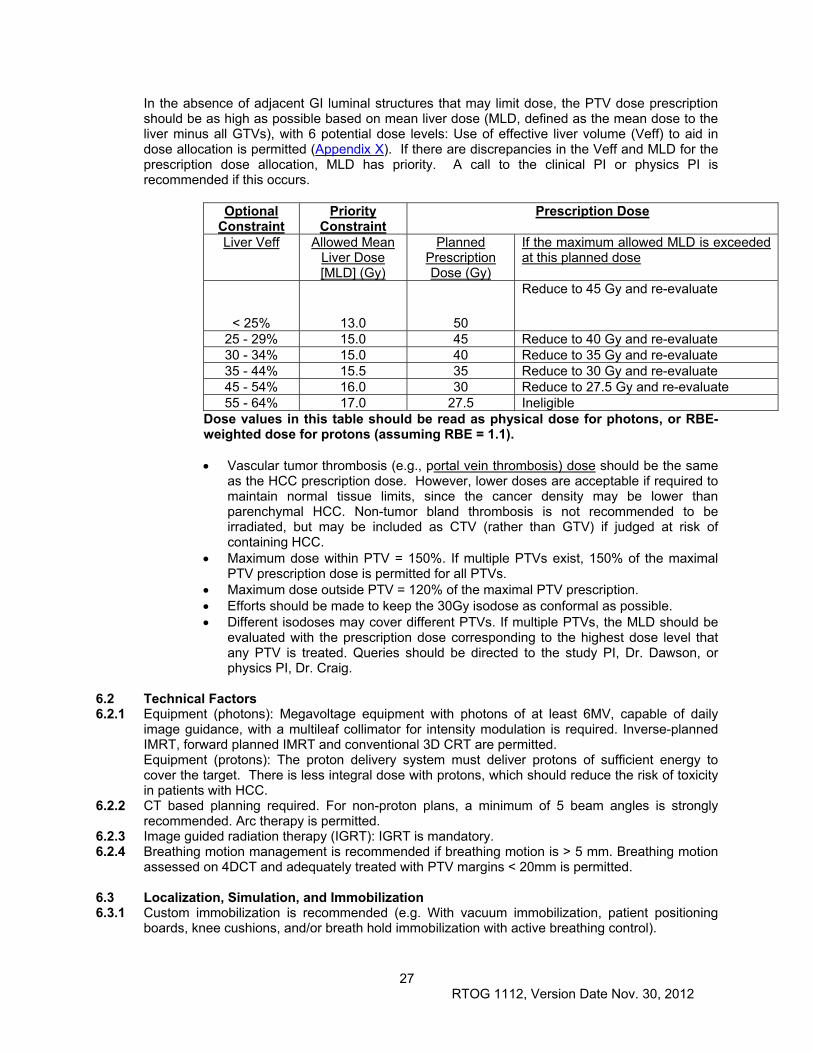
27 RTOG 1112, Version Date Nov. 30, 2012
In the absence of adjacent GI luminal structures that may limit dose, the PTV dose prescription should be as high as possible based on mean liver dose (MLD, defined as the mean dose to the liver minus all GTVs), with 6 potential dose levels: Use of effective liver volume (Veff) to aid in dose allocation is permitted (Appendix X). If there are discrepancies in the Veff and MLD for the prescription dose allocation, MLD has priority. A call to the clinical PI or physics PI is recommended if this occurs.
Optional
Constraint Priority
Constraint Prescription Dose
Liver Veff Allowed Mean Liver Dose [MLD] (Gy)
Planned Prescription Dose (Gy)
If the maximum allowed MLD is exceeded at this planned dose
< 25% 13.0 50
Reduce to 45 Gy and re-evaluate
25 - 29% 15.0 45 Reduce to 40 Gy and re-evaluate 30 - 34% 15.0 40 Reduce to 35 Gy and re-evaluate 35 - 44% 15.5 35 Reduce to 30 Gy and re-evaluate 45 - 54% 16.0 30 Reduce to 27.5 Gy and re-evaluate 55 - 64% 17.0 27.5 Ineligible
Dose values in this table should be read as physical dose for photons, or RBE-weighted dose for protons (assuming RBE = 1.1). Vascular tumor thrombosis (e.g., portal vein thrombosis) dose should be the same
as the HCC prescription dose. However, lower doses are acceptable if required to maintain normal tissue limits, since the cancer density may be lower than parenchymal HCC. Non-tumor bland thrombosis is not recommended to be irradiated, but may be included as CTV (rather than GTV) if judged at risk of containing HCC.
Maximum dose within PTV = 150%. If multiple PTVs exist, 150% of the maximal PTV prescription dose is permitted for all PTVs.
Maximum dose outside PTV = 120% of the maximal PTV prescription. Efforts should be made to keep the 30Gy isodose as conformal as possible. Different isodoses may cover different PTVs. If multiple PTVs, the MLD should be
evaluated with the prescription dose corresponding to the highest dose level that any PTV is treated. Queries should be directed to the study PI, Dr. Dawson, or physics PI, Dr. Craig.
6.2 Technical Factors 6.2.1 Equipment (photons): Megavoltage equipment with photons of at least 6MV, capable of daily
image guidance, with a multileaf collimator for intensity modulation is required. Inverse-planned IMRT, forward planned IMRT and conventional 3D CRT are permitted. Equipment (protons): The proton delivery system must deliver protons of sufficient energy to cover the target. There is less integral dose with protons, which should reduce the risk of toxicity in patients with HCC.
6.2.2 CT based planning required. For non-proton plans, a minimum of 5 beam angles is strongly recommended. Arc therapy is permitted.
6.2.3 Image guided radiation therapy (IGRT): IGRT is mandatory. 6.2.4 Breathing motion management is recommended if breathing motion is > 5 mm. Breathing motion
assessed on 4DCT and adequately treated with PTV margins < 20mm is permitted. 6.3 Localization, Simulation, and Immobilization 6.3.1 Custom immobilization is recommended (e.g. With vacuum immobilization, patient positioning
boards, knee cushions, and/or breath hold immobilization with active breathing control).
28 RTOG 1112, Version Date Nov. 30, 2012
6.3.2 Treatment planning CT scans will be required to define GTV. Multi-phasic IV contrast is recommended for the planning CT (arterial phase and/or delayed phase imaging recommended for GTV delineation, and venous phase for portal vein thrombosis delineation). If oral contrast is used at simulation, similar timing and volume of oral contrast is to be used at the time of treatment.
Exhale breath hold CT or average phase CT (from 4D CT) may be used as the baseline CT for radiation therapy planning. CT scans obtained during free breathing are strongly discouraged, but may be used if breath hold scanning is not possible for individual patients or if breathing motion is < 5 mm. CT scans used for target delineation are recommended to be multi-phase IV contrast scans obtained in breath hold. Exhale breath hold is preferred as it most often is closer to the average position than inhale breath hold, and exhale is more reproducible than inhale. If IV contrast scans cannot be obtained at the time of radiation planning, IV contrast CT scans from diagnostic radiology may be fused to the primary planning dataset to aid in target definition. If contraindications to IV CT contrast exist, contrast multiphase MR can be used to define GTV. Diagnostic imaging can be imported to the planning system to aid in target delineation. All scans used for target delineation should be fused to each other so that the livers are registered to each other for target delineation. Registration will be performed with the best fit liver-to-liver image registration, focusing on the region of the PTVs if deformation or rotation occurs between scans. Imaging details are in Appendix V.
6.3.3 Breathing motion assessment. Measurement of target/liver breathing motion is required, unless breath hold is to be used for liver immobilization. Motion may be assessed using 4D CT, fluoroscopy and/or cine MR.
4D CT: A 4D, or respiratory sorted, CT may be obtained for assessing motion if breath hold is not used for liver immobilization. Liver reproducibility of position in breath hold should be measured using fluoroscopy, CT or MRI.
6.3.4 Proton Specific Guidelines Localization, Simulation, and Immobilization Guidelines Patients must be simulated on a CT scanner, which has been commissioned for protons. Proton compatible immobilization devices are required, as is a motion management system. Immobilization devices will not extend to the lower thorax so as to minimize proton entrance through them. For contrast-enhanced CT simulations (either breath-hold or free-breathing), the initial CT sequence will be the non-contrast scan for proton planning purposes and the subsequent scan will be the contrast scan for contouring. All oral contrast Hounsfield units will be overridden during planning and replaced with a Hounsfield unit of 1. Hounsfield units for lipiodol, fiducials and clips will remain unchanged during planning. Where possible the proton beam will not exit into GI mucosa. Breathing motion management is recommended if breathing motion is > 5 mm. Breathing motion assessed on 4DCT and adequately treated with PTV margins < 20mm is permitted. Shadowing of dose along a beam behind radio-opaque fiducials is negligible and can be discounted during multi-beam proton planning. Where fiducials are used for breath-hold set-up, reproducibility of breathing amplitude on days of treatment compared to simulation will be confirmed prior to treatment delivery.
6.4 Treatment Planning/Target Volumes 6.4.1 The Gross Tumor Volume (GTV) is defined as all parenchymal and vascular HCC visualized
on contrast enhanced CT and/or MRI, most often best seen on arterial phase (as hyperintensity) and/or venous or delayed phase (as hypointensity relative to liver). GTVp1 should represent the ‘primary parenchymal (=p) dominant (=1)’ GTV, upon which primary QA will be based. Subsequent lesions can be labeled as GTVp2, GTVp3, GTVp4 and GTVp5). Vascular HCC thrombi (GTVv) most often are best seen on venous phase imaging as hypointensity relative to the contrast in the vessel, Vascular HCC may be combined with parenchymal HCC (labeled as GTVp or GTVpv) if they are to be treated to the same dose.
29 RTOG 1112, Version Date Nov. 30, 2012
Non-tumor thrombi should not be considered as GTV; they should be excluded from contouring or may be included in the CTV (as per 6.4.2). Non-tumor extrahepatic vascular thrombi should not be treated as GTV or CTV. Small enhancing GTV in adjacent lymph nodes are permitted to be irradiated only if normal tissue limits are not exceeded. They are not required to be irradiated, and no prophylactic nodal irradiation is allowed. The prescription dose should be annotated to each GTV after the final plan is complete (e.g. GTVp1_50 for a 50Gy target). Diagnostic contrast CT or MR imaging may be fused with the planning CT if there is no IV contrast used in the planning CT (liver-to-liver fusion is recommended).
6.4.2 The Clinical Target Volume (CTV): For each GTVp, the CTV is defined as the GTV (CTVp1… CTVp5), with no expansion. The minimal CTVv is the GTVv, with no expansion. It is expected that there will be no expansion from GTV to CTV for the majority of cases. However, CTV expansions to include regions at high risk for microscopic disease, including non-tumor vascular (v) thrombi (CTVv), prior TACE (t) sites (CTVt), or adjacent RFA (r) (or other ablation) sites (CTVr) are permitted. Such CTVs may be treated to a microscopic dose (27.5 Gy) or up to as high as the prescription dose, at the investigator’s discretion. Separate CTVs should be labeled CTVp1, CTVv2, CTVv3, CTVt4…etc. The prescription dose should be annotated to each CTV after the final plan is complete (e.g. CTVp1_50 for a 50Gy target, and CTVt2_27.5 for a CTV treated to 27.5 Gy).
6.4.3 The Planning Target Volume (PTV) The Photon PTV will provide a margin around each CTV to compensate for set-up and internal organ motion. PTV nomenclature should follow CTV nomenclature guidelines. For example, PTVv for the PTV around the CTVv and PTVp1 and PTVp2 for PTVs around CTVp1 and CTVp2. A minimum PTV margin of 4 mm around each CTV is required in all directions (for example if active breathing control is used with excellent reproducibility). The maximum permitted PTV margin is 20 mm, expected to be used uncommonly. PTV margins ≤ 10 mm are a goal. Asymmetric PTV margins are permitted. The actual PTV used will depend on motion management used, the patients’ motion and reproducibility. PTVs should not be manually modified due to proximity of adjacent OARs. The final PTVs should have dose annotated once the plan is final. Eg. PTVp1_50 and PTVv1_27.50 for targets treated to 50 cGy and 27.5Gy, respectively. The Proton PTV will provide a margin around each CTV to compensate for uncertainties including set-up and internal organ motion, aperture margin definitions, compensator smearing, range of individual beams, and modulation width of the SOBP. PTV nomenclature should follow CTV nomenclature guidelines, in a similar manner to the photon PTV. For example, PTVv_EN for the PTV around CTVv_EN. A minimum PTV margin of 4 mm around the CTV is required in all directions (for example if active breathing control is used with excellent reproducibility). The maximum permitted PTV margin is 20 mm. Asymmetric PTV margins are permitted, depending on institution motion management, individual patients’ motion and reproducibility. The final PTVs should have RBE-weighted dose annotated once the plan is final. Eg. PTVp1_50 and PTVv1_27.5. Additionally, the effect of variations in the set-up of the target with respect to tissue inhomogeneities (e.g., employing compensator smearing technique, beam-specific PTV etc.), or range uncertainties (e.g., by expanding the prescribed range and modulation, to create distal and proximal field margins) should be addressed in the design of treatment fields for each beam direction. As suggested in ICRU Report 78, paragraph 5.1.4.4, an adjustment must be made within the beam-design algorithm to take into account the margins needed to account for uncertainties along the beam direction (i.e. range uncertainties) and those included in the traditional PTV (i.e. lateral uncertainties). The proton distal target margin range will be
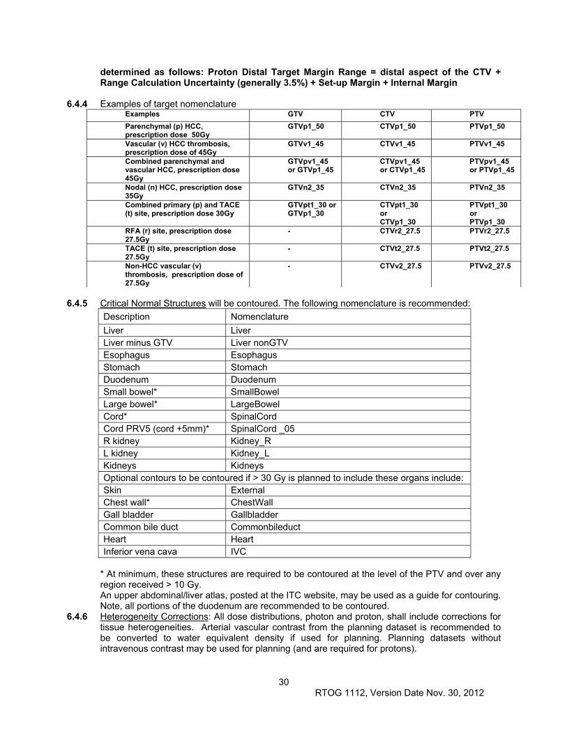
30 RTOG 1112, Version Date Nov. 30, 2012
determined as follows: Proton Distal Target Margin Range = distal aspect of the CTV + Range Calculation Uncertainty (generally 3.5%) + Set-up Margin + Internal Margin
6.4.4 Examples of target nomenclature Examples GTV CTV PTV
Parenchymal (p) HCC, prescription dose 50Gy
GTVp1_50 CTVp1_50 PTVp1_50
Vascular (v) HCC thrombosis, prescription dose of 45Gy
GTVv1_45 CTVv1_45 PTVv1_45
Combined parenchymal and vascular HCC, prescription dose 45Gy
GTVpv1_45 or GTVp1_45
CTVpv1_45 or CTVp1_45
PTVpv1_45 or PTVp1_45
Nodal (n) HCC, prescription dose 35Gy
GTVn2_35 CTVn2_35 PTVn2_35
Combined primary (p) and TACE (t) site, prescription dose 30Gy
GTVpt1_30 or GTVp1_30
CTVpt1_30 or CTVp1_30
PTVpt1_30 or PTVp1_30
RFA (r) site, prescription dose 27.5Gy
- CTVr2_27.5 PTVr2_27.5
TACE (t) site, prescription dose 27.5Gy
- CTVt2_27.5 PTVt2_27.5
Non-HCC vascular (v) thrombosis, prescription dose of 27.5Gy
- CTVv2_27.5 PTVv2_27.5
6.4.5 Critical Normal Structures will be contoured. The following nomenclature is recommended:
Description Nomenclature
Liver Liver Liver minus GTV Liver nonGTV Esophagus Esophagus Stomach Stomach Duodenum Duodenum Small bowel* SmallBowel Large bowel* LargeBowel Cord* SpinalCord Cord PRV5 (cord +5mm)* SpinalCord _05 R kidney Kidney_R L kidney Kidney_L Kidneys Kidneys Optional contours to be contoured if > 30 Gy is planned to include these organs include: Skin External Chest wall* ChestWall Gall bladder Gallbladder Common bile duct Commonbileduct Heart Heart Inferior vena cava IVC
* At minimum, these structures are required to be contoured at the level of the PTV and over any region received > 10 Gy. An upper abdominal/liver atlas, posted at the ITC website, may be used as a guide for contouring. Note, all portions of the duodenum are recommended to be contoured.
6.4.6 Heterogeneity Corrections: All dose distributions, photon and proton, shall include corrections for tissue heterogeneities. Arterial vascular contrast from the planning dataset is recommended to be converted to water equivalent density if used for planning. Planning datasets without intravenous contrast may be used for planning (and are required for protons).
31 RTOG 1112, Version Date Nov. 30, 2012
6.4.7 Goals of planning are to maximize dose to the target volumes, while maintaining all normal tissue constraints (as defined in Section 6.5). Reducing the maximal dose to all luminal gastrointestinal normal tissues should be a planning priority to reduce the risk of gastrointestinal toxicity. Beam angles may be individualized to minimize the pathlength through the liver and through adjacent organs at risk. Conformality of the 30 Gy isodose is another goal.
6.5 Critical Structures Maximal Doses (5/7/13) Dose values in this section should be read as physical dose for photons, or RBE-weighted
dose for protons, in 5 fractions (assuming RBE = 1.1). 6.5.1 Esophagus max (to 0.5 cc): 32 Gy 6.5.2 Stomach max (to 0.5 cc): 30 Gy 6.5.3 Duodenum max (to 0.5 cc): 30 Gy 6.5.4 Small bowel max (to 0.5 cc): 30 Gy 6.5.5 Large bowel max (to 0.5 cc): 30 Gy 6.5.6 Cord + 5 mm max (0.5cc): 25 Gy 6.5.7 Kidneys:
Bilateral mean dose < 10 Gy -OR- If one kidney mean dose > 10Gy, remaining (or only) kidney V10Gy < 10%
6.5.8 The following organ dose constraints are guidelines, not mandatory: Stomach (to 5 cc): < 25 Gy Duodenum (to 5 cc): < 25 Gy Small bowel (to 5 cc): < 25 Gy Liver minus all GTVs: > 700cc and V10Gy < 70% Heart max (30cc): < 30 Gy
Great vessel max (0.5 cc): < 60 Gy Skin (external) max (0.5 cc): < 32 Gy Chest wall max (0.5 cc): < 50 Gy Gallbladder max (0.5 cc): < 55 Gy Common bile duct max (0.5 cc) < 50 Gy (even though the bile duct is not often well visualized, it is always within the portal region and may be within high dose volumes for central targets, so efforts to reduce hot spots in this region are warranted)
6.6 Documentation Requirements 6.6.1 Quality Assurance Documentation
In patients randomized to the SBRT arm who do not receive radiation, the intended and/or best treatment plan should be submitted with an explanation for why the patient did not start radiation therapy. For each institution, the full 3D dosimetry plans for the first three registered for this study and randomized to the SBRT arm will be reviewed in a rapid review, PRIOR TO DELIVERY of radiation treatment. If these plans are within protocol compliance, then subsequent review of cases will be done in a timely review. Timely submission of all radiation plans is required for every patient treated on this study. The definition of timely submission for the patients requiring timely review is within 5 days of completion of radiation therapy. Liver protocol CT scan and/or MR showing the extent of the tumor with contrast is required to be submitted. If multiple phases of CT and/or MR imaging, and/or if diagnostic CTs or MR imaging, are helpful for target delineation, multiple phase imaging datasets should be submitted. A maximum of three datasets per patient is to be submitted. These datasets should be submitted, registered as they were used for target delineation, which should be with the best fit liver-to-liver image registration, focusing on the region of the PTVs if deformation or rotation occurs between scans.
6.6.2 Treatment Interruptions Treatment interruptions should be clearly documented in the patient’s treatment record. Total treatment time is recommended to be 10 days, with allowable total duration between 5 days and 15 days (see Section 6.7.1).
32 RTOG 1112, Version Date Nov. 30, 2012
6.6.3 Diagnostic CT/MR Submission Prior to Randomization The baseline diagnostic CT or MR or a planning CT must be uploaded to the ITC Database. The liver volume and the HCC and/or vascular thrombosis should be contoured in a radiation planning system or segmentation system prior to submission.
6.6.4 IGRT For each IGRT technology, in addition to each “as if patient” dataset where imaging and offsets are submitted for 2 sequential fractions, the images and offsets for all days for the first two actual patients treated on study should be submitted for timely review, so that feedback to the participating institution regarding IGRT can follow. For all subsequent patients, the IGRT images in treatment position for every fraction (and a table of subsequent ‘shifts’) are required to be submitted for subsequent future evaluation.
6.7 Compliance Criteria The review process for this protocol is aimed at assuring correct contouring of target and critical structures, as well as appropriate planning. These reviews should avoid violations and deviations for this protocol. Each treatment shall be judged according to the protocol guidelines, with variations and deviations defined below:
6.7.1 Total Treatment Duration Per protocol: All treatment falls within 15 calendar days Variation Acceptable: All treatments fall within 16 to 21 calendar days Deviation Unacceptable: All treatments that take 22 or more calendar days to complete
6.7.2 GTV Compliance Per protocol: no edits required Variation acceptable: Variations in GTV or CTV other than deviation unacceptable Deviation unacceptable: Definite HCC or enhancing thrombosis not contoured within GTV
6.7.3 PTV Compliance PTV Contouring Per protocol: PTV > 4 mm and < 20 mm Variation acceptable: PTV 3-4 mm or 20-25 mm Deviation unacceptable PTV < 3 mm or > 25 mm PTV Dosimetry Target coverage for each PTV should be considered on its own. If there are multiple tumors, the primary (dominant) PTV should be labeled #1. The intent is for prescription dose to cover 95% of each PTV. If PTVs are not treated as per guidelines, this is a deviation unacceptable. The PTV should be treated to as high a dose as possible, respecting normal tissue constraints (as per Section 6.1.5), as a dose response has been observed. Modifying required PTVs due to close proximity of adjacent OARs is not permitted. The following table describes variations and deviations in the prescription dose (dose covering 95% of the PTV). Treating “per protocol” should always be the planning intent.
Dose values in this section should be read as physical dose for photons, or RBE-weighted dose for protons (assuming RBE = 1.1). *Note that lower doses than the dose-allocation schedule are acceptable if they are required due to adjacent GI luminal structures that may limit the deliverable dose.
Dose to 95% PTV PTVs around GTVs * PTVs around non-GTV CTVs*
per protocol prescription dose +/- 5% prescription dose +/- 5% variation acceptable 90-95% or 105-110% of
prescription dose, and ≥ 25 Gy
85-95% or 105-115% of prescription dose and ≥ 25 Gy
deviation unacceptable <90% or >110% of prescription dose, or < 25 Gy
<85% or >115% of prescription dose, or <25Gy
Overall plan deviation unacceptable
< 25 Gy
33 RTOG 1112, Version Date Nov. 30, 2012
6.7.4 Compliance for Critical Structures (organs at risk, OARs)
If non-hepatic OARs limit the prescription dose, the highest dose (from the 6 prescription doses listed in Section 6.1.5) should be used, while maintaining OAR dose constraints.
Non-liver OARs per protocol variation acceptable deviation unacceptable Esophagus max (to 0.5 cc): Stomach max (to 0.5 cc): Duodenum max (to 0.5 cc): Small bowel max (to 0.5 cc): Large bowel max (to 0.5 cc): Cord + 5 mm max (0.5cc): Kidneys: Bilateral mean dose
32 Gy 30 Gy 30 Gy 30 Gy 32 Gy 25 Gy ≤10 Gy
> 32 but ≤34 Gy >30 but ≤32 Gy >30 but ≤32 Gy >30 but ≤32 Gy >32 but ≤34 Gy >25 but ≤28 Gy >10 but ≤12 Gy
> 34 Gy > 32 Gy > 32 Gy > 32 Gy > 34 Gy > 28 Gy > 12 Gy
Dose values in these tables should be read as physical dose for photons, or RBE-weighted dose for protons (assuming RBE = 1.1).
Prescription dose Liver (minus GTV) mean dose per protocol variation acceptable deviation unacceptable
50 Gy 45 Gy 40 Gy 35 Gy 30 Gy 27.5 Gy
≤13 Gy ≤15 Gy ≤15 Gy ≤15.5 Gy ≤16 Gy ≤17 Gy
13-13.2 Gy 15-15.2 Gy 15-15.2 Gy 15.5-15.7 Gy 16-16.2 Gy 17-17.2 Gy
> 13.2 Gy > 15.2 Gy > 15.2 Gy > 15.7 Gy > 16.2 Gy > 17.2 Gy
Dose values in these tables should be read as physical dose for photons, or RBE-weighted dose for protons (assuming RBE = 1.1).
6.8 Radiation Therapy and Imaging Quality Assurance Reviews 6.8.1 Rapid and Timely Review of RT Plans
For each technique (non-IMRT, non-proton SBRT, IMRT and/or protons), the first three patients to be treated at the site on this protocol will have a rapid review of their plans, i.e. the individual plan needs to be approved PRIOR to delivering any protocol treatment for patient or subsequent patients. After rapid review by the PI, suggestions regarding protocol compliance will be forwarded to the participating institution. Rapid review of all plans PRIOR to treatment delivery will continue until at least 3 plans have been submitted without deviations. After that point, treatment plans for the subsequent patients enrolled at an individual site will undergo a timely review. If any protocol deviations are found in the first 3 plans, rapid review will continue until at least at least three sequential plans have been approved without deviations. All subsequent plans are reviewed in a timely review. Thus, all radiation plans need to be submitted in a timely manner– ideally at least 5 days prior to planned start of therapy for rapid reviews and within 1 week (5 working days) post RT completion for timely reviews. All additional QA data (including actual treatment details and IGRT data for all fractions) is to be submitted within 1 week of RT end.
6.8.2 Planned Interim Analyses of Quality Assurance After first 50 patients are enrolled and/or first 25 patients randomized to the SBRT arm (whichever comes first), the Radiation Oncology Chair and Co-Chairs, along with a delegated team from RTOG, will summarize all QA results. All submitted imaging datasets for both arms of the study will be reviewed as will imaging, contouring and HCC:liver strata determination. Following this analysis, modifications to education material and/or the protocol to help prevent violations and deviation for future patients, may be recommended.
34 RTOG 1112, Version Date Nov. 30, 2012
Secondary reviews will occur after the first 100 and 200 patients are enrolled, again with a plan to improve education material and/or the study if needed, with individual feedback to participating institutions.
6.8.3 Pre-Randomization Imaging Submission for All Patients (including non-radiation patients) For all patients randomized, submission of IV contrast CT or MR with contouring of the HCC,
(including vascular thrombi) and calculation of the HCC GTV/liver volume stratification factor is required. This imaging may be done in radiation planning CT and/or may be done on diagnostic CT or MR imported to a radiation planning system or any platform that allows organ segmentation and data transfer. The first 3 cases per institution from patients randomized to the non-RT arm will be reviewed (timely) by the PI or designee. Any differences in the segmentation of tumor or liver or in the tumor:liver volume calculation will be recorded. Feedback to each institution will follow in a timely manner (regarding imaging quality or contouring). Rationale for use of the ratio of the GTV to liver volume as a stratification factor is that the ratio of HCC to liver volume is a known prognostic factor for HCC. Prognosis is best in patients with a small volume of HCC, and toxicity is increased in those with a smaller volume of liver. The GTV/liver volume takes into account both these factors. Furthermore, in the radiation arm, the radiation dose is expected to be associated with local control probability. Based on the PMH SBRT experience (Bujold 2012), dose is correlated with GTV/liver volume. Stratification categories were based on values from a sample of patients treated at PMH. Central review of assessment of the calculation of this stratification factor will help ensure the quality of calculation of GTV:liver, and will provide insight regarding the quality of imaging and selection of tumors for this study, even in the non-SBRT arm patients. Feedback to centers may help improve quality or patient selection for future cases.
6.9 Radiation Therapy Adverse Events
The criteria used for the grading of toxicities encountered in this study are Common Toxicity Criteria (CTC) version 4.0. Very likely (80-90%)
Fatigue (which generally goes away after the radiation therapy is completed) Skin irritation, redness, itchiness, discomfort Temporary changes in blood work (decrease in blood counts, increase in liver
enzymes), without symptoms Less likely (30%)
Nausea, vomiting (during therapy) – more common if stomach or gastrointestinal track irradiated
Gastric, esophagus, small bowel or large bowel irritation/ulceration, bleeding, fistula, obstruction or changes in motility following therapy (may require medications or surgery) (<10% permanent changes)
Chest wall pain, rib fracture (< 10%) Less likely, but serious (<20%)
Radiation-induced liver disease (RILD) (<5%). Classic RILD is a clinical diagnosis of anicteric ascites, hepatomegaly and elevation of alkaline phosphatase relative to other transaminases that may occur 2 weeks to 3 months following radiation to the liver.
Non-classic RILD includes elevation of liver enzymes and/or any decline in liver function within 12 weeks from start of therapy (~20%). RILD can lead to liver failure that could lead to death. There is an increased risk of liver toxicity in patients with large tumors and in patients with pre-existing liver disease.
Permanent thrombocytopenia (<1%); this may lead to bleeding Kidney injury (<1%); this may lead to changes on imaging and more rarely the need
for medication.
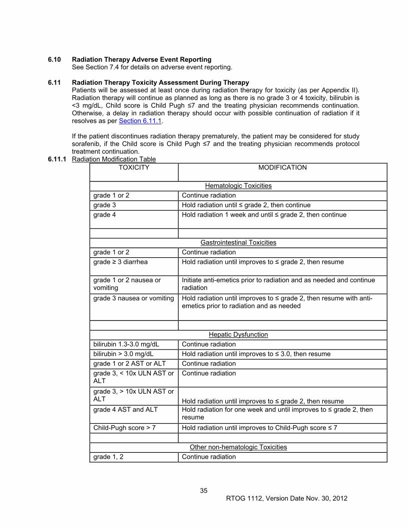
35 RTOG 1112, Version Date Nov. 30, 2012
6.10 Radiation Therapy Adverse Event Reporting
See Section 7.4 for details on adverse event reporting.
6.11 Radiation Therapy Toxicity Assessment During Therapy Patients will be assessed at least once during radiation therapy for toxicity (as per Appendix II). Radiation therapy will continue as planned as long as there is no grade 3 or 4 toxicity, bilirubin is <3 mg/dL, Child score is Child Pugh ≤7 and the treating physician recommends continuation. Otherwise, a delay in radiation therapy should occur with possible continuation of radiation if it resolves as per Section 6.11.1. If the patient discontinues radiation therapy prematurely, the patient may be considered for study sorafenib, if the Child score is Child Pugh ≤7 and the treating physician recommends protocol treatment continuation.
6.11.1 Radiation Modification Table TOXICITY MODIFICATION
Hematologic Toxicities
grade 1 or 2 Continue radiation
grade 3 Hold radiation until ≤ grade 2, then continue
grade 4 Hold radiation 1 week and until ≤ grade 2, then continue
Gastrointestinal Toxicities
grade 1 or 2 Continue radiation
grade ≥ 3 diarrhea Hold radiation until improves to ≤ grade 2, then resume
grade 1 or 2 nausea or vomiting
Initiate anti-emetics prior to radiation and as needed and continue radiation
grade 3 nausea or vomiting Hold radiation until improves to ≤ grade 2, then resume with anti-emetics prior to radiation and as needed
Hepatic Dysfunction
bilirubin 1.3-3.0 mg/dL Continue radiation
bilirubin > 3.0 mg/dL Hold radiation until improves to ≤ 3.0, then resume
grade 1 or 2 AST or ALT Continue radiation
grade 3, < 10x ULN AST or ALT
Continue radiation
grade 3, > 10x ULN AST or ALT Hold radiation until improves to ≤ grade 2, then resume grade 4 AST and ALT Hold radiation for one week and until improves to ≤ grade 2, then
resume
Child-Pugh score > 7 Hold radiation until improves to Child-Pugh score ≤ 7
Other non-hematologic Toxicities
grade 1, 2 Continue radiation
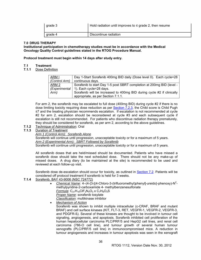
36 RTOG 1112, Version Date Nov. 30, 2012
grade 3 Hold radiation until improves to ≤ grade 2, then resume
grade 4 Discontinue radiation
7.0 DRUG THERAPY Institutional participation in chemotherapy studies must be in accordance with the Medical Oncology Quality Control guidelines stated in the RTOG Procedure Manual. Protocol treatment must begin within 14 days after study entry.
7.1 Treatment 7.1.1 Dose Definition
ARM I (Control Arm)
Day 1-Start Sorafenib 400mg BID daily (Dose level 0). Each cycle=28 continuous days.
ARM 2 (Experimental Arm)
Sorafenib to start Day 1-5 post SBRT completion at 200mg BID (level -1). Each cycle=28 days. Sorafenib will be increased to 400mg BID during cycle #2 if clinically appropriate, as per Section 7.1.1.
For arm 2, the sorafenib may be escalated to full dose (400mg BID) during cycle #2 if there is no dose limiting toxicity requiring dose reduction as per Section 7.2.3, the Child score is Child Pugh ≤7 and the treating physician recommends escalation. If escalation is not recommended at cycle #2 for arm 2, escalation should be reconsidered at cycle #3 and each subsequent cycle if escalation is still not recommended. For patients who discontinue radiation therapy prematurely, they should be considered for sorafenib, as per arm 2, according to the above guidelines.
7.1.2 Technique of Administration: Oral 7.1.3 Duration of Treatment
Arm 1 (Control Arm): Sorafenib Alone Sorafenib will continue until progression, unacceptable toxicity or for a maximum of 5 years. Arm 2 (Experimental Arm): SBRT Followed by Sorafenib Sorafenib will continue until progression, unacceptable toxicity or for a maximum of 5 years. All sorafenib doses that are held/missed should be documented. Patients who have missed a sorafenib dose should take the next scheduled dose. There should not be any make-up of missed doses. A drug diary (to be maintained at the site) is recommended to be used and reviewed at each follow-up visit. Sorafenib dose de-escalation should occur for toxicity, as outlined in Section 7.2. Patients will be considered off protocol treatment if sorafenib is held for 3 weeks.
7.1.4 Sorafenib, BAY 43-9006 (NSC 724772) Chemical Name: 4–(4-{3-[(4-Chloro-3-(trifluoromethyl)phenyl]-ureido}-phenoxy)-N2-
methylpyridine-2-carboxamide 4- methylbenzenesulfonate Formula: C21H16ClF3N4O3 x C7H8O3S Proper Name: sorafenib tosylate Classification: multikinase inhibitor
Mechanism of Action Sorafenib was shown to inhibit multiple intracellular (c-CRAF, BRAF and mutant BRAF) and cell surface kinases (KIT, FLT-3, RET, VEGFR-1, VEGFR-2, VEGFR-3, and PDGFR-ß). Several of these kinases are thought to be involved in tumour cell signaling, angiogenesis, and apoptosis. Sorafenib inhibited cell proliferation of the human hepatocellular carcinoma PLC/PRF/5 and HepG2 cell lines, and renal cell carcinoma (786-O cell line), and tumour growth of several human tumour xenografts (PLC/PRF/5 cell line) in immunocompromised mice. A reduction in tumour angiogenesis and increases in tumour apoptosis was seen in the xenograft
37 RTOG 1112, Version Date Nov. 30, 2012
models of human hepatocellular and renal cell carcinoma cell lines. Additionally, a reduction in Raf/MEK/ERK signaling was seen in human hepatocellular carcinoma PLC/PRF/5 and HepG2 cell lines.
Molecular M.W.: Sorafenib tosylate: 637 Daltons; sorafenib (free base): 465 Daltons Approximate Solubility: 0.19 mg/100 mL in 0.1 N HCl, 453 mg/100 mL in Ethanol, and 2971 mg/100 mL in PEG 400.
Supply: Commercially available Sorafenib tosylate is supplied as an immediate-release film-coated, round, and salmon color tablet containing 200 mg of the free base, sorafenib, and the excipients croscarmellose sodium, microcrystalline cellulose, hydroxypropylmethylcellulose, sodium lauryl sulfate, and magnesium stearate. The film-coat consists of hydroxypropylmethyl cellulose, polyethylene glycol, titanium dioxide and red iron oxide. The film coating has no effect on the rate of release of the active sorafenib tosylate. Sorafenib tosylate 200 mg tablets are supplied by manufacturer in bottles of 140 tablets.
o Non-Canadian International Institutions: Please refer to your LOI Approval Notification. Your institution will be responsible for acquiring any drug noted in the protocol as commercially available and not provided for the study.
Administration (Oral) Patients are to take sorafenib tablets 1 hour before or 2 hours after meals. When administered with high fat meals, sorafenib absorption was reduced by 29% compared with fasting.
7.1.5 Adverse Events and Potential Risks Cardiovascular
Very common: hypertension. Uncommon: hypertensive crisis, congestive heart failure, myocardial ischemia and/or infarction
Dermatologic Very common: erythema, rash, alopecia, hand-foot syndrome, pruritis Common: exfoliative dermatitis, acne, dry skin, flushing, skin desquamation Uncommon: folliculitis, eczema, erythema multiforme, Keratoacanthomas/squamous cell cancer of the skin
Digestive Very common: diarrhea, nausea, vomiting, increased lipase, increased amylase, weight loss Common: constipation, mucositis oral, stomatitis (including dry mouth and glossodynia), dyspepsia, dysphagia, abdominal pain Uncommon: ascites, constipation, pancreatitis, gastrointestinal reflux, gastritis, gastrointestinal perforations. Note that elevations in lipase are very common (41%); a diagnosis of pancreatitis should not be made solely on the basis of abnormal laboratory values
General Disorders Very common: asthenia, fatigue, bleeding events (hemorrhage including hematoma, epistaxis, mouth, pulmonary and respiratory tract, GI tract, and uncommon cases of cerebral hemorrhage), pain (including mouth pain, abdominal pain, headache, bone pain, and tumour pain) Common: decreased appetite, weight decreased, influenza-like illness, pyrexia Uncommon: infection, swelling of limbs
Hematologic Very common: lymphopenia, leucopenia Common: anemia, neutropenia, thrombocytopenia Uncommon: INR abnormal, prothrombin level abnormal
Hypersensitivity Uncommon: hypersensitivity reactions (including skin reactions and urticaria).
38 RTOG 1112, Version Date Nov. 30, 2012
Metabolic and Nutritional Very common: hypophosphatemia Common: transient increases in transaminases Uncommon: dehydration, hyponatremia, transient increases in alkaline phosphatase, increased bilirubin (including jaundice), hypothyroidism, increased cholesterol
Musculoskeletal Common: arthralgia, myalgia.
Nervous System and Psychiatric Common: sensory peripheral neuropathy, depression Uncommon: tinnitus, reversible posterior leukoencephalopathy, intracranial hemorrhage
Other Common: erectile dysfunction Uncommon: gynecomastia, hematuria, renal hemorrhage, ovarian, vaginal or prostatic hemorrhage
Respiratory Common: hoarseness Uncommon: rhinorrhea, epistaxis. Events may have a life-threatening or fatal outcome. Such events are uncommon.
7.1.6 Potential Drug Interactions CYP3A4 Inducers
Chronic concomitant administration of rifampin with a single dose of sorafenib resulted in a 24% decrease in the combined AUC of sorafenib and its active primary metabolite when rifampin was co-administered with sorafenib. The clinical significance of this overall decrease in drug exposure is unknown. Other inducers of CYP3A4 activity (eg, hypericum perforatum [also known as St. John’s wort], phenytoin, carbamazepine, phenobarbital, and dexamethasone) may also increase the metabolism of sorafenib and decrease its exposure.
CYP2C9 Substrates The possible effect of sorafenib on warfarin, a CYP2C9 substrate, was assessed in sorafenib-treated patients compared to placebo-treated patients. The concomitant treatment with sorafenib and warfarin did not result in changes in mean PT-INR compared to placebo. However, patients taking warfarin should have their INR checked regularly (see Product Monograph: WARNINGS AND PRECAUTIONS and PART II: DETAILED PHARMACOLOGY).
CYP Isoform-selective Substrates Concomitant administration of sorafenib and midazolam, dextromethorphan or omeprazole, which are substrates of cytochromes CYP3A4, CYP2D6 and CYP2C19, respectively, following 4 weeks of sorafenib administration did not alter the exposure of these agents. This indicates that sorafenib is neither an inhibitor nor an inducer of these cytochrome P450 isoenzymes. Therefore, clinical pharmacokinetic interactions of sorafenib with substrates of these enzymes are unlikely.
UGT1A9 Inhibitors An in vitro study has revealed a number of drugs affected UGT1A9-mediated sorafenib glucuronidation with an IC50 value below 100 μM. They were atorvastatin (IC50 = 67 μM), ketoconazole (87 μM), mefenamic acid (28 μM), erlotinib (69 μM), and niflumic acid (1.2 μM). The clinical relevance of these drug interactions has not been tested.
Combination with Other Antineoplastic Agents Sorafenib is approved only as monotherapy in the treatment of RCC and HCC (see Product Monograph: INDICATIONS AND CLINICAL USE). In clinical studies, sorafenib has been administered together with a variety of other antineoplastic agents at their commonly-used dosing regimens, including gemcitabine, oxaliplatin,
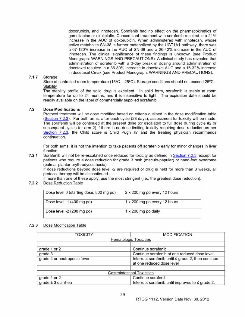
39 RTOG 1112, Version Date Nov. 30, 2012
doxorubicin, and irinotecan. Sorafenib had no effect on the pharmacokinetics of gemcitabine or oxaliplatin. Concomitant treatment with sorafenib resulted in a 21% increase in the AUC of doxorubicin. When administered with irinotecan, whose active metabolite SN-38 is further metabolized by the UGT1A1 pathway, there was a 67-120% increase in the AUC of SN-38 and a 26-42% increase in the AUC of irinotecan. The clinical significance of these findings is unknown (see Product Monograph: WARNINGS AND PRECAUTIONS). A clinical study has revealed that administration of sorafenib with a 3-day break in dosing around administration of docetaxel resulted in a 36-80% increase in docetaxel AUC and a 16-32% increase in docetaxel Cmax (see Product Monograph: WARNINGS AND PRECAUTIONS).
7.1.7 Storage Store at controlled room temperature (15ºC – 25ºC). Storage conditions should not exceed 25ºC. Stability The stability profile of the solid drug is excellent. In solid form, sorafenib is stable at room temperature for up to 24 months, and it is insensitive to light. The expiration date should be readily available on the label of commercially supplied sorafenib.
7.2 Dose Modifications Protocol treatment will be dose modified based on criteria outlined in the dose modification table (Section 7.2.3). For both arms, after each cycle (28 days), assessment for toxicity will be made. The sorafenib will be continued at the present dose (or escalated to full dose during cycle #2 or subsequent cycles for arm 2) if there is no dose limiting toxicity requiring dose reduction as per Section 7.2.3, the Child score is Child Pugh ≤7 and the treating physician recommends continuation. For both arms, it is not the intention to take patients off sorafenib early for minor changes in liver function.
7.2.1 Sorafenib will not be re-escalated once reduced for toxicity as defined in Section 7.2.3, except for patients who require a dose reduction for grade 3 rash (maculo-papular) or hand-foot syndrome (palmar-plantar erythrodysesthesia). If dose reductions beyond dose level -2 are required or drug is held for more than 3 weeks, all protocol therapy will be discontinued. If more than one of these apply, use the most stringent (i.e., the greatest dose reduction).
7.2.2 Dose Reduction Table
Dose level 0 (starting dose, 800 mg po) 2 x 200 mg po every 12 hours
Dose level -1 (400 mg po)
1 x 200 mg po every 12 hours
Dose level -2 (200 mg po) 1 x 200 mg po daily
7.2.3 Dose Modification Table
TOXICITY MODIFICATION
Hematologic Toxicities grade 1 or 2 Continue sorafenib grade 3 Continue sorafenib at one reduced dose level grade 4 or neutropenic fever Interrupt sorafenib until ≤ grade 2, then continue
at one reduced dose level
Gastrointestinal Toxicities grade 1 or 2 Continue sorafenib grade ≥ 3 diarrhea Interrupt sorafenib until improves to ≤ grade 2,
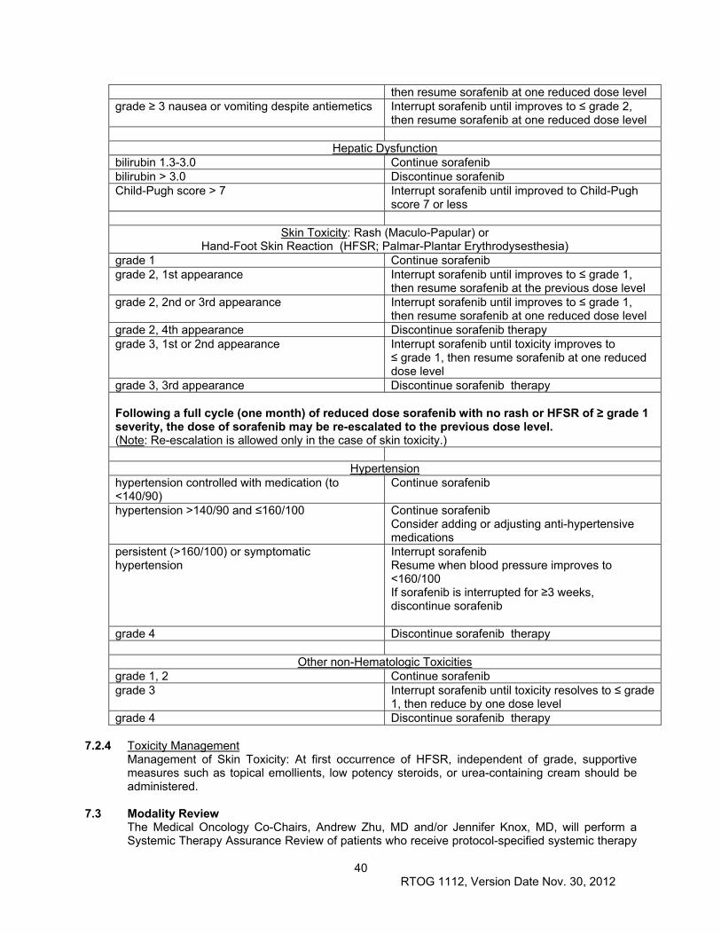
40 RTOG 1112, Version Date Nov. 30, 2012
then resume sorafenib at one reduced dose level grade ≥ 3 nausea or vomiting despite antiemetics Interrupt sorafenib until improves to ≤ grade 2,
then resume sorafenib at one reduced dose level
Hepatic Dysfunction bilirubin 1.3-3.0 Continue sorafenib bilirubin > 3.0 Discontinue sorafenib Child-Pugh score > 7 Interrupt sorafenib until improved to Child-Pugh
score 7 or less
Skin Toxicity: Rash (Maculo-Papular) or Hand-Foot Skin Reaction (HFSR; Palmar-Plantar Erythrodysesthesia)
grade 1 Continue sorafenib grade 2, 1st appearance Interrupt sorafenib until improves to ≤ grade 1,
then resume sorafenib at the previous dose level grade 2, 2nd or 3rd appearance Interrupt sorafenib until improves to ≤ grade 1,
then resume sorafenib at one reduced dose level grade 2, 4th appearance Discontinue sorafenib therapy grade 3, 1st or 2nd appearance Interrupt sorafenib until toxicity improves to
≤ grade 1, then resume sorafenib at one reduced dose level
grade 3, 3rd appearance Discontinue sorafenib therapy Following a full cycle (one month) of reduced dose sorafenib with no rash or HFSR of ≥ grade 1 severity, the dose of sorafenib may be re-escalated to the previous dose level. (Note: Re-escalation is allowed only in the case of skin toxicity.)
Hypertension hypertension controlled with medication (to <140/90)
Continue sorafenib
hypertension >140/90 and ≤160/100 Continue sorafenib Consider adding or adjusting anti-hypertensive medications
persistent (>160/100) or symptomatic hypertension
Interrupt sorafenib Resume when blood pressure improves to <160/100 If sorafenib is interrupted for ≥3 weeks, discontinue sorafenib
grade 4 Discontinue sorafenib therapy
Other non-Hematologic Toxicities grade 1, 2 Continue sorafenib grade 3 Interrupt sorafenib until toxicity resolves to ≤ grade
1, then reduce by one dose level grade 4 Discontinue sorafenib therapy
7.2.4 Toxicity Management
Management of Skin Toxicity: At first occurrence of HFSR, independent of grade, supportive measures such as topical emollients, low potency steroids, or urea-containing cream should be administered.
7.3 Modality Review The Medical Oncology Co-Chairs, Andrew Zhu, MD and/or Jennifer Knox, MD, will perform a Systemic Therapy Assurance Review of patients who receive protocol-specified systemic therapy
41 RTOG 1112, Version Date Nov. 30, 2012
in this trial. The goal of the review is to evaluate protocol compliance. The review process is contingent on timely submission of systemic therapy treatment data as specified in Section 12.1. The scoring mechanism is: Per Protocol/Acceptable Variation, Not Per Protocol, and Not Evaluable. A report is sent to each institution once per year to notify the institution about compliance for each case reviewed in that year. This Systemic Therapy Assurance Review will be performed on an ongoing basis (for example, the first review after RTOG Headquarters receives complete data for 20 cases and the next review after RTOG Headquarters receives complete data for 20 more cases). The final cases will be reviewed within 3 months after this study has reached the target accrual or as soon as RTOG Headquarters receives complete data for all cases, whichever occurs first.
7.4 Adverse Events
This study will utilize the NCI Common Terminology Criteria for Adverse Events (CTCAE) version 4.0 for AdEERS reporting of adverse events (AEs). All AE reporting on the study case report forms (CRFs) should follow grading criteria instructions on the specific CRF. The CTCAE version 4.0 is identified and located on the CTEP web site at: http://ctep.cancer.gov/protocolDevelopment/electronic_applications/ctc.htm. All appropriate treatment areas should have access to a copy of the CTCAE version 4.0. All adverse events (AEs) as defined in the tables below will be reported via the AdEERS (Adverse Event Expedited Reporting System) application accessed via the CTEP web site (https://webapps.ctep.nci.nih.gov/openapps/plsql/gadeers_main$.startup). Serious adverse events (SAEs) as defined in the tables below will be reported via AdEERS. Sites also can access the RTOG web site (http://www.rtog.org/ResearchAssociates/AdverseEventReporting.aspx) for this information. In order to ensure consistent data capture, serious adverse events reported on AdEERS reports also must be reported on an RTOG case report form (CRF). In addition, sites must submit CRFs in a timely manner after AdEERS submissions. Contact RTOG Operations Office phone number for instances when Internet fails. Once internet connectivity is restored, an AE report submitted by phone must be entered electronically into AdEERS by the original submitter at the site.
7.4.1 Adverse Events (AEs) Definition of an AE: Any unfavorable and unintended sign (including an abnormal laboratory finding), symptom, or disease temporally associated with the use of a medical treatment or procedure regardless of whether it is considered related to the medical treatment or procedure (attribution of unrelated, unlikely, possible, probable, or definite). [CTEP, NCI Guidelines: Adverse Event Reporting Requirements. February 29, 2012; http://ctep.cancer.gov/protocolDevelopment/electronic_applications/docs/aeguidelines.pdf] The following guidelines for reporting adverse events (AEs) apply to all NCI/RTOG research protocols. AEs, as defined above, experienced by patients accrued to this protocol should be reported on the AE section of the appropriate case report form (see Section 12.1). Note: AEs indicated in the AdEERS Expedited Reporting Requirements in text and/or table in Section 7.5 also must be reported via AdEERS. NOTE: If the event is a Serious Adverse Event (SAE) [see next section], further reporting will be required. Reporting AEs only fulfills Data Management reporting requirements.
7.4.2 Serious Adverse Events (SAEs) — All SAEs that fit any one of the criteria in the SAE definition below must be reported via AdEERS. Contact the AdEERS Help Desk if assistance is required. Certain SAEs as outlined below will require the use of the 24 Hour AdEERS Notification:
42 RTOG 1112, Version Date Nov. 30, 2012
• Phase II & III Studies: All unexpected potentially related SAEs Definition of an SAE: Any adverse experience occurring during any part of protocol treatment and 30 days after that results in any of the following outcomes:
Death; A life-threatening adverse drug experience; Inpatient hospitalization or prolongation of existing hospitalization; A persistent or significant disability/incapacity; A congenital anomaly/birth defect.
Important medical events that may not result in death, be life threatening, or require hospitalization may be considered an SAE, when, based upon medical judgment, they may jeopardize the patient and may require medical or surgical intervention to prevent one of the outcomes listed in the definition. Any pregnancy, including a male patient’s impregnation of his partner, occurring on study must be reported via AdEERS as a medically significant event. Pharmaceutically supported studies will require additional reporting over and above that which is required by CTEP.
SAEs (more than 30 days after last treatment) attributed to the protocol treatment (possible, probable, or definite) should be reported via AdEERS.
All supporting source documentation indicated as being provided in the Additional Information Section of the AdEERS Report must be properly labeled with the study/case numbers and the date of the event and must be faxed to the RTOG dedicated SAE FAX, 215-717-0990, before the five or ten-calendar-day deadline to allow RTOG to comply with the reporting requirements of the pharmaceutical company/companies supporting the RTOG trial. The RTOG Case Number without any leading zeros should be used as the Patient ID when reporting via AdEERS. Non-RTOG intergroup study and case numbers must also be included, when applicable. Submitted AdEERS Reports are forwarded to RTOG electronically via the AdEERS system. Use the patient’s case number as the patient ID when reporting via AdEERS. SAE reporting is safety related and separate and in addition to the Data Management reporting requirements as outlined in the previous AE reporting section. Any event that meets the above outlined criteria for an SAE but is assessed by the AdEERS System as “expedited reporting NOT required” must still be reported for safety reasons and to fulfill the obligations of RTOG to the pharmaceutical company/companies supporting the RTOG trial. Sites must bypass the “NOT Required” assessment and complete and submit the report. The AdEERS System allows submission of all reports regardless of the results of the assessment.
7.4.3 Acute Myeloid Leukemia (AML) or Myelodysplastic Syndrome (MDS) AML or MDS that is diagnosed during or subsequent to treatment in patients on NCI/CTEP-sponsored clinical trials must be reported via the AdEERS system within 30 days of AML/MDS diagnosis. If you are reporting in CTCAE version 4, the event(s) may be reported as either: 1) Leukemia secondary to oncology chemotherapy, 2) Myelodysplastic syndrome, or 3) Treatment-related secondary malignancy.
7.5 AdEERS Expedited Reporting Requirements
CTEP defines expedited AE reporting requirements for phase 2 and 3 trials as described in the table below. Important: All AEs reported via AdEERS also must be reported on the AE section of the appropriate case report form (see Section 12.1). Late Phase 2 and Phase 3 Studies: Expedited Reporting Requirements for Adverse Events that Occur on Studies under an IND/IDE within 30 Days of the Last Administration of the Commercially Available Agent 1, 2
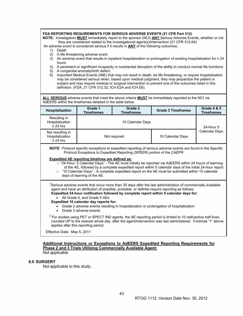
43 RTOG 1112, Version Date Nov. 30, 2012
FDA REPORTING REQUIREMENTS FOR SERIOUS ADVERSE EVENTS (21 CFR Part 312) NOTE: Investigators MUST immediately report to the sponsor (NCI) ANY Serious Adverse Events, whether or not
they are considered related to the investigational agent(s)/intervention (21 CFR 312.64) An adverse event is considered serious if it results in ANY of the following outcomes:
1) Death 2) A life-threatening adverse event 3) An adverse event that results in inpatient hospitalization or prolongation of existing hospitalization for ≥ 24
hours 4) A persistent or significant incapacity or substantial disruption of the ability to conduct normal life functions 5) A congenital anomaly/birth defect. 6) Important Medical Events (IME) that may not result in death, be life threatening, or require hospitalization
may be considered serious when, based upon medical judgment, they may jeopardize the patient or subject and may require medical or surgical intervention to prevent one of the outcomes listed in this definition. (FDA, 21 CFR 312.32; ICH E2A and ICH E6).
ALL SERIOUS adverse events that meet the above criteria MUST be immediately reported to the NCI via AdEERS within the timeframes detailed in the table below.
Hospitalization Grade 1 Timeframes
Grade 2 Timeframes
Grade 3 Timeframes Grade 4 & 5 Timeframes
Resulting in Hospitalization
≥ 24 hrs 10 Calendar Days
24-Hour 5 Calendar Days Not resulting in
Hospitalization ≥ 24 hrs
Not required 10 Calendar Days
NOTE: Protocol specific exceptions to expedited reporting of serious adverse events are found in the Specific Protocol Exceptions to Expedited Reporting (SPEER) portion of the CAEPR
Expedited AE reporting timelines are defined as: o “24-Hour; 5 Calendar Days” - The AE must initially be reported via AdEERS within 24 hours of learning
of the AE, followed by a complete expedited report within 5 calendar days of the initial 24-hour report. o “10 Calendar Days” - A complete expedited report on the AE must be submitted within 10 calendar
days of learning of the AE.
1Serious adverse events that occur more than 30 days after the last administration of commercially available agent and have an attribution of possible, probable, or definite require reporting as follows: Expedited 24-hour notification followed by complete report within 5 calendar days for:
All Grade 4, and Grade 5 AEs Expedited 10 calendar day reports for:
Grade 2 adverse events resulting in hospitalization or prolongation of hospitalization Grade 3 adverse events
2 For studies using PET or SPECT IND agents, the AE reporting period is limited to 10 radioactive half-lives, rounded UP to the nearest whole day, after the agent/intervention was last administered. Footnote “1” above applies after this reporting period.
Effective Date: May 5, 2011
Additional Instructions or Exceptions to AdEERS Expedited Reporting Requirements for Phase 2 and 3 Trials Utilizing Commercially Available Agent: Not applicable
8.0 SURGERY
Not applicable to this study.
44 RTOG 1112, Version Date Nov. 30, 2012
9.0 OTHER THERAPY 9.1 Permitted Supportive Therapy 9.1.1 All supportive therapy for optimal medical care will be given during the study period at the
discretion of the attending physician(s) within the parameters of the protocol and documented on each site’s source documents as concomitant medication.
9.1.2 Antiemetics (e.g. dopamine (D2) receptor antagonist or 5HT3 antagonists) should be considered to be used prior to each fraction of SBRT to prevent nausea if the stomach is anticipated to receive any radiation. They may also be used for symptomatic nausea or vomiting.
9.1.3 Antidiarrheals may be used to treat therapy induced diarrhea. 9.1.4 H2 blockers or proton pump inhibitors are strongly recommended if 20 Gy or more is delivered to
the luminal gastrointestinal tract or at the treating physicians discretion. They should start before completion of SBRT and continue for at least 6 months.
9.1.5 Analgesics may be used to treat tumor or therapy induced pain. NSAIDS are recommended to be avoided to reduce luminal GI irritation.
9.1.6 In the occurrence of liver toxicity (including classic RILD, non-classic RILD or any CTCAE 4.0 grade 4 toxicity or any sorafenib associated liver toxicity, occurring in the absence of HCC progression within 12 weeks of completion of radiation therapy), best supportive care and possible diuretics are recommended. Steroids may be used, and a referral to a hepatologist is recommended.
9.1.7 Topical creams, emollients, balms, low potency steroids or urea containing creams are recommended for discomfort due to hand foot syndrome.
9.1.8 Anti-hypertensives should be used for sorafenib-induced increase in blood pressure. Calcium channel blockers are recommended.
9.2 Non-permitted Supportive Therapy 9.2.1 Anticoagulants are not to be used to treat HCC related vascular thrombosis. 10.0 TISSUE/SPECIMEN SUBMISSION (5/7/13
Patients must be offered the opportunity to participate in the correlative components of the study, such as tissue/specimen submission or quality of life assessment. If the patient consents to participate in the tissue/specimen component of the study, the site is required to submit the patient’s specimens as specified in Section 10.0 of the protocol. Note: Sites are not permitted to delete the tissue/specimen component from the protocol or from the sample consent.
10.1 Tissue/Specimen Submission
The RTOG Biospecimen Resource at the University of California San Francisco acquires and maintains high quality specimens from RTOG trials. Tissue from each block is preserved through careful block storage and processing. The RTOG encourages participants in protocol studies to consent to the banking of their tissue. The RTOG Biospecimen Resource provides tissue specimens to investigators for translational research studies. Translational research studies integrate the newest research findings into current protocols to investigate important biologic questions. In this study, tissue will be submitted to the RTOG Biospecimen Resource for the purpose of tissue banking and translational research (recommended).
10.2 Specimen Collection for Tissue Banking and Translational Research For patients who have consented to participate in the tissue/blood component of the study. The following must be provided in order for the case to be evaluable for the Biospecimen Resource:
10.2.1 One H&E stained slide 10.2.2 A paraffin-embedded tissue block of the tumor (preferred) or 15 unstained slides (5 micron cut onto positive charged slides) of tumor tissue. Block or slides must be clearly labeled with the pathology identification number that corresponds to the Pathology Report. 10.2.3 A Pathology Report documenting that the submitted block or slides contains tumor. The report must include the RTOG protocol number and patient’s case number. The patient’s name and/or other identifying information should be removed from the report. The surgical pathology numbers and information must NOT be removed from the report.
45 RTOG 1112, Version Date Nov. 30, 2012
10.2.4 A Specimen Transmittal Form (ST) clearly stating that tissue is being submitted for the RTOG Biospecimen Resource; if for translational research, this should be stated on the form. The form must include the RTOG protocol number and patient’s case number.
For specimen collection: See Appendix IX for the specimen collection kits and instructions. The following materials must be provided to the RTOG Biospecimen Resource: An ST documenting the date of collection of the biospecimen; the RTOG protocol number, the patient’s case number, time point of study, and method of storage, for example, stored at -80 C, must be included.
10.2.5 Storage Conditions Store frozen specimens at -80 C (-70C to -90C) until ready to ship. If a -80C Freezer is not available:
Samples can be stored short term in a -20 C freezer (non-frost free preferred) for up to one week (please ship out Monday-Wednesday only; Canada: Monday-Tuesday).
OR: Samples can be stored in plenty of dry ice for up to one week, replenishing daily
(ship out Monday-Wednesday only; Canada: Monday-Tuesday). OR: Samples can be stored in liquid nitrogen vapor phase (ship out Monday-
Wednesday only; Canada: Monday-Tuesday). Please indicate on the ST the storage conditions used and time stored.
10.2.6 Specimen Collection Summary
Specimens for Tissue Banking/Translational Research Specimens taken from
patient: Collected when:
Submitted as: Shipped:
Representative H&E stained slides of the primary tumor
Pre-treatment H&E stained slide
Slide shipped ambient
A paraffin-embedded tissue block 15 unstained slides of the primary tumor taken before initiation of treatment
Pre-treatment Paraffin-embedded tissue block or 15 unstained slides (5 micron cut onto positively charged slides)
Block or slides shipped ambient
SERUM: 5-10 mL of whole blood in 1 red-top tube and centrifuge
Pre-treatment (baseline) 1 and 3 months post registration
Frozen serum samples containing 0.5 mL per aliquot in 1 mL cryovials (five to ten)
Serum sent frozen on dry ice via overnight carrier
PLASMA: 5-10 mL of anticoagulated whole blood in EDTA tube #1 (purple/ lavender top) and centrifuge
Pre-treatment (baseline) 1 and 3 months post registration At the time of progression (within 4 weeks)
Frozen plasma samples containing 0.5 mL per aliquot in 1 mL cryovials (five to ten)
Plasma sent frozen on dry ice via overnight carrier
Whole blood for DNA: 5-10 mL of anticoagulated
Pre-treatment Note: If site missed this
Frozen whole blood samples containing
Whole blood sent frozen on dry ice via
46 RTOG 1112, Version Date Nov. 30, 2012
whole blood in EDTA tube #2 (purple/lavender top) and mix
collection time point they may collect whole blood for DNA at a later time point instead but must note this on the ST.
1 mL per aliquot in 1 mL cryovials (three to five)
overnight carrier
10.2.7 Submit materials for Tissue Banking and Translational Research as follows:
U. S. Postal Service Mailing Address: For Non-frozen Specimens Only RTOG Biospecimen Resource University of California San Francisco UCSF Box 1800 2340 Sutter Street, Room S341 San Francisco, CA 94143-1800
Courier Address (FedEx, UPS, etc.): For Frozen Specimens RTOG Biospecimen Resource University of California San Francisco 2340 Sutter Street, Room S341 San Francisco, CA 94115
Questions: 415-476-RTOG (7864)/FAX 415-476-5271; [email protected]
10.3 Reimbursement RTOG will reimburse institutions for submission of protocol specified biospecimen materials sent to the Biospecimen Resource at the University of California San Francisco and other protocol-specified collection repositories/laboratories. After confirmation from the RTOG Biospecimen Resource or other designated repository/laboratory that appropriate materials have been received, RTOG Clinical Trials Administration will authorize payment according to the schedule posted with the Reimbursement and Case Credit Schedule found on the RTOG web site (http://www.rtog.org/LinkClick.aspx?fileticket=Csxzt1v1hEk%3d&tabid=323). Biospecimen payments will be processed quarterly and will appear on the institution’s summary report with the institution’s regular case reimbursement.
10.4 Confidentiality/Storage (See the RTOG Patient Tissue Consent Frequently Asked Questions, http://www.rtog.org/Researchers/BiospecimenResource/BiospecimenResourceFAQs.aspx for further details.)
10.4.1 Upon receipt, the specimen is labeled with the RTOG protocol number and the patient’s case number only. The RTOG Biospecimen Resource database only includes the following information: the number of specimens received, the date the specimens were received, documentation of material sent to a qualified investigator, type of material sent, and the date the specimens were sent to the investigator. No clinical information is kept in the database.
10.4.2 Specimens for tissue banking will be stored for an indefinite period of time. Specimens for the translational research component of this protocol will be retained until the study is terminated, unless the patient has consented to storage for future studies. If at any time the patient withdraws consent to store and use specimens, the material will be returned to the institution that submitted it.
11.0 PATIENT ASSESSMENTS
Patients must be offered the opportunity to participate in the correlative components of the study, such as quality of life (QOL) assessment. If the patient consents to participate in the QOL component of the study, sites are required to administer the baseline QOL and functional assessments prior to the start of protocol treatment.
47 RTOG 1112, Version Date Nov. 30, 2012
11.1 Study Parameters See Appendix I. Timing of all follow-up imaging and other assessments is from registration for both arms of the study.
11.2 Evaluation During Treatment 11.2.1 Once PD has occurred, patients need to follow the study on-treatment calendar. Documentation
of all subsequent therapies should occur. The follow-up schedule post-treatment is outlined in Appendix I.
11.3 Measurement of Response 11.3.1 See Appendix I (Study Parameter Table). Note, response in the irradiated volume is challenging
to assess before 3 months post radiation therapy due to radiation change in the surrounding liver. Even at 3 months, changes in the surrounding liver around the HCC may represent radiation treatment change, rather than tumor progression. Thus, review of images by experienced radiologists is required, as is importance of relaying radiation information to the radiologists, to avoid inaccurate labeling of progression when liver changes are due to radiation effect on the liver. It is strongly recommended to use the same method of assessment (i.e. comparable scanners and imaging techniques) from one scan to the subsequent scans. For example, multi-phasic CT scans should be used with the same slice thickness for each follow-up scan. It would not be appropriate to compare a pre-treatment non-contrast liver MRI on a 0.5T scanner with 0.5 cm slice thickness to a post-treatment gadolinium enhanced MRI on a 3T scanner with 0.2 cm slice thickness (nor vice versa). If a patient develops a contraindication to CT IV contrast, then contrast MR may be used to follow the patient. If a patient develops a contraindication to MR IV contrast, then non-contrast MR and/or US is recommended for follow-up. Imaging details are outlined in Appendix VI.
11.3.2 Response will be evaluated in this study using the international criteria proposed in the Reviewed Response Evaluation Criteria in Solid Tumors (RECIST) Guideline version 1.1 (Eur J Cancer: 2009: 45:228-247). Response will be assessed locally, with no planned central review. Overall response will be measured (based on assessment of target lesions), as well as irradiated lesion response (defined as response of the target measurable disease included in the radiation volume). Response measurements, including response assessment of tumor thrombosis as per Section 11.3.3, will take place every 3 months, according to the schedule in Appendix II. Measurable disease is defined as lesions that can be accurately measured in at least one dimension (longest diameter to be recorded), e.g. liver lesions ≥ 10mm by CT scan with slice thickness no greater than 5 mm, nodes ≥ 15mm in short axis by CT. All tumor measurements should be recorded in millimeters. Non-measurable disease is defined as all other lesions (or sites of disease), including small lesions (longest diameter < 10 mm or pathologic lymph node ≥ 10mm and ≤ 15 mm) and any vascular thrombosis. Other non-measureable disease includes ascites, pleural effusions.
Response criteria: Evaluation of target lesions Target lesions: All measureable lesions up to a maximum of two lesions per organ and five lesions in total, representative of all involved organs. Target lesions should be selected on the basis of their size (longest diameters) and their suitability for accurate repeated measurements. Vascular thrombi will not be included as target lesions. Target irradiated lesions (for SBRT arm only): All measureable lesions up to a maximum of two lesions in the liver, within the irradiated volume. Target lesions should be selected on the basis of their size (longest diameters) and their suitability for accurate repeated measurements. The sum of longest diameters (in any dimension) of target lesions will be used to characterize the objective tumor response. Vascular thrombi will not be included as target irradiated lesion,
48 RTOG 1112, Version Date Nov. 30, 2012
unless vascular HCC is the only HCC present, in which case it can be used as the target lesion. Response for vascular thrombi will follow Section 11.3.3. Complete response (CR): Disappearance of all measurable target lesions. Any pathological nodes (whether target or non-target) must have reduction in short axis to < 10mm. Partial response (PR): At least 30% decrease in the sum of the longest diameters of the target lesions, taking as reference the baseline sum diameters. Progressive disease (PD): At least 20% increase in sum of the longest diameters of target lesions, taking as reference the smallest sum on study (this includes the baseline sum if that is the smallest on study). In addition to the relative increase of 20%, the sum must also demonstrate an absolute increase of at least 5 mm. The appearance of one or more new lesions is also considered progression. Unequivocal, unambiguous progression of non-measurable disease is included as PD. For vascular thrombosis (consistent with Section 11.3.3), this is defined as:
a) Unequivocal, unambiguous new enhancing tumor thrombosis or b) Unequivocal, unambiguous increase in the volume of enhancing portion of thrombosis
Stable disease (SD): No change or small changes that do not meet the above criteria for PR or PD, taking as reference the smallest sum diameters while on study.
11.3.3 Vascular Thrombosis Response There are no validated guidelines to monitor and report vascular thrombosis response. Consistent with RECIST1.1, thrombosis will be considered non-measurable disease. Response of vascular thrombosis will be recorded as a secondary endpoint in this study, using the following guidelines:
CR thrombosis: Complete resolution of thrombosis, with recanalization of vessel. PR thrombosis: any of a) Partial recanalization of thrombosis (if prior complete blockage) b) Unequivocal reduction in the maximal girth of thrombosis
c) Unequivocal reduction in the volume, or elimination, of arterial enhancing portion of thrombosis
PD thrombosis: any unequivocal, unambiguous, a) New enhancing tumor thrombosis
b) Increase in the volume of enhancing portion of thrombosis Note that for “unequivocal progression” of thrombosis (non-measurable disease), the increase in overall tumor burden (enhancing thrombosis) must be comparable to the increase required for RECIST1.1 definition of PD of measurable disease (at least 20% increase and at least a 5 mm absolute increase). SD thrombosis: any of
a) No change or small changes that do not meet the above criteria for PR or PD, taking as reference the smallest sum diameters while on study.
b) Increase in the volume of non-enhancing thrombosis c) New bland non-enhancing thrombosis
11.4 Criteria for Discontinuation of Protocol Treatment 11.4.1 Protocol treatment may be discontinued for any of the following reasons:
Progression of disease, as defined in Section 11.3. Note that there are no accepted guidelines for assessing progression or response of vascular HCC to therapy. Thus, discontinuation of protocol treatment should only occur in reaction to changes in vascular thrombosis response if the progression is unequivocal and of substantial magnitude (estimated > 20% increase in enhancing vascular burden of HCC and > 5 mm absolute increase) and the investigator believes discontinuation is in the patient’s best interest. If there is any doubt about the ‘unequivocal’ nature of the vascular progression, patients should remain on therapy.
49 RTOG 1112, Version Date Nov. 30, 2012
Unacceptable adverse event(s), as defined in Section 6.0 and/or 7.0 and/or 13.5.4 Withdrawal of informed consent (subject’s decision to withdraw for any reason) Pregnancy Delay in protocol treatment > 3 weeks, as specified in Section 7.0.
Reasons for discontinuation from protocol treatment should be documented in the patient’s medical record and case report Form (CRF).
11.4.2 If protocol treatment is discontinued, follow up and data collection will continue as specified in the protocol.
11.4.3 Any salvage treatment will be recorded. If radiation therapy is offered for salvage post sorafenib in patients not randomized to SBRT, total dose, dose per fraction and overall time should be recorded and reported on the Salvage RT form. In this situation, radiation therapy is only recommended if the patient meets eligibility criteria similar to baseline eligibility of this study.
11.5 Health Related Quality of Life and Health Utility Assessments 11.5.1 Functional Assessment of Cancer Therapy-Hepatobiliary (FACT-Hep)
FACT-Hep version 4 is a 45-item self-report instrument designed to measure health-related quality of life (HRQL) in patients with hepatobiliary cancers. The FACT-Hep consists of the 27-item FACT-General (FACT-G), which assesses generic QOL concerns, and the newly validated 18-item Hepatobiliary Subscale (HS), which assesses disease-specific issues. Patients are asked to score each item for the past week on a 4-point likert scale (from 0 “not at all” and 4 “very much”). The total FACT-Hep score is the sum of the four sub-scale scores and ranges from 0 to 108; it takes less than 15 minutes to complete and is translated in 45 languages. The FACT-Hep is validated and presents good internal consistency, test–retest reliability, and convergent and discriminate validity in patients with hepatobiliary cancer and HCC Heffernan 2002, Steel 2006, Wang 2007, Steel 2004). The site research nurse or CRA is encouraged to be sensitive to each patient's demeanor. If patients appear particularly uncomfortable answering a question, they will be informed that they can skip that question. Similarly, interviewers will give patients a short break if the patient appears fatigued or otherwise in need of a few minutes break. Note: The FACT-Hep has been validly translated into many languages and will be available in languages beyond English in the RTOG 1112 Study Forms Section on the RTOG website. Patients eligible for QOL analyses need to provide informed consent, and translated FACT-Hep QOL questionnaires must be available in their primary language. FACT-Hep QOL will be administered at baseline, 3 months, 6 months and 12 months post initiation of protocol therapy. Patients will be included in the QOL analyses only if they have provided both baseline and at least 1 subsequent measurement.
11.5.2 EuroQol (EQ-5D) The EuroQol (EQ-5D) is a 2-part questionnaire measuring a patient’s utility or preference of their health state for the calculation of quality-adjusted survival that takes the patient approximately 5 minutes to complete (Schultz 2002). The first part consists of 5 items covering 5 dimensions, including: mobility, self-care, usual activities, pain/discomfort, and anxiety/depression. Each dimension can be graded on 3 levels including: 1-no problem, 2-moderate problems, and 3-extreme problems. There are 243 potential health states. The second part is a visual analogue scale (VAS) valuing current health state, measured on a 20 cm, 10 point-interval scale. Either the index score or the VAS score can be used in the quality-adjusted survival analysis (Wu 2002). The benefit of measuring quality-adjusted survival is that it can be compared to the outcomes of other interventions across disease sites and can be used by health policy makers to rank interventions. The EQ-5D will be administered at baseline, 3 months, 6 months and 12 months post initiation of protocol therapy. Patients will be included in the quality-adjusted survival analyses only if they have provided both baseline and at least 1 subsequent measurement.
11.5.3 Quality-Adjusted Survival The EQ-5D will be used to assess quality-adjusted survival. Quality-adjusted survival is the weighted sum of different time in different health states added up to a total quality-adjusted
50 RTOG 1112, Version Date Nov. 30, 2012
survival time [U=sum of quality (qi) of health states K times the duration (si) spent in each health state] (Glasziou 1990).
12.0 DATA COLLECTION 12.1 Medidata Rave®
This study will utilize Medidata Rave® for remote data capture (RDC) of all data. Access to the trial in Rave is granted through the iMedidata application (https://login.imedidata.com) to all persons with the appropriate roles in RSS. To access iMedidata/Rave the site user must have an active CTEP IAM account (https://eapps-ctep.nci.nih.gov/iam).
In addition, site users that are a member of the RTOG must have an up to date CTEP-IAM account and have been assigned the appropriate Rave roles (Rave CRA, Read-Only, Site Investigator) in RSS at the enrolling site. Each person responsible for data entry must be on the RTOG roster in order to receive access to Medidata Rave®. To be added to the RTOG roster, complete the RTOG Roster Update Form (http://www.rtog.org/LinkClick.aspx?fileticket=q61ShTwNbFQ%3d&tabid=217) and e-mail the completed form to [email protected]. The RTOG roster update form must be submitted at least 2 business days prior to the first patient registration. Upon initial site registration approval for the study in RSS (Regulatory Support System), all CRAs at participating sites will receive e-mail invitations from iMedidata ([email protected]) to activate their account. Once an account is activated, eLearning modules will be available for Rave RDC instructions. Site users must complete all modules before access to data entry in Rave is granted. Further training opportunities will be communicated through the web site. Users that have not previously activated their iMedidata/Rave accounts will also receive a separate invitation from iMedidata to activate their account. Account activation instructions are located on the CTSU website, Rave tab under the Rave resource materials (Medidata Account Activation and Study Invitation Acceptance). Additional information on iMedidata/Rave is available on the CTSU website under the Rave tab at www.ctsu.org/RAVE/ or by contacting the CTSU Help Desk at 1-888-823-5923 or by e-mail at [email protected].
12.1.1 Summary of Data Submission Adverse event data collection and reporting, which are required as part of every clinical trial, are done to ensure the safety of patients enrolled in the studies as well as those who will enroll in future studies using similar agents. Adverse events are reported in a routine manner at scheduled times during the trial using Medidata Rave. Additionally, certain adverse events must be reported in an expedited manner for more timely monitoring of patient safety and care. The following sections provide information about expedited reporting. For this trial the Protocol Specific Adverse Events and Other Adverse Events forms are used for routine AE reporting in Rave. Folder Form/Item Registration via the OPEN System Subject Enrollment Form Enrollment When pushed into RAVE there will be 5 forms representing registration
Demography Form Step Information Form Treatment Assignment Form Eligibility Checklist Form
Baseline folder Patient History Form (formerly known as the A5)
Work Up
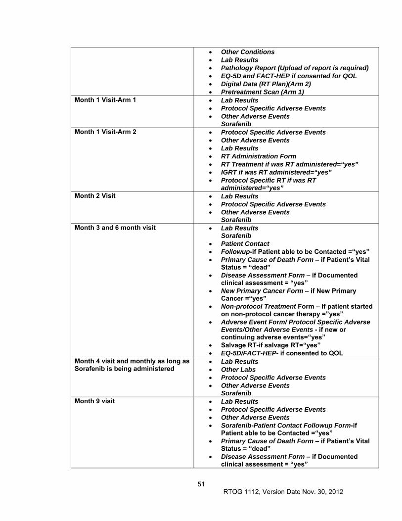
51 RTOG 1112, Version Date Nov. 30, 2012
Other Conditions Lab Results Pathology Report (Upload of report is required) EQ-5D and FACT-HEP if consented for QOL Digital Data (RT Plan)(Arm 2) Pretreatment Scan (Arm 1)
Month 1 Visit-Arm 1 Lab Results Protocol Specific Adverse Events Other Adverse Events
Sorafenib Month 1 Visit-Arm 2 Protocol Specific Adverse Events
Other Adverse Events Lab Results RT Administration Form RT Treatment if was RT administered=“yes” IGRT if was RT administered=“yes” Protocol Specific RT if was RT
administered=“yes” Month 2 Visit Lab Results
Protocol Specific Adverse Events Other Adverse Events
Sorafenib Month 3 and 6 month visit Lab Results
Sorafenib Patient Contact Followup-if Patient able to be Contacted =“yes” Primary Cause of Death Form – if Patient’s Vital
Status = “dead” Disease Assessment Form – if Documented
clinical assessment = “yes” New Primary Cancer Form – if New Primary
Cancer =“yes” Non-protocol Treatment Form – if patient started
on non-protocol cancer therapy =”yes” Adverse Event Form/ Protocol Specific Adverse
Events/Other Adverse Events - if new or continuing adverse events=“yes”
Salvage RT-if salvage RT=“yes” EQ-5D/FACT-HEP- if consented to QOL
Month 4 visit and monthly as long as Sorafenib is being administered
Lab Results Other Labs Protocol Specific Adverse Events Other Adverse Events
SorafenibMonth 9 visit Lab Results
Protocol Specific Adverse Events Other Adverse Events Sorafenib-Patient Contact Followup Form-if
Patient able to be Contacted =“yes” Primary Cause of Death Form – if Patient’s Vital
Status = “dead” Disease Assessment Form – if Documented
clinical assessment = “yes”
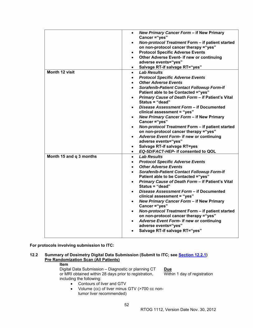
52 RTOG 1112, Version Date Nov. 30, 2012
New Primary Cancer Form – if New Primary Cancer =“yes”
Non-protocol Treatment Form – if patient started on non-protocol cancer therapy =“yes”
Protocol Specific Adverse Events Other Adverse Event- if new or continuing
adverse events=“yes” Salvage RT-if salvage RT=“yes”
Month 12 visit Lab Results Protocol Specific Adverse Events Other Adverse Events Sorafenib-Patient Contact Followup Form-if
Patient able to be Contacted =“yes” Primary Cause of Death Form – if Patient’s Vital
Status = “dead” Disease Assessment Form – if Documented
clinical assessment = “yes” New Primary Cancer Form – if New Primary
Cancer =“yes” Non-protocol Treatment Form – if patient started
on non-protocol cancer therapy =“yes” Adverse Event Form- if new or continuing
adverse events=“yes” Salvage RT-if salvage RT=yes EQ-5D/FACT-HEP- if consented to QOL
Month 15 and q 3 months Lab Results Protocol Specific Adverse Events Other Adverse Events Sorafenib-Patient Contact Followup Form-if
Patient able to be Contacted =“yes” Primary Cause of Death Form – if Patient’s Vital
Status = “dead” Disease Assessment Form – if Documented
clinical assessment = “yes” New Primary Cancer Form – if New Primary
Cancer =“yes” Non-protocol Treatment Form – if patient started
on non-protocol cancer therapy =“yes” Adverse Event Form- if new or continuing
adverse events=“yes” Salvage RT-if salvage RT=“yes”
For protocols involving submission to ITC: 12.2 Summary of Dosimetry Digital Data Submission (Submit to ITC; see Section 12.2.1)
Pre Randomization Scan (All Patients) Item Digital Data Submission – Diagnostic or planning CT Due or MRI obtained within 28 days prior to registration, Within 1 day of registration including the following:
Contours of liver and GTV Volume (cc) of liver minus GTV (>700 cc non-
tumor liver recommended)
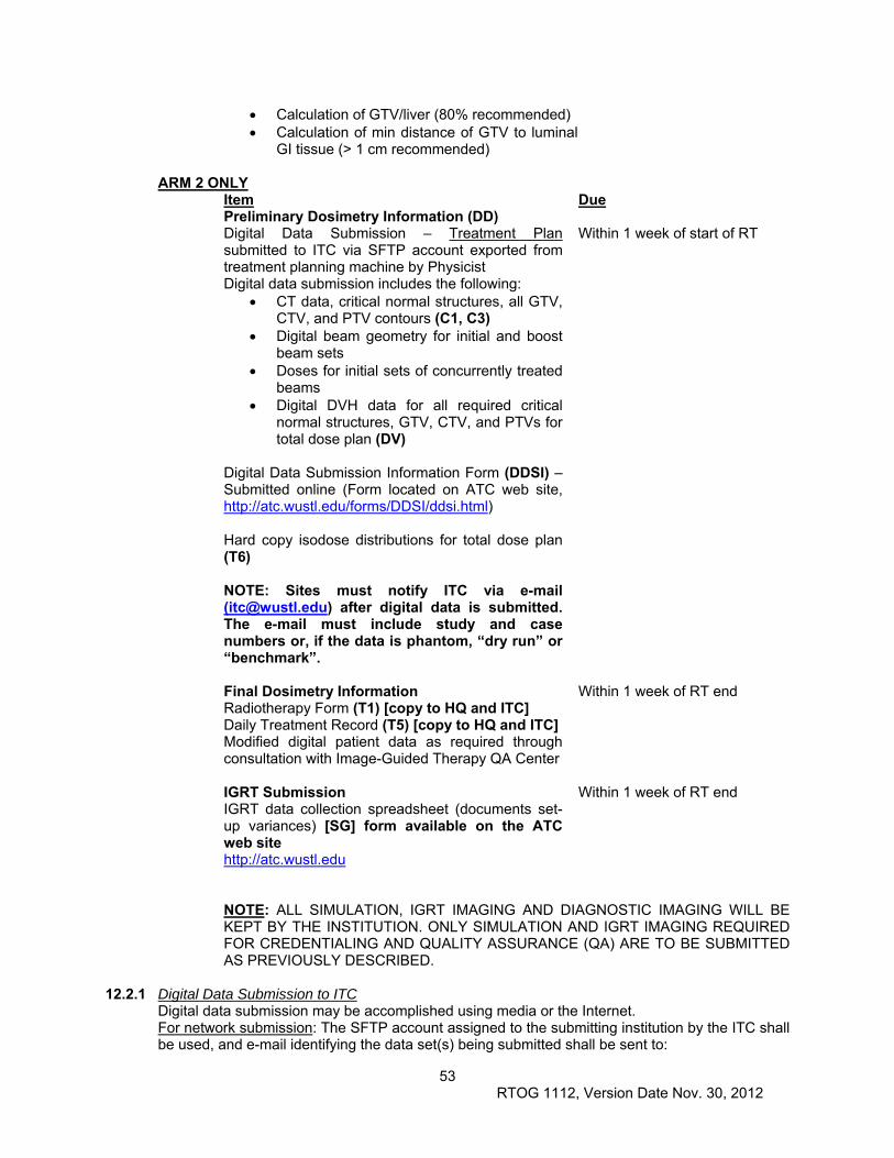
53 RTOG 1112, Version Date Nov. 30, 2012
Calculation of GTV/liver (80% recommended) Calculation of min distance of GTV to luminal
GI tissue (> 1 cm recommended)
ARM 2 ONLY Item Due Preliminary Dosimetry Information (DD) Digital Data Submission – Treatment Plan submitted to ITC via SFTP account exported from treatment planning machine by Physicist
Within 1 week of start of RT
Digital data submission includes the following: CT data, critical normal structures, all GTV,
CTV, and PTV contours (C1, C3)
Digital beam geometry for initial and boost beam sets
Doses for initial sets of concurrently treated beams
Digital DVH data for all required critical normal structures, GTV, CTV, and PTVs for total dose plan (DV)
Digital Data Submission Information Form (DDSI) – Submitted online (Form located on ATC web site, http://atc.wustl.edu/forms/DDSI/ddsi.html)
Hard copy isodose distributions for total dose plan (T6)
NOTE: Sites must notify ITC via e-mail ([email protected]) after digital data is submitted. The e-mail must include study and case numbers or, if the data is phantom, “dry run” or “benchmark”.
Final Dosimetry Information Within 1 week of RT end Radiotherapy Form (T1) [copy to HQ and ITC] Daily Treatment Record (T5) [copy to HQ and ITC] Modified digital patient data as required through consultation with Image-Guided Therapy QA Center
IGRT Submission Within 1 week of RT end IGRT data collection spreadsheet (documents set-up variances) [SG] form available on the ATC web site http://atc.wustl.edu
NOTE: ALL SIMULATION, IGRT IMAGING AND DIAGNOSTIC IMAGING WILL BE KEPT BY THE INSTITUTION. ONLY SIMULATION AND IGRT IMAGING REQUIRED FOR CREDENTIALING AND QUALITY ASSURANCE (QA) ARE TO BE SUBMITTED AS PREVIOUSLY DESCRIBED.
12.2.1 Digital Data Submission to ITC
Digital data submission may be accomplished using media or the Internet. For network submission: The SFTP account assigned to the submitting institution by the ITC shall be used, and e-mail identifying the data set(s) being submitted shall be sent to:
54 RTOG 1112, Version Date Nov. 30, 2012
For media submission: Please contact the ITC about acceptable media types and formats. Hardcopies accompanying digital data should be sent by mail or Federal Express and should be addressed to:
Image-Guided Therapy Center (ITC) ATTN: Roxana Haynes 4511 Forest Park, Suite 200 St. Louis, MO 63108 314-747-5415 FAX 314-747-5423
NOTE: Copies of simulation and port films and the complete RT daily treatment record for the previous original EBRT will be submitted to RTOG Headquarters ONLY if specifically requested.
13.0 STATISTICAL CONSIDERATIONS 13.1 Endpoints 13.1.1 Primary Endpoint
Overall survival (failure is death due to any cause) 13.1.2 Secondary Endpoints
Time to progression (failure is defined per Section 11.3) Progression free survival (failure is progression or death due to any cause) Toxicity: using Common Toxicity Criteria (CTCAE) version 4.0. Vascular thrombosis response Health Related Quality of Life [measured by the Functional Assessment of Cancer
Therapy–Hepatobiliary (FACT-Hep)] Quality adjusted survival
13.2 Stratification
Patients will be stratified before randomization with respect to Vascular involvement (IVC/main portal vein/right or left main branch portal vein vs. other vascular involvement vs. none), Hepatitis (B vs. C vs. other), Site (North American vs. non-North American), and HCC volume/liver volume (<10% vs. 10-40 vs. >40%). The treatment allocation scheme described by Zelen (1974) will be used because it balances patient factors other than institution.
13.3 Sample Size and Power Justification 13.3.1 The sample size calculations are based on the primary hypothesis that SBRT followed by
sorafenib will increase overall survival as compared to sorafenib alone for patients with hepatocellular carcinoma. It is projected that 75% of the accrual will come from North American sites and 25% of the accrual will come from non-North American sites. It is expected that approximately two-thirds of accrual will be patients with vascular thrombosis while one-third will be patients without vascular thrombosis. The sorafenib alone control arm median overall survival time (MST) is estimated to be 10.5 months, based on the SHARP sorafenib arm MST of 10.7 months (Llovet 2008) and a recent study of 1073 HCC patients randomized to sorafenib versus sunitinib; the median overall survival time (MST) of the patients in the sorafenib arm was 10.0 months (versus 8.1 in the sunitinib arm) [Cheng 2011]. Approximately 75% of these patients were Asian-Pacific, demonstrating that outcomes in Asia have improved since the original Asian-Pacific sorafenib randomized trial (where MST was 6.5 months for sorafenib alone [Cheng 2009]). The RT sorafenib combination arm MST is hypothesized to be 14.5 months, based on the Toronto RT alone MST of 17.0 months (Bujold 2012) in 102 patients with advanced HCC treated with RT alone on study, as well as a retrospective study of RT and sunitinib from Taiwan with a
55 RTOG 1112, Version Date Nov. 30, 2012
MST of 16 months (Chi 2010). Other reports of RT alone for HCC patients with portal vein thrombosis demonstrate a range of MSTs from 10 to 15 months, in the absence of sorafenib. The required sample size for the primary endpoint of OS is based on the following conditions:
OS times are exponentially distributed with (at least approximately) constant hazards in both treatment arms
The control arm will have a median OS of 10.5 months (monthly hazard of 0.066) The experimental arm will have a median OS of 14.5 months (monthly hazard of
0.0478) Hazard ratio (experimental/control) = 0.72 One-sided test at = 0.05 Statistical power of 85% 5 years of accrual (post ramp-up) with 1 year of follow-up Two interim significance tests and a final test are planned using the Haybittle-
Peto (Lan 1983; O’Brien 1979) rule for efficacy and a more aggressive futility rule suggested by Weiand et al (1994).
A total of 320 evaluable patients, using the group sequential design method (Pocock 1977) with 2 interim analyses, will provide the 277 OS events required to determine if the addition of SBRT to sorafenib alone improves overall survival from 10.5 to 14.5 months (HR=0.72). Given the conditions above, and adjusting for ineligible/lost patients or patients that cannot meet the RT planning requirements, a total sample size of 368 patients will be required to be accrued uniformly over 5 years, post ramp-up period, with an additional 1 year of follow-up.
13.3.2 Patient Accrual Patient accrual is projected to be 6 cases per month, with a ramp-up period in the first 6 months with no projected accrual. Following this ramp-up period, accrual will be completed in 5 years. Projected accrual was based on a survey submitted in June 2011 by the RTOG to all full RTOG members, the members of the RTOG GI Steering Committee and interested Asian centers. In brief, there were 43 respondents, including the 2 Asian centers. Twenty-eight centers planned to definitely open the study; 22 of these centers are already credentialed for RTOG lung or liver SBRT studies (excluding the 2 Asian centers). An additional 13 centers were considering opening the study (all of these centers were credentialed for RTOG lung or liver SBRT studies). Based on the 22 credentialed RTOG non-Asian centers, 12 patients per month were estimated to be accrued to this study. Including the Asian centers, 17 patients per month were estimated to participate. Accounting for some overestimation and lower than expected accrual in prior HCC studies, the actual expected accrual is 6 patients per month.
If the total accrual during months 13 through 18 of the study is ≥ 50% of the targeted accrual (18 or more cases), then the accrual will have met the NCI-CTEP Ph III accrual guidelines and will continue. If the total accrual during months 13 through 18 of the study is ≤ 20% of the targeted accrual (≤ 7 cases in total), then the protocol will be discontinued per NCI-CTEP accrual guidelines for phase III studies. If the total accrual during months 13 through 18 is between 21% and 49% (8 to 17 cases), then the protocol will continue to accrue subjects and will be evaluated again at the end of month 24. If the accrual during months 22 through 24 is at least 50% of the targeted accrual (≥ 9 cases in total), the NCI-CTEP accrual guidelines for phase III studies will have been met and the study will continue accrual; otherwise, the study will be discontinued.
13.4 Power Information for Health Reported Quality of Life – FACT-Hepatobiliary Module (FACT-
Hep) The Functional Assessment of Cancer Therapy – Hepatobiliary (FACT-Hep) will be used to measure HRQOL. Protocol-eligible patients will be included in the QOL analysis only if they have provided baseline and at least one subsequent measurement. The FACT-Hep will be collected on all cases participating in this portion of the trial and will be collected at 4 time points: baseline, 3 months, 6 months, and 12 months post initiation of protocol therapy.
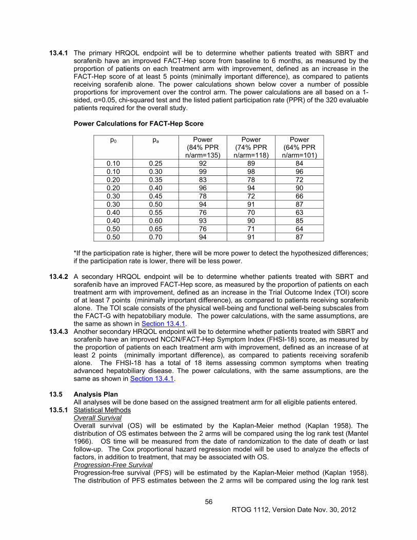
56 RTOG 1112, Version Date Nov. 30, 2012
13.4.1 The primary HRQOL endpoint will be to determine whether patients treated with SBRT and sorafenib have an improved FACT-Hep score from baseline to 6 months, as measured by the proportion of patients on each treatment arm with improvement, defined as an increase in the FACT-Hep score of at least 5 points (minimally important difference), as compared to patients receiving sorafenib alone. The power calculations shown below cover a number of possible proportions for improvement over the control arm. The power calculations are all based on a 1-sided, α=0.05, chi-squared test and the listed patient participation rate (PPR) of the 320 evaluable patients required for the overall study.
Power Calculations for FACT-Hep Score
p0 pa Power
(84% PPR n/arm=135)
Power (74% PPR n/arm=118)
Power (64% PPR n/arm=101)
0.10 0.25 92 89 84 0.10 0.30 99 98 96 0.20 0.35 83 78 72 0.20 0.40 96 94 90 0.30 0.45 78 72 66 0.30 0.50 94 91 87 0.40 0.55 76 70 63 0.40 0.60 93 90 85 0.50 0.65 76 71 64 0.50 0.70 94 91 87
*If the participation rate is higher, there will be more power to detect the hypothesized differences; if the participation rate is lower, there will be less power.
13.4.2 A secondary HRQOL endpoint will be to determine whether patients treated with SBRT and
sorafenib have an improved FACT-Hep score, as measured by the proportion of patients on each treatment arm with improvement, defined as an increase in the Trial Outcome Index (TOI) score of at least 7 points (minimally important difference), as compared to patients receiving sorafenib alone. The TOI scale consists of the physical well-being and functional well-being subscales from the FACT-G with hepatobiliary module. The power calculations, with the same assumptions, are the same as shown in Section 13.4.1.
13.4.3 Another secondary HRQOL endpoint will be to determine whether patients treated with SBRT and sorafenib have an improved NCCN/FACT-Hep Symptom Index (FHSI-18) score, as measured by the proportion of patients on each treatment arm with improvement, defined as an increase of at least 2 points (minimally important difference), as compared to patients receiving sorafenib alone. The FHSI-18 has a total of 18 items assessing common symptoms when treating advanced hepatobiliary disease. The power calculations, with the same assumptions, are the same as shown in Section 13.4.1.
13.5 Analysis Plan
All analyses will be done based on the assigned treatment arm for all eligible patients entered. 13.5.1 Statistical Methods
Overall Survival Overall survival (OS) will be estimated by the Kaplan-Meier method (Kaplan 1958). The distribution of OS estimates between the 2 arms will be compared using the log rank test (Mantel 1966). OS time will be measured from the date of randomization to the date of death or last follow-up. The Cox proportional hazard regression model will be used to analyze the effects of factors, in addition to treatment, that may be associated with OS. Progression-Free Survival Progression-free survival (PFS) will be estimated by the Kaplan-Meier method (Kaplan 1958).
The distribution of PFS estimates between the 2 arms will be compared using the log rank test
57 RTOG 1112, Version Date Nov. 30, 2012
(Mantel 1966). PFS time will be measured from the date of randomization to the date of first failure or last follow-up. The Cox proportional hazard regression model will be used to analyze the effects of factors, in addition to treatment, that may be associated with PFS. Time-to-Progression (TTP) Time-to-progression (TTP) will be estimated by the cumulative incidence method (Mantel 1966).
The distribution of TTP estimates between the 2 arms will be compared using Gray’s test (Gray 1988). TTP time will be measured from the date of randomization to the date of first failure or last follow-up. The Cox proportional hazard regression model will be used to analyze the effects of factors, in addition to treatment, that may be associated with TTP.
13.5.2 Interim Analysis to Monitor the Study Progress Interim reports will be prepared twice per year until the initial treatment results have been presented/published. In general, the interim reports will contain the following information:
Patient accrual rate with a projected completion date (while the study is still accruing)
Total patients accrued Distributions of important pretreatment and prognostic baseline variables The frequencies and severity of adverse events by treatment arm Compliance rates of treatment delivery
The interim reports will not contain the results from the treatment comparisons with respect to the primary endpoint, OS, or any secondary endpoints, with the exception of reporting of adverse events.
13.5.3 CDUS Reports This study will be monitored by the Clinical Data Update System (CDUS) version 3.0. Cumulative
CDUS data will be submitted quarterly by electronic means. Reports are due January 31, April 30, July 31, and October 31.
13.5.4 Significance Testing for Early Termination and/or Reporting Unacceptable Toxicity To address the safety of SBRT followed by sorafenib, the rate of unacceptable adverse events will be evaluated on both treatment arms. This analysis will focus on the following AEs occurring within 90 days from the start of protocol treatment definitely or probably related to protocol treatment: a) grade 4 or 5 hepatic b) grade 4 or 5 gastrointestinal c) grade 4 thrombocytopenia associated with any bleeding or grade 5 thrombocytopenia d) Any grade 5 treatment-related adverse event Assuming no more than a 10% rate of the above AEs on the sorafenib alone arm, the study chairs have determined that an increase to a rate of 30% or greater on the SBRT followed by sorafenib arm will be considered to be unacceptable. One-hundred and fifty-four patients provide 90% power to detect an increase in the rate of specified AEs from 10% to at least 30% with a 1-sided alpha of 0.05, using a Chi-squared test for difference in proportions. If the p-value from the test described above is ≤ 0.05, the conclusion will be that the treatment-related unacceptable AE rate for the SBRT followed by sorafenib arm is at least 30% and accrual to the study will be stopped. Primary Endpoint: Overall survival (OS) Two interim significance tests of treatment difference are planned. The timing of the interim analyses will be based on OS failure events, as described in Section 13.1.1. The maximum number of events required for the study is 277. Under the alternative hypothesis that the addition of SBRT will increase median OS from 10.5 months to 14.5 months, the projected numbers of events and the nominal significance levels for rejecting the H0 or the H1 for each of these two interim analyses are shown in the table below:
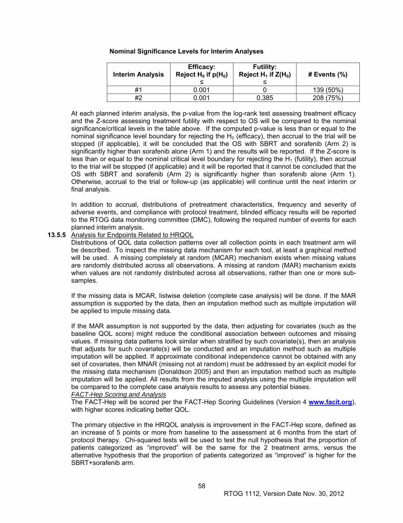
58 RTOG 1112, Version Date Nov. 30, 2012
Nominal Significance Levels for Interim Analyses
Interim Analysis
Efficacy: Reject H0 if p(H0)
≤
Futility: Reject H1 if Z(H0)
≤
# Events (%)
#1 0.001 0 139 (50%) #2 0.001 0.385 208 (75%)
At each planned interim analysis, the p-value from the log-rank test assessing treatment efficacy and the Z-score assessing treatment futility with respect to OS will be compared to the nominal significance/critical levels in the table above. If the computed p-value is less than or equal to the nominal significance level boundary for rejecting the H0 (efficacy), then accrual to the trial will be stopped (if applicable), it will be concluded that the OS with SBRT and sorafenib (Arm 2) is significantly higher than sorafenib alone (Arm 1) and the results will be reported. If the Z-score is less than or equal to the nominal critical level boundary for rejecting the H1 (futility), then accrual to the trial will be stopped (if applicable) and it will be reported that it cannot be concluded that the OS with SBRT and sorafenib (Arm 2) is significantly higher than sorafenib alone (Arm 1). Otherwise, accrual to the trial or follow-up (as applicable) will continue until the next interim or final analysis. In addition to accrual, distributions of pretreatment characteristics, frequency and severity of adverse events, and compliance with protocol treatment, blinded efficacy results will be reported to the RTOG data monitoring committee (DMC), following the required number of events for each planned interim analysis.
13.5.5 Analysis for Endpoints Related to HRQOL Distributions of QOL data collection patterns over all collection points in each treatment arm will be described. To inspect the missing data mechanism for each tool, at least a graphical method will be used. A missing completely at random (MCAR) mechanism exists when missing values are randomly distributed across all observations. A missing at random (MAR) mechanism exists when values are not randomly distributed across all observations, rather than one or more sub-samples. If the missing data is MCAR, listwise deletion (complete case analysis) will be done. If the MAR assumption is supported by the data, then an imputation method such as multiple imputation will be applied to impute missing data. If the MAR assumption is not supported by the data, then adjusting for covariates (such as the baseline QOL score) might reduce the conditional association between outcomes and missing values. If missing data patterns look similar when stratified by such covariate(s), then an analysis that adjusts for such covariate(s) will be conducted and an imputation method such as multiple imputation will be applied. If approximate conditional independence cannot be obtained with any set of covariates, then MNAR (missing not at random) must be addressed by an explicit model for the missing data mechanism (Donaldson 2005) and then an imputation method such as multiple imputation will be applied. All results from the imputed analysis using the multiple imputation will be compared to the complete case analysis results to assess any potential biases. FACT-Hep Scoring and Analysis The FACT-Hep will be scored per the FACT-Hep Scoring Guidelines (Version 4 www.facit.org), with higher scores indicating better QOL. The primary objective in the HRQOL analysis is improvement in the FACT-Hep score, defined as an increase of 5 points or more from baseline to the assessment at 6 months from the start of protocol therapy. Chi-squared tests will be used to test the null hypothesis that the proportion of patients categorized as “improved” will be the same for the 2 treatment arms, versus the alternative hypothesis that the proportion of patients categorized as “improved” is higher for the SBRT+sorafenib arm.
59 RTOG 1112, Version Date Nov. 30, 2012
Improvement in the FACT-Hep score, as defined above, will also be compared between the treatment arms for changes from baseline to both 3 and 12 months with the same methodology as listed above. Correlation of baseline FACT-Hep and survival will be evaluated.
EQ-5D Scoring and Analysis The quality-adjusted survival of each treatment will be evaluated and compared using EQ-5D if the primary endpoint supports the primary hypothesis. The EQ-5D is a 2-part self-assessment questionnaire. The first part consists of 5 items covering 5 dimensions (mobility, self care, usual activities, pain/discomfort, and anxiety/depression). Each dimension is measured by a 3-point likert scale (1-no problems, 2-moderate problems, and 3-extreme problems). The second part is a visual analog scale (VAS) valuing the current health state measured by a 100-point scale with a 10-point interval (0-worst imaginable health state, 100-best imaginable health state). We will transform the 5-item index score and VAS score into a utility score between 0 (worst health state) and 1 (best health state) for comparative purposes. Patients will complete the EQ-5D at the following time points: pretreatment (baseline), 3 months, 6 months, and 12 months post initiation of protocol therapy. To examine trade-offs between the survival time and QOL, they will be combined for each patient into a single measurement: quality-adjusted life years (QALY). If (and only if) the primary endpoint hypothesis is substantiated, a quality-adjusted survival analysis will be conducted. The quality- adjusted survival analysis will not be done until after the primary endpoint results are published. QALY is defined by the weighted sum of different time episodes added up to a total quality- adjusted survival time. QALY will be analyzed at 2 time points: at 6 and 12 months from start of treatment, using the EQ-5D.
13.5.6 Analysis for Reporting the Initial Treatment Results The primary hypothesis of this study is SBRT and sorafenib will increase the median OS from 10.5 months to 14.5 months as compared to sorafenib alone for patients with hepatobiliary carcinoma. This major analysis will occur after at least 277 OS failure events have been observed, unless an early stopping rule is satisfied. It will include:
Tabulation of all cases entered and those excluded from the analyses with the
reasons for exclusion given Distributions of important prognostic baseline variables The frequencies and severity of adverse events by treatment arm. Compliance rate of treatment delivery Observed results with respect to the primary and secondary endpoints
All eligible patients randomized will be included in the comparison and will be grouped by assigned treatment in the analysis. The primary hypothesis of treatment benefit will be tested using the log-rank statistic with a significance level of 0.05, given that the 2 interim analyses were carried out per Section 13.5.4. Additional analyses of treatment effect will be performed using the Cox proportional hazard model with the stratification factor included as a fixed covariate, as well as any factors that show an imbalance between the arms (eg, age, gender, race, Zubrod status, etc.).
13.6 Gender and Minorities Both men and women of all races and ethnic groups are eligible for this study. In conformance with the national Institutes of Health (NIH) Revitalization Act of 1993 with regard to inclusion of women and minorities in clinical research, possible interaction between race/ethnicity and treatment has been considered. It is projected that 80% of the patients will be men and 20% women; 2% will be of Hispanic or Latino ethnicity and 98% will not; racial distribution will be 73% white, 2% black or African American, and 25% Asian. Assuming no differences between the ethnicities, or among the races, the statistical power for detecting the hypothesized treatment
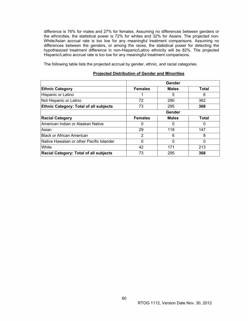
60 RTOG 1112, Version Date Nov. 30, 2012
difference is 76% for males and 27% for females. Assuming no differences between genders or the ethnicities, the statistical power is 72% for whites and 32% for Asians. The projected non-White/Asian accrual rate is too low for any meaningful treatment comparisons. Assuming no differences between the genders, or among the races, the statistical power for detecting the hypothesized treatment difference in non-Hispanic/Latino ethnicity will be 82%. The projected Hispanic/Latino accrual rate is too low for any meaningful treatment comparisons. The following table lists the projected accrual by gender, ethnic, and racial categories.
Projected Distribution of Gender and Minorities
Gender
Ethnic Category Females Males Total
Hispanic or Latino 1 5 6
Not Hispanic or Latino 72 290 362
Ethnic Category: Total of all subjects 73 295 368
Gender
Racial Category Females Males Total
American Indian or Alaskan Native 0 0 0
Asian 29 118 147
Black or African American 2 6 8
Native Hawaiian or other Pacific Islander 0 0 0
White 42 171 213
Racial Category: Total of all subjects 73 295 368
61 RTOG 1112, Version Date Nov. 30, 2012
REFERENCES Ben-Josef E, Normolle D, Ensminger WD, et al. Phase II trial of high-dose conformal radiation therapy with concurrent hepatic artery floxuridine for unresectable intrahepatic malignancies. J Clin Oncol. 23:8739-8747, 2005. Blomgren H, Lax I, Naslund I, et al. Stereotactic high dose fraction radiation therapy of extracranial tumors using an accelerator. Clinical experience of the first thirty-one patients. Acta Oncol. 34: 861-70, 1995. Brade A, Kim J, Brierley J. et al. Phase I Study of Sorafenib and Whole Liver Radiotherapy (WLRT) or Stereotactic body radiotherapy (SBRT) for Liver Metastases. abstract. ASTRO 2012. In press. Bujold A, Sykes J, Kim JJ, Brierley J, Cho C, Wong R, Dinniwell R, Kassam Z, Ringash J, Cummings B, Massey C, Sherman M, Knox JJ, Dawson LA. Results from Sequential Phase I and II Trials of Stereotactic Body Radiotherapy for Hepatocellular Carcinoma. Journal Clinical Oncology. In press online 2012 Bush DA, Hillebrand DJ, Slater JM, Slater JD. High-dose proton beam radiotherapy of hepatocellular carcinoma: preliminary results of a phase II trial. Gastroenterology. 127: S189-193, 2004. Cardenes HR, Price TR, Perkins SM, et al. Phase I feasibility trial of stereotactic body radiation therapy for primary hepatocellular carcinoma. Clin Transl Oncol. 12: 218-25, 2010. Carr BI, Carroll S, Muszbek N, et al. Economic evaluation of sorafenib in unresectable hepatocellular carcinoma. J Gastroenterol Hepatol. 25(11): 1739-46, 2010. Cella D et al; Validity of the FACT Hepatobiliary (FACT-Hep) Questionnaire for Assessing Disease-related Symptoms and Health-related QoL in Patients with Metastatic Pancreatic Cancer. Qual Life Res Jun 8, 2012 [Epub ahead of print]). Chang YS, Adnane J, Trail PA, et al. Sorafenib (BAY 43-9006) Inhibits Tumor Growth and Vascularization and Induces Tumor Apoptosis and Hypoxia in RCC Xenograft Models. Cancer Chemother Pharmacol. 59(5): 561-574, 2007. Cheng AL, Kang YK, Chen Z et al. Efficacy and safety of sorafenib in patients in the Asia-Pacific region with advanced hepatocellular carcinoma: a phase III randomized, double blind, placebo controlled trial. Lancet Oncol. 10(1): 25-34, 2009. Cheng A, Kong D, Lin J, et al. Phase III trial of sunitinib (Su) versus sorafenib (So) in advanced hepatocellular carcinoma (HCC). Abstract #4000. ASCO 2011. Cheng JC, Chuang VP, Cheng SH, et al. Local radiotherapy with or without transcatheter arterial chemoembolization for patients with unresectable hepatocellular carcinoma. Int J Radiat Oncol Biol Phys. 47: 435-442, 2000. Chi KH, Lias CS, Chang CC, et al. Angiogenic Blockade and Radiotherapy in Hepatocellular Carcinoma. Int J Radiat Oncol Biol Phys. 78(1): 188-193, 2010. Chiba T, Tokuuye K, Matsuzaki Y, et al. Proton beam therapy for hepatocellular carcinoma: a retrospective review of 162 patients. Clin Cancer Res. 11: 3799-3805, 2005. Dawson LA, Eccles C, Craig T. Individualized Image Guided Iso-NTCP based Liver Cancer SBRT. Acta Oncol. 45: 856 – 864, 2006. Dawson LA, Brade A, Cho C et al. Phase I Study of Sorafenib and SBRT for Advanced Hepatocellular Carcinoma. abstract. ASTRO 2012. In press. Donaldson GW, Moinpour CM. Learning to live with missing quality of life data in advanced-stage disease trials. J Clin Oncol. 2005;23:1-5. Fukumitsu N, Sugahara S, Nakayama H, et al. A prospective study of hypofractionated proton beam therapy for patients with hepatocellular carcinoma. International Journal of Radiation Oncology, Biology, Physics 74:831-836 2009. Glasziou PP, Simes RJ, Gelber RD. Quality adjusted survival analysis. Stat Med. 9: 1256-1276, 1990. Gray RJ. A class of K-samples test for comparing the cumulative incidence of a competing risk. Ann Statistics 1988; 16(3):1141-54. Guo WJ, Yu EX, Liu LM, et al. Comparison between chemoembolization combined with radiotherapy and chemoembolization alone for large hepatocellular carcinoma. World J Gastroenterol. 9: 1697-1701, 2003. Hata M, Tokuuye K, Sugahara S, et al. Proton beam therapy for hepatocellular carcinoma with portal vein tumor thrombus. Cancer. 104: 794-801, 2005. Hata M, Tokuuye K, Sugahara S, et al. Proton beam therapy for hepatocellular carcinoma with limited treatment options. Cancer. 107: 591-598, 2006.
62 RTOG 1112, Version Date Nov. 30, 2012
Heffernan N, Cella D, Webster K, et al. Measuring Health-Related Quality of Life in Patients With Hepatobiliary Cancers: The Functional Assessment of Cancer Therapy–Hepatobiliary Questionnaire . J Clin Oncol. 20(9): 2229-2239, 2002. Huang YJ, Hsu HC, Wang CY, et al. The treatment responses in cases of radiation therapy to portal vein thrombosis in advanced hepatocellular carcinoma. Int J Radiat Oncol Biol Phys. 73: 1155-63, 2009. Huo TI, Lin HC, Hsia CY, et al. The model for end-stage liver disease based cancer staging systems are better prognostic models for hepatocellular carcinoma: a prospective sequential survey. Am J Gastroenterol. 102: 1920-30, 2007. Jain N, Suit RK, Boucher HD, et al. Anti-Vascular endothelial growth factor treatment augments tumor radiation response under normoxic or hypoxic conditions. Cancer Res. 60: 5565-5570, 2000. Jemal A, Siegel R, Xu J, et al. Cancer statistics, 2010. CA: a Cancer Journal for Clinicians. 60: 277-300, 2010. Kaplan EL, Meier P. Nonparametric estimation from incomplete observations. J Stat Assoc. 53: 457-81, 1958. Kato H, Tsujii H, Miyamoto T, et al. Results of the first prospective study of carbon ion radiotherapy for hepatocellular carcinoma with liver cirrhosis. Int J Radiat Oncol Biol Phys. 59: 1468-1476, 2004. Kawashima M, Furuse J, Nishio T, et al. Phase II study of radiotherapy employing proton beam for hepatocellular carcinoma. J Clin Oncol. 23: 1839-1846, 2005. Kim TH, Kim DY, Park JW, et al. Three-dimensional conformal radiotherapy of unresectable hepatocellular carcinoma patients for whom transcatheter arterial chemoembolization was ineffective or unsuitable. Am J Clin Oncol. 29: 568-575, 2006. Koo JE, Kim JH, Lim YS, et al: Combination of transarterial chemoembolization and three-dimensional conformal radiotherapy for hepatocellular carcinoma with inferior vena cava tumor thrombus. Int J Radiat Oncol Biol Phys. 78: 180-187, 2010. Kwon JH, Bae SH, Kim JY, et al. Long-term effect of stereotactic body radiation therapy for primary hepatocellular carcinoma ineligible for local ablation therapy or surgical resection. Stereotactic radiotherapy for liver cancer. BMC Cancer. 10: 475, 2010. Lan KKG, DeMets ER. Discrete sequential boundaries for clinical trials. Biometrika. 1983;70: 659-663. Li B, Yu J, Wang L, et al. Study of local three-dimensional conformal radiotherapy combined with transcatheter arterial chemoembolization for patients with stage III hepatocellular carcinoma. Am J Clin Oncol. 26:e92-99, 2003. Liang SX, Zhu XD, Lu HJ, et al. Hypofractionated three-dimensional conformal radiation therapy for primary liver carcinoma. Cancer. 103: 2181-2188, 2005. Liu L, Cao Y, Chen C, Zhang X, et al. Sorafenib blocks the RAF/MEK/ERK pathway, inhibits tumor angiogenesis, and induces tumor cell apoptosis in hepatocellular carcinoma model PLC/PRF/5. Cancer Res. 66: 11851-11858, 2006. Liu MT, Li SH, Chu TC, et al: Three-dimensional conformal radiation therapy for unresectable hepatocellular carcinoma patients who had failed with or were unsuited for transcatheter arterial chemoembolization. Jpn J Clin Oncol 34: 532-539, 2004. Llovet JM, Real MI, Montana X, et al. Arterial embolisation or chemoembolisation versus symptomatic treatment in patients with unresectable hepatocellular carcinoma: a randomised controlled trial. Lancet. 359: 1734-1739, 2002. Llovet JM, Di Besceglie AM, Bruix J, et al. Design and endpoints of clinical trials in hepatocellular caricinoma. J Natl Cancer Inst. 100: 698-711, 2008. Llovet JM, Ricci S, Mazzaferro V, et al. Sorafenib in Advanced Hepatocellular Carcinoma. New Engl J Med. 359: 378-390, 2008. Lo CM, Ngan H, Tso WK, et al. Randomized controlled trial of transarterial lipiodol chemoembolization for unresectable hepatocellular carcinoma. Hepatology. 35: 1164-1171, 2002. Louis C, Dewas S, Mirabel X, et al. Stereotactic radiotherapy of hepatocellular carcinoma: preliminary results. Technol Cancer Res Treat. 9: 479-87, 2010. Mantel N. Evaluation of survival data and two new rank order statistics arising in its consideration. Cancer Chemother. Rep 50: 163-70, 1966. McIntosh A, Hagspiel KD, Al-Osaimi AM, et al. Accelerated treatment using intensity-modulated radiation therapy plus concurrent capecitabine for unresectable hepatocellular carcinoma. Cancer. 115: 5117-5125, 2009.
63 RTOG 1112, Version Date Nov. 30, 2012
Mendez-Romero A, Wunderink W, Hussain SM, et al. Stereotactic body radiation therapy for primary and metastatic liver tumors: A single institution phase i-ii study. Acta Oncol. 45: 831-837, 2006. Mizumoto M, Tokuuye K, Sugahara S, et al. Proton beam therapy for hepatocellular carcinoma adjacent to the porta hepatis. Int J Radiat Oncol Biol Phys. 71: 462-467, 2008. Mornex F, Girard N, Beziat C, et al. Feasibility and efficacy of high-dose three-dimensional-conformal radiotherapy in cirrhotic patients with small-size hepatocellular carcinoma non-eligible for curative therapies-mature results of the French Phase II RTF-1 trial. Int J Radiat Oncol Biol Phys. 66: 1152-1158, 2006. O'Brien PC, Fleming TR. A multiple testing procedure for clinical trials. Biometrics. 1979;35:549-556. Pan C, Dawson LA, Das SK, et al. Radiation-associated liver injury. Special QUANTEC Issue. Int J Radiat Oncol Biol Phys. 76(3): S94-S100, 2010. Plastaras JP, Kim SH, Liu YY, et al. Cell cycle dependent and schedule-dependent antitumor effects of sorafenib combined with radiation. Cancer Res. 67(19): 9443-54, 2007. Pocock SJ. Group sequential methods in the design and analysis of clinical trials. Biometrika. 1977;64:191-199. Raoul JL, Sangro B, Forner A, et al. Evolving strategies for the management of intermediate-stage hepatocellular carcinoma: Available evidence and export opinion on the use of transarterial chemoembolization. Cancer Treat Rev. 37(3): 212-220, 2011. Ringash J, Lockwood G, Lee M, et al. Responsiveness OF FACT-Hep and EORTC QLQ-C30 to Radiotherapy for Malignant Liver Tumors. Presented at ISOQOL. Abstract 1696, 2008. Schultz MW, Chen J, Woo HH, et al. A comparison of techniques for eliciting patient preference in patients with benign prostatic hyperplasia. J Urol. 168: 155-159, 2002. Seo YS, Kim MS, Yoo SY, et al: Preliminary result of stereotactic body radiotherapy as a local salvage treatment for inoperable hepatocellular carcinoma. J Surg Oncol 102:209-14, 2010 Seong J, Park HC, Han KH, et al. Clinical results and prognostic factors in radiotherapy for unresectable hepatocellular carcinoma: a retrospective study of 158 patients. Int J Radiat Oncol Biol Phys. 55:329-336, 2003. Seong J, Lee IJ, Shim SJ, et al. A multicenter retrospective cohort study of practice patterns and clinical outcome on radiotherapy for hepatocellular carcinoma in Korea. Liver Int. 29: 147-52, 2009. Shim SJ, Seong J, Han KH, et al. Local radiotherapy as a complement to incomplete transcatheter arterial chemoembolization in locally advanced hepatocellular carcinoma. Liver Int. 25: 1189-1196, 2005. Son SH, Choi BO, Ryu MR, et al. Stereotactic body radiotherapy for patients with unresectable primary hepatocellular carcinoma: dose-volumetric parameters predicting the hepatic complication. Int J Radiat Oncol Biol Phys. 78: 1073-80, 2010. Steel J, Baum A, Carr B. Quality of life in patients diagnosed with primary hepatocellular carcinoma: hepatic arterial infusion of cisplatin versus 90-yttrium microspheres (therasphere). Psychooncology. 13: 73–79, 2004. Steel JL, Eton DT, Cella D, et al. Clinically meaningful changes in health-related quality of life in patients diagnosed with hepatobiliary carcinoma. Ann Oncol 17(2): 304-312, 2006. Sugahara S, Oshiro Y, Nakayama H, et al. Proton beam therapy for large hepatocellular carcinoma. Int J Radiat Oncol Biol Phys. 76: 460-466, 2010. Toya R, Murakami R, Baba Y, et al: Conformal radiation therapy for portal vein tumor thrombosis of hepatocellular carcinoma. Radiother Oncol. 84: 266-71, 2007. Tse R, Kim, J, Cummings, B, et al. Phase I Study of Stereotactic Radiation Therapy for Primary Liver Cancer. J Clin Oncol. 26: 657-664, 2008. Tsujii H, Mizoe JE, Kamada T, et al. Overview of clinical experiences on carbon ion radiotherapy at NIRS. Radiother Oncol. 73(2):S41-49, 2004. Wang YB, Chen MH, Yan K, et al. Quality of life after radiofrequency ablation combined with transcatheter arterial chemoembolization for hepatocellular carcinoma: comparison with transcatheter arterial chemoembolization alone. Qual Life Res. 16: 389–397, 2007. Wieand, S., Schroeder, G. and O'Fallon, J. R. (1994), Stopping when the experimental regimen does not appear to help. Statist. Med., 13: 1453–1458. doi: 10.1002/sim.4780131321 Wilhelm SM, Carter C, Tang L., et al. BAY 43-9006 Exhibits Broad Spectrum Oral Antitumor Activity and Targets the RAF/MEK/ERK Pathway and Receptor Tyrosine Kinases Involved in Tumor Progression and Angiogenesis. Cancer Res. 64: 7099-7109, 2004.
64 RTOG 1112, Version Date Nov. 30, 2012
Winkler F, Kozin SV, Tong RT, et al. Kinetic os Vascular Normalization by VEGFR2 Blockade Governs Brain Tumor Response to Radiation: Role of Oxygenation, Antiopoietin-1, and Matrix Metalloproteinases. Cancer Cell. 6: 553-563, 2004. Wu AW, Jacobson KD, Frick DL, et al. Validity and responsiveness of the EQ-5D as a measure of health-related quality of life in people enrolled in an AIDS clinical trial. Qual Life Res. 11: 273-282, 2002. Yoon SM, Young-Suk L, Won HJ, et al. Radiotherapy plus transarterial chemoembolization for hepatocellular carcinoma invading the portal vein: Long term patient outcomes. Int J Radiat Oncol Biol Phys. 82: 2004-2011, 2012. Zelen M. The randomization and stratification of patients to clinical trials. J Chron Dis. 27: 365-75, 1974. Zeng ZC, Tang ZY, Fan J, et al. A comparison of chemoembolization combination with and without radiotherapy for unresectable hepatocellular carcinoma. Cancer. 10: 307-316, 2004.
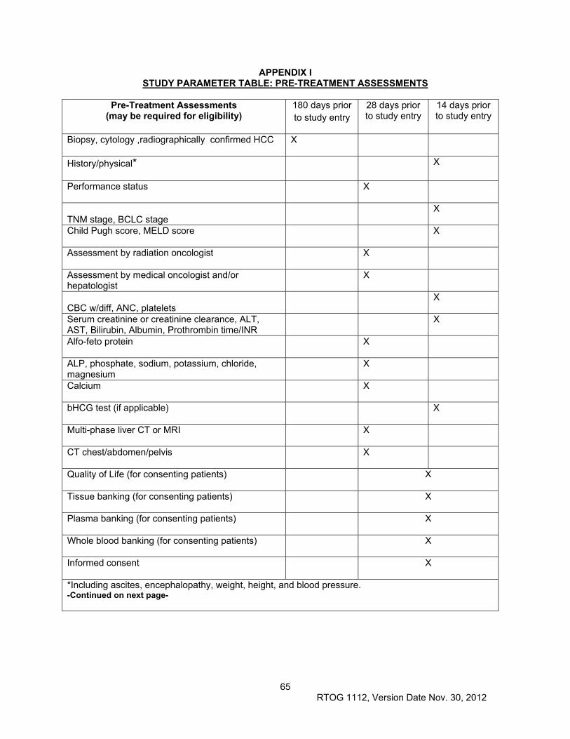
65 RTOG 1112, Version Date Nov. 30, 2012
APPENDIX I STUDY PARAMETER TABLE: PRE-TREATMENT ASSESSMENTS
Pre-Treatment Assessments
(may be required for eligibility) 180 days prior to study entry
28 days prior to study entry
14 days prior to study entry
Biopsy, cytology ,radiographically confirmed HCC X
History/physical* X
Performance status X
TNM stage, BCLC stage X
Child Pugh score, MELD score X
Assessment by radiation oncologist X
Assessment by medical oncologist and/or hepatologist
X
CBC w/diff, ANC, platelets X
Serum creatinine or creatinine clearance, ALT, AST, Bilirubin, Albumin, Prothrombin time/INR
X
Alfo-feto protein X
ALP, phosphate, sodium, potassium, chloride, magnesium
X
Calcium X
bHCG test (if applicable) X
Multi-phase liver CT or MRI X
CT chest/abdomen/pelvis X
Quality of Life (for consenting patients) X
Tissue banking (for consenting patients) X
Plasma banking (for consenting patients) X
Whole blood banking (for consenting patients) X
Informed consent X
*Including ascites, encephalopathy, weight, height, and blood pressure. -Continued on next page-
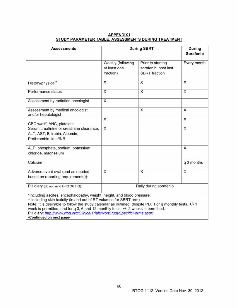
66 RTOG 1112, Version Date Nov. 30, 2012
APPENDIX I
STUDY PARAMETER TABLE: ASSESSMENTS DURING TREATMENT
Assessments During SBRT During Sorafenib
Weekly (following at least one fraction)
Prior to starting sorafenib, post last SBRT fraction
Every month
History/physical* X X X
Performance status X X X
Assessment by radiation oncologist X
Assessment by medical oncologist and/or hepatologist
X X
CBC w/diff, ANC, platelets X X
Serum creatinine or creatinine clearance, ALT, AST, Bilirubin, Albumin, Prothrombin time/INR
X X
ALP, phosphate, sodium, potassium, chloride, magnesium
X
Calcium q 3 months
Adverse event eval (and as needed based on reporting requirements)†
X X X
Pill diary (do not send to RTOG HQ) Daily during sorafenib
*Including ascites, encephalopathy, weight, height, and blood pressure. † Including skin toxicity (in and out of RT volumes for SBRT arm). Note: It is desirable to follow the study calendar as outlined, despite PD. For q monthly tests, +/- 1 week is permitted, and for q 3, 6 and 12 monthly tests, +/- 2 weeks is permitted. Pill diary: http://www.rtog.org/ClinicalTrials/NonStudySpecificForms.aspx -Continued on next page-
67 RTOG 1112, Version Date Nov. 30, 2012
APPENDIX I
STUDY PARAMETER TABLE: ASSESSMENTS IN FOLLOW-UP
Assessments Follow up (all patients)
q 3 mos from study entry x 3 yrs; q 6 mos x 2 yrs; then annually
q 6 mos x 2 yrs, then annually
See QOL & blood banking rows for details
History/physical* X
Performance status X
Child Pugh score, MELD score X
Assessment by radiation oncologist For SBRT patients
CBC w/diff, ANC, platelets X
Serum creatinine or creatinine clearance, ALT, AST, Bilirubin, Albumin, Prothrombin time/INR
X
Alfo-feto protein X
Multi-phase liver CT or MRI X
CT chest/abdomen/pelvis X
Adverse event eval (and as needed based on reporting requirements)†
X
Quality of Life (for consenting patients) 3, 6, 12 mos post study entry
Plasma banking (for consenting patients)
1 mo, 3 mos post study entry (see also Section 10.2.6)
Whole blood banking (for consenting patients)
1 mo, 3 mos post study entry
*Including ascites, encephalopathy, weight, height, and blood pressure. † Including skin toxicity (in and out of RT volumes for SBRT arm). Note: It is desirable to follow the study calendar as outlined, despite PD. For q monthly tests, +/- 1 week is permitted, and for q 3, 6 and 12 monthly tests, +/- 2 weeks is permitted.
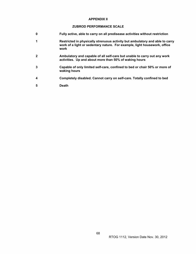
68 RTOG 1112, Version Date Nov. 30, 2012
APPENDIX II
ZUBROD PERFORMANCE SCALE
0 Fully active, able to carry on all predisease activities without restriction
1 Restricted in physically strenuous activity but ambulatory and able to carry work of a light or sedentary nature. For example, light housework, office work
2 Ambulatory and capable of all self-care but unable to carry out any work activities. Up and about more than 50% of waking hours
3 Capable of only limited self-care, confined to bed or chair 50% or more of waking hours
4 Completely disabled. Cannot carry on self-care. Totally confined to bed
5 Death
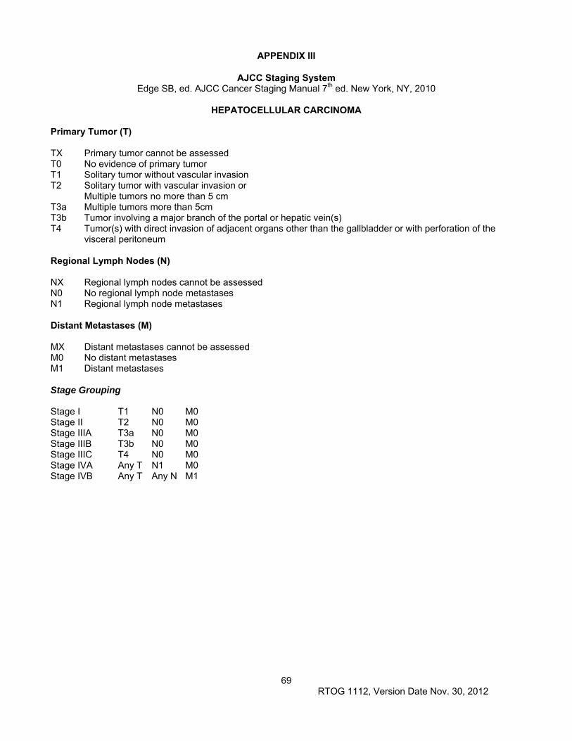
69 RTOG 1112, Version Date Nov. 30, 2012
APPENDIX III
AJCC Staging System Edge SB, ed. AJCC Cancer Staging Manual 7th ed. New York, NY, 2010
HEPATOCELLULAR CARCINOMA
Primary Tumor (T) TX Primary tumor cannot be assessed T0 No evidence of primary tumor T1 Solitary tumor without vascular invasion T2 Solitary tumor with vascular invasion or
Multiple tumors no more than 5 cm T3a Multiple tumors more than 5cm T3b Tumor involving a major branch of the portal or hepatic vein(s) T4 Tumor(s) with direct invasion of adjacent organs other than the gallbladder or with perforation of the
visceral peritoneum Regional Lymph Nodes (N) NX Regional lymph nodes cannot be assessed N0 No regional lymph node metastases N1 Regional lymph node metastases Distant Metastases (M) MX Distant metastases cannot be assessed M0 No distant metastases M1 Distant metastases Stage Grouping Stage I T1 N0 M0 Stage II T2 N0 M0 Stage IIIA T3a N0 M0 Stage IIIB T3b N0 M0 Stage IIIC T4 N0 M0 Stage IVA Any T N1 M0 Stage IVB Any T Any N M1
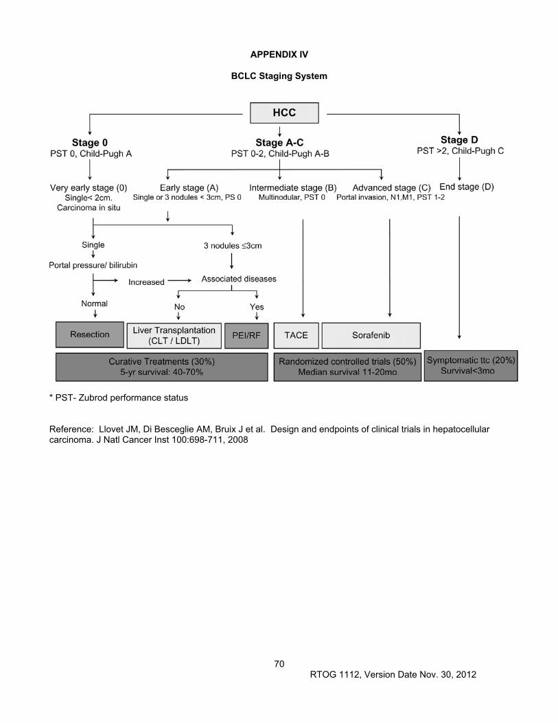
70 RTOG 1112, Version Date Nov. 30, 2012
APPENDIX IV
BCLC Staging System
* PST- Zubrod performance status Reference: Llovet JM, Di Besceglie AM, Bruix J et al. Design and endpoints of clinical trials in hepatocellular carcinoma. J Natl Cancer Inst 100:698-711, 2008
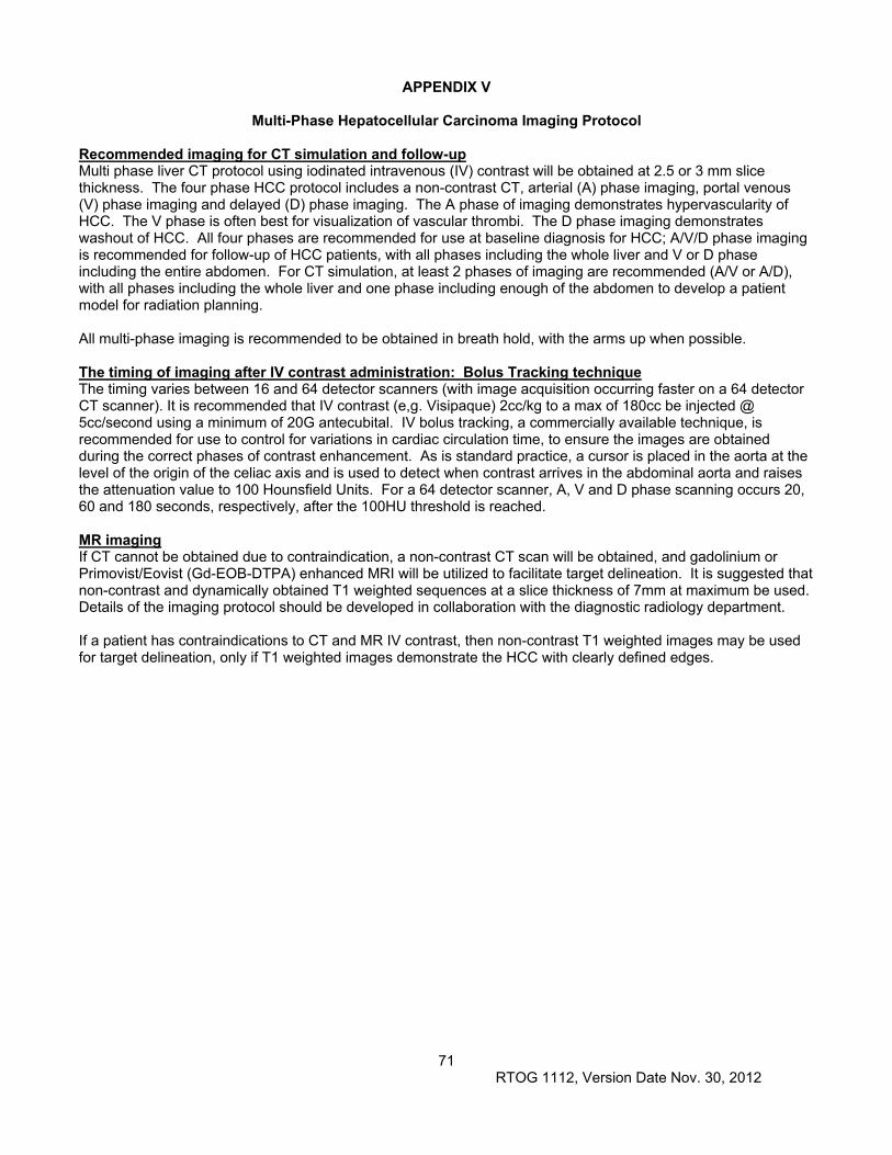
71 RTOG 1112, Version Date Nov. 30, 2012
APPENDIX V
Multi-Phase Hepatocellular Carcinoma Imaging Protocol
Recommended imaging for CT simulation and follow-up Multi phase liver CT protocol using iodinated intravenous (IV) contrast will be obtained at 2.5 or 3 mm slice thickness. The four phase HCC protocol includes a non-contrast CT, arterial (A) phase imaging, portal venous (V) phase imaging and delayed (D) phase imaging. The A phase of imaging demonstrates hypervascularity of HCC. The V phase is often best for visualization of vascular thrombi. The D phase imaging demonstrates washout of HCC. All four phases are recommended for use at baseline diagnosis for HCC; A/V/D phase imaging is recommended for follow-up of HCC patients, with all phases including the whole liver and V or D phase including the entire abdomen. For CT simulation, at least 2 phases of imaging are recommended (A/V or A/D), with all phases including the whole liver and one phase including enough of the abdomen to develop a patient model for radiation planning. All multi-phase imaging is recommended to be obtained in breath hold, with the arms up when possible. The timing of imaging after IV contrast administration: Bolus Tracking technique The timing varies between 16 and 64 detector scanners (with image acquisition occurring faster on a 64 detector CT scanner). It is recommended that IV contrast (e,g. Visipaque) 2cc/kg to a max of 180cc be injected @ 5cc/second using a minimum of 20G antecubital. IV bolus tracking, a commercially available technique, is recommended for use to control for variations in cardiac circulation time, to ensure the images are obtained during the correct phases of contrast enhancement. As is standard practice, a cursor is placed in the aorta at the level of the origin of the celiac axis and is used to detect when contrast arrives in the abdominal aorta and raises the attenuation value to 100 Hounsfield Units. For a 64 detector scanner, A, V and D phase scanning occurs 20, 60 and 180 seconds, respectively, after the 100HU threshold is reached. MR imaging If CT cannot be obtained due to contraindication, a non-contrast CT scan will be obtained, and gadolinium or Primovist/Eovist (Gd-EOB-DTPA) enhanced MRI will be utilized to facilitate target delineation. It is suggested that non-contrast and dynamically obtained T1 weighted sequences at a slice thickness of 7mm at maximum be used. Details of the imaging protocol should be developed in collaboration with the diagnostic radiology department. If a patient has contraindications to CT and MR IV contrast, then non-contrast T1 weighted images may be used for target delineation, only if T1 weighted images demonstrate the HCC with clearly defined edges.
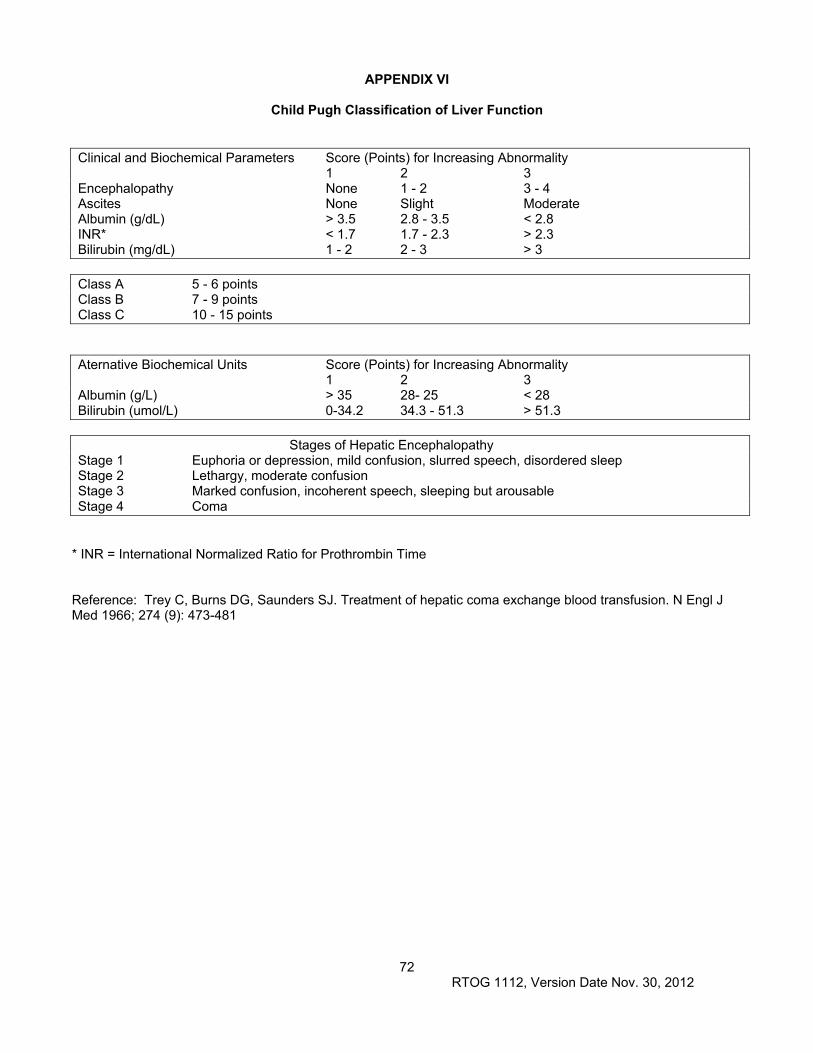
72 RTOG 1112, Version Date Nov. 30, 2012
APPENDIX VI
Child Pugh Classification of Liver Function
Clinical and Biochemical Parameters Score (Points) for Increasing Abnormality 1 2 3 Encephalopathy None 1 - 2 3 - 4 Ascites None Slight Moderate Albumin (g/dL) > 3.5 2.8 - 3.5 < 2.8 INR* < 1.7 1.7 - 2.3 > 2.3 Bilirubin (mg/dL) 1 - 2 2 - 3 > 3 Class A 5 - 6 points Class B 7 - 9 points Class C 10 - 15 points Aternative Biochemical Units Score (Points) for Increasing Abnormality 1 2 3 Albumin (g/L) > 35 28- 25 < 28 Bilirubin (umol/L) 0-34.2 34.3 - 51.3 > 51.3 Stages of Hepatic Encephalopathy Stage 1 Euphoria or depression, mild confusion, slurred speech, disordered sleep Stage 2 Lethargy, moderate confusion Stage 3 Marked confusion, incoherent speech, sleeping but arousable Stage 4 Coma
* INR = International Normalized Ratio for Prothrombin Time Reference: Trey C, Burns DG, Saunders SJ. Treatment of hepatic coma exchange blood transfusion. N Engl J Med 1966; 274 (9): 473-481
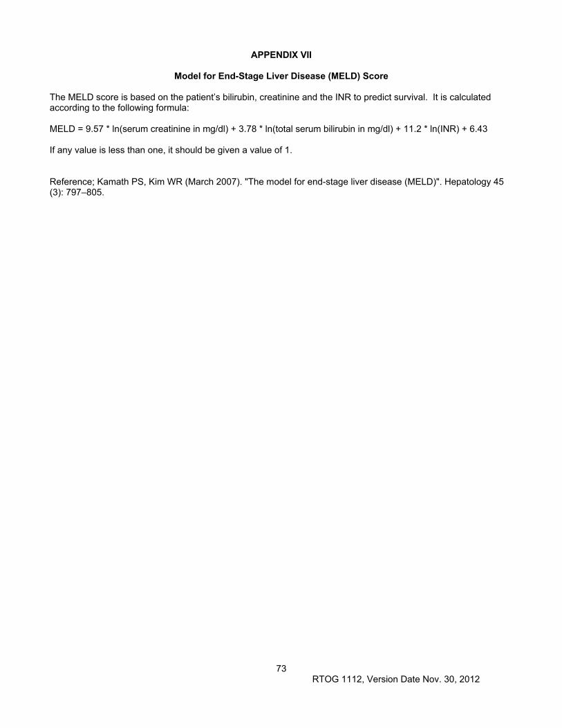
73 RTOG 1112, Version Date Nov. 30, 2012
APPENDIX VII
Model for End-Stage Liver Disease (MELD) Score
The MELD score is based on the patient’s bilirubin, creatinine and the INR to predict survival. It is calculated according to the following formula: MELD = 9.57 * ln(serum creatinine in mg/dl) + 3.78 * ln(total serum bilirubin in mg/dl) + 11.2 * ln(INR) + 6.43 If any value is less than one, it should be given a value of 1. Reference; Kamath PS, Kim WR (March 2007). "The model for end-stage liver disease (MELD)". Hepatology 45 (3): 797–805.
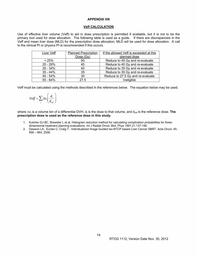
74 RTOG 1112, Version Date Nov. 30, 2012
APPENDIX VIII
Veff CALCULATION Use of effective liver volume (Veff) to aid in dose prescription is permitted if available, but it is not to be the primary tool used for dose allocation. The following table is used as a guide. If there are discrepancies in the Veff and mean liver dose (MLD) for the prescription dose allocation, MLD will be used for dose allocation. A call to the clinical PI or physics PI is recommended if this occurs.
Liver Veff Planned Prescription Dose (Gy)
If the allowed Veff is exceeded at this planned dose
< 25% 50 Reduce to 45 Gy and re-evaluate 25 - 29% 45 Reduce to 40 Gy and re-evaluate 30 - 34% 40 Reduce to 35 Gy and re-evaluate 35 - 44% 35 Reduce to 30 Gy and re-evaluate 45 - 54% 30 Reduce to 27.5 Gy and re-evaluate 55 - 64% 27.5 Ineligible
Veff must be calculated using the methods described in the references below. The equation below may be used.
Veff vi
di
dref
i
where ∆vi is a volume bin of a differential DVH, di is the dose to that volume, and dref is the reference dose. The prescription dose is used as the reference dose in this study.
1. Kutcher GJ BC, Brewster L et al. Histogram reduction method for calculating complication probabilities for three-dimensional treatment planning evaluations. Int J Radiat Oncol, Biol, Phys 1991;21:137-146.
2. Dawson LA, Eccles C, Craig T. Individualized Image Guided Iso-NTCP based Liver Cancer SBRT. Acta Oncol. 45: 856 – 864, 2006.
75 RTOG 1112, Version Date Nov. 30, 2012
APPENDIX IX (5/7/13) APPENDICES FOR RTOG BIOSPECIMEN COLLECTION
RTOG FFPE Specimen Plug Kit Collection
RTOG Blood Collection Kit Instructions Shipping Instructions:
U.S. Postal Service Mailing Address: For non-urgent FFPE or Non-frozen Specimens Only RTOG Biospecimen Resource University of California San Francisco Campus Box 1800 2340 Sutter Street, Room S341 San Francisco, CA 94143-1800
Courier Address (FedEx, UPS, etc.): For Frozen or Trackable Specimens RTOG Biospecimen Resource University of California San Francisco 2340 Sutter Street, Room S341 San Francisco, CA 94115
Include all RTOG paperwork in pocket of biohazard bag. Check that the Specimen Transmittal Form (ST) has the consent boxes checked off. Check that all samples are labeled with the RTOG study and case number, and include date of collection
as well as collection time point (e.g., pretreatment, post-treatment).
FFPE Specimens: o Slides should be shipped in a plastic slide holder/slide box. Place a small wad of padding in top of the
container. If you can hear the slides shaking it is likely that they will break during shipping. o FFPE Blocks can be wrapped with paper towel, or placed in a cardboard box with padding. Do not wrap
blocks with bubble wrap. Place padding in top of container so that if you shake the container the blocks are not shaking. If you can hear the slides shaking it is likely that they will break during shipping.
o Slides, Blocks, or Plugs can be shipped ambient or with a cold pack either by United States Postal Service (USPS) to the USPS address (94143) or by Courier to the Street Address (94115). Do NOT ship on Dry Ice.
Frozen Specimens:
o Multiple cases may be shipped in the same cooler, but make sure each one is in a separate bag and clearly identified.
o Place specimens and absorbent shipping material in Styrofoam cooler filled with dry ice (at least 7 lbs). There should be plenty of dry ice under and above the specimens. If the volume of specimens is greater than the volume of dry ice then ship in a larger Styrofoam box, or two separate boxes. Any Styrofoam box can be used, as long as it is big enough.
o Specimens received thawed due to insufficient dry ice or shipping delays will be discarded and the site will be notified.
o Send frozen specimens via overnight courier to the address above. Specimens should only be shipped Monday through Wednesday (Monday-Tuesday for Canada) to prevent thawing due to delivery delays. Saturday or holiday deliveries cannot be accepted. Samples can be stored frozen at -80 C until ready to ship.
For Questions regarding collection/shipping please contact the RTOG Biospecimen Resource by e-mail: [email protected] or phone: 415-476-RTOG(7864) or Fax: 415-476-5271.
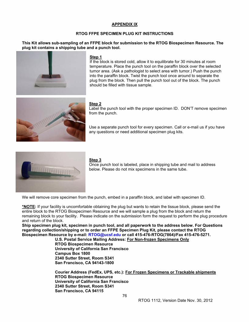
76 RTOG 1112, Version Date Nov. 30, 2012
APPENDIX IX
RTOG FFPE SPECIMEN PLUG KIT INSTRUCTIONS
This Kit allows sub-sampling of an FFPE block for submission to the RTOG Biospecimen Resource. The plug kit contains a shipping tube and a punch tool.
Step 1 If the block is stored cold, allow it to equilibrate for 30 minutes at room temperature. Place the punch tool on the paraffin block over the selected tumor area. (Ask a pathologist to select area with tumor.) Push the punch into the paraffin block. Twist the punch tool once around to separate the plug from the block. Then pull the punch tool out of the block. The punch should be filled with tissue sample. Step 2 Label the punch tool with the proper specimen ID. DON’T remove specimen from the punch. Use a separate punch tool for every specimen. Call or e-mail us if you have any questions or need additional specimen plug kits.
Step 3 Once punch tool is labeled, place in shipping tube and mail to address below. Please do not mix specimens in the same tube.
We will remove core specimen from the punch, embed in a paraffin block, and label with specimen ID. *NOTE: If your facility is uncomfortable obtaining the plug but wants to retain the tissue block, please send the entire block to the RTOG Biospecimen Resource and we will sample a plug from the block and return the remaining block to your facility. Please indicate on the submission form the request to perform the plug procedure and return of the block. Ship specimen plug kit, specimen in punch tool, and all paperwork to the address below. For Questions regarding collection/shipping or to order an FFPE Specimen Plug Kit, please contact the RTOG Biospecimen Resource by e-mail: [email protected] or call 415-476-RTOG(7864)/Fax 415-476-5271.
U.S. Postal Service Mailing Address: For Non-frozen Specimens Only RTOG Biospecimen Resource University of California San Francisco Campus Box 1800 2340 Sutter Street, Room S341 San Francisco, CA 94143-1800 Courier Address (FedEx, UPS, etc.): For Frozen Specimens or Trackable shipments RTOG Biospecimen Resource University of California San Francisco 2340 Sutter Street, Room S341 San Francisco, CA 94115
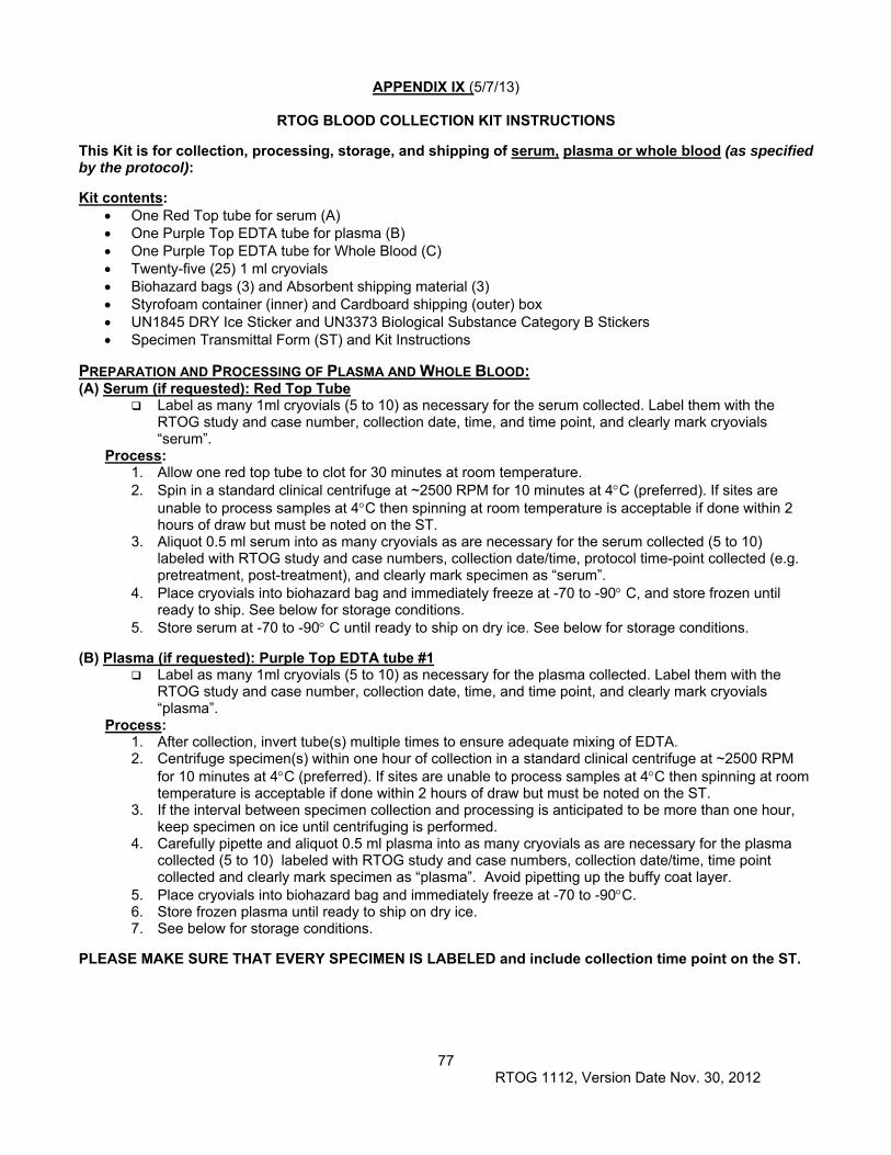
77 RTOG 1112, Version Date Nov. 30, 2012
APPENDIX IX (5/7/13)
RTOG BLOOD COLLECTION KIT INSTRUCTIONS This Kit is for collection, processing, storage, and shipping of serum, plasma or whole blood (as specified by the protocol): Kit contents:
One Red Top tube for serum (A) One Purple Top EDTA tube for plasma (B) One Purple Top EDTA tube for Whole Blood (C) Twenty-five (25) 1 ml cryovials Biohazard bags (3) and Absorbent shipping material (3) Styrofoam container (inner) and Cardboard shipping (outer) box UN1845 DRY Ice Sticker and UN3373 Biological Substance Category B Stickers Specimen Transmittal Form (ST) and Kit Instructions
PREPARATION AND PROCESSING OF PLASMA AND WHOLE BLOOD: (A) Serum (if requested): Red Top Tube
Label as many 1ml cryovials (5 to 10) as necessary for the serum collected. Label them with the RTOG study and case number, collection date, time, and time point, and clearly mark cryovials “serum”.
Process: 1. Allow one red top tube to clot for 30 minutes at room temperature. 2. Spin in a standard clinical centrifuge at ~2500 RPM for 10 minutes at 4C (preferred). If sites are
unable to process samples at 4C then spinning at room temperature is acceptable if done within 2 hours of draw but must be noted on the ST.
3. Aliquot 0.5 ml serum into as many cryovials as are necessary for the serum collected (5 to 10) labeled with RTOG study and case numbers, collection date/time, protocol time-point collected (e.g. pretreatment, post-treatment), and clearly mark specimen as “serum”.
4. Place cryovials into biohazard bag and immediately freeze at -70 to -90 C, and store frozen until ready to ship. See below for storage conditions.
5. Store serum at -70 to -90 C until ready to ship on dry ice. See below for storage conditions.
(B) Plasma (if requested): Purple Top EDTA tube #1 Label as many 1ml cryovials (5 to 10) as necessary for the plasma collected. Label them with the
RTOG study and case number, collection date, time, and time point, and clearly mark cryovials “plasma”.
Process: 1. After collection, invert tube(s) multiple times to ensure adequate mixing of EDTA. 2. Centrifuge specimen(s) within one hour of collection in a standard clinical centrifuge at ~2500 RPM
for 10 minutes at 4C (preferred). If sites are unable to process samples at 4C then spinning at room temperature is acceptable if done within 2 hours of draw but must be noted on the ST.
3. If the interval between specimen collection and processing is anticipated to be more than one hour, keep specimen on ice until centrifuging is performed.
4. Carefully pipette and aliquot 0.5 ml plasma into as many cryovials as are necessary for the plasma collected (5 to 10) labeled with RTOG study and case numbers, collection date/time, time point collected and clearly mark specimen as “plasma”. Avoid pipetting up the buffy coat layer.
5. Place cryovials into biohazard bag and immediately freeze at -70 to -90C. 6. Store frozen plasma until ready to ship on dry ice. 7. See below for storage conditions.
PLEASE MAKE SURE THAT EVERY SPECIMEN IS LABELED and include collection time point on the ST.
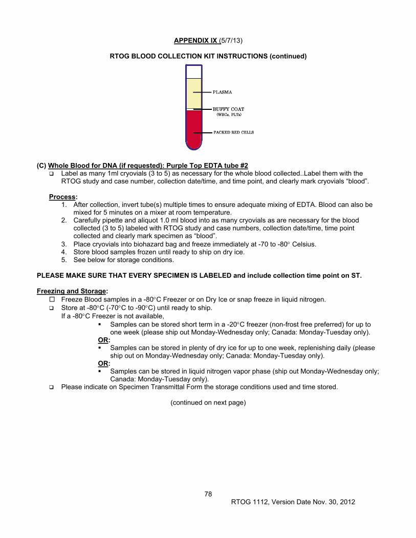
78 RTOG 1112, Version Date Nov. 30, 2012
APPENDIX IX (5/7/13)
RTOG BLOOD COLLECTION KIT INSTRUCTIONS (continued)
(C) Whole Blood for DNA (if requested): Purple Top EDTA tube #2
Label as many 1ml cryovials (3 to 5) as necessary for the whole blood collected..Label them with the RTOG study and case number, collection date/time, and time point, and clearly mark cryovials “blood”.
Process:
1. After collection, invert tube(s) multiple times to ensure adequate mixing of EDTA. Blood can also be mixed for 5 minutes on a mixer at room temperature.
2. Carefully pipette and aliquot 1.0 ml blood into as many cryovials as are necessary for the blood collected (3 to 5) labeled with RTOG study and case numbers, collection date/time, time point collected and clearly mark specimen as “blood”.
3. Place cryovials into biohazard bag and freeze immediately at -70 to -80 Celsius. 4. Store blood samples frozen until ready to ship on dry ice. 5. See below for storage conditions.
PLEASE MAKE SURE THAT EVERY SPECIMEN IS LABELED and include collection time point on ST. Freezing and Storage:
Freeze Blood samples in a -80C Freezer or on Dry Ice or snap freeze in liquid nitrogen. Store at -80C (-70C to -90C) until ready to ship.
If a -80C Freezer is not available, Samples can be stored short term in a -20C freezer (non-frost free preferred) for up to
one week (please ship out Monday-Wednesday only; Canada: Monday-Tuesday only). OR: Samples can be stored in plenty of dry ice for up to one week, replenishing daily (please
ship out on Monday-Wednesday only; Canada: Monday-Tuesday only). OR: Samples can be stored in liquid nitrogen vapor phase (ship out Monday-Wednesday only;
Canada: Monday-Tuesday only). Please indicate on Specimen Transmittal Form the storage conditions used and time stored.
(continued on next page)
79 RTOG 1112, Version Date Nov. 30, 2012
APPENDIX IX
RTOG BLOOD COLLECTION KIT INSTRUCTIONS (continued) Shipping/Mailing:
Ship specimens on Dry Ice overnight Monday-Wednesday (Monday-Tuesday from Canada) to prevent thawing due to delivery delays. Saturday and holiday deliveries cannot be accepted.
Include all RTOG paperwork in a sealed plastic bag and tape to the outside top of the Styrofoam box. Wrap frozen specimens of same type (i.e., all serum together, plasma together and whole bloods
together) in absorbent shipping material and place each specimen type in a separate biohazard bag. Place specimen bags into the Styrofoam cooler and fill with plenty of dry ice (7-10 lbs/3.5kg minimum). Add padding to avoid the dry ice from breaking the tubes.
Place Styrofoam coolers into outer cardboard box, and attach shipping label and UN3373 and UN1895 stickers to outer cardboard box.
Multiple cases may be shipped in the same cooler, but make sure each one is in a separate bag and that there is enough room for plenty of dry ice. Add padding to avoid the dry ice from breaking the tubes.
For questions regarding collection, shipping or to order a Blood Collection Kit, please e-mail [email protected] or call (415)476-7864.
Shipping Address:
Courier Address (FedEx, UPS, etc.): For all Frozen Specimens RTOG Biospecimen Resource University of California San Francisco 2340 Sutter Street, Room S341 San Francisco, CA 94115 For questions, call 415-476-RTOG (7864) or e-mail: [email protected]
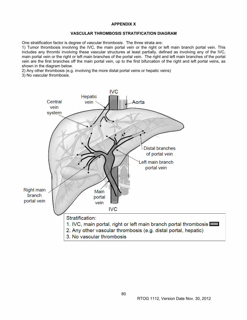
80 RTOG 1112, Version Date Nov. 30, 2012
APPENDIX X
VASCULAR THROMBOSIS STRATIFICATION DIAGRAM
One stratification factor is degree of vascular thrombosis. The three strata are: 1) Tumor thrombosis involving the IVC, the main portal vein or the right or left main branch portal vein. This includes any thrombi involving these vascular structures at least partially, defined as involving any of the IVC, main portal vein or the right or left main branches of the portal vein. The right and left main branches of the portal vein are the first branches off the main portal vein, up to the first bifurcation of the right and left portal veins, as shown in the diagram below. 2) Any other thrombosis (e.g. involving the more distal portal veins or hepatic veins) 3) No vascular thrombosis.