Embed Size (px)
Citation preview

Radiochemical synthesis of 18F-radiolabelled ProTides for
Positron Emission Tomography
A thesis submitted in accordance with the conditions
governing candidates for the degree of
Philosophiae Doctor in Cardiff University
by
Alessandra Cavaliere
January 2018
Cardiff School of Pharmacy and Pharmaceutical Science
Cardiff University

i
Acknowledgments
I want to take the opportunity to express my gratitude to all my supervisors for their great support provided during these years.
In particular, I would like to thank Prof. Andrew Westwell for the amazing positive attitude, patient guidance, encouragement and advice he has provided throughout my time as his student. I have been extremely lucky to have a supervisor who cared so much about my work and me and who responded always promptly to my queries.
I thank Dr. Katrin Probst for her great support as a supervisor and as a woman. She gave me the opportunity to gain, besides research skills, also an insight into clinical tracers development which opened to me so many opportunities.
I am going to miss you a lot Andrew and Katrin, you both have been an inspiration with your professionality and your ideas.
I want to thank Prof. Andrea Brancale for always being available, careful and incredibly friendly and Prof. Chris Marshall for his availability and kindness during my time
at PETIC. Furthermore, I want to express my gratitude to Prof. Chris McGuigan for being an inspirational guide during this journey.
I also want to thank Stephen Paisey for all the continuous support provided for my experimental work, all members from Andrew’s lab, the PETIC staff, Andrea’s and Chris’s lab for being always helpful and for becoming my friends making the work time incredibly pleasant moments. It’s thanks to all of you that I loved every single day of my work in Cardiff University.
I want to thank Prof. Franklin Aigbirhio and Abdul from Wolfson Brain Imaging Centre at Cambridge University and Rohan, Dr. Joachim Bugert and Prof. Arwin Jones for the fruitful collaborations.
I thank the Life Science Research Network of Wales and Cardiff University for their financial support and the opportunity to attend scientific conferences
Finally, I would like to express my sincerest appreciation to my amazing family
for always being there for me, for believing in me more than anyone else and for being
the best rule model I could possibly have. In addition, a huge thank to all my friends in
Italy and particularly in Cardiff (luckily you are so many that one page wouldn’t fit all
your names), and to my special supporter. You guys have been my family in these
years. Thanks for the support and the unique moments spent together. I will always
keep them in my mind and in my heart.

ii
Dedicated to mum, dad and Vivi
iii
Abstract
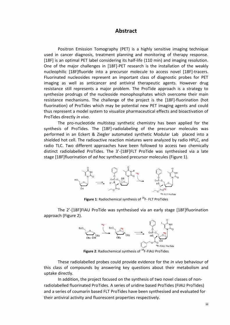
Positron Emission Tomography (PET) is a highly sensitive imaging technique used in cancer diagnosis, treatment planning and monitoring of therapy response. [18F] is an optimal PET label considering its half-life (110 min) and imaging resolution. One of the major challenges in [18F]-PET research is the installation of the weakly nucleophilic [18F]fluoride into a precursor molecule to access novel [18F]-tracers. Fluorinated nucleosides represent an important class of diagnostic probes for PET imaging as well as anticancer and antiviral therapeutic agents. However drug resistance still represents a major problem. The ProTide approach is a strategy to synthesize prodrugs of the nucleoside monophosphates which overcome their main resistance mechanisms. The challenge of the project is the [18F]-fluorination (hot fluorination) of ProTides which may be potential new PET imaging agents and could thus represent a model system to visualize pharmaceutical effects and bioactivation of ProTides directly in vivo.
The pro-nucleotide multistep synthetic chemistry has been applied for the synthesis of ProTides. The [18F]-radiolabeling of the precursor molecules was performed in an Eckert & Ziegler automated synthetic Modular Lab placed into a shielded hot cell. The radioactive reaction mixtures were analyzed by radio HPLC, and radio TLC. Two different approaches have been followed to access two chemically distinct radiolabelled ProTides. The 3’-[18F]FLT ProTide was synthesised via a late stage [18F]fluorination of ad hoc synthesised precursor molecules (Figure 1).
Figure 1: Radiochemical synthesis of 18
F- FLT ProTides
The 2’-[18F]FIAU ProTide was synthesised via an early stage [18F]fluorination approach (Figure 2).
Figure 2: Radiochemical synthesis of 18
F-FIAU ProTides
These radiolabelled probes could provide evidence for the in vivo behaviour of this class of compounds by answering key questions about their metabolism and uptake directly.
In addition, the project focused on the synthesis of two novel classes of non-
radiolabelled fluorinated ProTides. A series of uridine based ProTides (FIAU ProTides)
and a series of coumarin based FLT ProTides have been synthesised and evaluated for
their antiviral activity and fluorescent properties respectively.

iv
List of contents
LIST OF ABBREVIATIONS ........................................................................................................ VII
LIST OF FIGURES ......................................................................................................................... X
LIST OF TABLES ........................................................................................................................ XV
1. INTRODUCTION .................................................................................................................. 1
1.1 Positron Emission Tomography (PET) ....................................................................................... 1
1.1.1 Physical principles of PET ........................................................................................................... 3
1.1.2 [18F]fluorine ................................................................................................................................... 9
The main properties of [18F]fluorine are summarised in the table below (Table 1.3)17
. ........................ 9
1.1.2.1 Fluorination ........................................................................................................................... 11
1.1.2.2 [18F]FDG ................................................................................................................................ 17
1.1.2.3 [18F]-nucleosides .................................................................................................................. 18
1.1.2.4 Other [18F]-labelled PETprobes ............................................................................................ 22
1.1.3 Short lived radioisotopes .............................................................................................................. 23
1.1.4 Long-lived radioisotopes: Positron emitting metals ..................................................................... 24
1.1.5 PET imaging: application in drug discovery .................................................................................. 26
1.2 Nucleosides ....................................................................................................................................27
1.2.1 ProTides ........................................................................................................................................ 29
1.2.2 Fluorinated nucleosides and ProTides .......................................................................................... 32
1.2.2.1 Importance of fluorine in drug molecules ............................................................................. 33
2. RESEARCH AIMS AND OBJECTIVES ........................................................................ 47
3. RADIOCHEMICAL SYNTHESIS OF [18F]FLT PROTIDE ..................................... 50
3.1 Introduction ...................................................................................................................................50
3.2 [18F]fluorination (Hot): late stage approach ..................................................................................51
3.3 Synthesis of the precursor molecules .............................................................................................52
3.3.1 Synthesis of cyclised precursor (3.8) ............................................................................................. 52
3.3.2 Synthesis of the mesyl, tosyl and nosyl precursors ...................................................................... 57
3.4 Synthesis of the cold standard........................................................................................................64
3.5 Studies of ProTide stability .............................................................................................................67
3.6 [18F]fluorination ............................................................................................................................67
3.6.1 [18F]-Fluorination of the anhydride precursor (3.8) ..................................................................... 71
3.6.2 [18F]fluorination of the mesyl precursor (3.1) ............................................................................. 72
3.6.3 [18F]fluorination of the tosyl precursor (3.2) ............................................................................... 74
v
3.6.4 [18F]fluorination of the nosyl precursor (3.3) .............................................................................. 76
3.6.5 [18F]fluorination of the nosyl BOC protected precursor (3.4) ...................................................... 81
3.7 Conclusions ....................................................................................................................................85
4. RADIOCHEMICAL SYNTHESIS OF [18F]FIAUPROTIDE ................................................ 87
4.1 Introduction ...................................................................................................................................87
4.2 [18F]fluorination (Hot): early stage approach.................................................................................88
4.3 Synthesis of the cold standard (4.3) ...............................................................................................89
4.3 Synthesis of [18F]FIAU (1.16) ..........................................................................................................90
4.5 Synthesis of the [18F]FIAUProTide (2.2) .........................................................................................97
4.6 Evaluation of the radiotracer [18F]FIAU (1.16) on HSV-TK engineered HEK cell lines ......................99
4.7 Conclusions .................................................................................................................................. 101
5. SYNTHESIS OF NOVEL 2'-DEOXY-2'-FLUORO-5-IODOURIDINE PROTIDES AS NOVEL
ANTIVIRAL AGENTS .............................................................................................................. 103
5.1 Introduction ................................................................................................................................. 103
5.2 Synthesis of novel 2'-deoxy-2'-fluoro-5-iodouridine ProTides ...................................................... 106
5.3 Antiviral evaluation ...................................................................................................................... 113
5.4 Conclusion .................................................................................................................................... 116
6. SYNTHESIS OF NOVEL FLT CHIMERIC PROTIDES WITH FLUORESCENT PROBES . 117
6.1 Introduction ................................................................................................................................. 117
6.2 Synthesis of FLT chimeric ProTides with fluorescent probes ......................................................... 120
6.3 Fluorescence data ........................................................................................................................ 123
6.4 Conclusion .................................................................................................................................... 124
7. CONCLUSIONS AND FUTURE WORK .......................................................................... 125
7.1 General conclusions and future perspective ................................................................................. 125
8. EXPERIMENTAL ............................................................................................................. 127
8.1 General information ..................................................................................................................... 127
8.1.1 Analytics ...................................................................................................................................... 127
vi
8.1.2 Solvents and chemicals ............................................................................................................... 127
8.1.3 Radioactive source and equipment ............................................................................................ 128
8.2 Procedures ................................................................................................................................... 128
8.2.1 Standard procedure A1: synthesis of L-alanine ester hydrochloride salts.................................. 128
8.2.2 Standard procedure A2: synthesis of L-alanine ester sulfonate salts ......................................... 128
8.2.3 Standard procedure B: synthesis of aryloxy phosphorochloridates ........................................... 129
8.2.4 Standard procedure C: synthesis of phosphoramidates via NMI .............................................. 129
8.2.5 Standard procedure D: synthesis of phosphoramidates via tBuMgCl ........................................ 130
8.3 Spectroscopic data ....................................................................................................................... 131
8.3.1Experimental section from Chapter 3 .......................................................................................... 131
8.3.2 Experimental section from chapter 4 ......................................................................................... 149
8.3.3 Experimental section from chapter 5 ......................................................................................... 158
8.3.4 Experimental section from Chapter 6 ......................................................................................... 196
9. APPENDIX ....................................................................................................................... 204
10. BIBLIOGRAPHY .......................................................................................................... 207

vii
List of abbreviations
Å
BGO crystals
Angstrom [ 1Å = 10-10 m = 0.1nm ]
Bismuth Germanium Oxide Crystals
Bq
BOC
CAN
Bequerel [ 1 Bq= 1 decay/sec ]
Tert-Butyloxycarbonyl Protecting Group
Ceric Ammonium Nitrate
CDA Cytidine deaminase
Ci Curie [1 Ci = 3.7 x 10¹⁰ decays/sec =37 GBq]
CsF Cesium Fluoride
CT Computed Tomography
DAST Diethylaminosulfurtrifluoride
dCK Deoxycytidine Kinase
DCM Dichloromethane
dFdC 2,2’-Difluoro-2’-Deoxycytidine (Gemcitabine)
DIAD Diisopropylazodicarboxylate
DMF Dimethyl Formamide
DNA
EPD
Deoxyribonucleic Acid
Electronic Personal Dosimeters
ESI Electrospray Ionisation
EtOAc Ethyl Acetate
eV
E&Z
Electronvolt [J]
Eckert & Zigler Unit
FAC 2’-Fluoro-Deoxy-Arabinocytidine
FDG 2-Fluoro-Deoxy-Glucose
FGI Functional Group Interconversion
FIAU 2’-Fluoro-Deoxy-Arabino-5-Iodouridine
FLT 3’-Fluoro-Deoxy-L-Thymidine
FMAU
GC
Fluoro-Deoxy-Arabino-5-Methyluridine
Gas Chromatography
viii
GMP Good Manufacturing Practice
H
HBV
Hour
Hepatitis B Virus
HCl
HCV
Hydrogen Chloride
Hepatitis C Virus
hENT
HIV
Human Concentrative Nucleoside Transporter
Human Immunodeficiency Virus
HPLC High-Pressure Liquid Chromatography
HSV-TK-1 Herpes Simplex Virus Thymidine Kinase 1
Hz Hertz [1 Hz = s-1]
K222 Kryptofix 222 (Cryptant)
KF Potassium Fluoride
LC Liquid Chromatography
M Molar [Mol/L]
MeCN Acetonitrile
MeOH Methanol
mL Millilitre
MRI Magnetic Resonance Imaging
MS Mass Spectrometry
MsCl
NA
Methanesulfonyl (Mesyl) Chloride
Nucleoside Analogues
NaOH Sodium Hydroxide
NEt3/TEA
NMI
Triethylamine
1-Methylimidazole
NMR Nuclear Magnetic Resonance
NsCl
NTs
p-Nitrophenylsulfonyl (Nosyl) Chloride
Nucleoside Transporters
PET Positron Emission Tomography
PETIC Wales Research Diagnostic PET Imaging Centre
pKa Acid Dissociation Constant
ix
PPh3 Triphenylphosphine
ppm Parts Per Million
pTsOH p-Toluenesulfonic Acid
QC
R&D
Radio HPLC
Radio TLC
Quality Control
Research and Development
Radio High Performance Liquid Chromatography
Radio Thin Layer Chromatography
RCY
r.t.
Radiochemical Yield
Room Temperature
Rf Retardation Factor
RNA Ribonucleic Acid
RR Ribonucleotidereductase
Rt
SA
Retention Time
Specific Activity
SLC Solute Carrier Family (Nucleoside Transporter)
SM Starting Material
SN Nucleophilic Substitution
SPECT Single Photon Emission Computed Tomography
Sv Sievert (1 Sv = 1 joule/kilogram )
t1/2 Radiochemical Half-Life
TBAF Tetrabutyl Ammonium Fluoride
THF Tetrahydrofuran
TK Thymidine Kinase 1
TLC
TMSOTf
Thin-Layer Chromatography
Trimethylsyliltrifluoromethanesulfonate
TS Thymidylate Synthase
TsCl
US
Toluenesulfonyl (Tosyl) Chloride
Ultrasound
UV Ultraviolet

x
List of figures
Figure 1.1: Schematic illustration of an annihilation reaction and the subsequent coincidence detection
Figure 1.2: Schematic illustration of a cyclotron
Figure 1.3: Nuclear reaction short hand
Figure 1.4: Eckert& Ziegler automated modular lab
Figure 1.5: a) Fastlab from GE healthcare and b) Trasis
Figure 1.6: Structure of Kryptofix K[222]
Figure 1.7: Synthesis of [18F]FLT via two approaches
Figure 1.8: Metal catalyzed allylic fluorination reaction
Figure 1.9: Synthesis of the [18F]flutemetamol
Figure 1.10: Synthesis of [18F]fluorouracil (1.9) via transition-metal-mediated fluorination
Figure 1.11: Synthesis of the fluorinating agent Selectfluor
Figure 1.12: Electrophilic fluorination with Selectfluor
Figure 1.13: Synthesis of Clofarabine (1.13)
Figure 1.14: Synthesis of [18F]FIAU (1.16)
Figure1.15 Structure of the most widely used PET tracer [18F]FDG
Figure 1.16: Routine clinical synthesis of the radiotracer [18F]FDG
Figure 1.17: [18F] analogues: pyrimidine nucleosides
Figure 1.18: Thymidine salvage and de novo synthesis pathways
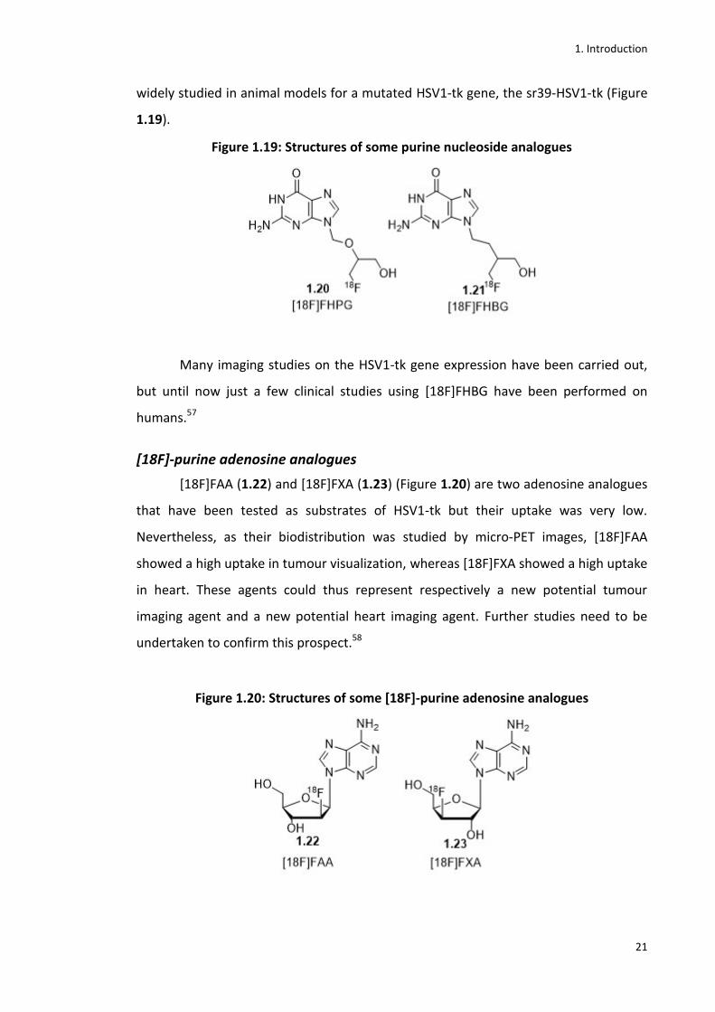
Figure 1.19: Structures of some purine nucleoside analogues
Figure 1.20: Structures of some [18F]-purine adenosine analogues
Figure 1.21: Structure of [18F]FAC
Figure 1.22: Structure of the Parkinson’s disease PET probe *18F+fluorodopa
Figure 1.23: Aprepitant (1.26) and a radiomarker of NK1 (1.27)
Figure 1.24: Structures of ribose and 2’deoxyribose nucleosides
Figure 1.25: Thymidine and cytidine and their analogues

xi
Figure 1.26: Pro-nucleotides mechanism of action
Figure 1.27: Proposed activation pathway of Protides
Figure 1.28: FDA-approved fluorinated anticancer and antiviral nucleoside analogues.
Figure 1.29: North and south conformation of two anomers of F-ddA
Figure 1.30: Fluorinated anticancer nucleobase, nucleoside and nucleotide analogues in clinical studies
Figure 1.31: Fluorinated antiviral nucleoside analogues and derivatives in clinical trials
Figure 1.32: Suggested mechanism by which FIAU causes widespread mitochondrial injury, and disturbance in metabolic processes
Figure 2.1: Late stage [18F]fluorination of ProTides
Figure 2.2: Early stage [18F]fluorination for the synthesis of ProTides
Figure 2.3: ProTide approach applied to the 2’-deoxy-2’ fluoro- 5-iodouridine
Figure 2.4: ProTide approach for the synthesis of hybrid ProTides
Figure 3.1: Structure of an [18F]FLT ProTide
Figure 3.2: Late stage fluorination approach for the synthesis of the [18F]FLT ProTide
Figure 3.3: General scheme for the synthesis of precursor molecules for the synthesis of [18F]FLT ProTide
Figure 3.4: Synthesis of the anhydride precursor
Figure 3.5: Mitsunobu reaction
Figure 3.6: Mechanism of the Mitsunobu reaction
Figure 3.7: Synthesis of Phosphorochloridate
Figure 3.8: Formation of the anhydride ProTide
Figure 3.9: Synthetic procedure for the mesyl, tosyl and nosyl precursors
Figure 3.10: Hydrolysis
Figure 3.11: ProTide coupling
Figure 3.12: Reaction mechanism for the final coupling step in the synthesis of the FLT ProTide
Figure 3.13 : Reaction mechanism of the Mesylation
Figure 3.14: Mesylation reaction
Figure 3.15: Nosylation and Tosylation
xii
Figure 3.16: N-Protection using the BOC group
Figure 3.17: Fluorination of the Mesyl precursor
Figure 3.18: Fluorination of the Nosyl precursor (2.3)
Figure 3.19: Synthesis of FLT ProTide using FLT as starting material
Figure 3.20: Mechanism of reaction of the Grignard reagent
Figure 3.21: Analytical HPLC (UV detector) of the cold [19F]FLT ProTide
Figure 3.22: Studies of stability of the FLT ProTide
Figure 3.23: Modular Lab Eckert and Ziegler (E&Z) at PETIC centre
Figure 3.24: Sketch of EZ Modular Lab
Figure 3.25: Flow chart of the synthetic steps in modular lab
Figure 3.26: General conditions for the hot fluorination of the precursors
Figure 3.27: [18F]fluorination of compound 3.8
Figure 3.28: [18F]fluorination of compound 3.1
Figure 3.29: [18F]fluorination of compound 3.2
Figure 3.30: [18F]fluorination of compound 3.3
Figure 3.31: GE Healthcare TracerlabFxFn
Figure 3.32: Sketch of TracerlabFxFn
Figure 3.33: Analytical HPLC chromatogram of the cold standard
Figure 3.34: [18F]fluorination of compound 3.3 with TracerLabFxFn
Figure 3.35: Hot fluorination of the nosyl derivative without the use of Kryptofix as reagent
Figure 3.36: Hot fluorination of the BOC protected nosyl derivative (3.4)
Figure 3.37: [18F]fluorination of compound 3.4
Figure 3.38: Deprotection of the BOC [18F]-intermediate
Figure 3.39: Deprotection of compound 3.12
Figure 3.40: Purified [18F]FLT ProTide
Figure 3.41: Radio-TLC chromatogram of the purified [18F]FLT ProTide
Figure 4.1: Structure of the2’-[18F]FIAU ProTide target (2.2)
Figure 4.2: Early stage fluorination approach for the synthesis of the

xiii
[18F]FIAU-ProTide (2.2)
Figure 4.3: Synthesis of the cold standard FIAU ProTide (4.3)
Figure 4.4: Iodination of the starting material (4.4)
Figure 4.5: Radiochemical synthesis of [18F]FIAU (1.16)
Figure 4.6: Synthesis of the [18F]-sugar (4.2)
Figure 4.7: [18F]fluorination of compound 4.1
Figure 4.8: Protection of the 5-Iodouracil (4.5)
Figure 4.9: Synthesis of protected β-anomer of [18F]FIAU (1.16) and the α-anomer (4.8)
Figure 4.10: Deprotection reaction
Figure 4.11: [18F]FIAU purification
Figure 4.12: Deprotection reaction when using TMSOTf and SnCl4 as catalysts
Figure 4.13: Synthesis of the [18F]FIAUProTide (2.2)
Figure 4.14: Synthesis of [18F]FIAU ProTide
Figure 4.15: Proposed retrosynthetic pathway to access the [18F]FIAU ProTide (2.2) via a late stage hot fluorination
Figure 5.1: Structures of 2’-deoxy-2’-β-fluoro-5-iodouridine (FIAU, 1.66) and its anomer 2’-deoxy-2’-α-fluoro-5-iodouridine (2.3)
Figure 5.2: Application of the ProTide strategy to fluorinated uridine based ProTides
Figure 5.3: General synthetic scheme for the 2’-deoxy- 2’-fluoro-5-iodouridine ProTides
Figure 5.4: Iodination of the α- and β-anomers
Figure 5.5: Formation of the naphtyl-phosphorodichloridate
Figure 5.6: Synthesis of the L-alanine ester salt via two methods
Figure 5.7: Synthesis of the phosphorochloridates
Figure 5.8: Synthesis of the ProTides
Figure 5.9: Antiviral effect on Zika virus at 10 µm on DEPG cells (n=3)
Figure 5.10: Antiviral effect of Zika virus at 10 µm on HUH7 cells (n=3)
Figure 5.11: Cytotoxicity at 10 µm on HUH7 cells (n=3)
Figure 5.12: Cytotoxicity at 10 µm on DBGRT cells (n=3)
Figure 6.1: Structure of hybrid coumarin derivatives of FLT ProTides

xiv
Figure 6.2: Structure of coumarin backbone
Figure 6.3: Structures of the coumarin derivatives tipranavir (6.1) and (+)-calanolide (6.2)
Figure 6.4: General synthetic scheme for FLT-coumarin derivatives ProTides
Figure 6.5: Synthesis of the phosphorodichloridates
Figure 6.6: Synthesis of the phosphorochloridate
Figure 6.7: Synthesis of the phosphoramidates
Figure 6.8: Fluorescence emission spectrum of compound 6.3, 6.4 and 6.6

xv
List of tables
Table 1.1: Selected isotopes used in PET imaging
Table 1.2: Nuclear reactions targets and products
Table 1.3: Main properties of [18F]fluorine
Table 1.4: Common PET Tracers
Table 1.5: Fluorinated NAs and their prodrugs in clinical use and clinical development for cancer
Table 1.5 (cont.): Fluorinated NAs and their prodrugs in clinical use or clinical development for viral infections
Table 3.1: Structures of mesyl, tosyl and nosyl leaving groups and pKa of their equivalent acids
Table 3.2: Attempts of cold fluorination of mesyl precursor
Table 3.3: [18F]fluorination attempts on the anhydride Precursor
Table 3.4: [18F]fluorination attempts on the mesyl precursor
Table 3.5: [18F]-fluorination attempts on the tosyl precursor
Table 3.6: [18F]fluorination attempts of the nosyl precursor
Table 4.1: Attempts for the hot glycosylation reaction
Table 4.2: Radiolabelling of wild HEK cells and tk engineered HEK cells
Table 4.3: Preliminary radiolabelling experiment results
Table 5.1: Structures and yields of 5.6a-5.6e
Table 5.2: Structures and yields of phosphorochloridates synthesised
Table 5.3: Structures and yields of 4.3, 5.8a-5

1. Introduction
1
1. Introduction
1.1 Positron Emission Tomography (PET) Positron Emission Tomography (PET) is a highly sensitive non-invasive nuclear
imaging technique widely used for cancer diagnosis and treatment planning. It is also
used for early detection and treatment of other diseases (i.e. Parkinson’s and
Alzheimer’s disease) by detecting metabolic changes within relevant cells.1
Diagnostic radiology is a field that emerged after the discovery of X-rays by
Wilhelm Rӧntgen in 1895. Nowadays, this technique is still used, together with CT
(Computed Tomography), MRI (Magnetic Resonance Imaging) and US (Ultrasound).
Although they can be useful in order to provide structural and anatomical information
on different medical conditions, their biggest limitation is that they give very poor
functional information about biological and metabolic processes.1
PET and SPECT (Single-Photon Emission Computed Tomography) instead give
the possibility to deeply understand the biological processes in different medical
diseases. PET, compared to SPECT, has several advantages such as the possibility of
having 3D and 2D physiological and biological information thank to the use of a wider
range of positron emitting radioelements and a higher sensitivity.1 PET, more recently,
has also been used for another important and crucial role in the drug discovery
industry. Indeed, PET gives the possibility to provide a wide range of useful information
on biological and pharmacological properties of several drug candidates through direct
radiolabeling of the drug molecule.2 PET thus represents an important tool for drug
discovery and personalized medicine, allowing for several clinical trials and monitoring
of patient response to treatments.3
Among different positron-emitting isotopes 18F, 11C, 13N and 15O are currently
the most widely used in PET.4 All of them are characterized by a low molecular weight
so that no difference in biological activity is observed compared with their non-labelled
counterparts.5 These radioelements decay by positron emission, an anti-electron
which encounters an electron and causes an annihilation event leading to the release

1. Introduction
2
of antiparallel gamma-rays4. For example, [18F]fluorine decays to [18O]oxygen, a
natural and stable isotope of oxygen, releasing a neutrino (ν) and a positron (β+)
(Equation 1.1)6.
Equation 1.1: Decay of 18F
18F 18O + β+ + ν
The time frame of β+ emission depends on the half-life of the radioactive
nucleus, which varies according to its specific decay constant (λ) (Equation 1.2)7.
Equation 1.2: Half-life of a radionuclide varies with the decay constant
t1/2 = 0.693
𝜆
The positron (β+) can encounter an electron (e-), its antiparticle, by travelling a
short distance to the surrounding tissues. An annihilation event thus occurs, and two
gamma-ray photons (γ) of 511 keV are generated from this event (Figure 1.1)8.
Figure 1.1.1: Schematic illustration of an annihilation reaction and the subsequent coincidence detection
The emitted -rays travel in the opposite direction with an angle of 180° and
PET scanners, which encircle the patient, detect the pair of photons simultaneously,
determining the precise 3D location of the radiolabelled compounds.
Radioelements 13N and 15O have very short half-lives (less than 10 minutes), so
they require a cyclotron (PET isotope generating particle accelerator) facility on site.
1. Introduction
3
11C has a half-life of 20.3 min, which is longer, but still often not long enough for a
multi-step tracer synthesis1.18F, on the contrary, is the most widely used radioisotope
in PET. With its half-life of 109.77 min, it allows for efficient multi-step synthesis of
radiolabelled tracer molecules (Table 1.1)9.
Table 1.1: Selected isotopes used in PET imaging9
Isotope Half life β-Positron emission
18O 2 min β+, 1732keV (99.9%)
15N 10 min β+,1199 keV (99.8%)
11C 20.39 min β+,960 keV (99.8%)
18F 109.8 β+,633 keV (96.7%)
64Cu 12.7 h β+,653 keV (17.9%), β-, 578.7 keV (39%)
124I 4.17 days β+,1199 keV (99.8%), β-, 2138 keV (11%)
Additionally, the longer half-life allows investigation of biological systems with
slower kinetics.10
1.1.1 Physical principles of PET
From the cyclotron to the PET scan
Many steps are required to obtain radiolabelled tracers for PET imaging. The
whole process starts with the production of proton rich radioisotopes produced by
bombardment of a target by proton sources. This process takes place at very high
velocity in cyclic particle accelerators named cyclotrons (Figure 1.2).
The cyclotron uses a combination of electric and magnetic fields. As soon as the
positively charged particle enters the gap between the two dees, it is accelerated by
the electric field towards one of them. The magnetic field makes the particle do semi-
circle movements and then moves it back to the gap. The electric field accelerates the
positive particle to the original dee, and this process is repeated with every transition
of the particle. Eventually, the particle exits the cyclotron at high speed to bombard
the target.11

1. Introduction
4
Figure 1.2: Schematic illustration of a cyclotron12
The positively charged particle, accelerated by the cyclotron, hits the target
isotope which emits another particle from its nucleus and thus produces the desired
radioisotope. Different targets and nuclear reactions can be used to obtain the desired
radioisotope.11
Details of a nuclear reaction are normally described as in the short hand in
Figure 1.3 where A represent the target isotope, b the bombarding particle, c the
emitted particle, D the isotope produced and z the atomic mass.
Figure 1.3: Nuclear reaction short hand
Listed below are some nuclear reactions associated with the most common targets
(Table 1.2)11,13.
1. Introduction
5
Table 1.2: Nuclear reactions targets and products11,13
TARGET NUCLEAR REACTION PRODUCT
N2 + 0.1% O2 14N(p,α)11C [11C]CO2
N2 + 5% H2 14N(p,α)11C [11C]CH4
N2 + 0.2% O2 14N(d,n)15O [15O]O2H2
18O 18O(p,n)18F [18F]F-(H2O)n
18O2 + 0.6% F2 18O(p,n)18F [18F]F2
20Ne 20Ne(d, α)18F [18F]F2
H2O/ETHANOL 16O(p,α)13N [13N]NH3
CO2 (TRACE N2) 12C(d,n)13N [13N]N2
Once a radioisotope is produced, it has to be incorporated into a precursor
molecule to obtain the desired radiotracer. Synthesis, purification and quality control
(QC) analyses need to be performed in a reasonable short time (typically less than 2 or
3 half-lives).
To achieve this, the radioisotope has to be in a reactive form. Sometimes it is
produced directly as its reactive species such as the [18F]F2 gas.14 Other times, it has to
be converted into a more reactive form. This is the case for [11C]MeI which can be
obtained from [11C]-CO2 or [11C]-CH4.15,16 In the case of the [18F]fluoride, because of
its weak nucleophilicity, its reactivity has to be increased by using crown ethers and
azeotropic drying.17
The reactive form of the radioisotope has to be introduced into a precursor
molecule at a late stage in the multi-step synthesis if possible. Therefore, precursors
are usually synthesized to be highly functionalized and activated so that the labeling
can be the last step or can be followed by a quick deprotection step only.10 To speed
up the reaction the precursor should be used in super-stoichiometric amounts
compared to the radioisotope.18 Radiotracers are also required to be enantiomerically
pure (more than 98%ee) whenever possible.18
In order to manipulate the reagents of the reaction when dealing with positron
emitting radioisotopes, an automated synthesizer is required so that the chemical
reaction can occur in a heavily shielded compartment called a hot cell. The radiotracer

1. Introduction
6
has to be injected into a patient in a short time from the beginning of the synthesis,
which means that the reaction has to be reproducible as its failure will lead to severe
inconvenience for patients.18
Besides these essential requirements, there are some extra desirable ones such
as a stable and easily synthesised precursor. A high radiochemical yield (RCY) is also a
desirable as more patients can be scanned and the tracer, accordingly to the kind of
radioisotope used, could also be transported offsite. To simplify the synthesis, ambient
temperature would be ideal as well as a high specific activity (SA).17
Specific activity is an import factor when it comes to radiotracers. It is defined
as the radioactivity per unit of material (grams or moles of tracers).The higher the SA,
the lower the concentration of cold harmful tracer that will be administered.19To
calculate the specific activity of a tracer, it should be taken into account that [19F]-
tracers could be present even if the fluorination was “no carrier-added”.20 Possible
sources of 19F could be Teflon tubing21 or ion exchange cartridges22.To calculate the
exact amount of tracer desired, HPLC with a UV detector can be used for UV sensitive
tracers. First, a calibration curve of the cold tracers should be done and then compared
with the concentration of the UV signal of the radioactive tracer.19
Automated modules for the synthesis
When dealing with radioactive tracers, the synthetic process is obviously
influenced by the short time available for the synthesis, purification and QC analyses
(usually not more than 3 half-lives of the radionuclide). For these reasons, for both
preclinical and clinical purposes, use of computer controlled synthetic automated
modules are required.22 This will, on one hand, reduce the exposure to radioactivity for
operators as modules will be placed into shielded hot cells but will also assure a
reproducible synthesis reducing the possibility of human errors and the overall
synthesis time. Many companies have developed in recent years automated modules
that can be adaptable for many kind of synthesis. The Eckert & Ziegler (E&Z) modular
lab (Figure 1.4) is currently one of the most commonly used for the routine synthesis
of the gold standard PET tracer [18F]FDG.
1. Introduction
7
Figure 1.4: Eckert & Ziegler automated modular lab22
Tracers like [18F]FDG, which require a nucleophilic fluorination followed by a
single deprotection step, can be synthesised by these automation modules. Other
automation modules like Fastlab from GE healthcare and Trasis (Figure 1.5) allow also
more complex synthesis with better radiochemical yields (RCY) and radiochemical
purity.
Figure 1.5: a) Fastlab from GE healthcare23 and b) Trasis24
a. b.
Analysis and quality control of radiotracers
Finally, before using the tracer, purification and quality control analyses have to
be performed. Again fast purification is required and it can be performed either by
preparative HPLC attached to automation modules, ideally just using purification
cartridges. Once purified, the tracer is usually isolated in a generally isotonic solution
and is passed through sterile filters. One of the samples produced is designated to the
1. Introduction
8
quality control section to make sure all the criteria required are met (this is particularly
true when the tracer has to be produced under GMP standards for clinical purposes).25
Listed below are some of the routine analyses performed for quality control of
radiopharmaceuticals:
- Dose calibration of the radioisotope.
- Filter test to determine its integrity.
- Visual inspection of the product.
- pH measures.
- Endotoxin test.
- Analytical HPLC to determine the radiochemical purity and identity of tracer and side
products.
- Radio TLC for RCY, purity and identity calculation.
- Analysis of residual Kryptofix left in the solution.
- GC to determine quantity of residual solvents in the product.26
When it comes to the development of novel tracers for R&D purposes, Radio-
TLC and Radio-HPLC are the main tools used for high quality analyses. Radio-HPLCs are
characterized by a UV, a refractive index or conductivity, and a radioactive detector.
This allows straightforward determination of the radioactive profile of the tracer.26
Real time analyses are provided either by flow-cell radioactivity measures or by the use
of microplates scintillation counting. The radioactivity is detected by using NaI or
Bismuth Germanium Oxide (BGO) scintillation crystals. They register the annihilation-
photons and re-emit low energy photons that are eventually converted into an
electrical signal thanks to coupled photomultiplier tubes.27
Radiation safety and monitoring
When dealing with radioactivity, health and safety legislation becomes more
stringent when compared to normal organic chemistry synthesis. All the staff must be
trained through a radiation safety and monitoring course prior to perform any kind of
work according to the current UK Ionisation Radiation Regulations (IRR99).28 In
addition, standard protective clothing and personal radiation film badges must be
worn to monitor torso, fingers and head. The IRR99 established a certain annual dose

1. Introduction
9
limit for operators; 200 mSV for the body, 500 mSV for hands, forearms and feet and
150 mSV for the lens of the eye. In order to reduce the risk of exposure, the amount of
time spent near to a radiation source must be reduced,28 the distance from the
radioactive material has to be increased as much as possible and lead shields should
also be used to minimize the exposure. Lab walls and hot cells should have a width of 4
cm to effectively shield the 511 keV photons.29 In conclusion, respecting these basic
rules as well as using electronic personal dosimeters (EPD) and personal film batch
dosimeters will protect the operator as well as the environment in which all the
process happens.
1.1.2 [18F]fluorine
Often the fluorination occurs in the last synthetic step but several synthetic
schemes plan it at an earlier stage since it needs to be followed by a deprotection step.
The fluorination, in fact, is often a non-regiospecific reaction, especially under the
conditions used in hot chemistry (i.e. high temperature), therefore usually precursor
molecules need to be protected in order to avoid degradation of the starting material
during the synthesis.19
The 18F half-life of 110 minutes, additionally, permits the [18F]-radiolabelled
pharmaceuticals to be transported out of the site of production and also gives the
possibility to use them for physiological studies that require scanning times of several
hours.17 Another huge advantage of using [18F]fluorine instead of other radioisotopes,
is that it is also characterized by a low percentage of positron emitting energy which is
the distance traveled by the positron before the annihilation event. The shorter the
distance, the better the image resolution.10
The main properties of [18F]fluorine are summarised in the table below (Table
1.3)17.

1. Introduction
10
Table 1.3: Main properties of [18F]fluorine17
PROPERTY VALUE
half life 110 min
maximum energy of e+ 0.64 MeV
mode of decay β+ (97%)
decay product 18O
Besides their advantageous half-life, [18F]-tracers provide high resolution images
thanks to the low positron energy emission of the 18F (0.64 MeV).17 This implies that,
once emitted, the positron has to travel a very short distance (≈2mm) before
encountering an electron in the surrounding tissue. From this annihilation event two
gamma rays of 511 KeV are generated and are detected by the PET scanner.22
[18F]fluorine is produced from the nuclear reaction 18O(p,n)18F in high yields
with low energy protons (<16 MeV) and high specific activity (Equation 1.3).1
Equation 1.3: [18F]fluorine production equation
18 8O + 11p 18
9F+ 10n+ γ
The high specific activity of [18F]-tracers allows their use in very low concentration
(picomolar range).23 [18F]fluorine can be obtained from the cyclotron or linear charged
particle accelerators. Most PET facilities accomplish the generation of [18F]fluorine by
irradiation of target molecules in a cyclotron.10
To perform a nucleophilic fluorination, the target of proton bombardment is
the oxygen-18 enriched water via the nuclear reaction 18O(p,n)18F. Once the [18F]F- is
generated, it is entrapped in an ion-exchange cartridge with a base such as KF to afford
[18F]KF. The aqueous fluoride thus produced, is a weak nucleophilic agent and, for this
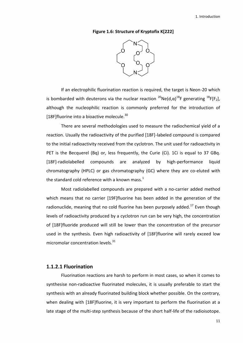
reason, a chelator agent like Kryptofix K[222] (Figure 1.6), is used to chelate the
potassium and release free [18F]fluoride ion which is thus activated and desolvated
ready for the nucleophilic attack.17
1. Introduction
11
Figure 1.6: Structure of Kryptofix K[222]
If an electrophilic fluorination reaction is required, the target is Neon-20 which
is bombarded with deuterons via the nuclear reaction 20Ne(d,α)18F generating 18F[F2],
although the nucleophilic reaction is commonly preferred for the introduction of
[18F]fluorine into a bioactive molecule.30
There are several methodologies used to measure the radiochemical yield of a
reaction. Usually the radioactivity of the purified [18F]-labeled compound is compared
to the initial radioactivity received from the cyclotron. The unit used for radioactivity in
PET is the Becquerel (Bq) or, less frequently, the Curie (Ci). 1Ci is equal to 37 GBq.
[18F]-radiolabelled compounds are analyzed by high-performance liquid
chromatography (HPLC) or gas chromatography (GC) where they are co-eluted with
the standard cold reference with a known mass.1
Most radiolabelled compounds are prepared with a no-carrier added method
which means that no carrier [19F]fluorine has been added in the generation of the
radionuclide, meaning that no cold fluorine has been purposely added.17 Even though
levels of radioactivity produced by a cyclotron run can be very high, the concentration
of [18F]fluoride produced will still be lower than the concentration of the precursor
used in the synthesis. Even high radioactivity of [18F]fluorine will rarely exceed low
micromolar concentration levels.31
1.1.2.1 Fluorination
Fluorination reactions are harsh to perform in most cases, so when it comes to
synthesise non-radioactive fluorinated molecules, it is usually preferable to start the
synthesis with an already fluorinated building block whether possible. On the contrary,
when dealing with [18F]fluorine, it is very important to perform the fluorination at a
late stage of the multi-step synthesis because of the short half-life of the radioisotope.
1. Introduction
12
In recent years many studies have been carried out to optimise late stage fluorination
showing better conditions to enable this step efficiently and rapidly.1,6,7,18
Nucleophilic Fluorination
Nucleophilic fluorinations are the most common kind of [18F]-fluorinations (hot
fluorination) in use today. The nucleophilic [18F]fluoride is produced by the nuclear
reaction on enriched water [18O]H2O through bombardment of protons. The
[18F]fluoride anion produced is a very weak nucleophile and a strategy has been
developed to increase its nucleophilicity. It is first dehydrated through a ion exchange
cartridge, and then activated by crown ethers such as Kryptofix K[222] and potassium
carbonate.6,7 Aliphatic nucleophilic fluorinations normally occur with an SN2
stereochemistry. Good leaving groups should be used for the precursors and aprotic
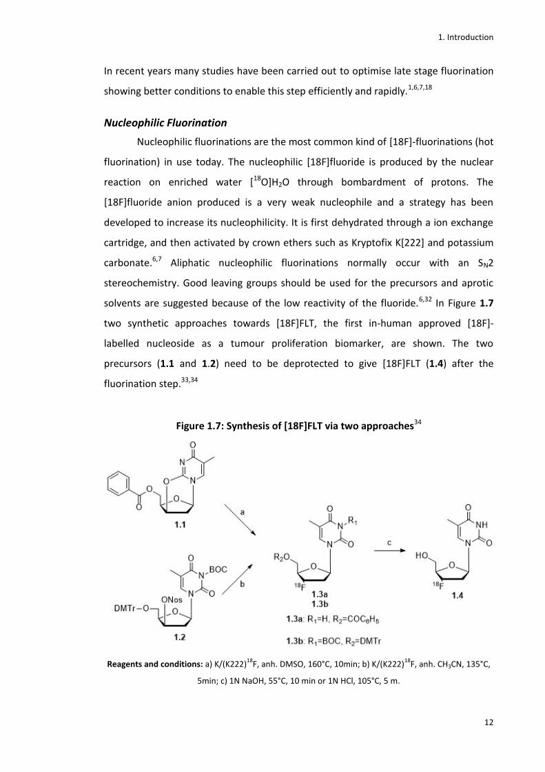
solvents are suggested because of the low reactivity of the fluoride.6,32 In Figure 1.7
two synthetic approaches towards [18F]FLT, the first in-human approved [18F]-
labelled nucleoside as a tumour proliferation biomarker, are shown. The two
precursors (1.1 and 1.2) need to be deprotected to give [18F]FLT (1.4) after the
fluorination step.33,34
Figure 1.7: Synthesis of [18F]FLT via two approaches34
Reagents and conditions: a) K/(K222)18
F, anh. DMSO, 160°C, 10min; b) K/(K222)18
F, anh. CH3CN, 135°C,
5min; c) 1N NaOH, 55°C, 10 min or 1N HCl, 105°C, 5 m.

1. Introduction
13
Recently many studies have been carried out to look for improved methods and
substrates for these [18F]-nucleophilic reactions. Modern organic chemistry
technology has been applied to radiochemistry and an example of the results obtained
by this combination is the use of a metal-catalyst for allylic fluorination reactions
(Figure 1.8).35,34
Figure 1.8: Metal catalysed allylic fluorination reaction
Nucleophilic aromatic fluorination is more challenging than aliphatic fluorination, and
is often used in PET radiochemistry for the synthesis of radiolabelled arenes. Harsh
conditions such as high reaction temperature and activated substrates with electron
withdrawing groups on the aromatic moieties in the ortho or para positions relative to
the position of fluorination are required. This becomes necessary to overcome the
normally disfavoured SNAr reaction. When there are electron rich groups on the
aromatic ring, a strategy has been developed to circumvent this issue. The electron-
donating group is masked as an electron withdrawing one using appropriate protecting
groups. An example of this strategy is the synthesis of [18F]flutemetamol (1.6), a PET
imaging agent of the brain used as diagnostic tool for Alzheimer’s disease (Figure
1.9).36,1
Figure 1.9: Synthesis of the [18F]flutemetamol
Reagents and conditions: a) K/ (K222)18
F, anh. DMSO, 150°C, 30min; b) 1N HCl, 105°C, 5 min.

1. Introduction
14
The radiolabeling of an ortho-nitroaniline was thus enabled by masking the
amine group as a formamide group. Therefore the displacement by [18F]fluoride of
the nitro group becomes possible.
Often [18F]fluoride is introduced into the aromatic moiety thanks to the use of
aryliodonium salts, to generate an extremely effective leaving group.7,37,38Recently to
improve the outcome of these types of fluorination, a number of transition metal
catalysed nucleophilic fluorinations have been developed. One of these uses pre-
catalysts such as nickel σ-aryl complexes to access [18F]fluorouracil (1.9), a PET tracer
for cancer imaging (Figure 1.10). This is the first time that a transition-metal-mediated
fluorination has been used for clinical application.34,39
Figure 1.10: Synthesis of [18F]fluorouracil (1.9) via transition-metal-mediated
fluorination34,39
Reagents and conditions: a) LnNiIIX2, pyridine, 70°C, 1h; b) PhI(4-OMe-pyridine)2(OTf)2, (18-c-6)K
18F,
CH3CN (0.5% H2O), 23°C, 1 min; c) HCl, EtOH, 23°C, 2 min.
Electrophilic Fluorination
Electrophilic fluorination reactions were commonly used for the preparation of
radiopharmaceuticals but they have been mostly replaced nowadays by nucleophilic
reactions where possible. Aromatic rings and/or electron rich-double bonds can be
readily fluorinated using electrophilic-fluorinating agents.40The reason for avoidance of
electrophilic fluorination is due to the high reactivity and toxicity of the hazardous
fluorine gas ([18F]F2), the simplest fluorinated reagent, generated by the nuclear
1. Introduction
15
reaction 20Ne(d,α)18F. This kind of reaction is characterized by a low chemical and
radiochemical yield and a poor selectivity.
Another method used to produce the fluorine gas ([18F]F2) is the bombardment
of oxygen-18(18O(p,n)18F) and a carrier fluorine gas. Also in this case the specificity and
the yields are very low because of the carrier-added production but mostly because of
the necessity to isolate by purification the desired radiolabelled compound which can
be a difficult procedure.41,7To increase both the yields and the selectivity of
electrophilic fluorination, several intermediate electrophilic reagents have been
produced. Among them [18F]XeF2 and [18F]AcOF are common even though they can
never reach a radiochemical yield greater than 50%.1
Since most applications preclude the use of fluorine gas, fluorine must bind to
an electronegative atom such as nitrogen, activated by a strongly withdrawing group
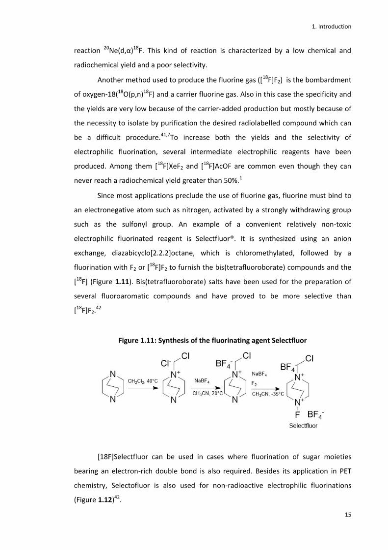
such as the sulfonyl group. An example of a convenient relatively non-toxic
electrophilic fluorinated reagent is Selectfluor®. It is synthesized using an anion
exchange, diazabicyclo[2.2.2]octane, which is chloromethylated, followed by a
fluorination with F2 or [18F]F2 to furnish the bis(tetrafluoroborate) compounds and the
[18F] (Figure 1.11). Bis(tetrafluoroborate) salts have been used for the preparation of
several fluoroaromatic compounds and have proved to be more selective than
[18F]F2.42
Figure 1.11: Synthesis of the fluorinating agent Selectfluor
[18F]Selectfluor can be used in cases where fluorination of sugar moieties
bearing an electron-rich double bond is also required. Besides its application in PET
chemistry, Selectofluor is also used for non-radioactive electrophilic fluorinations
(Figure 1.12)42.

1. Introduction
16
Figure 1.12: Electrophilic fluorination with Selectfluor
Reagents and conditions: a) Selectfluor, HOAc-H2O.
Another electrophilic fluorination agent has been recently synthesized is the
[18F]fluoride-derived palladium (IV). This is the first electrophilic agent derived directly
from [18F]fluoride. Its use has been limited because it involves a two-step sequence
reaction and because of the potentially sensitive organometallic reagents.42
Other synthetic strategies to access fluorinated molecules
When later-stage fluorination is either not possible or convenient, other
synthetic approaches involve the use of already fluorinated building blocks or, in the
case of PET chemistry, the early fluorination of a moiety of the molecule. N-
glycosylation reaction is for instance used to access some fluorinated nucleosides. It
consists of the reaction between already fluorinated nucleobases and sugar moieties.
This is considered a convergent approach and is particularly used to access 2’-β-fluoro
nucleosides. The fully protected sugar is first brominated to form the 1-α-glycosyl
bromide. The β-nucleoside is usually the most favoured product of this reaction as
reported in the synthesis of the antineoplastic agent Clofarabine (1.13)(Figure
1.13).43,34
Figure 1.13: Synthesis of Clofarabine (1.13)
Reagents and conditions: a) HBr, AcOH, rt; b) 2-chloroadenine, KOt-Bu, CH3CN, t-AmOH, DCE, 55C; c)
cat. NaOCH3, CH3OH, rt.

1. Introduction
17
As mentioned before, this synthetic approach is used also in PET chemistry to
access a number of [18F]-β nucleosides. [18F]FIAU (1.16), a reporter gene for the
expression of the herpes simplex virus type-1 thymidine kinase (HSV1-tk), is for
instance synthesised using the same synthetic convergent approach (Figure 1.14).44,45
Schinazi et al have recently reviewed synthetic strategies for the convergent
approach.46
Figure 1.14: Synthesis of [18F]FIAU (1.16)
Reagents and conditions: a) K/(K222)18
F, anh. CH3CN, 80°C, 30 min; b)5-iodouridine, HMDS, TMSOTf,
anh. CH3CN, 80°C, 1h; c)KOCH3, CH3OH, 80°C, 10 min.
1.1.2.2 [18F]FDG
[18F]FDG ( 2[18F]fluoro-2-deoxy-D-glucose) (1.17) is the most widely used PET
tracer, being used in perhaps as many as 90% of all PET oncology scans.
Figure 1.15: Structure of the most widely used PET tracer [18F]FDG
Glucose is a major source of energy for different biological systems. It is first
transported by membrane transporters (GLuT) into cells and through the BBB (blood
brain barrier). Glucose is there phosphorylated by a hexokinase using ATP as a source
of phosphate. [18F]FDG is transported inside the cells with high metabolic activity by

1. Introduction
18
the same transporter as for glucose and it is phosphorylated by the same kinase. But,
unlike glucose, it is not metabolized by the glucose-6-phosphate isomerase and it stays
entrapped into the cells. Once [18F] decays into [18O], the resulting 2-[18O]-
deoxyglucose-6-phosphate is metabolized while the [18F]FDG is excreted.47 [18F]FDG
has many applications in different areas such as oncology, neurology, and cardiology.48
It can be synthesized using both electrophilic and nucleophilic substitution reactions.
The electrophilic substitution can be performed using either [18F]F2 or
[18F]acetyl hypofluorite as the source of [18F]-fluorine. The hypofluorite gives better
yields than [18F]F2. The precursor molecule used is 3,4,6-tri-O-acetyl-D-glucan.49
Another approach for electrophilic substitution used for production of [18F]FDG uses
[18F]XeF2 as the source of electrophilic fluoride.50Unfortunately none of these methods
give very good yields so radiochemical facilities have abandoned them in favour of
nucleophilic substitutions.
Nucleophilic reactions can be performed using Cs[18F], Et4N[18F], or KH[F]F2 as
source, however 18F- in Kryptofix is the most commonly used for everyday routine
synthesis, using 1,3,4,6-tetra-O-acetyl-2-O-triflate-β-D-mannose as the precursor
molecule. Since the first time this synthesis was published, in 198651, several
improvements have been made and different modular systems have been used. Taking
into account the radioactive decay, radiochemical yields can be even higher than 60 %
and the total reaction time can be even less than 26 minutes.52 Figure 1.16 shows the
method mostly used for clinic production of [18F]FDG.
Figure 1.16: Routine clinical synthesis of the radiotracer [18F]FDG
1.1.2.3 [18F]-nucleosides
[18F]fluorouracil (5-[18F]FU) (1.9)
5-FU is a chemotherapeutic agent used in the treatment of solid tumours such
as breast and colon cancer, however because of its in vivo rapid catabolism, its use for
1. Introduction
19
therapeutic purposes is limited.53 5-[18F]FU (1.9) was synthesized to study the
behaviour of 5-FU in vivo and 5-ethyluracil, an analogue of 5-FU, was used to block the
catabolism of 5-[18F]FU. Through this experiment, it was possible to monitor the
transport of 5-FU in vivo and its anabolism. The study showed very useful information
to predict the response of the tumour to 5-FU. However 5-FU is not routinely used for
PET imaging because of its rapid catabolism.10
[18F]-analogues of pyrimidine nucleosides
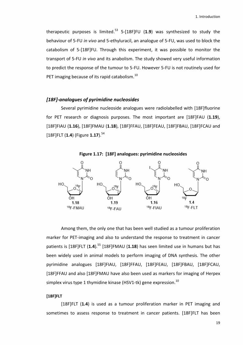
Several pyrimidine nucleoside analogues were radiolabelled with [18F]fluorine
for PET research or diagnosis purposes. The most important are [18F]FAU (1.19),
[18F]FIAU (1.16), [18F]FMAU (1.18), [18F]FFAU, [18F]FEAU, [18F]FBAU, [18F]FCAU and
[18F]FLT (1.4) (Figure 1.17).54
Figure 1.17: [18F] analogues: pyrimidine nucleosides
Among them, the only one that has been well studied as a tumour proliferation
marker for PET-imaging and also to understand the response to treatment in cancer
patients is [18F]FLT (1.4).55 [18F]FMAU (1.18) has seen limited use in humans but has
been widely used in animal models to perform imaging of DNA synthesis. The other
pyrimidine analogues [18F]FIAU, [18F]FFAU, [18F]FEAU, [18F]FBAU, [18F]FCAU,
[18F]FFAU and also [18F]FMAU have also been used as markers for imaging of Herpex
simplex virus type 1 thymidine kinase (HSV1-tk) gene expression.10
[18F]FLT
[18F]FLT (1.4) is used as a tumour proliferation marker in PET imaging and
sometimes to assess response to treatment in cancer patients. [18F]FLT has been

1. Introduction
20
widely studied on different types of tumours and is currently the subject of ongoing
clinical trials, having been widely investigated since 1998.10 Indeed it represents a valid
alternative to [18F]FDG when assessing tumours with high proliferation rates. Among
different types of cancer, it has been used especially for lung, brain (glioma) and
colorectal cancer.10
As it is an analogue of thymidine, [18F]FLT targets human thymidine kinase
(TK1). It is thus phosphorylated into [18F]FLT monophosphate which is trapped into
growing cells. The enzyme TK1 takes part in the synthesis of the DNA (deoxyribonucleic
acid) and in particular shows a very high activity during the S phase, the synthesis
phase of the cell cycle. [18F]FLT thus represents an indicator of cell proliferation rate
when accumulated into the cell. Lately it has been shown that [18F]FLT reflects
proliferative indices to potentially unreliable extents because it cannot discriminate
among high proliferative index tumour, which relies on thymidine salvage pathway,
from high proliferation tissues which instead rely especially upon the de novo synthesis
of thymidine (Figure 1.18).56
Figure 1.18: Thymidine salvage and de novo synthesis pathways56
[18F]-purine analogue: acycloguanosine
The first radiolabelled purine analogue nucleoside synthesized was [18F]FHPG
(1.20).57 After that, the PCV analogue [18F]FHBG (1.21) was developed and has been
1. Introduction
21
widely studied in animal models for a mutated HSV1-tk gene, the sr39-HSV1-tk (Figure
1.19).
Figure 1.19: Structures of some purine nucleoside analogues
Many imaging studies on the HSV1-tk gene expression have been carried out,
but until now just a few clinical studies using [18F]FHBG have been performed on
humans.57
[18F]-purine adenosine analogues
[18F]FAA (1.22) and [18F]FXA (1.23) (Figure 1.20) are two adenosine analogues
that have been tested as substrates of HSV1-tk but their uptake was very low.
Nevertheless, as their biodistribution was studied by micro-PET images, [18F]FAA
showed a high uptake in tumour visualization, whereas [18F]FXA showed a high uptake
in heart. These agents could thus represent respectively a new potential tumour
imaging agent and a new potential heart imaging agent. Further studies need to be
undertaken to confirm this prospect.58
Figure 1.20: Structures of some [18F]-purine adenosine analogues
1. Introduction
22
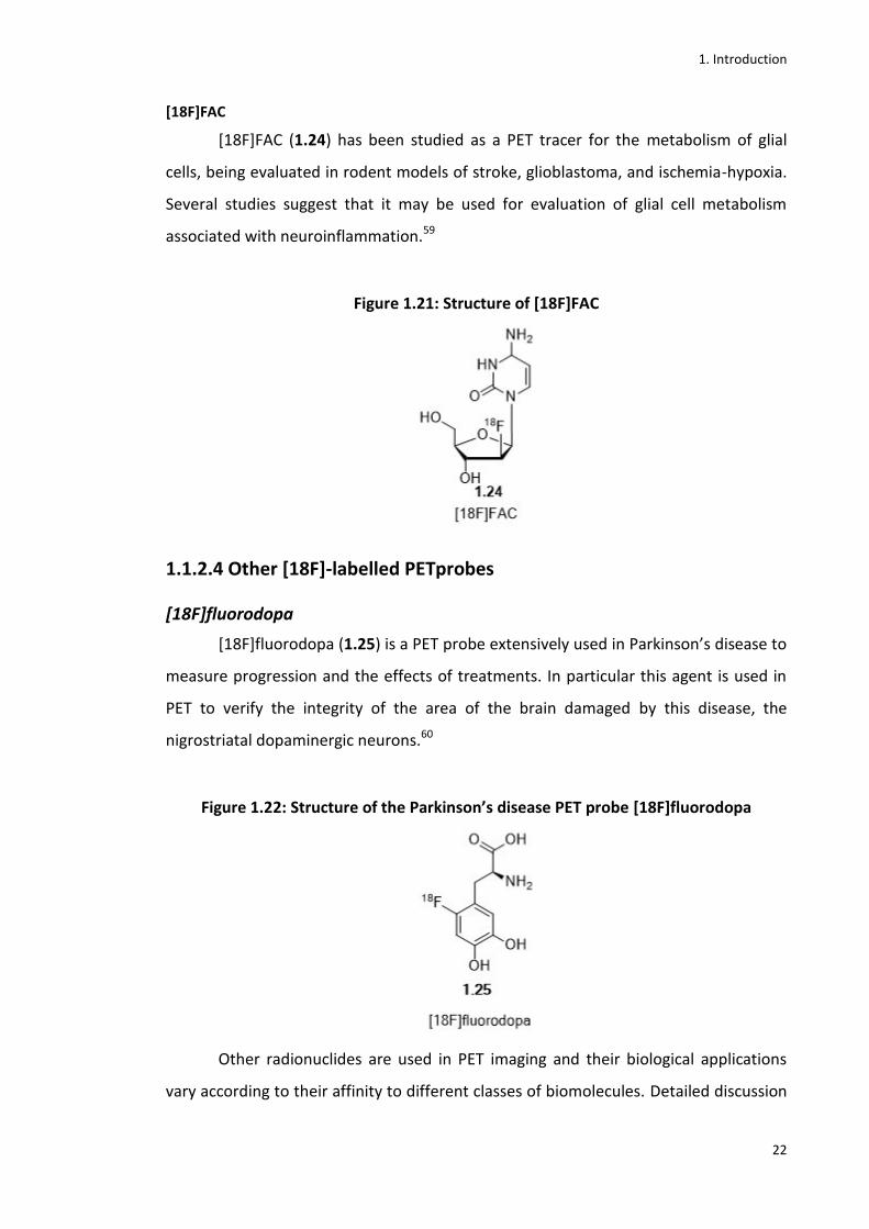
[18F]FAC
[18F]FAC (1.24) has been studied as a PET tracer for the metabolism of glial
cells, being evaluated in rodent models of stroke, glioblastoma, and ischemia-hypoxia.
Several studies suggest that it may be used for evaluation of glial cell metabolism
associated with neuroinflammation.59
Figure 1.21: Structure of [18F]FAC
1.1.2.4 Other [18F]-labelled PETprobes
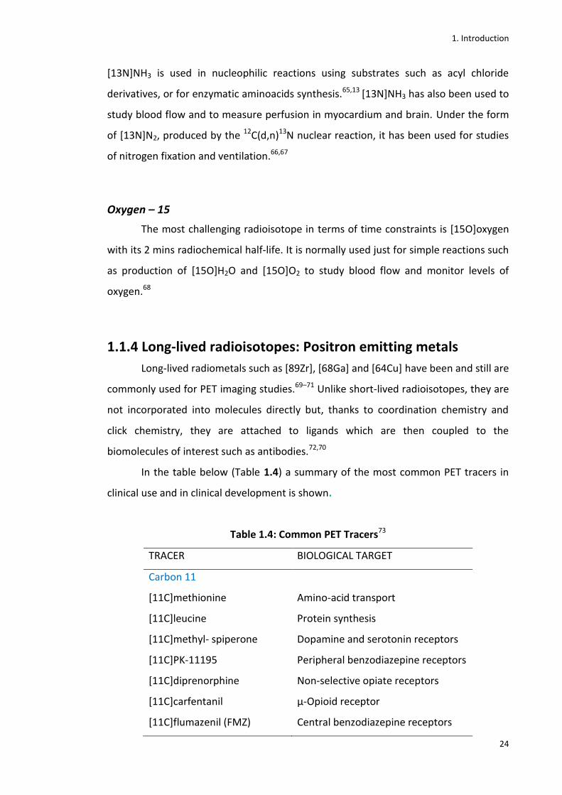
[18F]fluorodopa
[18F]fluorodopa (1.25) is a PET probe extensively used in Parkinson’s disease to
measure progression and the effects of treatments. In particular this agent is used in
PET to verify the integrity of the area of the brain damaged by this disease, the
nigrostriatal dopaminergic neurons.60
Figure 1.22: Structure of the Parkinson’s disease PET probe [18F]fluorodopa
Other radionuclides are used in PET imaging and their biological applications
vary according to their affinity to different classes of biomolecules. Detailed discussion

1. Introduction
23
of further types [18F]-labelled PET tracers is beyond the scope of this introductory
chapter.
1.1.3 Short lived radioisotopes
Carbon – 11
Another commonly used radioisotope in PET imaging is [11C]carbon. The
nuclear reaction most widely used for its production is 14N(p,α)11C. When this reaction
happens in the presence of oxygen, the product is [11C]CO2, whereas [11C]CH4 is
obtained in the presence of hydrogen. These two radiolabelled compounds can be
used as reagents for many other chemical reactions.61
[11C]CO2 is used as intermediate to synthesise [11C]CH3I, the most widely used
reagent to perform methylation of heteroatoms to obtain a large number of [11C]-
tracers. [11C]CH3I can be produced by two methods known as wet or dry methods. The
wet method uses a reducing agent such as LiAlH4 to reduce [11C]CO2. This step is
followed by an iodination step with HI to finally obtain the methylating reagent
[11C]CH3I.60 The dry method uses instead high temperature to perform iodination with
I2 using as a substrate [11C]CH4.16,62 The ‘dry or gas’ method is nowadays the method
of choice for the synthesis of [11C]CH3I because of the high SA of the radiolabelled
product obtained.63
Nitrogen – 13
Because of its very short half life (9.96 min), [13N]nitrogen has a limited
application in PET chemistry and, together with [15O]oxygen and other short lived
radioisotopes, is commonly used for perfusion studies. However, many attractive
applications of [13N]-tracers have incentivised the development of many synthetic
strategies for its incorporation into drug molecules. This is due to the fact that nitrogen
is present in a large number of natural and synthetic molecules with biologically
interesting activities.
[13N]nitrogen is normally incorporated into molecules using the reagent
[13N]NH3. The radioisotope is commonly produced by the nuclear reaction 16O(p,α)13N
using water as a target. A mixture of [13N]NO3, [13N]NO2 and [13N]NH3 are thus
produced. By using DeVarda’s alloy, nitrate and nitrite are reduced to ammonia.64
1. Introduction
24
[13N]NH3 is used in nucleophilic reactions using substrates such as acyl chloride
derivatives, or for enzymatic aminoacids synthesis.65,13 [13N]NH3 has also been used to
study blood flow and to measure perfusion in myocardium and brain. Under the form
of [13N]N2, produced by the 12C(d,n)13N nuclear reaction, it has been used for studies
of nitrogen fixation and ventilation.66,67
Oxygen – 15
The most challenging radioisotope in terms of time constraints is [15O]oxygen
with its 2 mins radiochemical half-life. It is normally used just for simple reactions such
as production of [15O]H2O and [15O]O2 to study blood flow and monitor levels of
oxygen.68
1.1.4 Long-lived radioisotopes: Positron emitting metals
Long-lived radiometals such as [89Zr], [68Ga] and [64Cu] have been and still are
commonly used for PET imaging studies.69–71 Unlike short-lived radioisotopes, they are
not incorporated into molecules directly but, thanks to coordination chemistry and
click chemistry, they are attached to ligands which are then coupled to the
biomolecules of interest such as antibodies.72,70
In the table below (Table 1.4) a summary of the most common PET tracers in
clinical use and in clinical development is shown.
Table 1.4: Common PET Tracers73
TRACER BIOLOGICAL TARGET
Carbon 11
[11C]methionine Amino-acid transport
[11C]leucine Protein synthesis
[11C]methyl- spiperone Dopamine and serotonin receptors
[11C]PK-11195 Peripheral benzodiazepine receptors
[11C]diprenorphine Non-selective opiate receptors
[11C]carfentanil μ-Opioid receptor
[11C]flumazenil (FMZ) Central benzodiazepine receptors
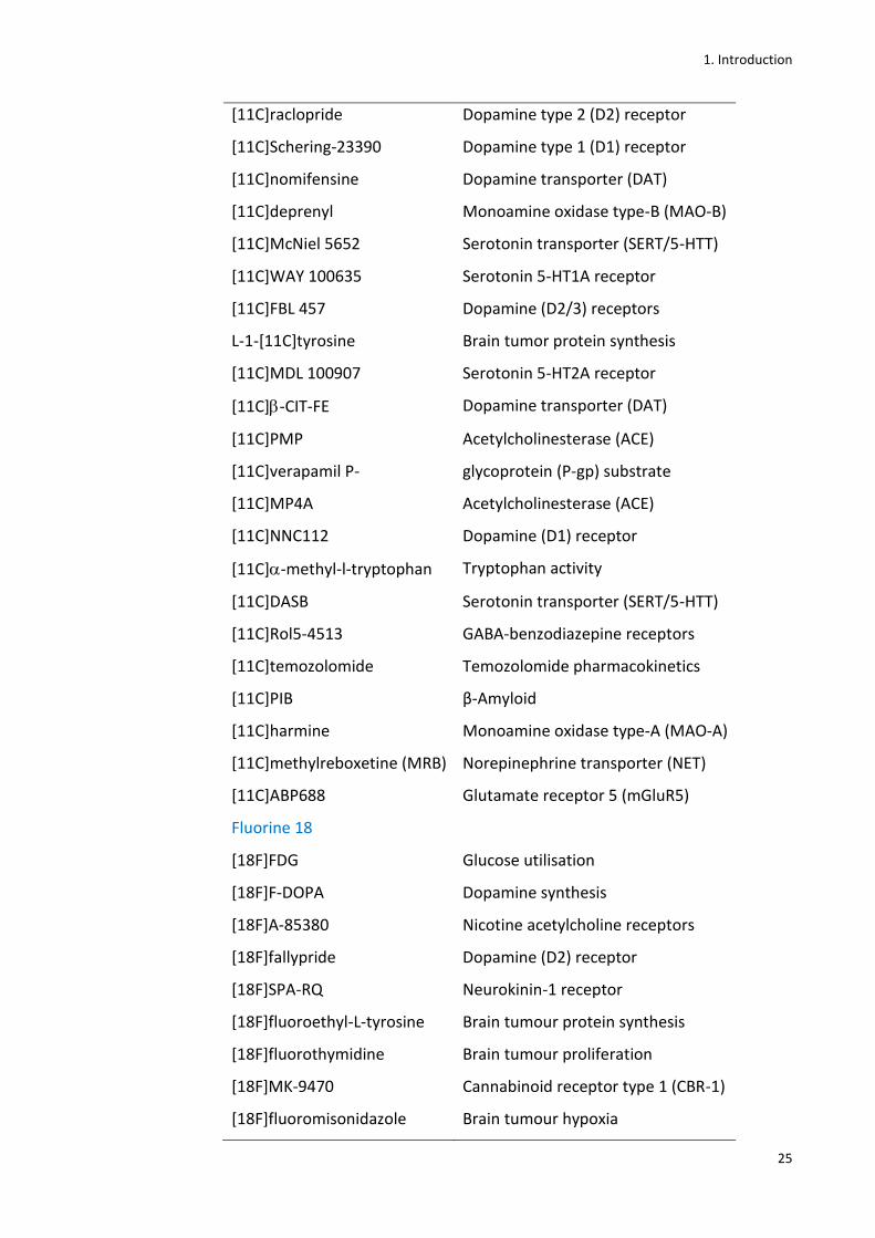
1. Introduction
25
[11C]raclopride Dopamine type 2 (D2) receptor
[11C]Schering-23390 Dopamine type 1 (D1) receptor
[11C]nomifensine Dopamine transporter (DAT)
[11C]deprenyl Monoamine oxidase type-B (MAO-B)
[11C]McNiel 5652 Serotonin transporter (SERT/5-HTT)
[11C]WAY 100635 Serotonin 5-HT1A receptor
[11C]FBL 457 Dopamine (D2/3) receptors
L-1-[11C]tyrosine Brain tumor protein synthesis
[11C]MDL 100907 Serotonin 5-HT2A receptor
[11C]-CIT-FE Dopamine transporter (DAT)
[11C]PMP Acetylcholinesterase (ACE)
[11C]verapamil P- glycoprotein (P-gp) substrate
[11C]MP4A Acetylcholinesterase (ACE)
[11C]NNC112 Dopamine (D1) receptor
[11C]-methyl-l-tryptophan Tryptophan activity
[11C]DASB Serotonin transporter (SERT/5-HTT)
[11C]Rol5-4513 GABA-benzodiazepine receptors
[11C]temozolomide Temozolomide pharmacokinetics
[11C]PIB β-Amyloid
[11C]harmine Monoamine oxidase type-A (MAO-A)
[11C]methylreboxetine (MRB) Norepinephrine transporter (NET)
[11C]ABP688 Glutamate receptor 5 (mGluR5)
Fluorine 18
[18F]FDG Glucose utilisation
[18F]F-DOPA Dopamine synthesis
[18F]A-85380 Nicotine acetylcholine receptors
[18F]fallypride Dopamine (D2) receptor
[18F]SPA-RQ Neurokinin-1 receptor
[18F]fluoroethyl-L-tyrosine Brain tumour protein synthesis
[18F]fluorothymidine Brain tumour proliferation
[18F]MK-9470 Cannabinoid receptor type 1 (CBR-1)
[18F]fluoromisonidazole Brain tumour hypoxia
1. Introduction
26
Oxygen
[15O]oxygen Oxygen utilisation
[15O]water Blood flow
1.1.5 PET imaging: application in drug discovery
PET Imaging has been used not just as a diagnostic tool but more recently as a
means to answer key questions in drug discovery on biological and pharmacological
behaviour of certain compounds in vivo. Matthews and co-workers have recently
reviewed this area of study.74 By radiolabelling certain drug candidates, it is possible to
gain useful information on their biodistribution in vivo such as whether or not they
reach the target tissue primarily in rodents and primates, and secondarily in humans.
This technique is also useful to understand whether some compounds can cause side
effects by accumulating at non-target sites and additionally biodistribution studies can
give information on dose-limiting toxicity. These kinds of studies can also give the
possibility, through PET scans, to have data on pharmacokinetics and
pharmacodynamics of drug candidates by comparing radio-TLC and radio-HPLC of the
parent compound to the metabolism products.1
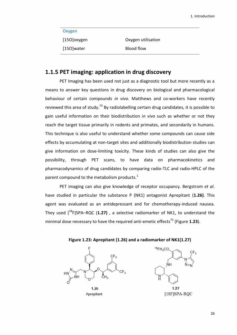
PET imaging can also give knowledge of receptor occupancy. Bergstrom et al.
have studied in particular the substance P (NK1) antagonist Aprepitant (1.26). This
agent was evaluated as an antidepressant and for chemotherapy-induced nausea.
They used [18F]SPA–RQC (1.27) , a selective radiomarker of NK1, to understand the
minimal dose necessary to have the required anti-emetic effects75 (Figure 1.23).
Figure 1.23: Aprepitant (1.26) and a radiomarker of NK1(1.27)
1. Introduction
27
Bergstrom et al. understood that 100 mg/day was an effective dose for its
antiemetic purpose but that this dose was not effective as an antidepressant. Indeed
Merck was evaluating Aprepitant’s application as an anti-depressive in Phase III clinical
trial where the dose of 300 mg/day was still a not effective dose to have the anti-
depressive effect. The PET studies, which demonstrated that that dose was definitely
enough to achieve the occupancy of the target receptor, saved Merck several million
dollars through interruption of the ongoing Phase III clinical trial on this compound.1
1.2 Nucleosides
Nucleosides are endogenous compounds critically involved in DNA and RNA
synthesis but also in enzyme regulation, metabolism and cell signaling.76,77 Several
nucleoside and nucleotide analogues have been synthesized to act as antimetabolites
of their physiological counterparts in several biochemical pathways. They inhibit viral
replication and cellular metabolism by incorporation into DNA and RNA and thus they
represent cornerstones in the treatment of patients with cancer and viral infections.
Nucleosides can inhibit several essential enzymes like kinase, pyrimidine and purine
nucleoside phosphorylase, several viral and human polymerases, DNA methyl
transferase, thymidylate synthase, and ribonucleotidereductase. Chemically they
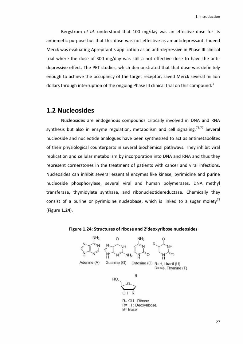
consist of a purine or pyrimidine nucleobase, which is linked to a sugar moiety78
(Figure 1.24).
Figure 1.24: Structures of ribose and 2’deoxyribose nucleosides

1. Introduction
28
When nucleosides have a phosphate group in the 5’ position of the sugar
moiety they are named nucleotides. Their activity depends on certain modifications on
the nucleobase that gives them different specific properties. Nucleosides can act as
antimetabolites, where the metabolic pathway of these synthetic nucleosides and
nucleotides is the same one of their endogenous counterparts.79 After cellular uptake
performed by nucleoside-transporter systems, NAs become substrates for specific
nucleo(s)(t)ide kinase enzymes which convert them to the 5’-mono, -di and
triphosphate forms.26 The active triphosphate form can interfere with the de novo
synthesis of DNA/RNA precursors, leading to the inhibition of DNA and RNA synthesis
and to suppression of cell growth and division.26
The first phosphorylation is the rate-limiting phosphorylation step of their
activation pathway. Thymidine kinase (TK1) or deoxycytidine kinase (dCK) can carry out
this phosphorylation according to the substrate. For instance, TK1 is specific for
thymidine (1.28) and its analogues, while dCK is specific for cytidine (1.30) and its
analogues (e.g. gemcitabine 1.31).
Figure 1.25: Thymidine and cytidine and their analogues
When nucleosides are monophosphorylated, they are effectively entrapped
inside the cellular cytoplasm as charged nucleotides. Nucleosides are hydrophilic
molecules that diffuse slowly across cell membranes, therefore cells have developed a
complex transport system to facilitate membrane transport. This system consists of
multiple carrier proteins, the nucleoside transporters (NTs), which facilitate their
cellular uptake for nucleotide biosynthesis. The monophosphorylated nucleosides are
therefore further phosphorylated by other kinases to form the diphosphates and the
triphosphates which finally take part in the biosynthesis of DNA and RNA.80
1. Introduction
29
1.2.1 ProTides
Despite the enormous importance of nucleoside analogues as both antiviral
and anticancer agents, drug resistance still represents a major issue for their clinical
application. As already described above, to exert their therapeutic activity, nucleoside
analogues need to be phosphorylated by intracellular kinases to the 5’-mono-, di- and
triphosphate forms. Many synthetic strategies have been developed to access directly
the active 5’-triphosphate form, but these compounds are not viable drug candidates
because of their chemical instability and high polarity which disallow their effective
crossing through cell membranes. Nevertheless, the first phosphorylation is considered
to be rate-limiting step of their activation pathway, therefore many approaches to
develop prodrugs of nucleoside monophosphate forms have been developed.81
The phosphoramidate-based technology, also known as the ProTide
(PronucleoTide) approach, developed by McGuigan and co-workers in 1995 is
considered one of the most effective pro-nucleotide strategies currently used in the
clinic.82,83 It consists in masking the negatively charged 5’-O-monophosphate form as a
phosphoramidate. This masked derivative, after passive diffusion through cell
membrane and subsequent intracellular metabolism, delivers the 5’-O-
monophosphate form 84 which is thus trapped into the cell and available for further
phosphorylations (Figure 1.26).82,83

1. Introduction
30
Figure 1.26: Pro-nucleotides mechanism of action81
Chemically ProTides are aryloxyphosphoramidate prodrugs synthesized
through a coupling reaction between a phosphorochloridate and the 5’-OH of the
nucleoside. The phosphorus atom is attached to an amino acid alkyl ester and to an aryloxy
group enabling passive transmembrane transport and masking the negative charge.84,85
1. Introduction
31
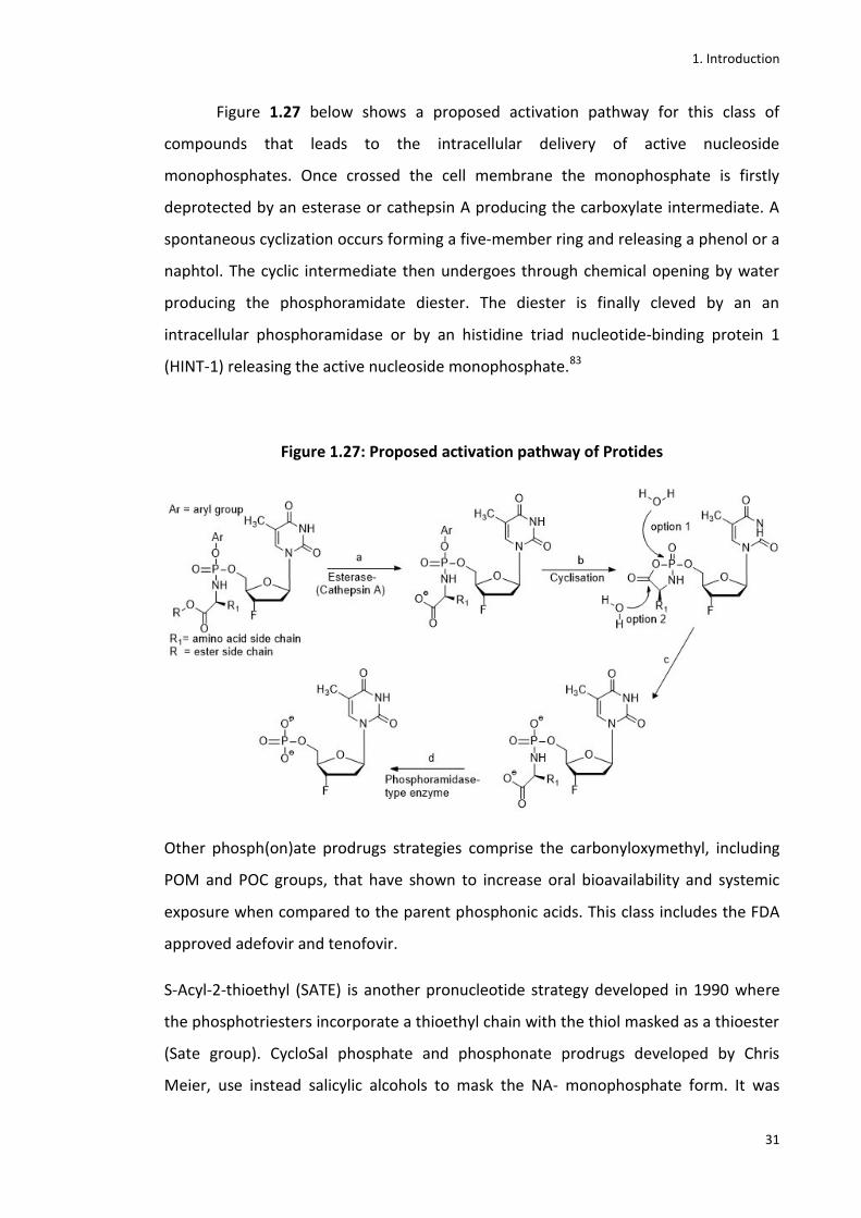
Figure 1.27 below shows a proposed activation pathway for this class of
compounds that leads to the intracellular delivery of active nucleoside
monophosphates. Once crossed the cell membrane the monophosphate is firstly
deprotected by an esterase or cathepsin A producing the carboxylate intermediate. A
spontaneous cyclization occurs forming a five-member ring and releasing a phenol or a
naphtol. The cyclic intermediate then undergoes through chemical opening by water
producing the phosphoramidate diester. The diester is finally cleved by an an
intracellular phosphoramidase or by an histidine triad nucleotide-binding protein 1
(HINT-1) releasing the active nucleoside monophosphate.83
Figure 1.27: Proposed activation pathway of Protides
Other phosph(on)ate prodrugs strategies comprise the carbonyloxymethyl, including
POM and POC groups, that have shown to increase oral bioavailability and systemic
exposure when compared to the parent phosphonic acids. This class includes the FDA
approved adefovir and tenofovir.
S-Acyl-2-thioethyl (SATE) is another pronucleotide strategy developed in 1990 where
the phosphotriesters incorporate a thioethyl chain with the thiol masked as a thioester
(Sate group). CycloSal phosphate and phosphonate prodrugs developed by Chris
Meier, use instead salicylic alcohols to mask the NA- monophosphate form. It was

1. Introduction
32
successfully applied to some antiviral nucleotides such as AZT, d4t and acyclovir. All
these and other pronucleotide strategies have been recently reveiwed by Scinazi et al.
1.2.2 Fluorinated nucleosides and ProTides
An important class of antiviral and anticancer drugs is represented by
fluorinated 2’- and 3’-substituted nucleosides where one or more hydrogens on the
sugar moiety are substituted by fluorine atoms. Figure 1.28 shows all the FDA-
approved fluorinated anticancer and antiviral nucleoside analogues.34

1. Introduction
33
Figure 1.28: FDA-approved fluorinated anticancer and antiviral nucleoside analogues.
1.2.2.1 Importance of fluorine in drug molecules
Despite the widespread occurrence of fluorine as an element on earth,
organofluorine compounds are almost absent in nature. Nevertheless, 20-25% of
synthetic drugs are thought to contain in their structure at least one fluorine.86,87,34
Fluorine plays a crucial rule in the optimisation of clinical candidates. It increases the
lipophilicity (logP) which is a major factor, together with the molecular size, that
influences membrane permeability that can lead to an optimization of drug
candidates’ oral bioavailability. Modulation of pKa can also have a huge impact on
compounds bioavailability as well as on their absorbance. Fluorine is the most

1. Introduction
34
electronegative element (3.98 on the Pauling scale) and, when included into a
molecule, can strongly affect the acidity or the basicity of the surrounding functional
groups in the molecule.34,88,89
Additionally, when the fluorine is bonded to the carbon, the highly polarised
bond formed can provide a remarkable stability to the molecule itself. All these factors
can have a positive impact on selectivity, toxicity, potency, and on pharmacokinetic
and pharmacodynamic properties of the molecule. The size of fluorine (van der Waals
radius of 1.47Å) is also very similar to that of hydrogen (1.20 Å), therefore it is
frequently used to replace the H or the hydroxyl group in drug molecules without
causing steric perturbation. However, from the chemical point of view its replacement
does not lead to dramatic changes whereas from the biological point of view it can
block oxidative metabolism and therefore block the formation of unwanted
metabolites. Drug molecules are indeed usually metabolised by enzymes such as
Cytocrome P450 (CYP450) monooxygenases that decrease compounds lipophilicity and
therefore increase their clearance. Fluorine substitution bypasses that problem by
blocking these sites from enzymatic cleavage and, therefore, increases the metabolic
stability of many drug molecules.34,88,89
Fluorine has a major impact on nucleosides and when placed on the ribose ring
of nucleoside analogues, it can significantly affect the conformation of nucleosides. It
causes changes in the dipole-dipole and gauche interactions, on the F-base
interactions and influences the anomeric effect. Generally, when placed in the 2’--
position of the ribose, fluorine favours a south conformation of the molecule, which
has often conferred to the molecule an enhanced anti-HIV activity, whereas the
fluorine in the 2’--position favours a north conformation (Figure 1.29). Nevertheless,
a general rule that correlates the position of the fluorine with the biological activity of
the molecule is difficult to define. A research group working on pro-nucleotide CycloSal
triesters of 2′-fluorinated-2′,3′-dideoxyadenosines (F-ddA) studied the effects induced
by two opposite and configurations of the fluorine at C2′ of the dideoxyribose
moiety. They showed that 2’--F-ddA is active as an anti-HIV agent, whereas the -
analogue did not show any antiviral activity. Interestingly, when the -configured
phosphotriesters of the inactive 2’--F -ddA were tested, they showed a higher anti-
HIV activity compared to 2’--F- ddA (Figure 1.29).34,90
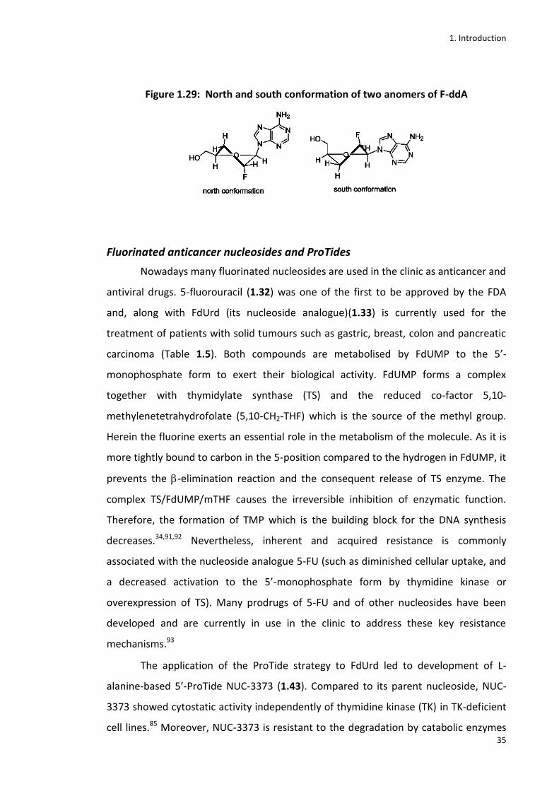
1. Introduction
35
Figure 1.29: North and south conformation of two anomers of F-ddA
Fluorinated anticancer nucleosides and ProTides
Nowadays many fluorinated nucleosides are used in the clinic as anticancer and
antiviral drugs. 5-fluorouracil (1.32) was one of the first to be approved by the FDA
and, along with FdUrd (its nucleoside analogue)(1.33) is currently used for the
treatment of patients with solid tumours such as gastric, breast, colon and pancreatic
carcinoma (Table 1.5). Both compounds are metabolised by FdUMP to the 5’-
monophosphate form to exert their biological activity. FdUMP forms a complex
together with thymidylate synthase (TS) and the reduced co-factor 5,10-
methylenetetrahydrofolate (5,10-CH2-THF) which is the source of the methyl group.
Herein the fluorine exerts an essential role in the metabolism of the molecule. As it is
more tightly bound to carbon in the 5-position compared to the hydrogen in FdUMP, it
prevents the -elimination reaction and the consequent release of TS enzyme. The
complex TS/FdUMP/mTHF causes the irreversible inhibition of enzymatic function.
Therefore, the formation of TMP which is the building block for the DNA synthesis
decreases.34,91,92 Nevertheless, inherent and acquired resistance is commonly
associated with the nucleoside analogue 5-FU (such as diminished cellular uptake, and
a decreased activation to the 5’-monophosphate form by thymidine kinase or
overexpression of TS). Many prodrugs of 5-FU and of other nucleosides have been
developed and are currently in use in the clinic to address these key resistance
mechanisms.93
The application of the ProTide strategy to FdUrd led to development of L-
alanine-based 5’-ProTide NUC-3373 (1.43). Compared to its parent nucleoside, NUC-
3373 showed cytostatic activity independently of thymidine kinase (TK) in TK-deficient
cell lines.85 Moreover, NUC-3373 is resistant to the degradation by catabolic enzymes

1. Introduction
36
such as thymidine phosphorylase (TP), which is often upregulated in tumour cells, and
dihydropyrimidine dehydrogenase (DPD), an enzyme overexpressed in the liver. In
addition, within in vitromodels, NUC-3373 generates up to 363-fold higher intracellular
levels of FdUMP compared to 5-FU in the human colorectal cancer cell line HT29.94In
vivo studies showed greater tumour volume reduction in the human colorectal cancer
HT29 mouse xenograft model when NUC-3373 was tested compared to 5-FU.95A Phase
I clinical study of NUC-3373 started in 2016 on patients with advanced solid tumours
(Table 1.5).34,96
Another clinically widely used fluorinated anticancer agent is gemcitabine (2’-
deoxy-2’,2’-difluorocytidine, dFdC) (1.31) which has been approved for the treatment
of pancreatic, non-small cell lung, ovarian and breast cancers.97,98 Again its clinical
effectiveness is reduced because of resistance mechanisms such as poor cellular
uptake and poor conversion of gemcitabine into active metabolites by deoxycytidine
kinases, plus rapid deamination by cytidine deaminase into the inactive and toxic by-
product dFdU.99–101 The triphosphate metabolite (dFdC-TP), which is the active form of
dFdC, is thus incorporated into the growing DNA chain in the S-phase of cell cycle and
therefore causes chain termination of DNA synthesis leading eventually to cell death.
The diphosphate form (dFdC-DP) of dFdCalso contributes significantly to its anticancer
activity by inhibiting ribonucleotidereductase (RNR). As a result dFdC-TP is
incorporated as a substrate for DNA polymerase instead of natural
deoxynucleotides.34,102
To improve the clinical efficacy of gemcitabine many modifications have been
applied to its original chemical structure. Novel gemcitabine-based prodrugs such as
LY2334737 (1.48), CO-101(1.49) and more interestingly the phosphoramidate NUC-
1031 (1.46) have therefore been developed. NUC-1031 is a phosphoramidate L-
alanine-based ProTide of gemcitabine designed to overcome the resistance
mechanisms of the parent nucleoside. Its cellular uptake is indeed independent of
nucleoside transporters compared to the parent lead. Once into the cell, it releases the
5’-monophosphate form via consecutive steps which are mediated by a
carboxyesterase-type enzyme and phosphoramidase-type enzyme that catalyses the P-
N bond cleavage.34,103 Phase I clinical studies conducted in patients with advanced solid
tumours in 2012 showed promising pharmacokinetics and a favourable safety profile

1. Introduction
37
and efficacy. NUC-1031 demonstrated the ability to achieve 217x higher intracellular
dFd-CTP levels compared to the parent compound. The plasma half-life of this ProTide
was also much more favourable (7.3 h) than the one reported with gemcitabine (1.5
hours). In conclusion these studies indicated that NUC1031 was able to achieve a
durable disease control in a high number of patients including the ones who were
refractory to, or who relapsed on prior gemcitabine treatment. NUC-1031 is currently
investigated in several clinical trials for the treatment of patients with ovarian, biliary
and pancreatic cancers.104,34 Figure 1.30 shows all anticancer fluorinated nucleobase,
nucleoside and nucleotide analogues in clinical development.
Figure 1.30: Fluorinated anticancer nucleobase, nucleoside and nucleotide
analogues in clinical studies34
1. Introduction
38
Purine-based anticancer fluorinated nucleoside analogues
Fludarabine (FAMP) (1.34) is the 5’-monophosphate form of the purine
nucleoside analogue 2’-fluoro-arabinofuranosyladenine used for the treatment of B-
cell chronic lymphocytic leukaemia (B-CLL). It is also used in chemoimmunotherapy in
combination with the monoclonal antibody rituximab and the DNA-alkylating agent
cyclophosphamide, to treat CLL patients.34,105 As it is a monophosphate, it is negatively
charged at physiological pH, therefore is unable to enter cells. To reach the active 5’-
triphosphate form (F-ara-ATP), it is therefore first dephosphorylated to F-ara-A,
incorporated into the cell by nucleoside transport systems, and re-phosphorylated by
deoxycytidine kinase to the original 5’-monophosphate form. Subsequently the
adenylate kinase and nucleoside diphosphate kinase catalyse the phosphorylation to
the 5’-di and 5’-triphosphate forms, respectively. F-ara-ATP thus inhibits DNA
polymerases by acting as an alternative substrate to the natural deoxynucleotide
(dATP).34,106,107 Interestingly here the fluorine is essential as it increases the relative
resistance of F-ara-A to the deamination performed by adenosine deaminase.34,107
Nevertheless, some clinical trials of fludarabine showed severe central nervous
toxicity, somnolence and an increase of liver enzyme levels. This is probably due to the
phosphorolytic cleavage of F-ara-A which releases the metabolite 2-fluoroadenine (F-
Ade) that can accumulate as the toxic triphosphate F-AdeTP. Many resistance
mechanisms have been associated with the nucleoside analogue fludarabine. Mainly
they are related to alterations in membrane nucleoside transporters, deoxycytidine
kinase and cytoplasmic 5-nucleotidase cN-II activities, and to changes in the expression
of miR-34a, a small non-coding RNA molecule which mediates post-transcriptional
gene silencing.34,107,109
Clofarabine (Cl-F-ara-A)(1.36) is a second-generation chemotherapeutic agent
similar in structure to fludarabine, which compared to fludarabine, shows resistance to
phosphorolytic cleavage and deamination. Clofarabine is currently approved for clinical
use in the treatment of relapsed and refractory paediatric acute lymphoblastic
leukaemia. Clofarabine triphosphate is the active form of the nucleoside analogue and
it exerts its anticancer activity by inhibition of ribonucleotidereductase and inhibition
of DNA synthesis after incorporation into the DNA chain thus inducing
apoptosis.34,107,109 The fluorine atom in clofarabine has also an essential rule as it has

1. Introduction
39
been postulated that, because of the electron-withdrawing properties, it modifies the
reactivity of the 3’-OH group and the three-dimensional structure of DNA so that
incorporation of other nucleotide analogues and extension of the DNA chain is
inhibited.34,107,109 The resistance mechanisms associated with the other nucleoside
analogues have also been reported for clofarabine. However, recent studies showed
that this agent, besides its anticancer activity, has also been reported to inhibit DNA
polymerase activity of HIV-1 reverse transcriptase and to limit the dNTP substrates
pool for the synthesis of viral DNA.34,110,111
Antiviral fluorinated nucleosides and ProTides
Nucleoside analogues also represent a significant class of antiviral therapeutic
agents. They are currently used in the clinic for the treatment of hepatitis b virus
(HBV), hepatitis c virus (HCV), human immunodeficiency virus (HIV), human respiratory
syncytial virus (HRSV), human cytomegalovirus (HCMV), and Varicella zoster virus
(VZV). Compared to the anticancer nucleoside analogues, they are chemically more
diverse and they are characterised by a better tolerance profile due to low levels of
activity on mammalian enzymes.34,110 The active antiviral form is again the 5’-
triphosphate nucleoside analogue. They are classified as nucleoside reverse
transcriptase inhibitors (NRTis) including both compounds mimicking the endogenous
natural nucleosides and those that need to be phosphorylated to their 5’-triphosphate
form to be active. Their targets are the catalytic residues of the viral polymerase that
interact with the template, the primer and the incoming nucleoside 5’-triphosphates.
The introduction of fluorine in their chemical structure helped to improve their
biological profiles and to provide an improved metabolic stability.110 Figure 1.31
reports the fluorinated antiviral nucleoside analogues and derivatives available in the
clinic as well as some that are currently undergoing clinical trials.34

1. Introduction
40
Figure 1.31: Fluorinated antiviral nucleoside analogues and derivatives in clinical
trials34
Pyrimidine-based antiviral fluorinated nucleosides and prodrugs
One of the first antiviral fluorinated nucleoside analogue synthesised in 1971
was the 3'-fluoro-3'-deoxythymidine (FLT)(1.29), known as Alovudine. Its anti-HIV
activity was only discovered in 1988, when studies showed that this agent had a major
potency when compared to the established anti-HIV agent azidothymidine
(AZT).34,112In vitro studies proved that FLT inhibits replication of highly resistant
nucleoside reverse transcriptase inhibitor (NRTI) HIV strains. However FLT did not

1. Introduction
41
progress further in clinical trials because of dose-dependent safety concerns.113
ProTides of FLT have also been synthesised and early in vitro studies showed potent
inhibition of HIV-1 and HIV-2 replication, although they showed less potency when
compared to the parent nucleoside therefore did not proceed to the clinical stage.34,114
FIAU (Fialuridine) (1.61) was another 2’-F-nucleoside analogue that was selected as a
Phase I clinical trial candidate for the treatment of HBV, but it showed mitochondrial
toxicity resulting in lactic acidosis and hepatic failure (Figure 1.32).113
Figure 1.32: Suggested mechanism by which FIAU causes widespread mitochondrial
injury, and disturbance in metabolic processes115
After its failure in a Phase II clinical trial in 1993, [18F]FIAU (1.16) has been used
in PET imaging as a reporter-gene of HSV1-TK.116 Emtricitabine (FTC) is an anti HIV
agent included into the list of the essential medications compiled by the World Health
Organization. Chemically emtricitabine is a 5-fluorodeoxycytidine derivative
characterised by an oxathiolane ring as sugar. Its active triphosphate form acts by
inhibiting the reverse transcriptase (NRTI). Compared to the antiviral agent
Lamivudine, the presence of the fluorine atom boosts its bioavailability and its half-

1. Introduction
42
life.117–119Mericitabine (RG-7128)(1.53) is another potent anti-HCV agent that has
recently successfully completed Phase I and II clinical trials. Chemically mericitabine is
a 3’,5’-diisobutyrate prodrug of the well studied 2′-deoxy-2′-fluorocytidine (FdC).120
Sofosbuvir (Sovaldi®, GS7977)(1.51) is an anti-HCV phosphoramidate prodrug
of -D-2’-deoxy-2’--fluoro-2’--C-methyluridine. It is an inhibitor of the NS5B RNA
polymerase, a protein responsible for the synthesis of both positive and negative-
strand genomic RNA. Sofosbuvir mimics the natural substrate of NS5B polymerase, is
incorporated into the growing RNA strand and thus induces chain termination. It was
initially synthesized and commercialised as a diastereoisomeric mixture (GS-9851),
which comprised Sofosbuvir (GS-7977, Sp isomer) and GS-491241 (RP isomer).
Cathepsin A (CatA) and carboxyl esterase 1 (CES1) convert GS-9851 into an inactive and
achiral intermediate that is then hydrolysed by a histidine triad nucleotide-binding
protein 1 (Hint1) into the 5’-monophosphate form. A second phosphorylation
performed by uridine-monophosphate-cytidine-monophosphate kinase (UMP-CMP)
leads to the 5’-diphosphate form which is eventually converted into the active 5’-
triphosphate form by nucleoside diphosphate kinase.34,121,122 The single stereoisomer
drug Sofosbuvir, compared to the diastereoisomeric mixture, is characterised by a
more specific metabolism that leads to a lower potential for cytochrome P450-
mediated drug-drug interactions. Compared to other anti-HCV agents, Sofosbuvir
showed a high genetic barrier to resistance and remarkable sustained virological
response rates (SVR) (over 90% of patients). Sofosbuvir was approved by FDA 2013 in
the USA for the treatment of chronic HCV infection in patients with genotypes 1, 2, 3
or 4. In combination with Ribavirin, it showed effects also in co-infected subjects with
HIV. In addition, Sofosbuvir prevented a recurrence of HCV infection in most of the
patients awaiting liver transplant.34,122,123 Recently, Sofosbuvir has been also evaluated
for the treatment of Zika virus and first in vitro studies showed promising results but
further studies need to be undertaken.124
Purine-based fluorinated antiviral nucleosides and prodrugs
Many fluorinated purine-based nucleosides and prodrugs have also been
synthesised and, among them, PSI-353661 (1.56) and PSI-352938 (1.57) (prodrugs of β-
D-2'-deoxy-2'-α-fluoro-2'-β-C-methylguanosine-5'-monophosphate), have shown a
promising in vitro anti-HCV activity. Both of them showed a unique resistance profile

1. Introduction
43
making them good candidates for a combination therapy with other anti-HCV agents
including other nucleosides analogues.125 The phosphoramidate agent GS9131 (1.58) is
another 2’-F’-purine based prodrug which has been shown to inhibit HIV-1 reverse
transcriptase (RT) together with a remarkable resistance profile toward N(t)RTI
resistance mutations. GS9131 is currently under clinical evaluation.34,126
Table 1.5: Fluorinated NAs and their prodrugs in clinical use and clinical development
for cancer34
Drug Name Originator/Developer Phase Disease Target
Fluorouracil (5-FU, 1.32)
Roche Approved (1962)
Colorectal, breast, pancreatic, stomach cancer
TS
Floxuridine (FdUrd, 1.33)
Roche Approved (1970)
Liver metastasis TS
Capecitabine (1.35) Roche Approved (1998)
Metastatic breast, colorectal cancer
TS
Tegafur (1.40) + Uracil
Taiho Pharmaceutical
Used in Japan, Taiwan II II/III
Advanced GI cancers Colon, combination therapy for hepatocellular carcinoma Gastric cancer
TS, DNA synthesis inhibition
Tegafur (1.40) + Gimeracil + Oteracil
Taiho Pharmaceutical III
Advanced gastric cancer in combination with cisplatin
TS
Doxifluridine (1.41) Aida Pharmaceuticals III Combination therapy for GI cancer
TS
FdCyd(1.45) + THU National Cancer Institute
I and II Neoplasms DNA methyltransferase
Flucytosine (1.44) Tocagen I/II I
Combination therapy for solid tumours Brain tumours
TS
NUC-3373 (1.43) NuCana I Colorectal and breast cancer
TS

1. Introduction
44
Gemcitabine (1.31) Eli Lilly Approved (1996)
Non-small cell lung, breast, pancreatic,ovarian, soft tissue sarcoma
DNA polymerase RNR, dCMP deaminase
LY2334737 (1.48) Eli Lilly Discontinued Malignant and metastatic solid tumours
DNA synthesis inhibition
CO-101 (1.49) Clavis Pharma Discontinued
Advanced solid tumours Metastatic pancreatic dual adenocarcinoma
DNA synthesis inhibition
NUC-1031 (1.46) NuCana III II I
Pancreatic cancer Ovarian cancer Combination therapy for ovarian and biliary tract cancers
DNA synthesis inhibition
RX-3117 (1.47) Rexahn Pharmaceuticals/TEVA Pharmaceuticals
I/II I
Solid tumours Combination therapy for ovarian cancer
DNA methyltransferase
Tezacitabine (1.50) Aventis/Chiron Corporation
Discontinued Hematological malignancies
RNR
Trifluorothymidine (1.37) + Tipiracil HCl (TAS-102)
Taiho Pharmaceutical Approved (2015)
Metastatic colorectal cancer
TS
Fludarabine (1.34)
Southern Research Institute/Bayer HealthCare Pharmaceuticals
Approved (1991)
Hairy cell leukemia, B-cell CLL
DNA polymerase, RNR, DNA primase
Clofarabine (1.36) Bioenvasion Approved (2004)
Pediatric refractory ALL
DNA polymerase, RNR
Information about status of clinical trials available from www.clinicaltrials.gov.
1. Introduction
45
Table 1.5 (cont.): Fluorinated NAs and their prodrugs in clinical use or clinical
development for viral infections34
Drug Name Originator/Developer
Phase Disease Viral Target
Trifluorothymidine (1.38) Viroptic
GlaxoSmithKline Approved (1998)
Herpes Simplex Virus (HSV)
DNA polymerase
Emtricitabine Emtriva
Emory University/Gilead
Approved (2003)
Human Immunodeficiency Virus (HIV)
Nucleoside reverse transcriptase
Sofosbuvir (1.51) Sovaldi
Gilead Approved (2013)
Hepatitis C (HCV)
NS5B RNA-dependent RNA polymerase
Mericitabine (1.53)
Pharmasset/Hoffmann-LaRoche
II HCV
NS5B RNA-dependent RNA polymerase
Favipiravir (1.55) Toyama Chemical/MediVector
Approved in Japan (2014) III JIKI trial
As stockpiling against Influenza Pandemics Influenza Ebola
RNA-dependent RNA polymerase
Fiacitabine (1.52, FIAC)
Memorial Sloan-Kettering Cancer Center/Oclassen Pharmaceuticals
II Cytomegalovirus and HIV infections
DNA polymerase RNR
Elvucitabine (1.39)
Yale University/Achillion Pharmaceuticals
II Chronic HIV infections
Nucleoside reverse transcriptase
Racivir (1.54) Emory University/Pharmasset
II HIV
Nucleoside reverse transcriptase
Fialuridine (1.61, FIAU)
Oclassen Pharmaceuticals/Eli Lilly
Discontinued
HSV, HIV, and Hepatitis B (HBV)
DNA polymerase
Clevudine (1.60, Bukwand/Pharmasse Discontinue HBV DNA

1. Introduction
46
L-FMAU) t d polymerase
Alovudine (1.29, FLT)
Medivir/Beijing Mefuvir Medicinal Technology
Discontinued
HIV DNA polymerase
PSI 353661 (1.56) Pharmasset Preclinical development
HCV
NS5B RNA-dependent RNA polymerase
PSI 352938 (1.57) Pharmasset I HCV
NS5B RNA-dependent RNA polymerase
GS9131 (1.58) Gilead I HCV
Nucleoside reverse transcriptase
Information about status of clinical trials available from www.clinicaltrials.gov.

2.Research aims and objectives
47
2. Research aims and objectives
As a proof of concept, this project aims to develop [18F]-radiolabelled ProTides
that will be used as PET imaging agents and a model system to visualize
pharmaceutical and biological effects of ProTides directly in vivo. Although many
synthetic routes towards [18F]-radiolabelled nucleosides have been accomplished in
the past,127 to our knowledge the synthesis of [18F]-radiolabelled ProTides has never
been achieved until now. Two different ProTides will be synthesised as model
standards of two classes of fluorinated ProTides, the 2’ and the 3’-fluorinated ProTides.
The 3’-[18F]FLT ProTide (2.1) and the 2’-[18F]FIAU ProTide(2.2) will be synthesised
exploring different synthetic routes using both late stage and early stage fluorination
following different synthetic pathways.
In order to obtain the [18F]FLT ProTide, suitable precursor molecules will first be
synthesised using cold chemistry methods.128 The main goal will consist in finding good
leaving groups that could be easily replaced by the weak anhydrous nucleophile
[18F]fluoride. Several leaving groups will thus be explored and tested for
radiofluorination compatibility under different reaction conditions. (Figure 2.1)
Figure 2.1: Late stage [18F]fluorination of ProTides
A second approach could consist in synthesising the 2’-[18F]FIAU ProTide
through an early stage hot fluorination. Although more challenging because of the
short half-life of the 18F- allowing a limited time to perform the multistep synthesis, this
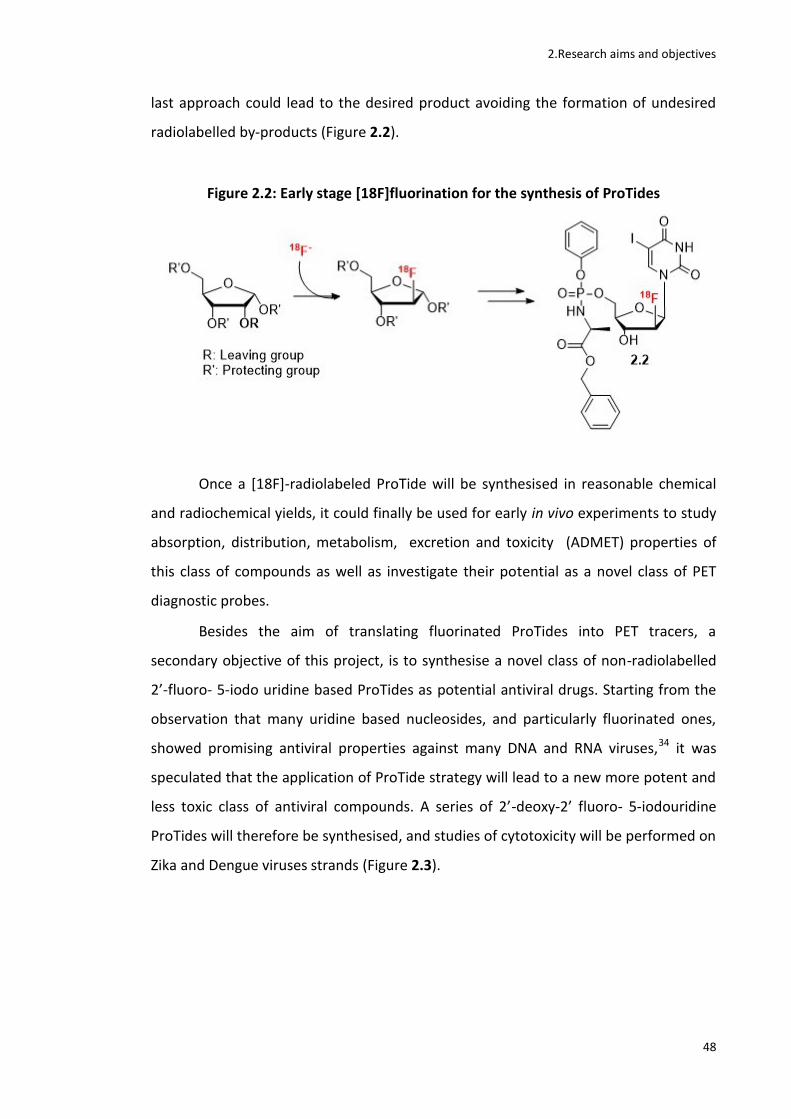
2.Research aims and objectives
48
last approach could lead to the desired product avoiding the formation of undesired
radiolabelled by-products (Figure 2.2).
Figure 2.2: Early stage [18F]fluorination for the synthesis of ProTides
Once a [18F]-radiolabeled ProTide will be synthesised in reasonable chemical
and radiochemical yields, it could finally be used for early in vivo experiments to study
absorption, distribution, metabolism, excretion and toxicity (ADMET) properties of
this class of compounds as well as investigate their potential as a novel class of PET
diagnostic probes.
Besides the aim of translating fluorinated ProTides into PET tracers, a
secondary objective of this project, is to synthesise a novel class of non-radiolabelled
2’-fluoro- 5-iodo uridine based ProTides as potential antiviral drugs. Starting from the
observation that many uridine based nucleosides, and particularly fluorinated ones,
showed promising antiviral properties against many DNA and RNA viruses,34 it was
speculated that the application of ProTide strategy will lead to a new more potent and
less toxic class of antiviral compounds. A series of 2’-deoxy-2’ fluoro- 5-iodouridine
ProTides will therefore be synthesised, and studies of cytotoxicity will be performed on
Zika and Dengue viruses strands (Figure 2.3).
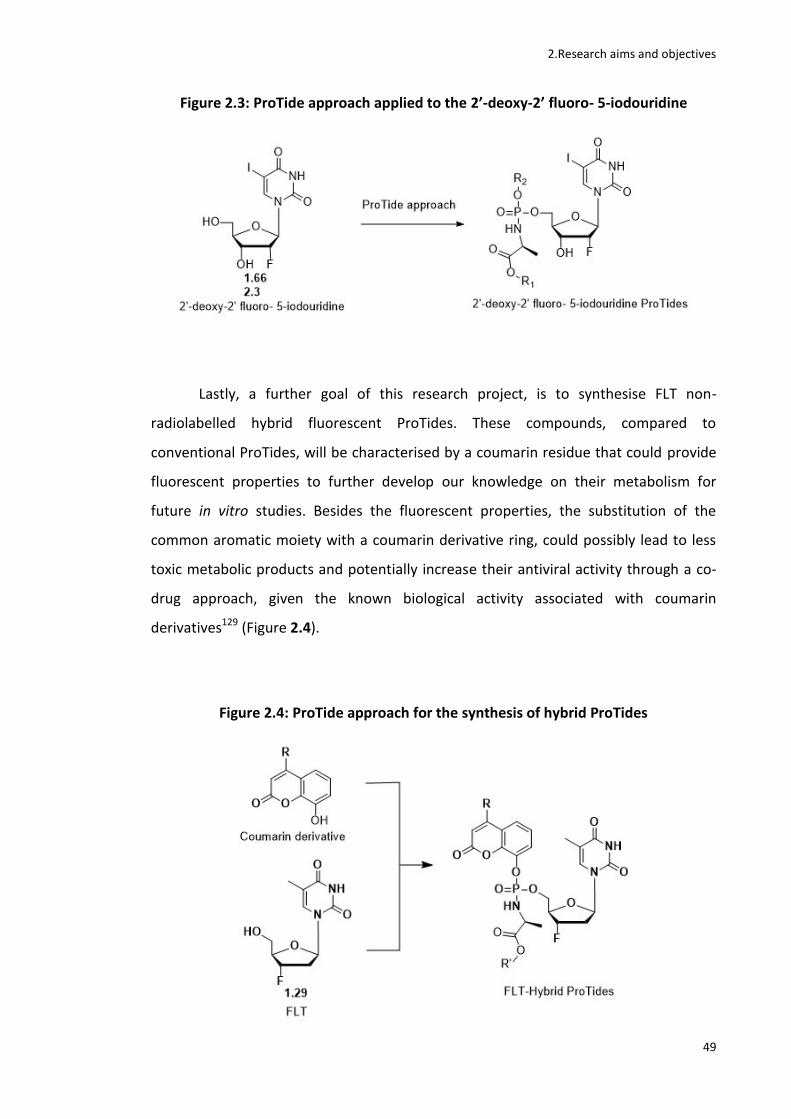
2.Research aims and objectives
49
Figure 2.3: ProTide approach applied to the 2’-deoxy-2’ fluoro- 5-iodouridine
Lastly, a further goal of this research project, is to synthesise FLT non-
radiolabelled hybrid fluorescent ProTides. These compounds, compared to
conventional ProTides, will be characterised by a coumarin residue that could provide
fluorescent properties to further develop our knowledge on their metabolism for
future in vitro studies. Besides the fluorescent properties, the substitution of the
common aromatic moiety with a coumarin derivative ring, could possibly lead to less
toxic metabolic products and potentially increase their antiviral activity through a co-
drug approach, given the known biological activity associated with coumarin
derivatives129 (Figure 2.4).
Figure 2.4: ProTide approach for the synthesis of hybrid ProTides

3. Radiochemical synthesis of [18F]FLT ProTide
50
3. Radiochemical synthesis of [18F]FLT ProTide
3.1 Introduction The main goal of this research project is the synthesis of [18F]-radiolabeled
ProTides as a proof of concept for the ProTide strategy through future in vivo
biodistribution studies. Several ProTides are currently either approved therapies or
under clinical evaluation and have proved to be more active than their parent
nucleoside analogues by circumventing their main resistance mechanisms.83
Radiolabelling these compounds with a radioisotope with a relatively short half life,
such as [18F]fluorine, would enable their use as PET imaging probes in order to have a
deeper understanding of their ADMET properties directly in vivo. Eventually, this could
be a first step towards the future of a Protide based personalized medicine.
Two [18F]-radiolabelled ProTides have been chosen as targets for this project,
the [18F]FLT ProTide (2.1) and the [18F]FIAU ProTide (2.2), as a model standard of 3’-
fluorinated and 2’-fluorinated ProTides respectively, following two different synthetic
approaches. In this chapter the late stage fluorination approach for the synthesis of
the [18F]FLT ProTide (2.1) will be discussed.
Figure 3.1: Structure of an [18F]FLT ProTide
[18F]FLT ProTide has been chosen as a model standard for the class of the 3’-
fluorinated ProTides for the reasons listed below:
- It already contains a fluorine in its original structure hence radiolabelling this
Protide with [18F]fluorine comes as a natural and convenient choice.
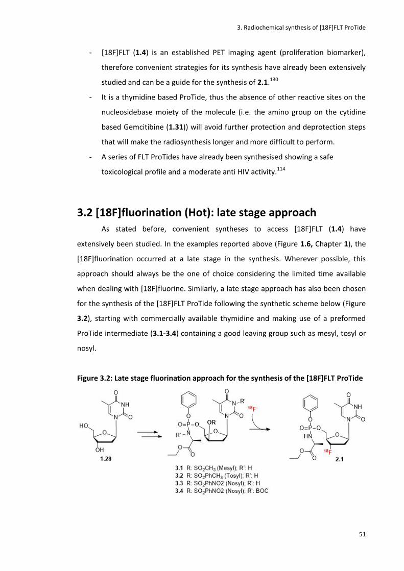
3. Radiochemical synthesis of [18F]FLT ProTide
51
- [18F]FLT (1.4) is an established PET imaging agent (proliferation biomarker),
therefore convenient strategies for its synthesis have already been extensively
studied and can be a guide for the synthesis of 2.1.130
- It is a thymidine based ProTide, thus the absence of other reactive sites on the
nucleosidebase moiety of the molecule (i.e. the amino group on the cytidine
based Gemcitibine (1.31)) will avoid further protection and deprotection steps
that will make the radiosynthesis longer and more difficult to perform.
- A series of FLT ProTides have already been synthesised showing a safe
toxicological profile and a moderate anti HIV activity.114
3.2 [18F]fluorination (Hot): late stage approach As stated before, convenient syntheses to access [18F]FLT (1.4) have
extensively been studied. In the examples reported above (Figure 1.6, Chapter 1), the
[18F]fluorination occurred at a late stage in the synthesis. Wherever possible, this
approach should always be the one of choice considering the limited time available
when dealing with [18F]fluorine. Similarly, a late stage approach has also been chosen
for the synthesis of the [18F]FLT ProTide following the synthetic scheme below (Figure
3.2), starting with commercially available thymidine and making use of a preformed
ProTide intermediate (3.1-3.4) containing a good leaving group such as mesyl, tosyl or
nosyl.
Figure 3.2: Late stage fluorination approach for the synthesis of the [18F]FLT ProTide
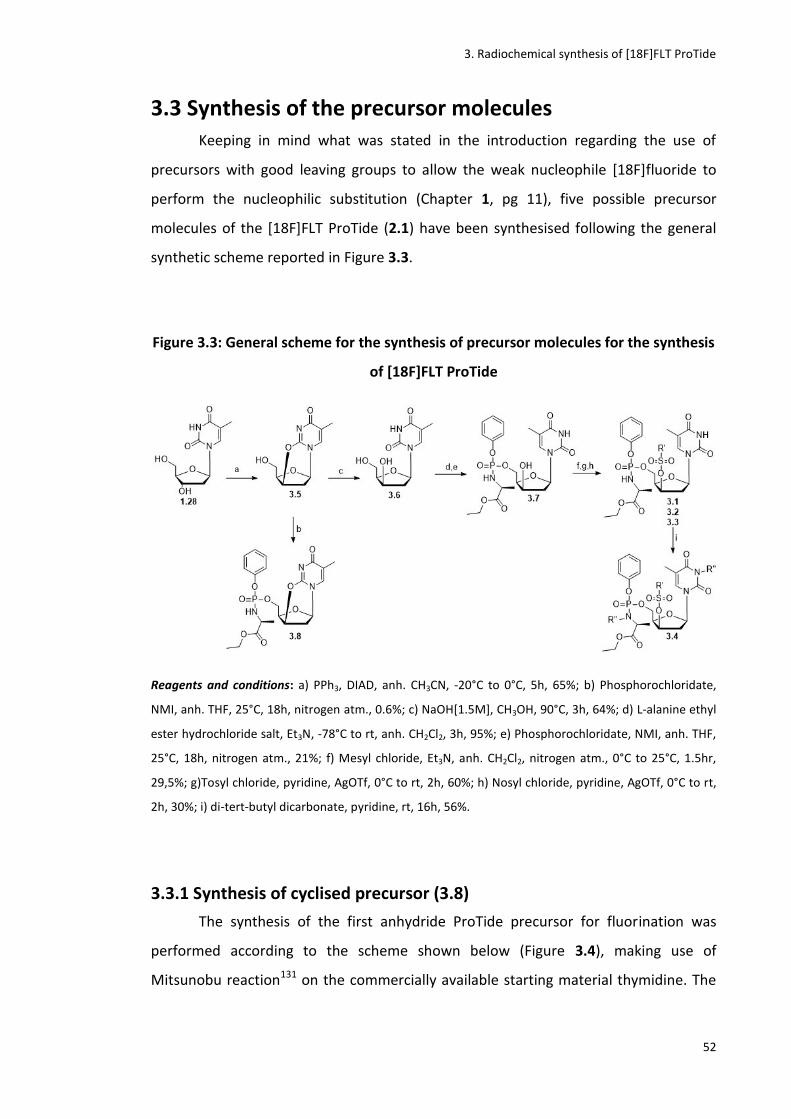
3. Radiochemical synthesis of [18F]FLT ProTide
52
3.3 Synthesis of the precursor molecules Keeping in mind what was stated in the introduction regarding the use of
precursors with good leaving groups to allow the weak nucleophile [18F]fluoride to
perform the nucleophilic substitution (Chapter 1, pg 11), five possible precursor
molecules of the [18F]FLT ProTide (2.1) have been synthesised following the general
synthetic scheme reported in Figure 3.3.
Figure 3.3: General scheme for the synthesis of precursor molecules for the synthesis
of [18F]FLT ProTide
Reagents and conditions: a) PPh3, DIAD, anh. CH3CN, -20°C to 0°C, 5h, 65%; b) Phosphorochloridate,
NMI, anh. THF, 25°C, 18h, nitrogen atm., 0.6%; c) NaOH[1.5M], CH3OH, 90°C, 3h, 64%; d) L-alanine ethyl
ester hydrochloride salt, Et3N, -78°C to rt, anh. CH2Cl2, 3h, 95%; e) Phosphorochloridate, NMI, anh. THF,
25°C, 18h, nitrogen atm., 21%; f) Mesyl chloride, Et3N, anh. CH2Cl2, nitrogen atm., 0°C to 25°C, 1.5hr,
29,5%; g)Tosyl chloride, pyridine, AgOTf, 0°C to rt, 2h, 60%; h) Nosyl chloride, pyridine, AgOTf, 0°C to rt,
2h, 30%; i) di-tert-butyl dicarbonate, pyridine, rt, 16h, 56%.
3.3.1 Synthesis of cyclised precursor (3.8)
The synthesis of the first anhydride ProTide precursor for fluorination was
performed according to the scheme shown below (Figure 3.4), making use of
Mitsunobu reaction131 on the commercially available starting material thymidine. The

3. Radiochemical synthesis of [18F]FLT ProTide
53
“anhydro” intermediate (3.5) was then reacted with the ProTide precursor phosphoryl
chloride (3.10) according to a previously reported ProTide protocol.128
Figure 3.4: Synthesis of the anhydride precursor
Reagents and conditions: a) PPh3, DIAD, anh. CH3CN, -20°C to 0°C, 5h, 65%; b) L-alanine ethyl ester
hydrochloride salt, Et3N, -78°C to rt, anh. CH2Cl2, 3h, 95%; c) Phosphorochloridate, NMI, anh. THF, 25°C,
18h, nitrogen atm.,0.6%.
Firstly it was investigated whether 2’ and 3’ fluorinated nucleoside analogues
can be accessed via fluorination of anhydrous precursors with applications also in
radiochemistry.132 For this reason a Mitsunobu reaction was performed using
thymidine (1.28) as starting material in order to obtain the six membered 2,3’-
anhydride derivative via an intramolecular cyclisation. For this purpose DIAD
(diisopropylazadicarboxylate) and PPh3 (triphenylphosphine) were used (Figure 3.5).133
PPh3 combines with DIAD to form a zwitterionic phosphonium intermediate which
deprotonates the 3’-OH on the thymidine to form the anionic nuclephile. This
intermediate binds to the zwitterionic phosphonium ion to perform an SN2 attack
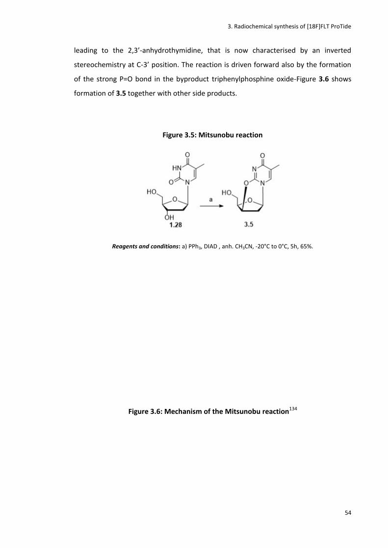
3. Radiochemical synthesis of [18F]FLT ProTide
54
leading to the 2,3’-anhydrothymidine, that is now characterised by an inverted
stereochemistry at C-3’ position. The reaction is driven forward also by the formation
of the strong P=O bond in the byproduct triphenylphosphine oxide-Figure 3.6 shows
formation of 3.5 together with other side products.
Figure 3.5: Mitsunobu reaction
Reagents and conditions: a) PPh3, DIAD , anh. CH3CN, -20°C to 0°C, 5h, 65%.
Figure 3.6: Mechanism of the Mitsunobu reaction134
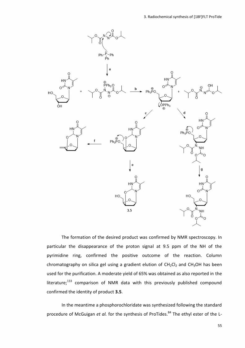
3. Radiochemical synthesis of [18F]FLT ProTide
55
The formation of the desired product was confirmed by NMR spectroscopy. In
particular the disappearance of the proton signal at 9.5 ppm of the NH of the
pyrimidine ring, confirmed the positive outcome of the reaction. Column
chromatography on silica gel using a gradient elution of CH2Cl2 and CH3OH has been
used for the purification. A moderate yield of 65% was obtained as also reported in the
literature;133 comparison of NMR data with this previously published compound
confirmed the identity of product 3.5.
In the meantime a phosphorochloridate was synthesized following the standard
procedure of McGuigan et al. for the synthesis of ProTides.84 The ethyl ester of the L-
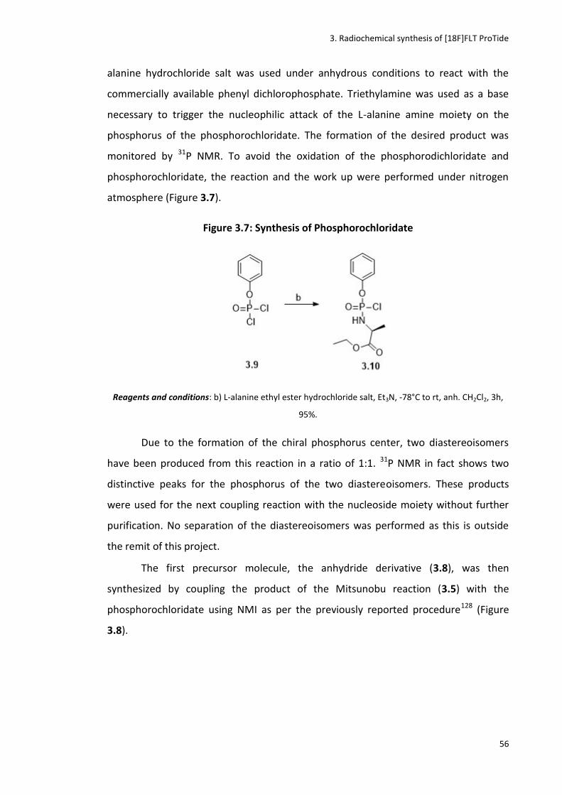
3. Radiochemical synthesis of [18F]FLT ProTide
56
alanine hydrochloride salt was used under anhydrous conditions to react with the
commercially available phenyl dichlorophosphate. Triethylamine was used as a base
necessary to trigger the nucleophilic attack of the L-alanine amine moiety on the
phosphorus of the phosphorochloridate. The formation of the desired product was
monitored by 31P NMR. To avoid the oxidation of the phosphorodichloridate and
phosphorochloridate, the reaction and the work up were performed under nitrogen
atmosphere (Figure 3.7).
Figure 3.7: Synthesis of Phosphorochloridate
Reagents and conditions: b) L-alanine ethyl ester hydrochloride salt, Et3N, -78°C to rt, anh. CH2Cl2, 3h,
95%.
Due to the formation of the chiral phosphorus center, two diastereoisomers
have been produced from this reaction in a ratio of 1:1. 31P NMR in fact shows two
distinctive peaks for the phosphorus of the two diastereoisomers. These products
were used for the next coupling reaction with the nucleoside moiety without further
purification. No separation of the diastereoisomers was performed as this is outside
the remit of this project.
The first precursor molecule, the anhydride derivative (3.8), was then
synthesized by coupling the product of the Mitsunobu reaction (3.5) with the
phosphorochloridate using NMI as per the previously reported procedure128 (Figure
3.8).
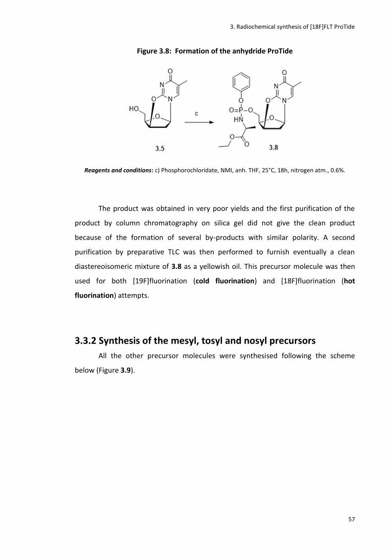
3. Radiochemical synthesis of [18F]FLT ProTide
57
Figure 3.8: Formation of the anhydride ProTide
Reagents and conditions: c) Phosphorochloridate, NMI, anh. THF, 25°C, 18h, nitrogen atm., 0.6%.
The product was obtained in very poor yields and the first purification of the
product by column chromatography on silica gel did not give the clean product
because of the formation of several by-products with similar polarity. A second
purification by preparative TLC was then performed to furnish eventually a clean
diastereoisomeric mixture of 3.8 as a yellowish oil. This precursor molecule was then
used for both [19F]fluorination (cold fluorination) and [18F]fluorination (hot
fluorination) attempts.
3.3.2 Synthesis of the mesyl, tosyl and nosyl precursors
All the other precursor molecules were synthesised following the scheme
below (Figure 3.9).
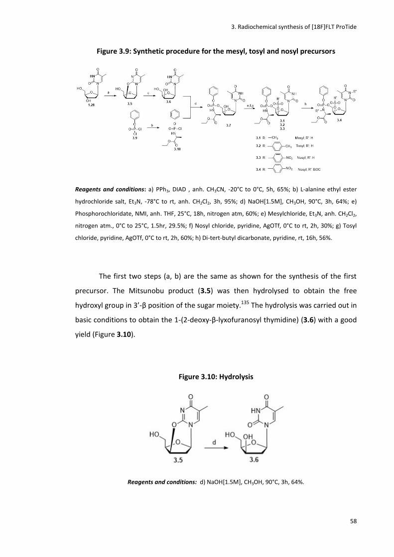
3. Radiochemical synthesis of [18F]FLT ProTide
58
Figure 3.9: Synthetic procedure for the mesyl, tosyl and nosyl precursors
Reagents and conditions: a) PPh3, DIAD , anh. CH3CN, -20°C to 0°C, 5h, 65%; b) L-alanine ethyl ester
hydrochloride salt, Et3N, -78°C to rt, anh. CH2Cl2, 3h, 95%; d) NaOH[1.5M], CH3OH, 90°C, 3h, 64%; e)
Phosphorochloridate, NMI, anh. THF, 25°C, 18h, nitrogen atm, 60%; e) Mesylchloride, Et3N, anh. CH2Cl2,
nitrogen atm., 0°C to 25°C, 1.5hr, 29.5%; f) Nosyl chloride, pyridine, AgOTf, 0°C to rt, 2h, 30%; g) Tosyl
chloride, pyridine, AgOTf, 0°C to rt, 2h, 60%; h) Di-tert-butyl dicarbonate, pyridine, rt, 16h, 56%.
The first two steps (a, b) are the same as shown for the synthesis of the first
precursor. The Mitsunobu product (3.5) was then hydrolysed to obtain the free
hydroxyl group in 3’-β position of the sugar moiety.135 The hydrolysis was carried out in
basic conditions to obtain the 1-(2-deoxy-β-lyxofuranosyl thymidine) (3.6) with a good
yield (Figure 3.10).
Figure 3.10: Hydrolysis
Reagents and conditions: d) NaOH[1.5M], CH3OH, 90°C, 3h, 64%.

3. Radiochemical synthesis of [18F]FLT ProTide
59
The phosphophorochloride (3.10) was again synthesised as a diastereoisomeric
mixture following the standard procedure.84
These products were used for the next coupling reaction with the nucleoside
moiety without further purification. A coupling reaction was then performed between
the phosphorochloridates and the nucleobase moiety (Figure 3.11).
Figure 3.11: ProTide coupling
Reagents and conditions: e) Phosphorochloridate, NMI, anh. THF, 25°C, 18h, nitrogen atm., 18%.
For this synthetic step 1-methylimidazole (NMI) was used as a base to form an
imidazolium intermediate with the phosphorochloridate that can then readily react
with the nucleoside. Below is shown the mechanism of action of this reaction when
using the base NMI (Figure 3.12).
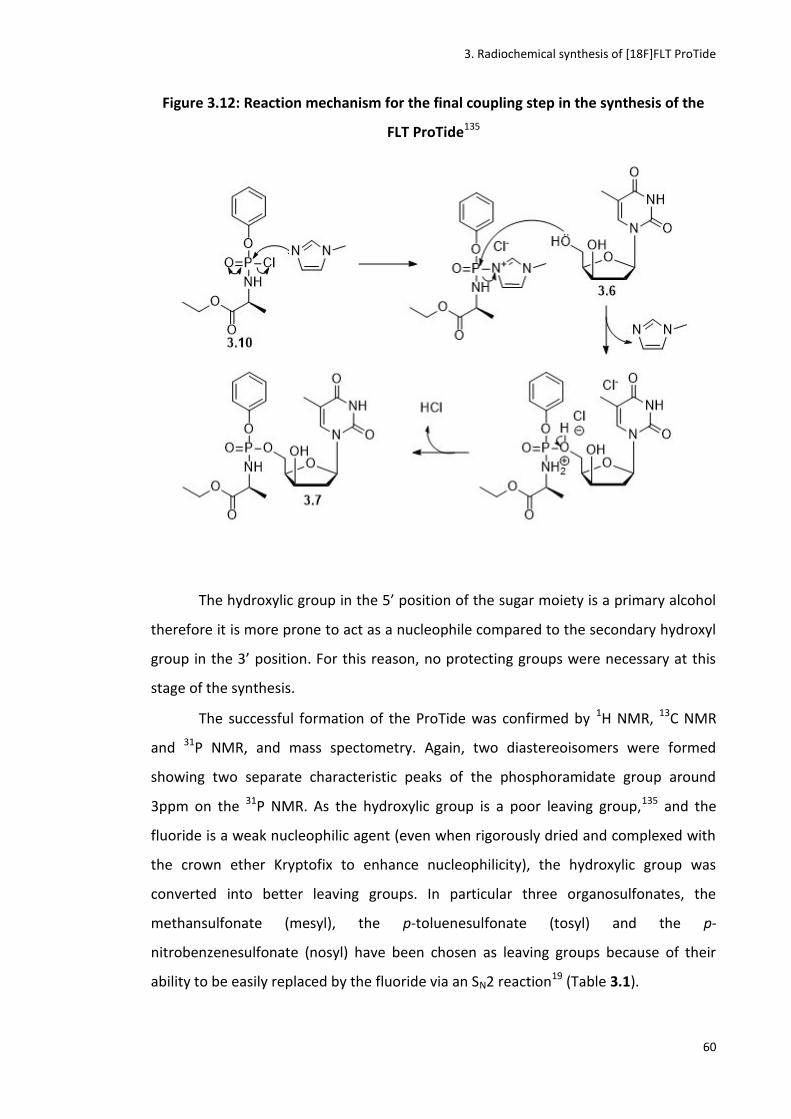
3. Radiochemical synthesis of [18F]FLT ProTide
60
Figure 3.12: Reaction mechanism for the final coupling step in the synthesis of the
FLT ProTide135
The hydroxylic group in the 5’ position of the sugar moiety is a primary alcohol
therefore it is more prone to act as a nucleophile compared to the secondary hydroxyl
group in the 3’ position. For this reason, no protecting groups were necessary at this
stage of the synthesis.
The successful formation of the ProTide was confirmed by 1H NMR, 13C NMR
and 31P NMR, and mass spectometry. Again, two diastereoisomers were formed
showing two separate characteristic peaks of the phosphoramidate group around
3ppm on the 31P NMR. As the hydroxylic group is a poor leaving group,135 and the
fluoride is a weak nucleophilic agent (even when rigorously dried and complexed with
the crown ether Kryptofix to enhance nucleophilicity), the hydroxylic group was
converted into better leaving groups. In particular three organosulfonates, the
methansulfonate (mesyl), the p-toluenesulfonate (tosyl) and the p-
nitrobenzenesulfonate (nosyl) have been chosen as leaving groups because of their
ability to be easily replaced by the fluoride via an SN2 reaction19 (Table 3.1).
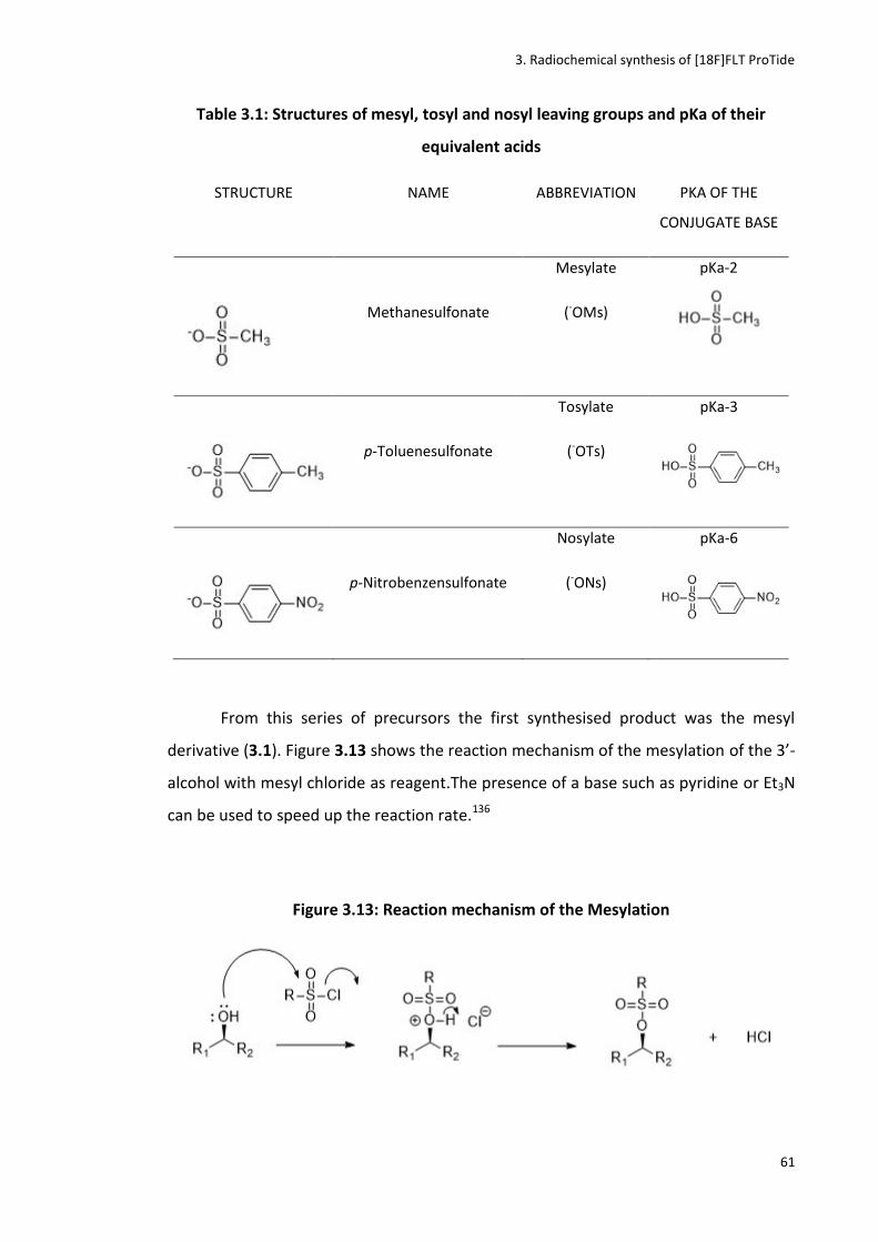
3. Radiochemical synthesis of [18F]FLT ProTide
61
Table 3.1: Structures of mesyl, tosyl and nosyl leaving groups and pKa of their
equivalent acids
STRUCTURE NAME ABBREVIATION PKA OF THE
CONJUGATE BASE
Methanesulfonate
Mesylate
(-OMs)
pKa-2
p-Toluenesulfonate
Tosylate
(-OTs)
pKa-3
p-Nitrobenzensulfonate
Nosylate
(-ONs)
pKa-6
From this series of precursors the first synthesised product was the mesyl
derivative (3.1). Figure 3.13 shows the reaction mechanism of the mesylation of the 3’-
alcohol with mesyl chloride as reagent.The presence of a base such as pyridine or Et3N
can be used to speed up the reaction rate.136
Figure 3.13: Reaction mechanism of the Mesylation
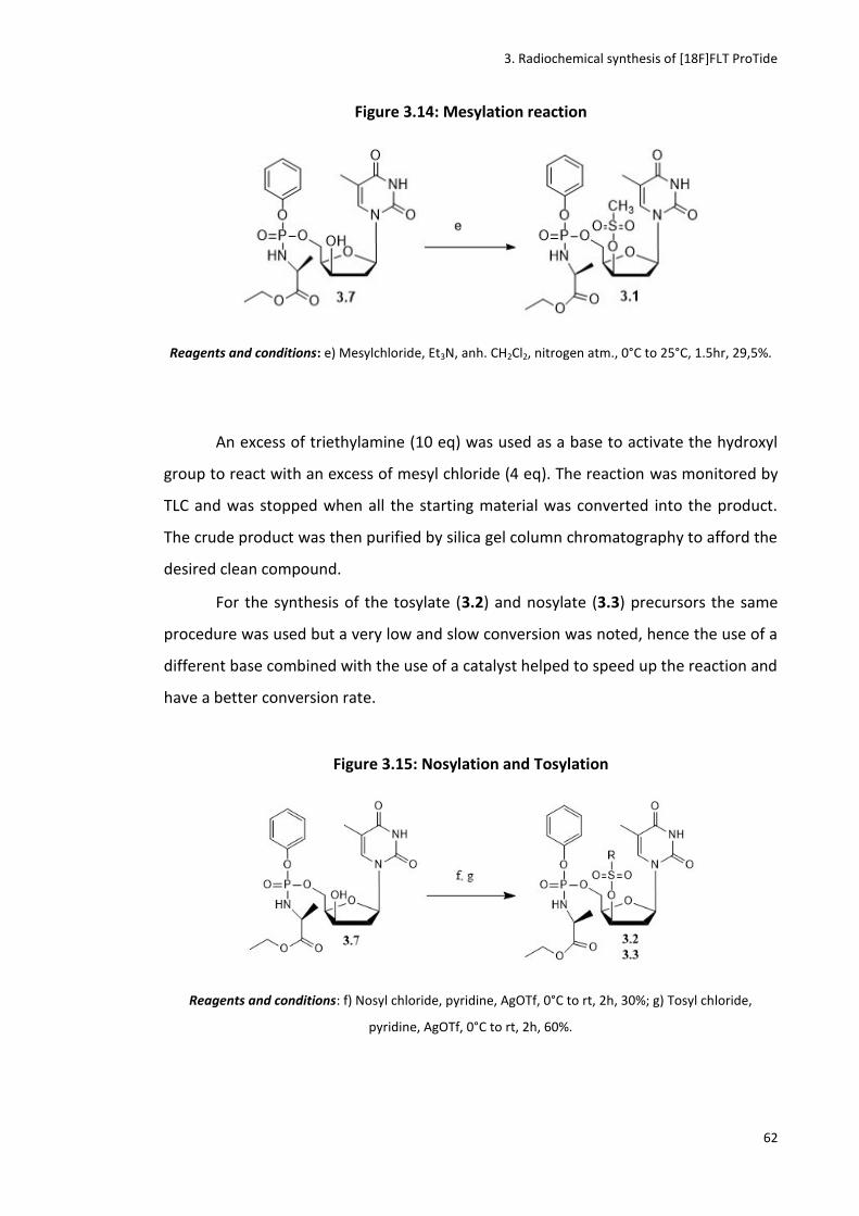
3. Radiochemical synthesis of [18F]FLT ProTide
62
Figure 3.14: Mesylation reaction
Reagents and conditions: e) Mesylchloride, Et3N, anh. CH2Cl2, nitrogen atm., 0°C to 25°C, 1.5hr, 29,5%.
An excess of triethylamine (10 eq) was used as a base to activate the hydroxyl
group to react with an excess of mesyl chloride (4 eq). The reaction was monitored by
TLC and was stopped when all the starting material was converted into the product.
The crude product was then purified by silica gel column chromatography to afford the
desired clean compound.
For the synthesis of the tosylate (3.2) and nosylate (3.3) precursors the same
procedure was used but a very low and slow conversion was noted, hence the use of a
different base combined with the use of a catalyst helped to speed up the reaction and
have a better conversion rate.
Figure 3.15: Nosylation and Tosylation
Reagents and conditions: f) Nosyl chloride, pyridine, AgOTf, 0°C to rt, 2h, 30%; g) Tosyl chloride,
pyridine, AgOTf, 0°C to rt, 2h, 60%.
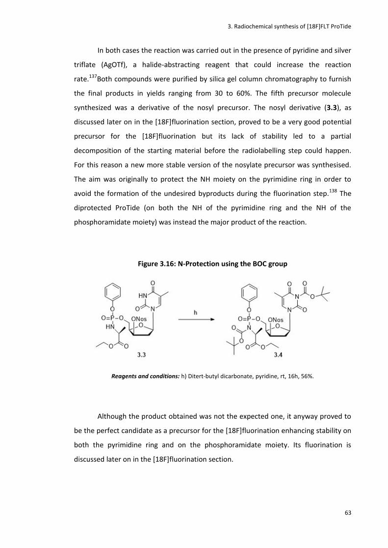
3. Radiochemical synthesis of [18F]FLT ProTide
63
In both cases the reaction was carried out in the presence of pyridine and silver
triflate (AgOTf), a halide-abstracting reagent that could increase the reaction
rate.137Both compounds were purified by silica gel column chromatography to furnish
the final products in yields ranging from 30 to 60%. The fifth precursor molecule
synthesized was a derivative of the nosyl precursor. The nosyl derivative (3.3), as
discussed later on in the [18F]fluorination section, proved to be a very good potential
precursor for the [18F]fluorination but its lack of stability led to a partial
decomposition of the starting material before the radiolabelling step could happen.
For this reason a new more stable version of the nosylate precursor was synthesised.
The aim was originally to protect the NH moiety on the pyrimidine ring in order to
avoid the formation of the undesired byproducts during the fluorination step.138 The
diprotected ProTide (on both the NH of the pyrimidine ring and the NH of the
phosphoramidate moiety) was instead the major product of the reaction.
Figure 3.16: N-Protection using the BOC group
Reagents and conditions: h) Ditert-butyl dicarbonate, pyridine, rt, 16h, 56%.
Although the product obtained was not the expected one, it anyway proved to
be the perfect candidate as a precursor for the [18F]fluorination enhancing stability on
both the pyrimidine ring and on the phosphoramidate moiety. Its fluorination is
discussed later on in the [18F]fluorination section.

3. Radiochemical synthesis of [18F]FLT ProTide
64
3.4 Synthesis of the cold standard For the synthesis of the cold standard (3.11) a few attempts to perform cold
fluorination of the mesyl precursor (3.1) have been carried out but none of them gave
the desired product. Tetrabutyl ammonium fluoride tri-hydrate (TBAF.3H2O), KF or CsF
were used as source of fluorine. Kryptofix was used to enhance the weak
nucleophilicity of the fluorine. None of the reactions gave the desired fluorinated
compound returning only unreacted starting material and other by-products (Table
3.2).1
Figure 3.17: Fluorination of the Mesyl precursor
Table 3.2: Attempts of cold fluorination of mesyl precursor (3.1)
Starting
material
T °C Fluorine
source
Solvent Reaction
time
Product
3.1 70 °C TBAF THF 1h No
3.1 120 °C KF DMF 1h No
3.1 120 °C CsF DMF 1h No
When the fluorination was performed on the nosyl derivative (3.3), the
formation of the FLT ProTide (3.11) was observed even if the yield was low.

3. Radiochemical synthesis of [18F]FLT ProTide
65
Figure 3.18: Fluorination of the Nosyl precursor (3.3)
Reagents and conditions: a) TBAF 1M in THF, anh.DMF, rt, 1h, 3%.
The poor yield of this reaction was due to the formation of many by-products.
Another synthetic strategy was also used to access the FLT ProTide (3.11) in better
yields (Figure 3.19).
Figure 3.19: Synthesis of FLT ProTide using FLT as starting material
Reagents and conditions: a) Phosphorochloridate, t-BuMgCl, anh. THF, 18h, 23.5%.
The commercially available FLT (1.29) was used as starting material and the
Grignard reagent t-BuMgCl was used to perform the coupling reaction with the
phosphorochloride (Figure 3.19, 3.20) giving the desired compound in better
yields.114,128

3. Radiochemical synthesis of [18F]FLT ProTide
66
Figure 3.20: Mechanism of reaction of the Grignard reagent
This compound was eventually used as cold standard for the [18F]fluorination
step. It has indeed to be co-spiked with the radiolabelled compound on an HLPC
system in order to compare the retention time of the cold and the hot FLT ProTide. The
same HPLC gradient elution (H2O-CH3CN 99%-1% to 50%-50%) was used for the
analyses of the [18F]fluorination reactions.
Figure 3.21: Analytical HPLC (UV detector) of the cold [19F]FLT ProTide.
(H2O-CH3CN 99%-1% to 50%-50%). RT: 9.7 min
0:00 5:00 10:00 15:00 mm:ss
0.0
500.0
1000.0
1500.0
2000.0
2500.0
mAU

3. Radiochemical synthesis of [18F]FLT ProTide
67
3.5 Studies of ProTide stability Studies of stability of the FLT-ProTide (3.11) were then performed in order to
prove that, once formed, the [18F]FLT ProTide is stable under the harsh conditions
used in radiochemistry (i.e high temperature, excess of a fluoride source and
Kryptofix).
Figure 3.22: Studies of stability of the FLT ProTide
Reagents and conditions: a) KF(10eq), Kryptofix(10 eq), anh.solvent, 40-120 °C.
To a stirring solution of FLT-ProTide in anhydrous DMF, a solution of KF and
Kryptofix in DMF anhydrous was added at rt. The reaction mixture was heated for 1h
at 40°C, 80°C, 100°C and 120°C. Each reaction was monitored by TLC, 31P NMR, and 19F
NMR to observe the potential degradation of the starting material and eventually the
formation of side products. No decomposition was observed up to 120°C which is the
maximum temperature that was going to be used for the [18F]fluorination step.
The reaction mixture was then purified by silica gel column chromatography
and the starting material was recovered and was analyzed by 1H NMR, 13C NMR, 19F
NMR, 31P NMR, which all confirmed its stability and purity.
3.6 [18F]fluorination
Before starting attempts of hot fluorination of the precursor molecules
synthesised, the fully automated synthetic system (Eckert and Ziegler Nuclear Interface
Module) for the introduction of 18F- into the precursor molecule had to be been
configured. This system, located into a shielded hot cell, was set up as shown in Figure
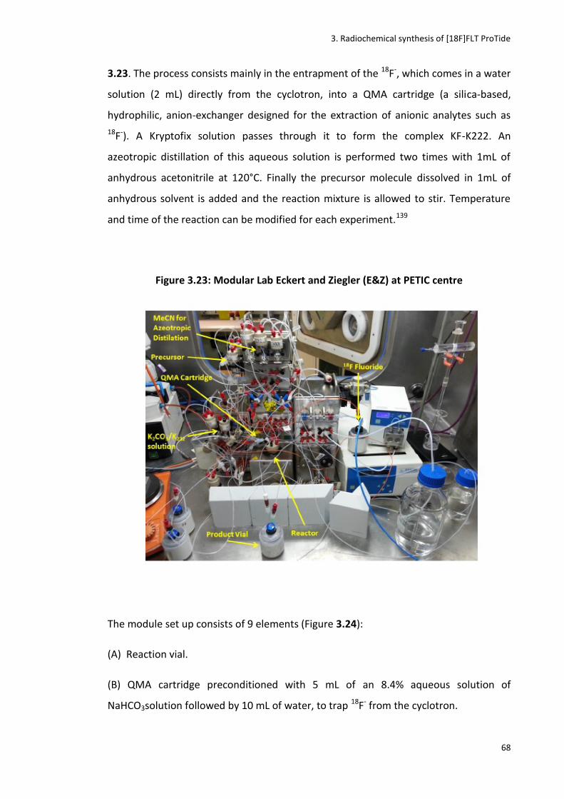
3. Radiochemical synthesis of [18F]FLT ProTide
68
3.23. The process consists mainly in the entrapment of the 18F-, which comes in a water
solution (2 mL) directly from the cyclotron, into a QMA cartridge (a silica-based,
hydrophilic, anion-exchanger designed for the extraction of anionic analytes such as
18F-). A Kryptofix solution passes through it to form the complex KF-K222. An
azeotropic distillation of this aqueous solution is performed two times with 1mL of
anhydrous acetonitrile at 120°C. Finally the precursor molecule dissolved in 1mL of
anhydrous solvent is added and the reaction mixture is allowed to stir. Temperature
and time of the reaction can be modified for each experiment.139
Figure 3.23: Modular Lab Eckert and Ziegler (E&Z) at PETIC centre
The module set up consists of 9 elements (Figure 3.24):
(A) Reaction vial.
(B) QMA cartridge preconditioned with 5 mL of an 8.4% aqueous solution of
NaHCO3solution followed by 10 mL of water, to trap 18F- from the cyclotron.

3. Radiochemical synthesis of [18F]FLT ProTide
69
(C) Kryptofix [2.2.2] vial.
(D) Anhydrous acetonitrile vial for the azeotropic evaporation.
(E) Precursor vial filled with the precursor dissolved in the reaction solvent.
(F) Acid vial filled with 0.05M HCl for eventual deprotection.
(G) Base vial filled with 0.25M NaOH for eventual neutralization.
(H) Vacuum pump for solvent removal.
(I) Final product vessel for product isolation.
Figure 3.24: Sketch of EZ Modular Lab
A flow chart of the process performed by the software program is shown in Figure
3.25.

3. Radiochemical synthesis of [18F]FLT ProTide
70
Figure 3.25: Flow chart of the synthetic steps in modular lab
Several conditions have been explored for the hot fluorination. In particular the
three essential requirements that can deeply influence the outcome of the
radiolabelling are the time of the reaction (15-30 mins), the absolute anhydrous
conditions (i.e absence of water and use of anhydride solvents) and the high
temperature (90-120°C).
Reaction mixtures were analyzed by using a radio TLC and a radio HPLC. The
reaction mixture was co-eluted with the cold reference standard in order to compare
the retention time of the cold standard (detected with the UV detector) and of the
radiolabelled compound (detected with the radioactive detector) on the HPLC
system.52,140
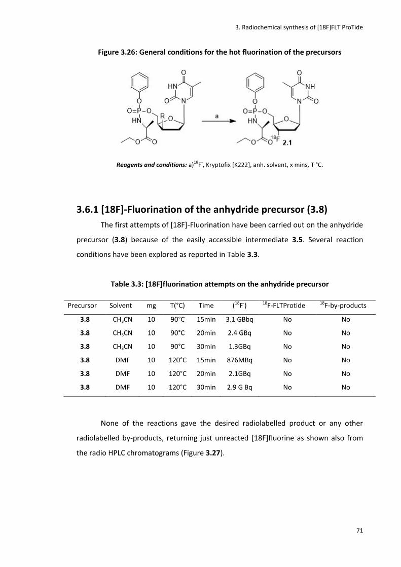
3. Radiochemical synthesis of [18F]FLT ProTide
71
Figure 3.26: General conditions for the hot fluorination of the precursors
Reagents and conditions: a)18
F-, Kryptofix [K222], anh. solvent, x mins, T °C.
3.6.1 [18F]-Fluorination of the anhydride precursor (3.8)
The first attempts of [18F]-Fluorination have been carried out on the anhydride
precursor (3.8) because of the easily accessible intermediate 3.5. Several reaction
conditions have been explored as reported in Table 3.3.
Table 3.3: [18F]fluorination attempts on the anhydride precursor
Precursor Solvent mg T(°C) Time (18F-) 18F-FLTProtide 18F-by-products
3.8 CH3CN 10 90°C 15min 3.1 GBbq No No
3.8 CH3CN 10 90°C 20min 2.4 GBq No No
3.8 CH3CN 10 90°C 30min 1.3GBq No No
3.8 DMF 10 120°C 15min 876MBq No No
3.8 DMF 10 120°C 20min 2.1GBq No No
3.8 DMF 10 120°C 30min 2.9 G Bq No No
None of the reactions gave the desired radiolabelled product or any other
radiolabelled by-products, returning just unreacted [18F]fluorine as shown also from
the radio HPLC chromatograms (Figure 3.27).

3. Radiochemical synthesis of [18F]FLT ProTide
72
Figure 3.27: [18F]fluorination of compound 3.8
a) Radioactive chromatogram of the reaction mixture showing free 18F- at 3min; b) UV
chromatogram of the reaction mixture co-spiked with the cold standard (3.11).(Rt of
compound 3.11: 10 min)
a)
b)
No other attempts have been performed as this precursor didn’t show any
reactivity towards the [18F]fluorination. This could be due to the poor reactivity of the
3’-position suggesting that the use of better leaving groups such as the mesyl, tosyl or
nosyl is required for this type of reaction.
3.6.2 [18F]fluorination of the mesyl precursor (3.1)
The second precursor tested was the mesyl precursor (3.1) because of its
synthetic accessibility and because of its balance between reactivity and stability as
leaving group. Similar reaction conditions to the ones used for the anhydride derivative
have been tested for this step (Table 3.4).
0:00 5:00 10:00 15:00 mm:ss
0.0
10000.0
20000.0
30000.0
Counts
0:00 5:00 10:00 15:00 mm:ss
0.0
100.0
200.0
300.0
mAU

3. Radiochemical synthesis of [18F]FLT ProTide
73
Table 3.4: [18F]fluorination attempts on the mesyl precursor
Precursor Solvent Mg T(°C) Time (18F-) 18F-FLTProtide 18F-by-products
3.1 DMF 10mg 120°C 15min 810 MBq No No
3.1 DMF 10mg 120°C 20min 2.35 GBq No No
3.1 DMF 10mg 120°C 30min 910 MBq No No
3.1 DMF 20 mg 120°C 15min 580 MBq No No
3.1 DMF 20 mg 120°C 20min 970 MBq No No
3.1 DMF 20 mg 120°C 30min 780 MBq No No
Figure 3.28: [18F]fluorination of compound 3.1
a)Radioactive chromatogram of the reaction mixture showing a major peak of free 18F-
at 3min; b)UV chromatogram of the reaction mixture co-spiked with the cold standard
(3.11). (Rt of compound 3.11: 10 min)
a)
b)
As again no radiolabelled products were formed and only [18F]fluorine was
returned unreacted, a larger amount of starting material was used (20 mg) to test
whether this variable could influence the outcome of the reaction. In this case, the
0:00 5:00 10:00 15:00 mm:ss
0.0
10000.0
20000.0
30000.0
Counts
0:00 5:00 10:00 15:00 mm:ss
0.0
1000.0
2000.0
3000.0
mAU
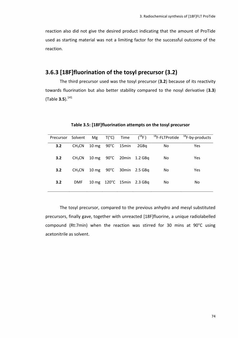
3. Radiochemical synthesis of [18F]FLT ProTide
74
reaction also did not give the desired product indicating that the amount of ProTide
used as starting material was not a limiting factor for the successful outcome of the
reaction.
3.6.3 [18F]fluorination of the tosyl precursor (3.2)
The third precursor used was the tosyl precursor (3.2) because of its reactivity
towards fluorination but also better stability compared to the nosyl derivative (3.3)
(Table 3.5).141
Table 3.5: [18F]fluorination attempts on the tosyl precursor
Precursor Solvent Mg T(°C) Time (18F-) 18F-FLTProtide 18F-by-products
3.2 CH3CN 10 mg 90°C 15min 2GBq No Yes
3.2 CH3CN 10 mg 90°C 20min 1.2 GBq No Yes
3.2 CH3CN 10 mg 90°C 30min 2.5 GBq No Yes
3.2 DMF 10 mg 120°C 15min 2.3 GBq No No
The tosyl precursor, compared to the previous anhydro and mesyl substituted
precursors, finally gave, together with unreacted [18F]fluorine, a unique radiolabelled
compound (Rt:7min) when the reaction was stirred for 30 mins at 90°C using
acetonitrile as solvent.
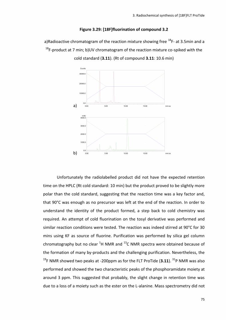
3. Radiochemical synthesis of [18F]FLT ProTide
75
Figure 3.29: [18F]fluorination of compound 3.2
a)Radioactive chromatogram of the reaction mixture showing free 18F- at 3.5min and a
18F-product at 7 min; b)UV chromatogram of the reaction mixture co-spiked with the
cold standard (3.11). (Rt of compound 3.11: 10.6 min)
a)
b)
Unfortunately the radiolabelled product did not have the expected retention
time on the HPLC (Rt cold standard: 10 min) but the product proved to be slightly more
polar than the cold standard, suggesting that the reaction time was a key factor and,
that 90°C was enough as no precursor was left at the end of the reaction. In order to
understand the identity of the product formed, a step back to cold chemistry was
required. An attempt of cold fluorination on the tosyl derivative was performed and
similar reaction conditions were tested. The reaction was indeed stirred at 90°C for 30
mins using KF as source of fluorine. Purification was performed by silica gel column
chromatography but no clear 1H NMR and 13C NMR spectra were obtained because of
the formation of many by-products and the challenging purification. Nevertheless, the
19F NMR showed two peaks at -200ppm as for the FLT ProTide (3.11). 31P NMR was also
performed and showed the two characteristic peaks of the phosphoramidate moiety at
around 3 ppm. This suggested that probably, the slight change in retention time was
due to a loss of a moiety such as the ester on the L-alanine. Mass spectrometry did not
0:00 5:00 10:00 15:00 mm:ss
0.0
10000.0
20000.0
30000.0
Counts
0:00 5:00 10:00 15:00 mm:ss
0.0
1000.0
2000.0
3000.0
4000.0mAU

3. Radiochemical synthesis of [18F]FLT ProTide
76
confirm this hypothesis showing a major peak at 453 that does not correspond to any
of the substructures hypothesised.
3.6.4 [18F]fluorination of the nosyl precursor (3.3)
The fourth precursor, the nosyl derivative (3.3), was then used for the
[18F]fluorination attempt. Among all precursors synthesised, although the nosyl is
supposed to be the more reactive one, it is likely characterised by a lack of stability
that could be a deterrent for its usage. All the conditions explored for this reaction are
reported in Table 3.6.
Table 3.6: [18F]fluorination attempts of the nosyl precursor
Precursor Solvent Mg T(°C) Time (18F-) 18F-FLTProtide 18F-by-products
3.3 CH3CN 10 mg 90°C 15min 1.2 GBq No Yes
3.3 CH3CN 10 mg 90°C 20min 1.5 GBq Yes Yes
3.3 CH3CN 10 mg 90°C 30min 2.3 GBq Yes Yes
3.3 CH3CN 10 mg 90°C 40min 2.2 GBq Yes Yes
3.3 DMF 10 mg 120°C 15min 734 MBq No Yes
3.3 DMF 10 mg 120°C 20min 1.1 GBq No Yes
Similarly, to the fluorination of the tosyl derivative, the major radiolabelled
products had retention times of 5 and 8 mins as the major products of the reaction,
together with many other more polar by-products. However, when the reaction was
performed at 90°C for 30 mins in acetonitrile, also the desired [18F]FLT ProTide (2.1)
was formed although in poor yields (Figure 3.30).

3. Radiochemical synthesis of [18F]FLT ProTide
77
Figure 3.30: [18F]fluorination of compound 3.3
a) Radioactive chromatogram of the reaction mixture showing free 18F- at 3min, [18F]-
by-products at 5 and 8 min and [18F]FLT ProTide at 10 min; b) UV chromatogram of
the reaction mixture co-spiked with the cold standard (3.11). (Rt of compound 3.7: 10
min)
a)
b)
To re-confirm this positive result, the same reaction was performed also on
another automated synthetic modular lab (the GE FxFnTracerlab), at the Molecular
Imaging Chemistry Laboratory (MICL) in the Wolfson Brain Imaging Centre of
Cambridge University to prove the reproducibility of this reaction with different
automated synthesisers (Figure 3.31).

3. Radiochemical synthesis of [18F]FLT ProTide
78
Figure 3.31: GE Healthcare TracerlabFxFn142
The set up of this synthesiser was similar to the one of the E&Z one (Figure 3.32):
(A) Reaction vial.
(B) QMA cartridge preconditioned with 5 mL of a 8.4% aqueous solution of NaHCO3
solution followed by 10 mL of water, to trap 18F- from the cyclotron.
(C) Kryptofix [2.2.2] vial.
(D) Anhydrous acetonitrile vial for the azeotropic evaporation.
(E) Precursor vial filled with the precursor dissolved in the reaction solvent.
(F) Vacuum pump for solvent removal.
(G) Final product vessel for product isolation.
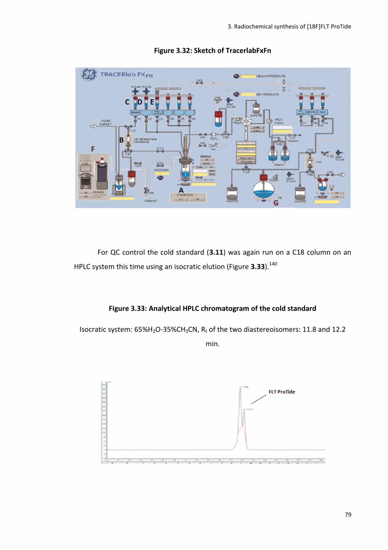
3. Radiochemical synthesis of [18F]FLT ProTide
79
Figure 3.32: Sketch of TracerlabFxFn
For QC control the cold standard (3.11) was again run on a C18 column on an
HPLC system this time using an isocratic elution (Figure 3.33).140
Figure 3.33: Analytical HPLC chromatogram of the cold standard
Isocratic system: 65%H2O-35%CH3CN, Rt of the two diastereoisomers: 11.8 and 12.2
min.

3. Radiochemical synthesis of [18F]FLT ProTide
80
Again the results of the hot fluorination were the same obtained as with the
E&Z modular lab. A slight decrease in the yield of the reaction was observed but results
obtained with the E&Z synthesiser were generally confirmed (Figure 3.34).
Figure 3.34: [18F]fluorination of compound 3.3 with TracerLabFxFn
a)Radioactive chromatogram of the reaction mixture showing many 18F-by-products
and [18F]-FLT ProTide at 12 min; b)UV chromatogram of the reaction mixture co-
spiked with the cold standard (3.11). (Rt of compound 3.11: 11.8-12-2 min)
a)
b)
Bearing in mind the future desired clinical application of the tracer, another
variable was then introduced in the system to understand whether the reaction could
be performed even without the use of Kryptofix which, despite being a good enhancer
for the nucleophilicity of the [18F]-fluorine, is considered to be toxic when used in high
quantity.143
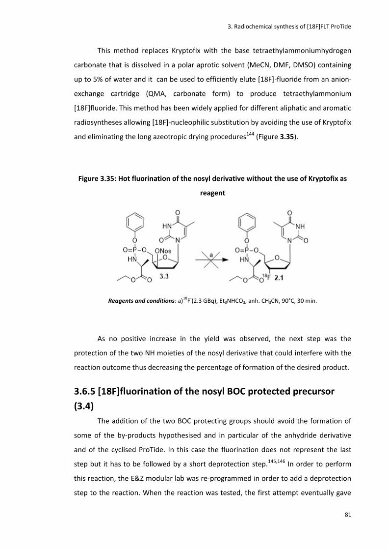
3. Radiochemical synthesis of [18F]FLT ProTide
81
This method replaces Kryptofix with the base tetraethylammoniumhydrogen
carbonate that is dissolved in a polar aprotic solvent (MeCN, DMF, DMSO) containing
up to 5% of water and it can be used to efficiently elute [18F]-fluoride from an anion-
exchange cartridge (QMA, carbonate form) to produce tetraethylammonium
[18F]fluoride. This method has been widely applied for different aliphatic and aromatic
radiosyntheses allowing [18F]-nucleophilic substitution by avoiding the use of Kryptofix
and eliminating the long azeotropic drying procedures144 (Figure 3.35).
Figure 3.35: Hot fluorination of the nosyl derivative without the use of Kryptofix as
reagent
Reagents and conditions: a)18
F-(2.3 GBq), Et3NHCO3, anh. CH3CN, 90°C, 30 min.
As no positive increase in the yield was observed, the next step was the
protection of the two NH moieties of the nosyl derivative that could interfere with the
reaction outcome thus decreasing the percentage of formation of the desired product.
3.6.5 [18F]fluorination of the nosyl BOC protected precursor
(3.4)
The addition of the two BOC protecting groups should avoid the formation of
some of the by-products hypothesised and in particular of the anhydride derivative
and of the cyclised ProTide. In this case the fluorination does not represent the last
step but it has to be followed by a short deprotection step.145,146 In order to perform
this reaction, the E&Z modular lab was re-programmed in order to add a deprotection
step to the reaction. When the reaction was tested, the first attempt eventually gave

3. Radiochemical synthesis of [18F]FLT ProTide
82
the desired radiolabelled [18F]-BOC protected FLT ProTide under the following
conditions (Figure 3.36).
Figure 3.36: Hot fluorination of the BOC protected nosyl derivative (3.4)
Reagents and conditions: a)18
F-(2.5GBq), Kryptofix, anh. CH3CN, 90°C, 30 min.
After 30 mins of radiolabelling at 90°C, the reaction mixture was passed
through an Al cartridge to remove the excess unreacted 18F-. An aliquot of the reaction
mixture was then taken and injected into the HPLC showing a major product with a
retention time at around 15 mins. This suggested that the desired BOC radiolabelled
product was formed therefore supporting the hypothesis that the BOC protection
provides a better stability to the nosyl precursor.
Figure 3.37: [18F]fluorination of compound 3.4
a)Radioactive chromatogram of the reaction mixture showing BOC-protected [18F]FLT
ProTide at 15 min
a)
CH3CNDMSO
Bkg 1
Region 1
Bkg 2
Region 2
Bkg 3
0:00 5:00 10:00 15:00 mm:ss
0.0
10000.0
20000.0
30000.0
Counts
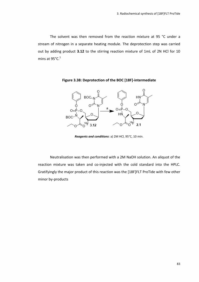
3. Radiochemical synthesis of [18F]FLT ProTide
83
The solvent was then removed from the reaction mixture at 95 °C under a
stream of nitrogen in a separate heating module. The deprotection step was carried
out by adding product 3.12 to the stirring reaction mixture of 1mL of 2N HCl for 10
mins at 95°C.1
Figure 3.38: Deprotection of the BOC [18F]-intermediate
Reagents and conditions: a) 2M HCl, 95°C, 10 min.
Neutralisation was then performed with a 2M NaOH solution. An aliquot of the
reaction mixture was taken and co-injected with the cold standard into the HPLC.
Gratifyingly the major product of this reaction was the [18F]FLT ProTide with few other
minor by-products

3. Radiochemical synthesis of [18F]FLT ProTide
84
Figure 3.39: Deprotection of compound 3.12
a)Radioactive chromatogram of the reaction mixture showing [18F]FLT ProTide as
major peak at 10.8 min; b)UV chromatogram of the reaction mixture co-spiked with
the cold standard (3.11). (Rt of compound 3.11: 10.7 min)
a)
b)
The compound was then purified by semi-preparative HPLC and was eluted
after 35 minutes at a flow rate of 3.5 mL/min using 30% CH3CN/70% H2O as the mobile
phase. The HPLC solvent was then removed from the mixture under a stream of
nitrogen. The radioactive product was taken up in saline and subsequently flushed
through a sterility filter to obtain a sterile and clean aqueous solution of [18F]FLT
ProTide.
An aliquot of the purified sample was analysed by HPLC (Figure 3.39) via co-
elution with the cold standard. Radiochemical reactions were carried out using starting
activities between 1.5-8 GBq, leading to final product activities of 240-480MBq in a
highly respectable RCY of 15-30% (n=5, decay-corrected from end of bombardment
(EoB)), with high radiochemical purities (97%) and specific activities_1800 mCi/μmol.
The total synthesis time was 130 min after the end of bombardment (EoB).
0:00 5:00 10:00 15:00 mm:ss
0.0
5000.0
10000.0
15000.0
Counts
0:00 5:00 10:00 15:00 mm:ss
0.0
500.0
1000.0
1500.0
2000.0
2500.0
mAU

3. Radiochemical synthesis of [18F]FLT ProTide
85
Figure 3.40: Purified [18F]FLT ProTide
a) Radioactive chromatogram of the purified [18F]FLT ProTide with Rt: 10.8 min ,
b) UV chromatogram of the reaction mixture co-spiked with the cold standard
with Rt: 10.7 min.
a)
b)
Figure 3.41: Radio-TLC chromatogram of the purified [18F]FLT ProTide
3.7 Conclusions Five potential precursor molecules of the [18F]FLTProTide (2.1) and a cold
reference standard, the [19F]FLT ProTide (3.11), have been synthesised using cold

3. Radiochemical synthesis of [18F]FLT ProTide
86
chemistry methods. Late stage [18F]fluorination was performed on all the precursors
synthesised exploring different reaction conditions. The nosyl derivate (3.3) furnished
the desired product in poor yield therefore protection of the NH moieties of the
molecule was performed in order to minimize the formation of undesired side
products. This protection markedly increased the yield of the reaction furnishing the
desired radiolabelled BOC protected compound. This compound was then deprotected
to finally furnish the desired target compound 2.1. This is the first time to our
knowledge that a radiolabelled ProTide has been synthesised.
However, the results here presented should be regarded as first trials of the
synthesis of [18F]radiolabelled ProTides. Further investigations with different set-ups
(e.g. different automated synthetic modules, microwave-assisted synthesis) should be
carried out to validate the reproducibility and clinical potential application of this
radiosynthesis.9,45,145

4. Radiochemical synthesis of [18F]FIAU ProTide
87
4. Radiochemical synthesis of [18F]FIAUProTide
4.1 Introduction Besides the late stage [18F]fluorination approach discussed in Chapter 3,
another synthetic pathway involving an early stage [18F]fluorination has been followed
to access a different class of radiolabelled compounds, the 2’-[18F]FIAUProTides
(Figure 4.1).
Figure 4.1: Structure of the2’-[18F]FIAU ProTide target (2.2)
[18F]FIAU (1.16) is already a well known PET imaging probe used as a
biomarker for imaging HSV1-tkgene expression.146 Reporter gene imaging is a non
invasive technique used in live subjects to determine location, duration and extent of
expression of the gene of interest.147–150 In particular Herpes simplex virus-1 thymidine
kinase (HSV1-tk) is one of the genes most widely used as a reporter in molecular
imaging to visualise and monitor many biological processes including lymphocyte
migration151, transcriptional regulation152, and stem-cell tracking.153,154 Radiolabelled
nucleoside analogues, such as [18F]FIAU, can be used for monitoring HSV1-tk gene
expression using PET imaging.54
*18F+FIAU ProTide has been chosen as a model standard for the class of the 2’-
fluorinated ProTides for the following reasons:
- Fluorine is placed in position 2’ on the sugar moiety therefore it can be used as
model standard for the radiochemical synthesis of other 2’-[18F]ProTides.

4. Radiochemical synthesis of [18F]FIAU ProTide
88
- [18F]FIAU (1.16) is a PET imaging probe and many synthetic strategies have
already been extensively explored45,116,132,155 and can be used as a guide for the
synthesis of its ProTide. Additionally studies of retention of [18F]-FIAU on HEK
(human equilibrative kidney) cells engineered to express HSV- tk1 can be useful
to prove the successful synthesis of this tracer.
- It is a uridine based ProTide, thus the absence of other reactive sites on the
nucleobase moiety of the molecule will avoid further protection and
deprotection steps.
4.2 [18F]fluorination (Hot): early stage approach Among many synthetic approaches, the only one reported in the literature
which gives [18F]FIAU in good yields consists of an early stage hot fluorination of the
sugar moiety followed by a multi-step synthesis155 that has to be performed in a
relative short time taking into account the half life of the [18F]fluorine (an ideal
synthesis should not exceed three half lives)1. A similar synthetic pathway to that of
[18F]FIAU will be applied for the synthesis of its ProTide according to the scheme
outlined in Figure 4.2.
Figure 4.2: Early stage fluorination approach for the synthesis of the
[18F]FIAU ProTide (2.2)
Although the early stage hot fluorination is considered not to be ideal when a
multi-step synthesis has to be performed, the advantage consists in the radiolabelling

4. Radiochemical synthesis of [18F]FIAU ProTide
89
step that does not give any by-products thus providing a compound that can be used
for the next steps without further purification.
4.3 Synthesis of the cold standard (4.3)
Again a cold standard, the FIAU ProTide (4.3), was synthesised in order to have
a reference compound to co-inject into the analytical HPLC together with the
radiolabelled compound as a proof of the successful outcome of the reaction.140 As the
synthesis of [18F]FIAU is well known, no attempts at cold fluorination were performed
to test the reactivity towards nucleophilic substitution performed by the fluoride on
the triflate group of the precursor molecule 4.1. Therefore the synthesis of the cold
standard was accomplished following the synthetic scheme in Figure 4.3.
Figure 4.3: Synthesis of the cold standard FIAU ProTide (4.3)
Reagents and conditions: a) I2, Ceric ammonium nitrate, CH3CN, 75°C, 1h, 60%; b) L-alanine benzyl ester
hydrochloride salt, Et3N, -75°C to rt, anh. CH2Cl2, 3h, 88%; c) NMI, anh. THF, 0°C to rt, 16h, 12%.
The commercially available 2'-deoxy-2'-β-fluoro-uridine (4.4) was used as
starting material of the synthesis (Figure 4.4). I2 was used as reagent to perform the
iodination reaction in position 5 of the pyrimidine ring and ceric ammonium nitrate
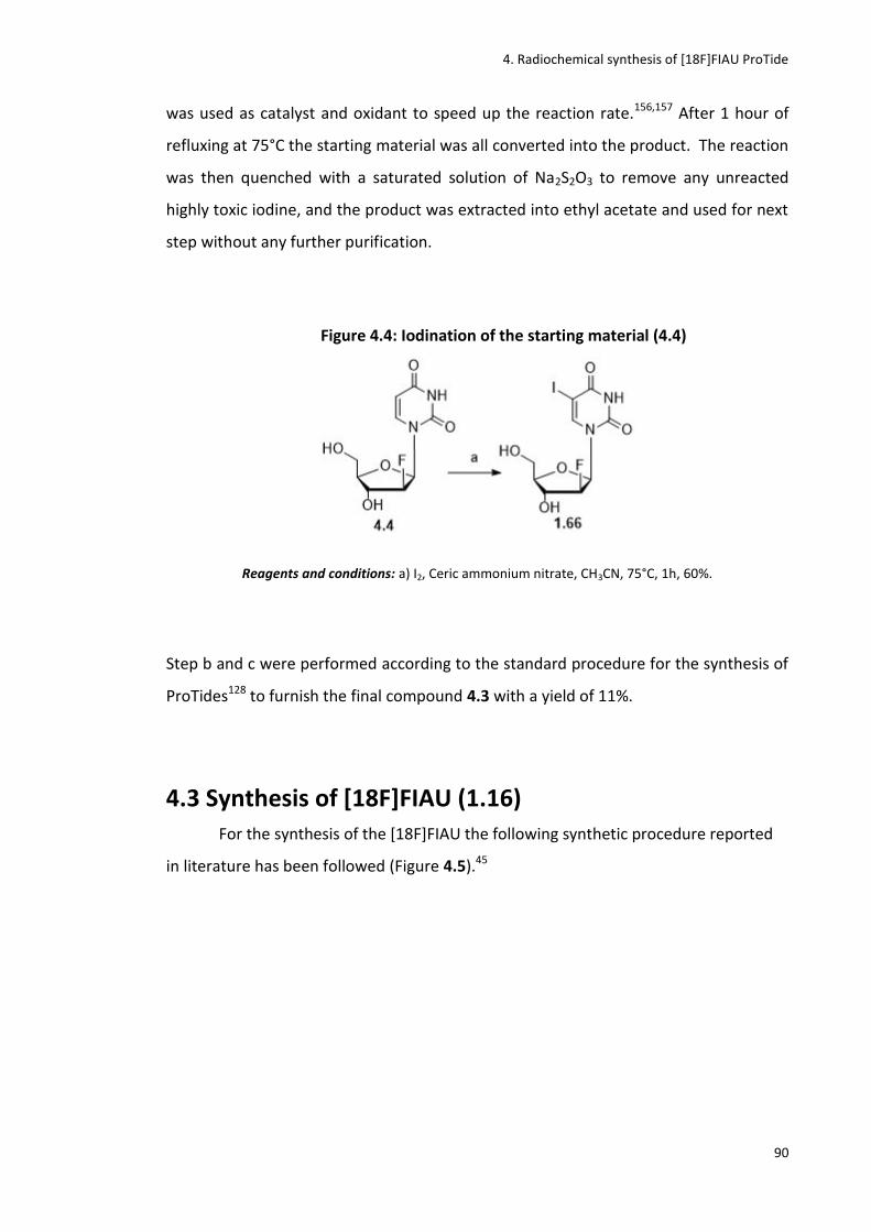
4. Radiochemical synthesis of [18F]FIAU ProTide
90
was used as catalyst and oxidant to speed up the reaction rate.156,157 After 1 hour of
refluxing at 75°C the starting material was all converted into the product. The reaction
was then quenched with a saturated solution of Na2S2O3 to remove any unreacted
highly toxic iodine, and the product was extracted into ethyl acetate and used for next
step without any further purification.
Figure 4.4: Iodination of the starting material (4.4)
Reagents and conditions: a) I2, Ceric ammonium nitrate, CH3CN, 75°C, 1h, 60%.
Step b and c were performed according to the standard procedure for the synthesis of
ProTides128 to furnish the final compound 4.3 with a yield of 11%.
4.3 Synthesis of [18F]FIAU (1.16)
For the synthesis of the [18F]FIAU the following synthetic procedure reported
in literature has been followed (Figure 4.5).45

4. Radiochemical synthesis of [18F]FIAU ProTide
91
Figure 4.5: Radiochemical synthesis of [18F]FIAU (1.16)
Reagents and conditions: a) 18
F- , Kryptofix, anh. CH3CN, 95°C, 30 min; b) Hexamethyldisilaxane, TMSOTf,
anh. dichloroethane, 85°C, 2h; c) anh. CH3CN, 85°C, 1h; d) NaOCH3/CH3OH, 80°C, 10 min.
Compared to the other reported radiosyntheses of this tracer, this one has the
advantage of avoiding the bromination step in position 2 of compound 4.2 before the
coupling with 4.7.54,155Therefore a shorter synthesis is obtained with also the
advantage of avoiding the usage of the highly toxic Br2 reagent.
The first step consists of the hot fluorination of the commercially available
starting material sugar 4.1. In this case the leaving group is the triflate, one of the best
leaving groups amongst the organosulfonates.158 The reaction was carried out using
the E&Z modular lab following the same procedure described in Chapter 3. The
conditions for the radiolabelling are reported below.

4. Radiochemical synthesis of [18F]FIAU ProTide
92
Figure 4.6: Synthesis of the [18F]-sugar (4.2)
Reagents and conditions: a) 18
F- , Kryptofix, anh. CH3CN, 95°C, 30 min.
After purification with an alumina cartridge (characterized by an extremely
polar surface for analyte retention) to eliminate the excess of free 18F-,41 the
radiolabelled sugar (4.2) was produced and used for next step without further
purification. When an aliquot of the hot mixture was co-spiked with the cold standard
(commercially available), it showed the same retention time at around 8 min as shown
in Figure 4.7.
Figure 4.7: [18F]fluorination of compound 4.1
a) Radioactive chromatogram of the reaction mixture showing the [18F]-sugar 4.2
as the unique product of the reaction with Rt of 8.2 min; b) UV chromatogram
of the reaction mixture co-spiked with the cold standard. (Rt of commercially
available cold standard: 7.9 min). b) HPLC system: 98% CH3CN, 2% H20.
a)
b)
0:00 5:00 10:00 15:00 mm:ss
0.0
5000.0
10000.0
15000.0
20000.0
25000.0
Counts
0:00 5:00 10:00 15:00 mm:ss
0.0
100.0
200.0
300.0
400.0
mAU

4. Radiochemical synthesis of [18F]FIAU ProTide
93
The second step consists in the protection of the base moiety that was freshly
synthesised while the hot fluorination was ongoing in a second heating module placed
into the hot cell next to E&Z modular lab. Hexamethyldisilaxane was used as protecting
reagent and trimethylsilyltrifluoromethanesulfonate (TMSOTf) was used as catalyst to
activate the following glycosylation reaction45(Figure 4.8). To confirm that the reaction
was complete, HPLC and LC-MS were performed.
Figure 4.8: Protection of the 5-Iodouracil (4.5)
Reagents and conditions: b) Hexamethyldisilaxane, TMSOTf, anh. DCE, 85°C, 2h.
Compound 4.7 was dried at 90°C under compressed air to remove the
dichloroethane solvent, obtaining a yellowish oil. The radiolabelled sugar (4.2) (in
CH3CN solution) was then delivered directly from the E&Z unit to the vial containing
the protected uracil (4.7) to perform the glycosylation reaction followed by a quick
deprotection step as shown in Figure 4.9. The glycosylation is known to be a non-
stereoselective reaction because of the formation of a new sterogenic centre.159
Indeed two anomers were formed from this reaction, the β-anomer (the [18F]FIAU)
(1.16) and the α-anomer (4.7).155
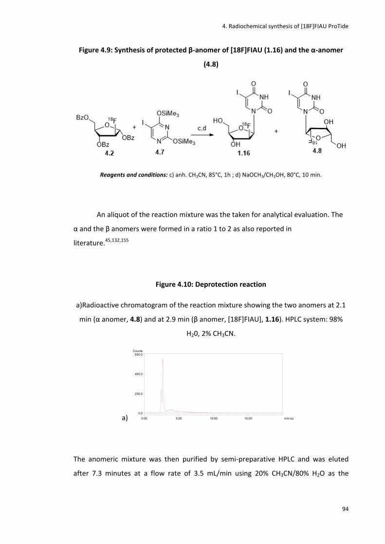
4. Radiochemical synthesis of [18F]FIAU ProTide
94
Figure 4.9: Synthesis of protected β-anomer of [18F]FIAU (1.16) and the α-anomer
(4.8)
Reagents and conditions: c) anh. CH3CN, 85°C, 1h ; d) NaOCH3/CH3OH, 80°C, 10 min.
An aliquot of the reaction mixture was the taken for analytical evaluation. The
α and the β anomers were formed in a ratio 1 to 2 as also reported in
literature.45,132,155
Figure 4.10: Deprotection reaction
a)Radioactive chromatogram of the reaction mixture showing the two anomers at 2.1
min (α anomer, 4.8) and at 2.9 min (β anomer, *18F+FIAU+, 1.16). HPLC system: 98%
H20, 2% CH3CN.
a)
The anomeric mixture was then purified by semi-preparative HPLC and was eluted
after 7.3 minutes at a flow rate of 3.5 mL/min using 20% CH3CN/80% H2O as the
0:00 5:00 10:00 15:00 mm:ss
0.0
200.0
400.0
600.0Counts

4. Radiochemical synthesis of [18F]FIAU ProTide
95
mobile phase. An aliquot of the purified compound was taken for analytical HPLC
evaluation and co-spiked with the cold standard.
Figure 4.11: [18F]FIAU purification
a) Radioactive chromatogram of the [18F]FIAU (1.16) showing the product with Rt of
2.3 min; b) UV chromatogram of the reaction mixture co-spiked with the cold standard
(1.61). (Rt of cold standard: 2.1 min). HPLC system: 98% H20, 2% CH3CN.
a)
b)
The radiolabelled compound showed the same retention time as the standard
and also literature strongly supports that the elution time of the β-anomer is longer
than the α.45 Nevertheless an additional biological evaluation of the tracer was
performed to support the isolation of the correct anomer (see section 4.6).
In order to optimise and speed up the glycosylation step, several conditions
and combination of catalysts were explored (Table 4.1).10,45
0:00 5:00 10:00 15:00 mm:ss
0.0
500.0
1000.0
1500.0
Counts
0:00 5:00 10:00 15:00 mm:ss
0.0
1000.0
2000.0
3000.0
mAU
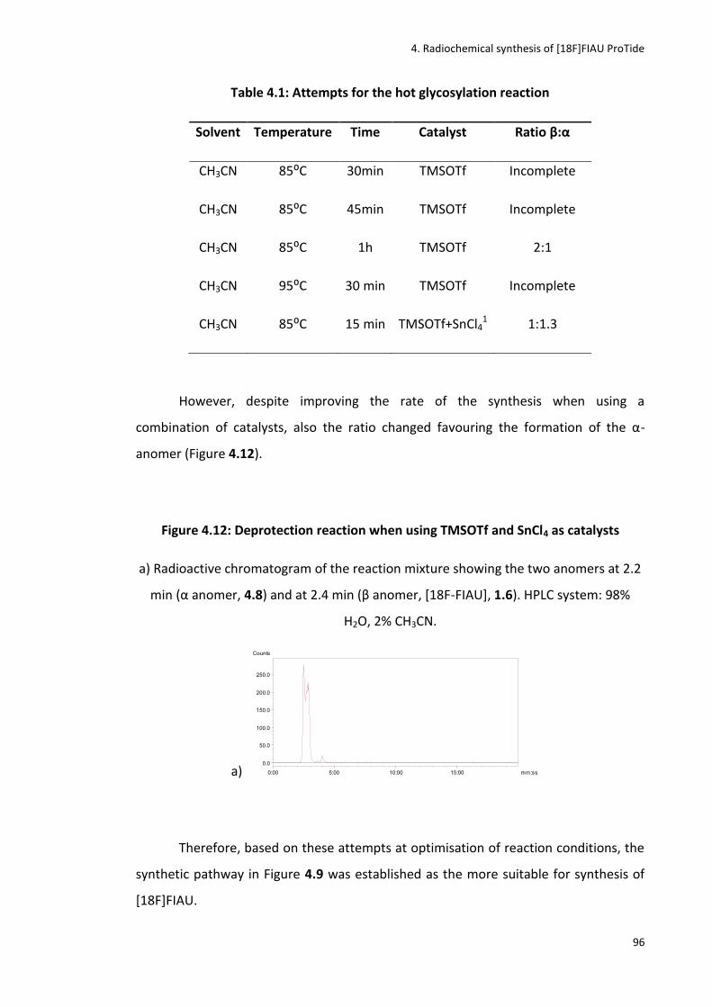
4. Radiochemical synthesis of [18F]FIAU ProTide
96
Table 4.1: Attempts for the hot glycosylation reaction
Solvent Temperature Time Catalyst Ratio β:α
CH3CN 85:C 30min TMSOTf Incomplete
CH3CN 85:C 45min TMSOTf Incomplete
CH3CN 85:C 1h TMSOTf 2:1
CH3CN 95:C 30 min TMSOTf Incomplete
CH3CN 85:C 15 min TMSOTf+SnCl41 1:1.3
However, despite improving the rate of the synthesis when using a
combination of catalysts, also the ratio changed favouring the formation of the α-
anomer (Figure 4.12).
Figure 4.12: Deprotection reaction when using TMSOTf and SnCl4 as catalysts
a) Radioactive chromatogram of the reaction mixture showing the two anomers at 2.2
min (α anomer, 4.8) and at 2.4 min (β anomer, *18F-FIAU], 1.6). HPLC system: 98%
H2O, 2% CH3CN.
a)
Therefore, based on these attempts at optimisation of reaction conditions, the
synthetic pathway in Figure 4.9 was established as the more suitable for synthesis of
[18F]FIAU.
0:00 5:00 10:00 15:00 mm:ss
0.0
50.0
100.0
150.0
200.0
250.0
Counts
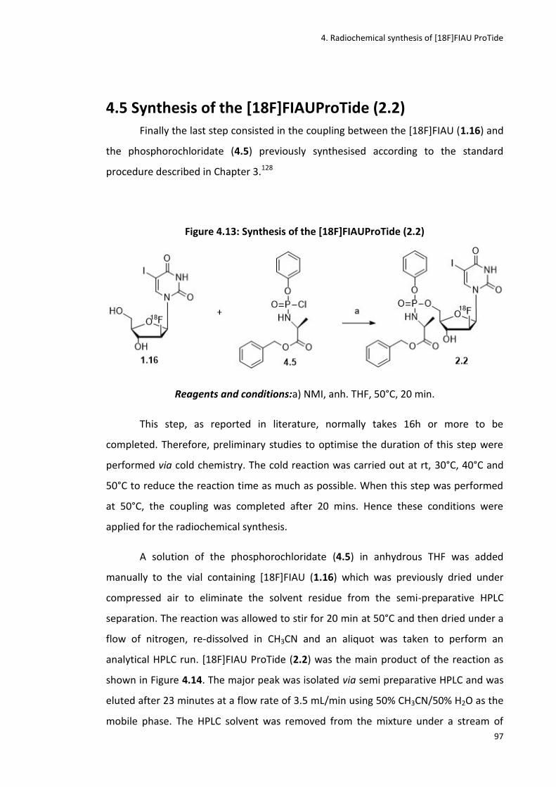
4. Radiochemical synthesis of [18F]FIAU ProTide
97
4.5 Synthesis of the [18F]FIAUProTide (2.2)
Finally the last step consisted in the coupling between the [18F]FIAU (1.16) and
the phosphorochloridate (4.5) previously synthesised according to the standard
procedure described in Chapter 3.128
Figure 4.13: Synthesis of the [18F]FIAUProTide (2.2)
Reagents and conditions:a) NMI, anh. THF, 50°C, 20 min.
This step, as reported in literature, normally takes 16h or more to be
completed. Therefore, preliminary studies to optimise the duration of this step were
performed via cold chemistry. The cold reaction was carried out at rt, 30°C, 40°C and
50°C to reduce the reaction time as much as possible. When this step was performed
at 50°C, the coupling was completed after 20 mins. Hence these conditions were
applied for the radiochemical synthesis.
A solution of the phosphorochloridate (4.5) in anhydrous THF was added
manually to the vial containing [18F]FIAU (1.16) which was previously dried under
compressed air to eliminate the solvent residue from the semi-preparative HPLC
separation. The reaction was allowed to stir for 20 min at 50°C and then dried under a
flow of nitrogen, re-dissolved in CH3CN and an aliquot was taken to perform an
analytical HPLC run. [18F]FIAU ProTide (2.2) was the main product of the reaction as
shown in Figure 4.14. The major peak was isolated via semi preparative HPLC and was
eluted after 23 minutes at a flow rate of 3.5 mL/min using 50% CH3CN/50% H2O as the
mobile phase. The HPLC solvent was removed from the mixture under a stream of

4. Radiochemical synthesis of [18F]FIAU ProTide
98
nitrogen. The radioactive product was taken up in saline and subsequently flushed
through a sterility filter to obtain a sterile and clean aqueous solution of [18F]FIAU
ProTide.
An aliquot of the purified sample was analysed by analytical HPLC via co-elution
with the cold standard as shown in Figure 4.14. Radiochemical reactions were carried
out using starting activities between 7-15 GBq, leading to final product activities of 8-
56 MBq in a RCY of 1-5% (n=7, decay-corrected from end of bombardment (EoB)), with
high radiochemical purities (98%) and specific activities of 1800 mCi/μmol. The total
synthesis time was 240 min after the end of bombardment (EoB).
Figure 4.14: Synthesis of [18F]FIAU ProTide
a) Radioactive chromatogram of the [18F]FIAU ProTide reaction mixture showing a
major peak with Rt of 13 min; b) Radioactive chromatogram of the [18F]FIAU ProTide
purified with Rt of 12.3 min; c) UV chromatogram of the [18F]FIAU ProTide co-spiked
with the cold standard (4.3). (Rt of cold standard: 12.2 min). HPLC system: 90% H2O-
10% CH3CN to 50% H2O-50% CH3CN.
a)
b)
0:00 5:00 10:00 15:00 mm:ss
0.0
10.0
20.0
30.0
40.0
Counts
0:00 5:00 10:00 15:00 mm:ss
0.0
100.0
200.0
300.0
400.0
Counts
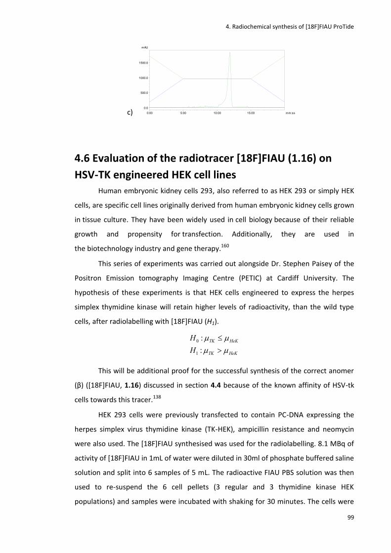
4. Radiochemical synthesis of [18F]FIAU ProTide
99
c)
4.6 Evaluation of the radiotracer [18F]FIAU (1.16) on
HSV-TK engineered HEK cell lines
Human embryonic kidney cells 293, also referred to as HEK 293 or simply HEK
cells, are specific cell lines originally derived from human embryonic kidney cells grown
in tissue culture. They have been widely used in cell biology because of their reliable
growth and propensity for transfection. Additionally, they are used in
the biotechnology industry and gene therapy.160
This series of experiments was carried out alongside Dr. Stephen Paisey of the
Positron Emission tomography Imaging Centre (PETIC) at Cardiff University. The
hypothesis of these experiments is that HEK cells engineered to express the herpes
simplex thymidine kinase will retain higher levels of radioactivity, than the wild type
cells, after radiolabelling with [18F]FIAU (H1).
HeKTK
HeKTK
H
H
:
:
1
0
This will be additional proof for the successful synthesis of the correct anomer
(β) (*18F+FIAU, 1.16) discussed in section 4.4 because of the known affinity of HSV-tk
cells towards this tracer.138
HEK 293 cells were previously transfected to contain PC-DNA expressing the
herpes simplex virus thymidine kinase (TK-HEK), ampicillin resistance and neomycin
were also used. The [18F]FIAU synthesised was used for the radiolabelling. 8.1 MBq of
activity of [18F]FIAU in 1mL of water were diluted in 30ml of phosphate buffered saline
solution and split into 6 samples of 5 mL. The radioactive FIAU PBS solution was then
used to re-suspend the 6 cell pellets (3 regular and 3 thymidine kinase HEK
populations) and samples were incubated with shaking for 30 minutes. The cells were
0:00 5:00 10:00 15:00 mm:ss
0.0
500.0
1000.0
1500.0
mAU
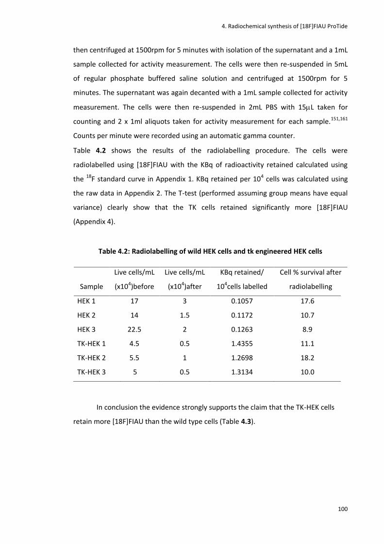
4. Radiochemical synthesis of [18F]FIAU ProTide
100
then centrifuged at 1500rpm for 5 minutes with isolation of the supernatant and a 1mL
sample collected for activity measurement. The cells were then re-suspended in 5mL
of regular phosphate buffered saline solution and centrifuged at 1500rpm for 5
minutes. The supernatant was again decanted with a 1mL sample collected for activity
measurement. The cells were then re-suspended in 2mL PBS with 15L taken for
counting and 2 x 1ml aliquots taken for activity measurement for each sample.151,161
Counts per minute were recorded using an automatic gamma counter.
Table 4.2 shows the results of the radiolabelling procedure. The cells were
radiolabelled using [18F]FIAU with the KBq of radioactivity retained calculated using
the 18F standard curve in Appendix 1. KBq retained per 104 cells was calculated using
the raw data in Appendix 2. The T-test (performed assuming group means have equal
variance) clearly show that the TK cells retained significantly more [18F]FIAU
(Appendix 4).
Table 4.2: Radiolabelling of wild HEK cells and tk engineered HEK cells
Sample
Live cells/mL
(x104)before
Live cells/mL
(x104)after
KBq retained/
104cells labelled
Cell % survival after
radiolabelling
HEK 1 17 3 0.1057 17.6
HEK 2 14 1.5 0.1172 10.7
HEK 3 22.5 2 0.1263 8.9
TK-HEK 1 4.5 0.5 1.4355 11.1
TK-HEK 2 5.5 1 1.2698 18.2
TK-HEK 3 5 0.5 1.3134 10.0
In conclusion the evidence strongly supports the claim that the TK-HEK cells
retain more [18F]FIAU than the wild type cells (Table 4.3).
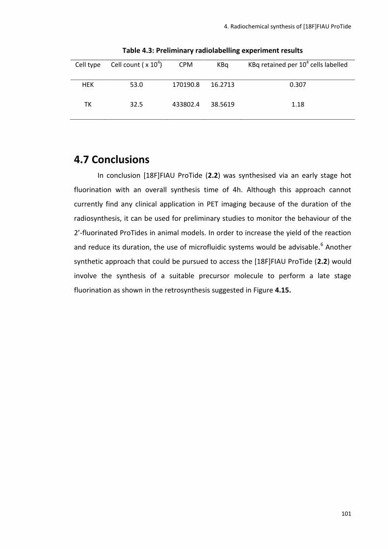
4. Radiochemical synthesis of [18F]FIAU ProTide
101
Table 4.3: Preliminary radiolabelling experiment results
Cell type Cell count ( x 104) CPM KBq KBq retained per 104 cells labelled
HEK 53.0 170190.8 16.2713 0.307
TK 32.5 433802.4 38.5619 1.18
4.7 Conclusions In conclusion [18F]FIAU ProTide (2.2) was synthesised via an early stage hot
fluorination with an overall synthesis time of 4h. Although this approach cannot
currently find any clinical application in PET imaging because of the duration of the
radiosynthesis, it can be used for preliminary studies to monitor the behaviour of the
2’-fluorinated ProTides in animal models. In order to increase the yield of the reaction
and reduce its duration, the use of microfluidic systems would be advisable.6 Another
synthetic approach that could be pursued to access the [18F]FIAU ProTide (2.2) would
involve the synthesis of a suitable precursor molecule to perform a late stage
fluorination as shown in the retrosynthesis suggested in Figure 4.15.

4. Radiochemical synthesis of [18F]FIAU ProTide
102
Figure 4.15: Proposed retrosynthetic pathway to access the [18F]FIAU ProTide (2.2)
via a late stage hot fluorination
Additionally studies of retention of [18F]FIAU on HEK cells engineered to express
HSV-TK1 were performed and further evidenced the successful synthesis of this tracer
showing a major retention compared to wild type cells.

5. Synthesis of novel 2’-deoxy-2’-fluoro-5-iodouridine Protides as novel antiviral agents
103
5. Synthesis of novel 2'-deoxy-2'-fluoro-5-iodouridine ProTides as novel antiviral agents
5.1 Introduction Viral infections can be classified into three main groups. The first class includes
life-threatening chronic viruses like the Human Immunodeficiency Virus (HIV), the
Hepatitis B virus (HBV), and Hepatitis C Virus (HCV). The second class comprises acute
viral infections that are generally non-lethal and self-resolving in otherwise healthy
patients such as influenza viruses. The third class includes non-lethal viral infections
but with a significant economic impact, such as the common cold caused by
rhinoviruses.162 In the last two decades important achievements have been made in
the treatment of these viral infections. Nevertheless the spread of new viral diseases
such as Zika, Ebola and new strains of hepatitis and herpes viruses has characterised
recent years with potential for a pandemic outbreak,163–165 therefore there is
significant unmet medical need for new antiviral agents.
Nucleoside analogues (NA), particularly fluorinated nucleosides, represent an
important class of antiviral agents and act as inhibitors of nucleoside reverse
transcriptase (NRTis). Their active form is the 5’-triphosphate form and their targets
are the catalytic residues of the viral polymerase that interact with the template, the
primer and the incoming nucleoside 5’-triphosphates.34,166–169Uridine based
nucleosides and ProTides have been of particular interest for the treatment of viral
infections.34,81,122,170
For example, Sofosbuvir (1.72), the ProTide of -D-2’-deoxy-2’--fluoro-2’--C-
methyluridine, is a recently approved anti-HCV agent characterised by a uridine
backbone, and a fluorine in the 2’--C position of the ribose moiety.122,123,171 FIAU
(Fialouridine) was another 2’-F-nucleoside analogue (Figure 5.1) that was selected as a
Phase I clinical trial candidate for the treatment of HBV, however it showed
mitochondrial toxicity resulting in lactic acidosis and hepatic failure.172,173
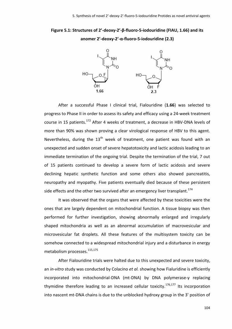
5. Synthesis of novel 2’-deoxy-2’-fluoro-5-iodouridine Protides as novel antiviral agents
104
Figure 5.1: Structures of 2’-deoxy-2’-β-fluoro-5-iodouridine (FIAU, 1.66) and its
anomer 2’-deoxy-2’-α-fluoro-5-iodouridine (2.3)
After a successful Phase I clinical trial, Fialouridine (1.66) was selected to
progress to Phase II in order to assess its safety and efficacy using a 24-week treatment
course in 15 patients.172 After 4 weeks of treatment, a decrease in HBV-DNA levels of
more than 90% was shown proving a clear virological response of HBV to this agent.
Nevertheless, during the 13th week of treatment, one patient was found with an
unexpected and sudden onset of severe hepatotoxicity and lactic acidosis leading to an
immediate termination of the ongoing trial. Despite the termination of the trial, 7 out
of 15 patients continued to develop a severe form of lactic acidosis and severe
declining hepatic synthetic function and some others also showed pancreatitis,
neuropathy and myopathy. Five patients eventually died because of these persistent
side effects and the other two survived after an emergency liver transplant.174
It was observed that the organs that were affected by these toxicities were the
ones that are largely dependent on mitochondrial function. A tissue biopsy was then
performed for further investigation, showing abnormally enlarged and irregularly
shaped mitochondria as well as an abnormal accumulation of macrovesicular and
microvesicular fat droplets. All these features of the multisystem toxicity can be
somehow connected to a widespread mitochondrial injury and a disturbance in energy
metabolism processes.115,175
After Fialouridine trials were halted due to this unexpected and severe toxicity,
an in-vitro study was conducted by Colacino et al. showing how Fialuridine is efficiently
incorporated into mitochondrial-DNA (mt-DNA) by DNA polymerase-γ replacing
thymidine therefore leading to an increased cellular toxicity.176,177 Its incorporation
into nascent mt-DNA chains is due to the unblocked hydroxy group in the 3' position of

5. Synthesis of novel 2’-deoxy-2’-fluoro-5-iodouridine Protides as novel antiviral agents
105
the deoxyribose moiety that allows the addition of nucleotides to the growing mt-DNA
chain (and to a lesser extent on the nuclear-DNA (N-DNA)). This results in the
formation of non-functional mt-DNA and N-DNA thus affecting gene expression,
essential for the cells survival and correct functioning.176
After the failure in 1993 of Fialouridine in Phase II clinical trial no further
studies were conducted on its therapeutic activity or its toxicity. It was instead
investigated as a diagnostic probe leading to the PET tracer agent [18F]FIAU (1.16)
discussed in Chapter 4. Furthermore, no studies have been conducted on its isomer 2'-
deoxy-2'-α-fluoro-5-iodouridine that therefore represents a novel and interesting
chemical entity. As the uridine backbone represents an already validated starting point
to design antiviral agents, uridine based Protides and particularly fluorinated ones,
could represent novel antiviral agents for the treatment of new viral diseases such as
Zika, Ebola and new strains of hepatitis and herpes viruses for which there is no
established treatment.163,178
The aim of this project is to synthesise a novel class of 2'-deoxy-2'-fluoro-5-
iodouridine ProTides and evaluate them as antiviral agents for the treatment of newly
arising viral diseases. It is expected that the application of the ProTide strategy could
potentially lead to potent antiviral agents with an increased antiviral activity compared
to their parent NA and reduced toxicity. It will be also discussed how the fluorine in the
α- or β-2’-C position of the ribose can play a role in the activity of these NAs and their
prodrugs.90 The synthesis of a series of 2'-deoxy-2'-α-fluoro-5-iodouridine ProTides will
be the main target of the project as the α-anomer (2.3) represents a novel and more
convenient starting material for the synthesis compared to the β-anomer (FIAU, 1.66).
Nevertheless a ProTide of the β-anomer (FIAU, 1.66), will also be synthesised to
understand whether the position of the fluorine can lead to any change in their
antiviral activity (Figure 5.2).

5. Synthesis of novel 2’-deoxy-2’-fluoro-5-iodouridine Protides as novel antiviral agents
106
Figure 5.2: Application of the ProTide strategy to fluorinated uridine based ProTides
5.2 Synthesis of novel 2'-deoxy-2'-fluoro-5-iodouridine ProTides
The synthesis of 2’-deoxy-2’-fluoro-5-iodouridine ProTides was based on
standard ProTide chemistry84 discussed earlier in the thesis, via five major steps as
shown in Figure 5.3.
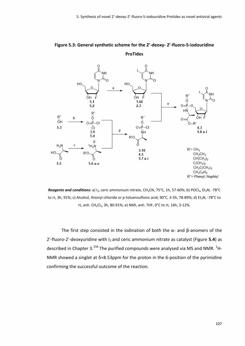
5. Synthesis of novel 2’-deoxy-2’-fluoro-5-iodouridine Protides as novel antiviral agents
107
Figure 5.3: General synthetic scheme for the 2’-deoxy- 2’-fluoro-5-iodouridine
ProTides
Reagents and conditions: a) I2, ceric ammonium nitrate, CH3CN, 75°C, 1h, 57-60%; b) POCl3, Et3N, -78°C
to rt, 3h, 91%; c) Alcohol, thionyl chloride or p-toluensulfonic acid, 90°C, 3-5h, 78-89%; d) Et3N, -78°C to
rt, anh. CH2Cl2, 3h, 80-91%; e) NMI, anh. THF, 0°C to rt, 16h, 3-12%.
The first step consisted in the iodination of both the α- and β-anomers of the
2'-fluoro-2'-deoxyuridine with I2 and ceric ammonium nitrate as catalyst (Figure 5.4) as
described in Chapter 3.156 The purified compounds were analysed via MS and NMR. 1H-
NMR showed a singlet at δ=8.53ppm for the proton in the 6-position of the pyrimidine
confirming the successful outcome of the reaction.

5. Synthesis of novel 2’-deoxy-2’-fluoro-5-iodouridine Protides as novel antiviral agents
108
Figure 5.4: Iodination of the α- and β-anomers
Reagents and conditions: a) I2, ceric ammonium nitrate, CH3CN, 75°C, 1h, 57-60%.
Whereas phenylphosphorochloride is commercially available, the naphthyl
derivative is not. Therefore compound 5.4 (R’’ = naphthyl) was synthesised by coupling
naphthol with phosphorus oxychloride using triethylamine as the base and salt
scavenger. Anhydrous solvent and nitrogen atmosphere were essential for the positive
outcome of the reaction. The crude reaction mixture was reduced in vacuo and used
without further purification for next step (Figure 5.5).128
Figure 5.5: Formation of the naphtyl-phosphorodichloridate
Reagents and conditions: b) POCl3, Et3N, -78°C to rt, 3h, 91%.
L-alanine was chosen as the aminoacid for the phosphoramidate moiety
because, as widely reported in literature, the replacement of natural amino acids with
other amino acids such as the D-alanine179, or simple amines leads to a slight or
complete loss of activity of these derivatives in most ProTide cases.180 L-alanine is the

5. Synthesis of novel 2’-deoxy-2’-fluoro-5-iodouridine Protides as novel antiviral agents
109
most commonly used natural amino acid used in ProTide chemistry.181 Several esters
of the L-alanine were used and, among them, methyl, ethyl and tert-butyl L-alanine
esters were commercially available therefore were used directly for the coupling with
the phosphorodichloridate.182 Isopropyl, neopentyl and benzyl L-alanine ester were
instead synthesised via an esterification reaction of the L-alanine.
Two methods were used to access two kinds of salts, the chloride and sulfonate
salts of the L-alanine esters (Figure 5.6).103 The first method consisted in the reaction
between the alcohols with L-alanine and thionyl chloride to obtain the chloride salts.
However, sulfonate salts are considered to be more stable then the chloride salts183
therefore the alcohols were also reacted with L-alanine and para-toluene sulfonic acid
(p-TSA) to access also the equivalent L-alanine ester sulfonate salts (Figure 5.6). In this
second method, to shift the equilibrium to the right and therefore to push the reaction
to completion, Dean-stark apparatus was used to condense and thus collect the water
produced during the reaction. To monitor the reaction, thin layer chromatography
(TLC) was used. To confirm the formation of the product, a ninhydrin solution capable
of detecting the amino groups of the aminoacid, was sprayed on the TLC plate eluted
in a system of 2-propanol/water (7:3). A different Rf between the L-alanine and the
ester salt was observed. All the L-alanine ester salts were thereafter used for the next
step without further purification.
Figure 5.6: Synthesis of the L-alanine ester salt via two methods
Reagents and conditions: c) Alcohol, thionyl chloride or p-toluenesulfonic acid, 90°C, 3-5h, 75-89%.

5. Synthesis of novel 2’-deoxy-2’-fluoro-5-iodouridine Protides as novel antiviral agents
110
Table 5.1: Structures and yields of 5.6a-e
ID R’-X Yield
5.6a Neopentyl chloride 75%
5.6b Neopentyl sulfonate 83%
5.6c Isopropyl chloride 89%
5.6d Isopropyl sulfonate 78%
5.6e Tert-butyl chloride 84%
The phosphorochloridates were synthesised according to the standard
procedure by the reaction of the L-alanine ester salts with aryl dichlorophosphonates
at low temperatures under nitrogen atmosphere with TEA used as a base84 (Figure
5.7). Formation of phosphorochloridates was confirmed by 31P-NMR. Once the
reaction was complete, the solvent was evaporated under reduced pressure and the
resulting solid was re-dissolved in anhydrous diethyl ether and filtered under nitrogen.
The filtrate was then evaporated in vacuo to obtain yellowish oils that were used for
the next step without further purification.
Figure 5.7: Synthesis of the phosphorochloridates
Reagents and conditions: d) L-alanine ester salt, Et3N, -78°C to rt, anh. CH2Cl2, 3h, 80- 95%.

5. Synthesis of novel 2’-deoxy-2’-fluoro-5-iodouridine Protides as novel antiviral agents
111
Table 5.2: Structures and yields of Phosphorochloridates synthesised
ID R R’ Yield
3.10 Ethyl Phenyl 95%
4.5 Benzyl Phenyl 88%
5.7a Methyl Phenyl 80%
5.7b Isopropyl Phenyl 83%
5.7c Neopentyl Phenyl 91%
5.7d Tert-butyl Phenyl 82%
5.7e Methyl Phenyl 85%
5.7f Ethyl Naphtyl 94%
5.7g Isopropyl Naphtyl 82%
5.7 h Neopentyl Naphtyl 80%
5.7i Tert-butyl Naphtyl 81%
Finally, the methyl, ethyl, isopropyl, benzyl, neopentyl and tert-butyl
phosphorochloridates were reacted with 2'-deoxy-2'-α-fluoro-5-iodouridine in
anhydrous THF using N-methylimidazole to form an imidazolium intermediate with the
phosphorochloridates that can readily react with 2'-deoxy-2'-fluoro-5-iodouridine to
form the ProTides (Figure 5.8).
After 24 h the reactions were stopped and the solvent was evaporated in
vacuo. The residues were extracted with CH2Cl2 and 0.5N HCl necessary to neutralise
the base used in the reaction. The organic layer was then concentrated under reduced
pressure to obtain yellowish oils. All ProTides were purified by column
chromatography using a gradient system of CH2Cl2/CH3OH (100% to 95% CH2Cl2) and,
where necessary, also by the use of preparative TLC plates.

5. Synthesis of novel 2’-deoxy-2’-fluoro-5-iodouridine Protides as novel antiviral agents
112
Figure 5.8: Synthesis of the ProTides
Reagents and conditions: e) NMI, anh. THF, 0°C to rt, 16h, 3-12%.
Table 5.3: Structures and yields of 4.3, 5.8a-5.8l
ID Nucleoside R R’ Yield
4.3 β Benzyl Phenyl 9%
5.8a α Methyl Phenyl 7%
5.8b α Ethyl Phenyl 12%
5.8c α Isopropyl Phenyl 3%
5.8d α Neopentyl Phenyl 3%
5.8e α Tert-butyl Phenyl 6%
5.8f α Benzyl Phenyl 11%
5.8g α Methyl Naphtyl 4%
5.8h α Ethyl Naphtyl 7%
5.8i α Isopropyl Naphtyl 5%
5.8l α Benzyl Naphtyl 3%
The yields of the ProTides were poor for several reasons. Firstly synthesis and
purification consisted of several steps that normally lead to an expected loss of
products. Secondly, some intermediates were used as crude compounds without any
purification resulting in the formation of several by-products. Finally, the hydroxyl
group in the 3’ position of the ribose can compete with the one in 5’ position when
5. Synthesis of novel 2’-deoxy-2’-fluoro-5-iodouridine Protides as novel antiviral agents
113
reacting with the phosphorochloridates causing the formation of several by-products.
The use of NMI in this final step can reduce the formation of by-products. Indeed the
Grignard reagent t-BuMgCl has proved to be very successful for the phosphorylation of
many nucleoside analogues,85 but its use can lead to phosphorylation of the other free
hydroxyl groups in the molecule. This can result in the formation of mixtures of 5'-
mono and 3',5'-bisphosphorylated by-products, thereby reducing the overall yield of
the reaction. Although mixtures can be separable by column chromatography, the
isomeric mixtures can be very difficult to separate resulting in a loss of pure product.
5.3 Antiviral evaluation
Initially antiviral evaluation was performed against Zika virus within the facility
of Dr. Joachim Bugert at the Institut für Mikrobiologie der Bundeswehr, in Munich. Zika
is a mosquito-borne flavivirus transmitted primarily by Aedes mosquitoes. It was first
identified in 1947 in monkeys and later in humans in 1952 in Uganda and the United
Republic of Tanzania. Recent outbreaks of Zika virus disease have been recorded in
Africa, the Americas, Asia and the Pacific. Typical symptoms are similar to other
arbovirus infections such as dengue, and include mild fever, skin rash, conjunctivitis,
muscle and joint pain, malaise or headache. It can also lead to microcephaly and
Guillain-Barré syndrome, and other neurological complications are also being
investigated. There is currently no vaccine available.184
Zika antiviral effect was first tested on DBGRT cells, glioma cells derived from
an adult female with glioblastoma multiforme who had been treated with local brain
irradiation and multidrug chemotherapy184,185. When the Zika antiviral effect was
tested on DBGRT cells, none of the compounds tested (1.66, 2.29, 2.9, 4.3, 5.8a-l)
reached the threshold of 50% protection at 10 M. Lead compounds of this series
were interestingly the two nucleosides 2.29 and 1.66. Ribavirin (Riba) was used as
positive control at 100 µM (Figure5.9).

5. Synthesis of novel 2’-deoxy-2’-fluoro-5-iodouridine Protides as novel antiviral agents
114
Figure 5.9: Antiviral effect on Zika virus at 10 m on DEPG cells (n=3)
HUH7 cells have been chosen as second line for the antiviral testing of
compounds 1.61, 2.29, 4.3, 5.8a-l. They are well differentiated hepatocyte-
derived carcinoma cell lines, originally taken from a liver tumour in a 57-year-old
Japanese male in 1982.184,186 They are extensively used in hepatitis C and dengue virus
and more recently in Zika virus research conducted by Bluemling et al.187–189 When
tested in HUH7 cells, none of the compounds synthesised reached the threshold of
50% protection at 10M. The lead compound was again FIAU (1.66). Ribavirin (Riba)
was again used as control at a concentration of 100M (Figure 5.10).

5. Synthesis of novel 2’-deoxy-2’-fluoro-5-iodouridine Protides as novel antiviral agents
115
Figure 5.10: Antiviral effect of Zika virus at 10µm on HUH7 cells (n=3)
Cytotoxicity essays have also been performed on both cell lines. When the toxic
effect was tested at 10M in HUH7cells, none of the compounds showed toxicity
(Figure 5.11) but, when tested on DBGRT at 10µM, some of the compounds showed
toxicity although the CC50 was >10M (Figure5.12).
Figure 5.11: Cytotoxicity at 10µm on HUH7 cells (n=3)

5. Synthesis of novel 2’-deoxy-2’-fluoro-5-iodouridine Protides as novel antiviral agents
116
Figure 5.12: Cytotoxicity at 10µm on DBGRT cells (n=3)
5.4 Conclusion
In conclusion a novel class of 2’-deoxy-2’-fluoro-5-iodouridine ProTides has
been synthesised. The yields of the final ProTides were low because of purification
issues associated with the intermediates and because of the presence of the hydroxyl
group in the 3’-position of the sugar that can compete with the hydroxyl in the 5’-
position when reacting with the phosphorochloridates leading to the formation of by-
products.
Initial antiviral tests show no activity against Zika virus when compared to the
antiviral agent Ribavirin. No particular difference in activity has been noticed between
the α- and β-anomers showing that position of fluorine in this particular case does not
influence either the cytotoxicity or the antiviral activity of these compounds and
interestingly, no increase in activity has been observed in the ProTides when compared
to the parent lead nucleosides. Future plans will consist of performing antiviral tests
against HSV and Dengue viruses that could be other potential targets of these novel
fluorinated uridine based pro-nucleotides.189
6. Synthesis of novel FLT chimeric ProTides with fluorescent probes
117
6. Synthesis of novel FLT chimeric ProTides with
fluorescent probes
6.1 Introduction The scientific importance of ProTide technology in the anticancer and antiviral
drug discovery setting has already been discussed in Chapter 1.82 Nevertheless
questions remain surrounding the mechanism of in vivo ProTide activation,
biodistribution and the potential toxicity of their metabolites. Although PET would be
the most sophisticated and sensitive imaging tool to answer these questions, other
imaging techniques could be used as a short-term approach that would give a first
insight of the ProTide activation pathway and metabolism within in vivo models.190–192
Fluorescence spectroscopy is a widely used technique that analyzes the
fluorescence of molecules by using a beam of light, typically ultraviolet light, to excite
the electrons in molecules and cause them to emit light. Measurement of fluorescence
is taken by devices called fluorimeters.193 With the rapid development of fluorescence
analysis technology, small-molecule fluorescent probes have been widely applied to
track biological changes with many advantages such as high sensitivity and selectivity
compared to other imaging tools.193,194
The aim of this project is indeed to synthesise a small series of ProTides as
fluorescent probes by replacing the normally used and potentially toxic phenyl and
naphtyl groups with aromatic moieties known to have characteristic fluorescent as
well as therapeutic properties. In particular the fluorescent probes chosen as targets
for this project were the coumarin derivatives of FLT ProTides (Figure 6.1).

6. Synthesis of novel FLT chimeric ProTides with fluorescent probes
118
Figure 6.1: Structure of hybrid coumarin derivatives of FLT ProTides
Coumarins, naturally occurring fluorophores from the benzopyrone family,195 have
been chosen for this co-drug approach196 for several reasons:
- They are non-polar cell permeable molecules with drug-like properties.197,198
- The coumarin backbone is particularly advantageous because of its stability,
synthetic accessibility and light emitting properties. When substituted in
position 7 with electron-donating groups the intensity of blue fluorescence
emitted increases remarkably197,199 (Figure 6.2).
Figure 6.2: Structure of coumarin backbone
- The 4-hydroxycoumarin pharmacophore has been shown to be a potent
inhibitor of HIV protease leading to the approval of tipranavir (6.1),198 an anti-
HIV agent. 198,200,201 Synthetic (+)-calanolide (6.2), a non-nucleoside reverse
transcriptase inhibitor (NNRTI) of HIV-1, has also completed phase I/II clinical
trials as combination therapy for HIV infection (Figure 6.3).202,203 Many other
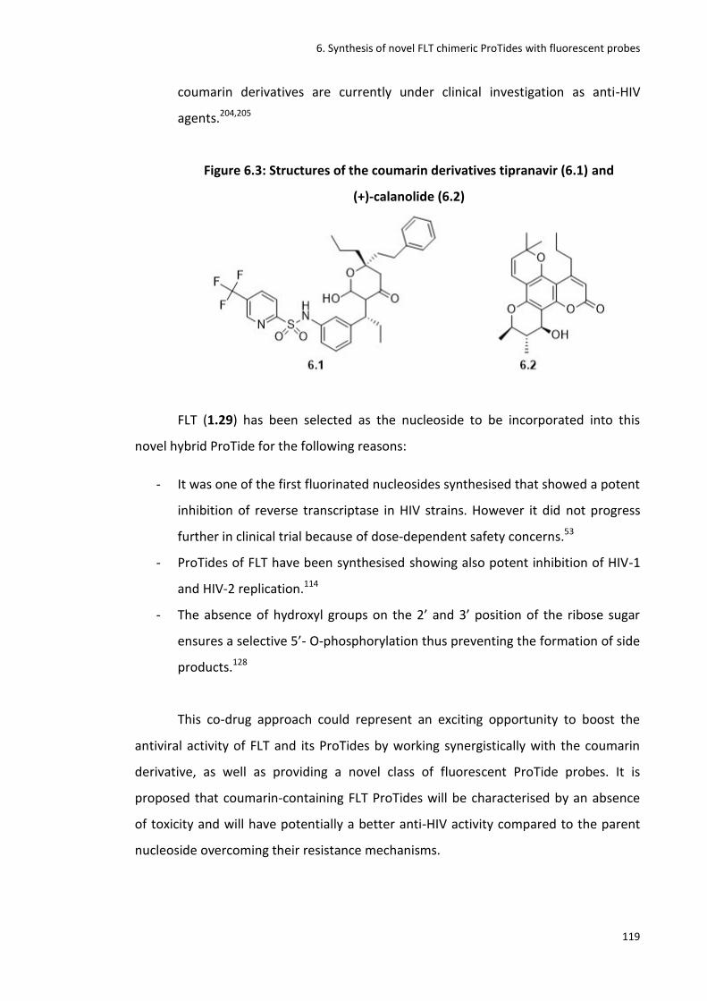
6. Synthesis of novel FLT chimeric ProTides with fluorescent probes
119
coumarin derivatives are currently under clinical investigation as anti-HIV
agents.204,205
Figure 6.3: Structures of the coumarin derivatives tipranavir (6.1) and
(+)-calanolide (6.2)
FLT (1.29) has been selected as the nucleoside to be incorporated into this
novel hybrid ProTide for the following reasons:
- It was one of the first fluorinated nucleosides synthesised that showed a potent
inhibition of reverse transcriptase in HIV strains. However it did not progress
further in clinical trial because of dose-dependent safety concerns.53
- ProTides of FLT have been synthesised showing also potent inhibition of HIV-1
and HIV-2 replication.114
- The absence of hydroxyl groups on the 2’ and 3’ position of the ribose sugar
ensures a selective 5’- O-phosphorylation thus preventing the formation of side
products.128
This co-drug approach could represent an exciting opportunity to boost the
antiviral activity of FLT and its ProTides by working synergistically with the coumarin
derivative, as well as providing a novel class of fluorescent ProTide probes. It is
proposed that coumarin-containing FLT ProTides will be characterised by an absence
of toxicity and will have potentially a better anti-HIV activity compared to the parent
nucleoside overcoming their resistance mechanisms.

6. Synthesis of novel FLT chimeric ProTides with fluorescent probes
120
6.2 Synthesis of FLT chimeric ProTides with fluorescent
probes
The synthesis of coumarin-derivatives of FLT ProTides was accomplished
according to the general scheme shown in Figure 6.4.
Figure 6.4: General synthetic scheme for FLT-coumarin derivative ProTides
Reagents and conditions: a) POCl3, Et3N, -78°C to rt, 1h, 86-86%; b) L-alanine ester salt, anh. CH2Cl2,
Et3N, -78°C to rt, 2h, 71-81%; c) Phosphorochloridate, tBuMgCl, anh. THF, 0°C to rt, 16 h, 5-11%.
Two coumarin derivatives have been chosen as starting materials to form the
phosphorochloridate, the 4-methylumbelliferone (4-MU) (6.3a, R = Me) and the 7-
hydroxy-4-(trifluoromethyl)coumarin (4-TF) (6.3b, R = CF3).
4-MU (6.3a), besides being a pH-sensitive fluorescent indicator, is also currently
used for the treatment of biliary spasm. It inhibits hyaluronic acid synthesis that is a
factor often associated with diseases such as tumour progression.205–207 4-MU is
indeed under investigation as an anticancer agent because of its ability to inhibit
proliferation, migration and invasion of cancer cells.207,208 It also showed activity
against chronic hepatitis C and B and is currently in Phase II clinical trials under the
registered name of Heparvit.209 Recent studies also suggest its potential for future HIV-
1 reservoir eradication strategies.210

6. Synthesis of novel FLT chimeric ProTides with fluorescent probes
121
4-TF (6.3b) is used as fluorescent probe for the imaging of enzymes such as
endoplasmic reticulum carboxylesterases, enzymes involved in the cleavage of ester-
containing prodrugs.211,212 Although 4-TF has not shown any therapeutic activity, its
application to ProTide strategy could be useful for in vivo imaging of the ProTide
activation pathway into cells.
The first step consisted of the synthesis of phosphorochloridates according to
the standard procedure shown in Chapter 3 and 5.128 In this case the appropriate
coumarin derivative was coupled with phosphorus oxychloride using triethylamine as
the base, and HCl scavenger. Anhydrous conditions were again essential for the
successful outcome of the reaction therefore all the synthetic procedures have been
conducted under nitrogen atmosphere. Compounds were obtained in good yields and
were used for the next step without further purification (Figure 6.5).
Figure 6.5: Synthesis of the phosphorodichloridates
Reagents and conditions: a) POCl3, Et3N, -78°C to rt, 1h, 86-88%.
Step b consisted of the formation of the phosphorochloridates by reacting the
phosphorodichloridates with the appropriate L-alanine esters using TEA as the base
and anhydrous conditions as per the previously reported standard procedure.128 The
reaction mixture was monitored by 31P NMR and after 1h at rt the conversion of the
starting material into the product was complete, therefore the reaction was stopped,
reduced under vacuum and stored in the freezer under a strict nitrogen atmosphere
(Figure 6.6).
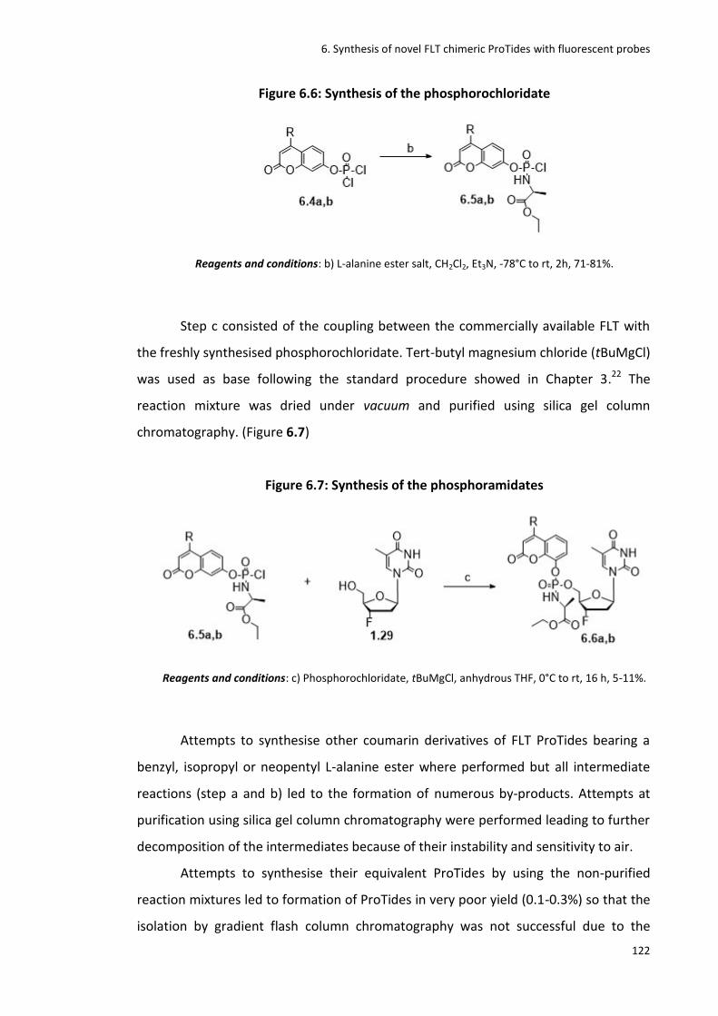
6. Synthesis of novel FLT chimeric ProTides with fluorescent probes
122
Figure 6.6: Synthesis of the phosphorochloridate
Reagents and conditions: b) L-alanine ester salt, CH2Cl2, Et3N, -78°C to rt, 2h, 71-81%.
Step c consisted of the coupling between the commercially available FLT with
the freshly synthesised phosphorochloridate. Tert-butyl magnesium chloride (tBuMgCl)
was used as base following the standard procedure showed in Chapter 3.22 The
reaction mixture was dried under vacuum and purified using silica gel column
chromatography. (Figure 6.7)
Figure 6.7: Synthesis of the phosphoramidates
Reagents and conditions: c) Phosphorochloridate, tBuMgCl, anhydrous THF, 0°C to rt, 16 h, 5-11%.
Attempts to synthesise other coumarin derivatives of FLT ProTides bearing a
benzyl, isopropyl or neopentyl L-alanine ester where performed but all intermediate
reactions (step a and b) led to the formation of numerous by-products. Attempts at
purification using silica gel column chromatography were performed leading to further
decomposition of the intermediates because of their instability and sensitivity to air.
Attempts to synthesise their equivalent ProTides by using the non-purified
reaction mixtures led to formation of ProTides in very poor yield (0.1-0.3%) so that the
isolation by gradient flash column chromatography was not successful due to the
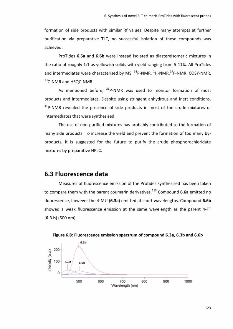
6. Synthesis of novel FLT chimeric ProTides with fluorescent probes
123
formation of side products with similar Rf values. Despite many attempts at further
purification via preparative TLC, no successful isolation of these compounds was
achieved.
ProTides 6.6a and 6.6b were instead isolated as diastereisomeric mixtures in
the ratio of roughly 1:1 as yellowish solids with yield ranging from 5-11%. All ProTides
and intermediates were characterised by MS, 31P-NMR, 1H-NMR,19F-NMR, COSY-NMR,
13C-NMR and HSQC-NMR.
As mentioned before, 31P-NMR was used to monitor formation of most
products and intermediates. Despite using stringent anhydrous and inert conditions,
31P-NMR revealed the presence of side products in most of the crude mixtures of
intermediates that were synthesised.
The use of non-purified mixtures has probably contributed to the formation of
many side products. To increase the yield and prevent the formation of too many by-
products, it is suggested for the future to purify the crude phosphorochloridate
mixtures by preparative HPLC.
6.3 Fluorescence data Measures of fluorescence emission of the Protides synthesised has been taken
to compare them with the parent coumarin derivatives.213 Compound 6.6a emitted no
fluorescence, however the 4-MU (6.3a) emitted at short wavelengths. Compound 6.6b
showed a weak fluorescence emission at the same wavelength as the parent 4-FT
(6.3.b) (500 nm).
Figure 6.8: Fluorescence emission spectrum of compound 6.3a, 6.3b and 6.6b
6. Synthesis of novel FLT chimeric ProTides with fluorescent probes
124
These preliminary data suggest that these ProTides could be used as
fluorescent Probes as they possess different UV absorption fluorescence wavelengths
and intensity compared to their parent coumarin derivatives. Therefore, a change in
UV fluorescence could be observed during metabolism in vivo when the coumarin is
cleaved off the ProTide. This change could be useful to visualise ProTide uptake in vivo,
and thus confirm whether ProTides are activated inside the cells as speculated.
6.4 Conclusion In conclusion two novel ProTides of FLT containing coumarin derivatives and L-
alanine esters as masking groups (6.6a, 6.6b), have been synthesised as models of a
new class of ProTide fluorescent probes. These novel compounds are characterised by
fluorescence properties that may allow the visualisation of their uptake thus
demonstrating their activation mechanism. This co-drug approach with coumarin
derivatives could also boost the anti-HIV properties of the parent NA FLT. Another six
ProTides were synthesised but couldn’t be isolated because of the formation of
numerous side products, instability issues, poor yield and difficult purification process.
All ProTides and by-products have been characterised by MS and NMR spectroscopy.
It is recommended for the future to test these compounds in in vitro models to
assess their efficacy and potency as anti-HIV and more generally as antiviral agents. If
efficacious, they could be optimised by altering the coumarin moiety with other
coumarins known to have anti-HIV activity or simply by increasing the lipophilicity of
the parent coumarin to facilitate their cell permeability. This could increase the anti-
HIV efficacy of FLT ProTides even further. The drawback of this co-drug approach, as
for any other co-drug approaches, could be the issue with dosing.196,214 Therapeutic
doses for FLT and for the coumarin derivative could be different so that the
administration of both drugs as one co-drug may not be feasible. Nevertheless, UV
absorbance and fluorescence data could still be used to visualise uptake and activation
mechanism of this class of novel FLT chimeric ProTides with fluorescent probes in in
vivo models.

7. Conclusions and future work
125
7. Conclusions and future work
7.1 General conclusions and future perspective
The phosphoramidate (ProTide) technology developed by McGuigan and co-
workers from 1995 onwards is considered to be the most effective pro-nucleotide
approach currently used in the clinic.17 Fluorinated nucleosides and ProTides represent
a significant class of anticancer and antiviral agents used in the clinic and in clinical
development. Fluorine plays a crucial rule in the optimization of these clinical agents
and candidates by improving their selectivity, toxicity, potency, and pharmacokinetic
and pharmacodynamic properties.34
In order to visualize pharmaceutical and biological effects of fluorinated
ProTides directly in vivo, [18F]-radiolabelled ProTides have been synthesised for the
first time as PET imaging probes. These imaging biomarkers represent a proof of
concept for the ProTide strategy and a model system to visualize their pharmaceutical
and biological effects directly in vivo. Two [18F]-radiolabelled ProTides have been
synthesised as model standards for two classes, the 2’- and the 3’-fluorinated ProTides.
The 3’-[18F]FLT ProTide (2.1) was synthesised via a convenient late stage
[18F]fluorination of ad hoc designed precursor molecules. Among the five precursor
molecules synthesised, the nosyl derivative furnished the desired product in poor
yields therefore protection of the NH moieties of the molecule with BOC protecting
groups was performed to minimize the formation of undesired by-products. This
protection increased remarkably the yield of the reaction furnishing the desired
radiolabelled BOC protected compound that was then deprotected to finally furnish
the desired target compound 2.1 in good radiochemical yield and radiochemical purity.
The 2’-[18F]FIAU ProTide (2.2) was synthesised following a different synthetic
approach involving an early stage [18F]-fluorination of the sugar moiety with an overall
synthetic time of 4h.
This was the first time that [18F]-radiolabelled ProTides have ever been
synthesised. Although these results have to be regarded as first trials of their synthesis,
these radiolabelled probes could provide evidence for the in vivo behaviour of this

7. Conclusions and future work
126
class of compounds answering key questions about their metabolism and uptake
directly.
For the future it is recommended to investigate different set-ups such as the
use of different automated synthetic modules, microwave-assisted synthesis and
microfluidic systems in order to validate the potential clinical application of these
radiosyntheses. These radiolabelled ProTides not only could represent a first step
towards ProTide personalised medicine but, on the other hand, could also represent a
novel class of PET imaging diagnostic probes.
Other imaging techniques could also be used to provide insight into ProTide
activation pathways in vivo.215 For this reason novel ProTides of FLT containing
coumarin derivatives (6.6a, 6.6b), have been synthesised as new potential fluorescent
probes and therapeutic agents. By incorporating the coumarin moiety, these new FLT
ProTides are characterised by fluorescence properties, which should allow the
visualisation of their uptake thus demonstrating their activation mechanism. As some
coumarin derivatives are also characterised by interesting antiviral therapeutic
properties, this could also be considered as a co-drug approach that could boost the
anti HIV properties of the parent NA FLT. Future work will investigate their efficacy and
potency as anti-HIV and more generally as antiviral agents. Where efficacious,
optimization of the coumarin moiety with respect to anti-HIV properties could be
performed in order to boost their therapeutic activity. UV absorbance and
fluorescence data will be used to visualise uptake and the activation mechanism of this
class of novel FLT chimeric ProTides with fluorescent probes within in vivo models.
Besides the use of imaging techniques to study the mechanism of action of
ProTides, a novel class of cold, non-fluorescent 2’-deoxy-2’-fluoro-5-iodouridine
ProTides (2.9, 4.3, 5.8a-l) have been synthesised and tested as antiviral agents in
response to the high demand of novel antiviral agents against the outbreak of new
viral diseases or against novel strains of well-known viruses.178 Initial antiviral tests
against Zika virus did not show any activity of these fluorinated uridine based ProTides
when compared to Ribavirin, but further investigation on other viruses such as Dengue
and HSV are currently ongoing.

8. Experimental
127
8. Experimental
8.1 General information
8.1.1 Analytics 1H NMR spectra were measured on a Bruker Avance Ultra Shield spectrometer
(500 MHz) at ambient temperature. Data were recorded as follows: chemical shift in
δppm from internal standard tetramethylsilane; multiplicity (s =singlet; d = doublet; t =
triplet; m = multiplet); coupling constant (Hz), integration and assignment. 13C NMR
spectra were measured on a Bruker Avance Ultra Shield spectrometer (125 MHz) at
ambient temperature. Chemical shifts were recorded in ppm from the solvent
resonance employed as the internal standard (e.g. CDCl3 at 77.00 ppm). 31P NMR
spectra were recorded on a Bruker Avance Ultra Shield spectrometer (202 MHz) at
ambient temperature. 19F NMR spectra were recorded on a Bruker Avance Ultra Shield
(474 MHz) spectrometer at ambient temperature. Highperformance liquid
chromatography (HPLC) analysis was conducted on an Agilent Technology 1200 Series
System with an analytical reversed phase column (Phenomenex Synergi 4μ Hydro- RP
80, C-18, 4.6×250 mm) and with a semi-preparative reversed phase column
(Phenomenex Synergi 4μ Hydro-RP 80, C-18, 10×250 mm) both coupled with a RAM/
RAM Model 4 detector (Lablogic System, Ltd.) for radio-HPLC purposes. Thin-layer
chromatography (TLC) was conducted on pre-coated silica gel 60 GF254 plates.
Preparative TLC plates (20x20 cm, 500-2000 silica) were purchased from Merck. Radio-
TLC was performed on a Canberra UNISPEC 125. Mass spectrometry analysis (LC-ESI-
MS) was performed on a Bruker micro-TOF or on an Agilent 6430 T-Quadrupole
spectrometer. High-resolution mass spectrometry (ESI-HRMS was performed at the
EPSRC National Mass Spectrometry facility at Swansea University.
8.1.2 Solvents and chemicals All the anhydrous solvents and reagents were purchased from Sigma-Aldrich
and they were used without further purification. Some nucleosides used as starting
materials were purchased from Carbosynth Ltd. UK. Fluka silica gel (35-70 mm) was

8. Experimental
128
used as stationary phase for column chromatography. Radioactive material was
purified with QMA and Al cartridges (Waters corp., Milford, MA, USA).
8.1.3 Radioactive source and equipment 18F-Fluoride was produced in an IBA Cyclon 18/9 cyclotron using the 18O(p,n)18F
nuclear reaction. 18O-Enriched water (enrichment grade 98%, 2.2 mL, Nukem GmbH
Germany) was irradiated with 18MeV protons. An Eckert & Ziegler module system was
used to perform the radiofluorinations. A vacuum pump N820 (Neuberger, Freiburg,
Germany) was used for the drying process. Smartline pump 100 126 was used for the
semi-preparative HPLC attached to the Eckert & Ziegler module system.
8.2 Procedures Described below are the standard procedures followed in the synthesis of L-
alanine ester salts, phosphorochloridates and the ProTides.128
8.2.1 Standard procedure A1: synthesis of L-alanine ester
hydrochloride salts Thionyl chloride (2.0 mol/eq) was added dropwise to a stirring solution of the
appropriate alcohol (15.0 mol/eq) at 0°C under nitrogen atmosphere. The reaction was
allowed to stir at 0°C for 0.5 hr then slowly allowed to warm to room temperature (r.t).
L-alanine (1.0 mol/eq) was added dropwise and the mixture was heated under reflux
overnight. The reaction was monitored by TLC using 2-propanol/water (7:3) and
ninhydrin solution. The solvent was removed in vacuo with last traces of solvent
removed by azeotropic co-evaporation to give a solid crude product.
8.2.2 Standard procedure A2: synthesis of L-alanine ester
sulfonate salts A mixture of L-alanine (1.0 mol/eq), the appropriate alcohol (15.0 mol/eq) and
para-toluene sulfonic acid (p-TSA) monohydrate (1.1 mol/eq) in toluene was heated

8. Experimental
129
under reflux overnight using the Dean-Stark apparatus. The reaction was monitored by
TLC using 2-propanol/water (7:3) and ninhydrin solution. The solvent was then
removed in vacuo with last traces of solvent removed by azeotropic co-evaporation to
give a solid crude product.
8.2.3 Standard procedure B: synthesis of aryloxy
phosphorochloridates To a solution of an appropriate amino acid ester salt (1.0molequivalent) and
phenyl dichlorophosphate (1.0 mol/eq) in anhydrous CH2Cl2, anhydrous triethylamine
(TEA) (2.0 mol/eq) was added dropwise at -78°C under a nitrogen atmosphere. The
reaction mixture was then stirred at -78°C for 1h, then at r.t for 3h. The reaction was
monitored by 31P-NMR. The solvent was removed under reduced pressure and the
residue triturated with anhydrous diethyl ether. The precipitate was filtered under
nitrogen and the filtrate was concentrated to produce the product as an oil.
8.2.4 Standard procedure C: synthesis of phosphoramidates via
NMI To a stirring solution of an appropriate nucleoside (1.0 mol equivalent) in
anhydrous tetrahydrofuran (THF) at 0°C under nitrogen atmosphere, N-
methylimidazole (NMI) (5.0mol equivalent) was added and the reaction was stirred for
30 min. A solution of an appropriate phosphorochloridate (3.0mol equivalent)
dissolved in THF was then added dropwise and the reaction was stirred at r.t for 18h.
The reaction was monitored by TLC with CH2Cl2/CH3OH (9:1). The solvent was then
removed in vacuo to give a residue that was re-dissolved in CH2Cl2 and washed twice
with 0.5N HCl. The organic phase was dried over MgSO4, filtered and reduced to
dryness to give a crude product that was purified by flash column chromatography
eluting with CH2Cl2/CH3OH in different proportions depending on the specific analogue
being synthesised.
8. Experimental
130
8.2.5 Standard procedure D: synthesis of phosphoramidates via
tBuMgCl
To a stirring solution of the appropriate nucleoside (1 mol/eq) in anhydrous
THF under a nitrogen atmosphere, tBuMgCl (1.1 mol/eq) was added, and stirred for 10
min. The appropriate phosphorochloridate (1.2 mol/eq) dissolved in anhydrous THF
was added to the reaction mixture dropwise. The reaction mixture was left stirring at
rt for 24 h, then anhydrous THF solvent removed by evaporation in vacuo at rt. The
crude product was purified by flash column chromatography eluting with
CH2Cl2/CH3OH in different proportions depending on the specific analogue being
synthesised.

8. Experimental
131
8.3 Spectroscopic data
8.3.1Experimental section from Chapter 3
(3R,5R)-3-(hydroxymethyl)-8-methyl-2,3-dihydro-5H,9H-2,5-methanopyrimido[2,1-
b][1,5,3]dioxazepin-9-one. (3.5)
MF: C10H12N2O4
MW: 224.22
A mixture of thymidine (1.28)(1eq, 0.250g, 1.032mmol) and triphenylphosphine
(Ph3P) (2eq, 0.541g, 2.064mmol) was suspended in anhydrous acetonitrile (20mL) and
cooled down to -15°C. To this mixture diisoproprylazadicarboxylate (DIAD) (2eq,
0.406mL, 0.417g, 2.64 mmol) was added dropwise maintaining the temperature
below -5°C with vigorous stirring. After the addition the reaction was left to stir for 5h
at 0°C. Following this, the mixture was again cooled down to -20°C and ethyl acetate
(20mL) was added at the same temperature and stirred for other 15min. The white
precipitate formed, was collected by Buchner filtration and washed with cold ethyl
acetate. The filtrate solution was reduced to dryness and the resulting compound was
purified by silica g e l column chromatography using 9 0 % CH2Cl2-10%CH3OH as
eluent to obtain the product 3.5 as a white solid. Yield: 52%. Rf: 0.5.
1H-NMR (500 MHz, DMSO-d6): δppm 7.55 (d, J=1.2Hz, 1H, ArH), 5.80 (d, J=3.9Hz, 1H,
H-1’), 5.23 (brs, 1H, H-3’), 5.01 (t, 1H, C-5’OH), 4.20 (m, 1H, H-4’), 3.51 (m, 2H, H-5’, H-
5”), 2.55 (d, J=1.2Hz, H-2’,1H), 2.47 (ddd, J1,8=19Hz, J1,4=6.7Hz, J1,2=3Hz, 1H, H-2”), 1.76
(d, J=1.1Hz, 3H, CH3). , .
Note: Data agrees with previously published papers.216
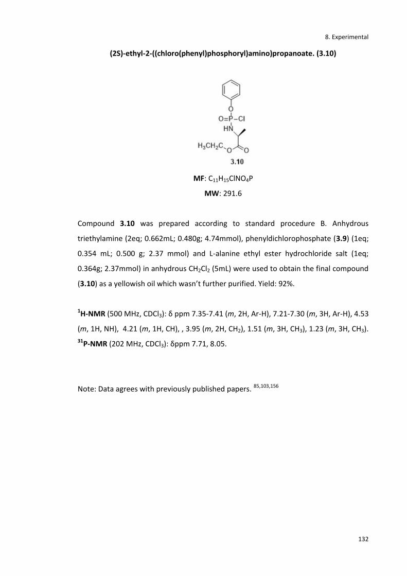
8. Experimental
132
(2S)-ethyl-2-((chloro(phenyl)phosphoryl)amino)propanoate. (3.10)
MF: C11H15ClNO4P
MW: 291.6
Compound 3.10 was prepared according to standard procedure B. Anhydrous
triethylamine (2eq; 0.662mL; 0.480g; 4.74mmol), phenyldichlorophosphate (3.9) (1eq;
0.354 mL; 0.500 g; 2.37 mmol) and L-alanine ethyl ester hydrochloride salt (1eq;
0.364g; 2.37mmol) in anhydrous CH2Cl2 (5mL) were used to obtain the final compound
(3.10) as a yellowish oil which wasn’t further purified. Yield: 92%.
1H-NMR (500 MHz, CDCl3): δ ppm 7.35-7.41 (m, 2H, Ar-H), 7.21-7.30 (m, 3H, Ar-H), 4.53
(m, 1H, NH), 4.21 (m, 1H, CH), , 3.95 (m, 2H, CH2), 1.51 (m, 3H, CH3), 1.23 (m, 3H, CH3).
31P-NMR (202 MHz, CDCl3): δppm 7.71, 8.05.
Note: Data agrees with previously published papers. 85,103,156
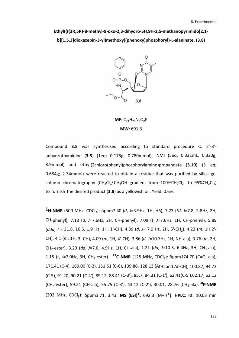
8. Experimental
133
Ethyl((((3R,5R)-8-methyl-9-oxo-2,3-dihydro-5H,9H-2,5-methanopyrimido[2,1-
b][1,5,3]dioxazepin-3-yl)methoxy)(phenoxy)phosphoryl)-L-alaninate. (3.8)
MF: C21H26N3O8P
MW: 691.3
Compound 3.8 was synthesised according to standard procedure C. 2’-3’-
anhydrothymidine (3.5) (1eq; 0.175g; 0.780mmol), NMI (5eq; 0.331mL; 0.320g;
3.9mmol) and ethyl(2chloro(phenyl)phosphorylamino)propanoate (3.10) (3 eq;
0.684g; 2.34mmol) were reacted to obtain a residue that was purified by silica gel
column chromatography (CH2Cl2/ CH3OH gradient from 100%CH2Cl2 to 95%CH2Cl2)
to furnish the desired product (3.8) as a yellowish oil. Yield: 0.6%.
1H-NMR (500 MHz, CDCl3): δppm7.40 (d, J=3.9Hz, 1H, H6), 7.23 (td, J=7.8, 2.8Hz, 2H,
CH-phenyl), 7.13 (d, J=7.6Hz, 2H, CH-phenyl), 7.09 (t, J=7.6Hz, 1H, CH-phenyl), 5.89
(ddd, J = 31.8, 16.5, 1.9 Hz, 1H, 1'-CH), 4.39 (d, J= 7.0 Hz, 2H, 5'-CH2), 4.22 (m, 1H,2'-
CH), 4.1 (m, 1H, 3'-CH), 4.09 (m, 1H, 4'-CH), 3.86 (d, J=10.7Hz, 1H, NH-ala), 3.76 (m, 2H,
CH2-ester), 3.29 (dd, J=7.0, 4.9Hz, 1H, CH-ala), 1.21 (dd, J=10.3, 6.4Hz, 3H, CH3-ala),
1.15 (t, J=7.0Hz, 3H, CH3-ester). 13C-NMR (125 MHz, CDCl3): δppm174.70 (C=O, ala),
171.41 (C-4), 169.00 (C-2), 151.51 (C-6), 139.86, 128.13 (Ar-C and Ar-CH), 100.87, 94.73
(C-5), 91.20, 90.21 (C-4'), 89.12, 88.41 (C-3'), 85.7, 84.31 (C-1'), 63.41(C-5'),62.17, 62.12
(CH3-ester), 59.21 (CH-ala), 55.75 (C-3’), 43.12 (C-2’), 30.01, 28.76 (CH3-ala). 31P-NMR
(202 MHz, CDCl3): δppm3.71, 3.43. MS (ESI)+: 692.3 [M+H+]. HPLC: Rt: 10.03 min

8. Experimental
134
(95%). *Gradient: (0’) 95%H2O/5%CH3CN - (5’) 50% H2O/50%CH3CN- (15’) 50%
H2O/50% CH3CN- (20’) 95% H2O/5% CH3CN].
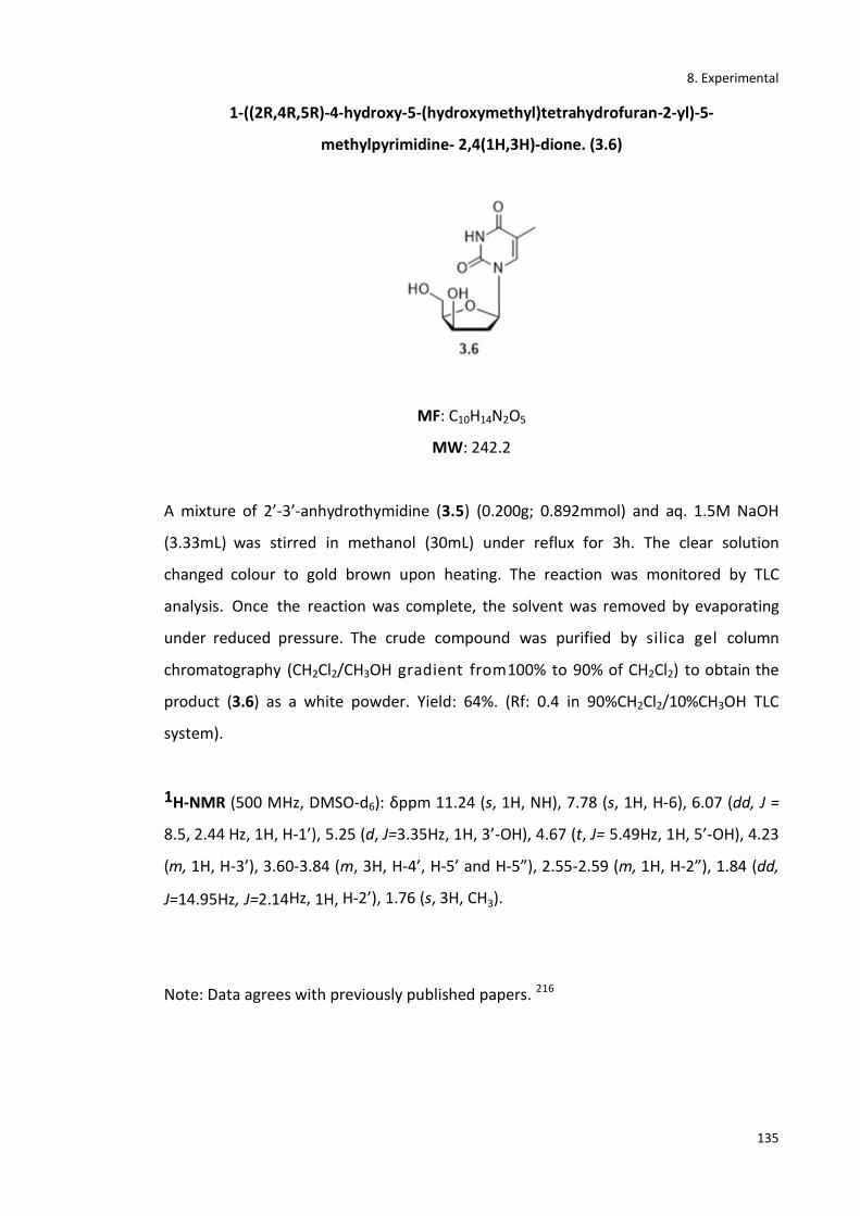
8. Experimental
135
1-((2R,4R,5R)-4-hydroxy-5-(hydroxymethyl)tetrahydrofuran-2-yl)-5-
methylpyrimidine- 2,4(1H,3H)-dione. (3.6)
MF: C10H14N2O5
MW: 242.2
A mixture of 2’-3’-anhydrothymidine (3.5) (0.200g; 0.892mmol) and aq. 1.5M NaOH
(3.33mL) was stirred in methanol (30mL) under reflux for 3h. The clear solution
changed colour to gold brown upon heating. The reaction was monitored by TLC
analysis. Once the reaction was complete, the solvent was removed by evaporating
under reduced pressure. The crude compound was purified by silica gel column
chromatography (CH2Cl2/CH3OH gradient from100% to 90% of CH2Cl2) to obtain the
product (3.6) as a white powder. Yield: 64%. (Rf: 0.4 in 90%CH2Cl2/10%CH3OH TLC
system).
1H-NMR (500 MHz, DMSO-d6): δppm 11.24 (s, 1H, NH), 7.78 (s, 1H, H-6), 6.07 (dd, J =
8.5, 2.44 Hz, 1H, H-1’), 5.25 (d, J=3.35Hz, 1H, 3’-OH), 4.67 (t, J= 5.49Hz, 1H, 5’-OH), 4.23
(m, 1H, H-3’), 3.60-3.84 (m, 3H, H-4’, H-5’ and H-5”), 2.55-2.59 (m, 1H, H-2”), 1.84 (dd,
J=14.95Hz, J=2.14Hz, 1H, H-2’), 1.76 (s, 3H, CH3).
Note: Data agrees with previously published papers. 216

8. Experimental
136
(2S)-methyl2-(((((2R,3R,5R)-3-hydroxy-5-(5-methyl-2,4-dioxo-3,4-dihydropyrimidin-
1(2H)- yl)tetrahydrofuran-2-yl)methoxy)(phenoxy)phosphoryl)amino)propanoate.
(3.7b)
MF: C20H26N3O9P
MW: 483.4
Compound 3.7b was synthesised according to the standard procedure C. 1-(2-deoxy-β-
lyxofuranoxyl thymidine) (3.6) (1eq; 0.109g; 0.45mmol), NMI (5eq; 0.177mL; 0.183g,
2.23mmol) and methyl-(2-chloro(phenyl)phosphorylamino)propanoate (5.7a) (3eq;
0.350g; 1.34mmol) were reacted to obtain a residue that was purified by silica gel
column chromatography (CH2Cl2/CH3OH gradient from 100% CH2Cl2 to 95% CH2Cl2) to
afford the product 3.7b as a white solid. Yield: 9.3%.
1H NMR(500 MHz, CDCl3): δppm 7.56 (d, J = 7 Hz, 1H, H-6), 7.33-7.41 (m, 2H, Ar- H),
7.22-7.31 (m, 3H, Ar-H), 6.24-6.31 (m, 1H, H-1’), 5.25 (m, 1H, 3’-OH), 4.98 (m, 1H, H-
3’), 4.30-4.51 (m, 2H, NH, H-4’), 3.94-4.02 (m, 1H, CH), 3.67 (s, 3H, OCH3), 3.38 (d, J =
16 Hz, 2H, H-5’, H-5”), 2.45-2.47 (m, 1H, H-2”), 2.07-2.23 (m, 1H, H-2’), 1.850 (d, J = 10
Hz, 3H, CH3 -Thy), 1.15-1.38 (m, 3H, CH3 -Ala). 31P NMR: (202 MHz, CDCl3): δppm 5.34,
4.89

8. Experimental
137
(2S)-ethyl2-(((((2R,3R,5R)-3-hydroxy-5-(5-methyl-2,4-dioxo-3,4-dihydropyrimidin-
1(2H)- yl)tetrahydrofuran-2-yl)methoxy)(phenoxy)phosphoryl)amino)propanoate
MF: C21H28N3O9P
MW: 497.4
Compound 3.7 was prepared according to standard procedure C. 1-(2-deoxy-β-
lyxofuranoxyl thymidine) (3.6) (1eq; 0.175g; 0.721mmol), NMI (5eq; 0.287mL; 0.297g,
3.62mmol) and ethyl-(2-chloro(phenyl)phosphorylamino)propanoate (3.10) (3eq;
0.633g; 2.17mmol) were reacted to obtain a residue that was purified by silica gel
column chromatography (97% CH2Cl2/3% CH3OH) to afford the product as a white solid.
Yield: 18.4 %. Rf: 0.4
1H-NMR (500 MHz, CDCl3): δppm7.58 (d, J=7 Hz , 1H, H-6), 7.35-7.43 (m, 2H, Ar-H),
7.20-7.29 (m, 3H, Ar-H), 6.25-6.32 (m, 1H, H-1’), 5.22 (m, 1H, 3’-OH), 4.94 (m, 1H, H-
3’), 4.31-4.52 (m, 2H, NH, H-4’), 3.95-4.03 (m, 1H, CH), 3.68 (d, J=7Hz,2H, CH2-ester),
3.35 (d, J=16Hz, 2H, H-5’, H-5”), 2.48-2.51 (m, 1H, H-2”), 2.09-2.25 (m, 1H, H-2’), 1.85
(d, J=10Hz, 3H, CH3-thy), 1.16-1.23 (m, 6H, CH3-ester). 31P-NMR (200 MHz, CDCl3):
δppm5.34, 4.98.

8. Experimental
138
(2S)-ethyl2-(((((2R,3R,5R)-5-(5-methyl-2,4-dioxo-3,4-dihydropyrimidin-1(2H)-yl)-3-
((methylsulfonyl)oxy)tetrahydrofuran-2-yl)methoxy)(phenoxy)phosphoryl)amino)
propanoate. (3.1)
MF: C22H30N3O11PS
MW: 575.5
Triethylamine (10eq; 1.06mL; 0.769g; 7.6mmol) and mesyl chloride (4 eq; 0.235mL;
0.348g; 3.04mmol) were added to a solution of the compound 3.7 (1eq; 0.378g;
0.76mmol) in dry CH2Cl2 (20mL) at 0°C. The mixture was stirred at this temperature for
additional 10 minutes before it was allowed to warm up to rt and it was stirred for 1.5 h.
After full conversion of the starting material, the mixture was diluted with sat. NaHCO3-
solution and extracted with CH2Cl2 (2x 20mL). After drying over Na2SO4, the solution
was reduced in vacuo and the resulting compound was purified by silica gel column
chromatography (CH2Cl2/CH3OH gradient from 100% CH2Cl2 to 95% CH2Cl2) to afford
the product (3.1) as a white solid. Yield: 29.5%. Rf: 0.45 in 90%CH2Cl2/10%CH3OH TLC
system.
1H NMR (500 MHz, CDCl3): δppm 8.93-8.96(s, 1H, NH, thy), 7.33-7.32 (d, J=7 Hz, 1H,
H-6), 7.24-7.28 (m, 2H, Ar-H), 7.10-7.15 (m, 3H, Ar-H), 6.23-6.21 (m, 2H, H-1’), 5.19-5.15
(s, 1H, NH-ala), 3.84-4.37 (m, 1H, H-3’; 1H, H-4’; 1H, CH; 2H, H-5’, H-5”; 4H, CH2-ala),
2.96-3.01 (s, 3H, SO2CH3), 2.72-2.75 (m, 1H, H-2”), 2.38-2.42 (m, 2H, H-2’), 1.87 (d,
J=1Hz, 3H, CH3-thy), 1.28-1.33 (m, 3H , CH3-ala), 1.19-1.16 (m, 3H, CH3-ala). 13C NMR
(125 MHz, CDCl3) δppm 173.7-173.4 (C=O, acetyl), 163.6 (C-2), 150.42 (C-1), 135.1 (C-4),
129.8 (C-2; C-6Ar), 125.2 (C-4Ar), 120.2 (C-3; C-5Ar), 111.6 (C-3), 83.5 (C-1’), 79.9(C-
8. Experimental
139
3’), 77.3 (C-4’), 63.7 (C-5’), 61.7 (CH2-Ala), 50.8 (CH-ala), 39.2 (C-2’), 38.8 (CH3-mesyl),
21.34 (CH3-ethyl), 14.04 (CH3-ala), 12.76 (CH3-thy). 31P NMR (202MHz, CDCl3): δppm
2.92, 2.63. MS(ESI+): 576.2 [M+H+]. HPLC: Rt: 13.2 min (98%). *Gradient: (0’)
95%H2O/5%CH3CN - (5’) 50% H2O/50%CH3CN- (15’) 50% H2O/50% CH3CN- (20’) 95%
H2O/5% CH3CN].
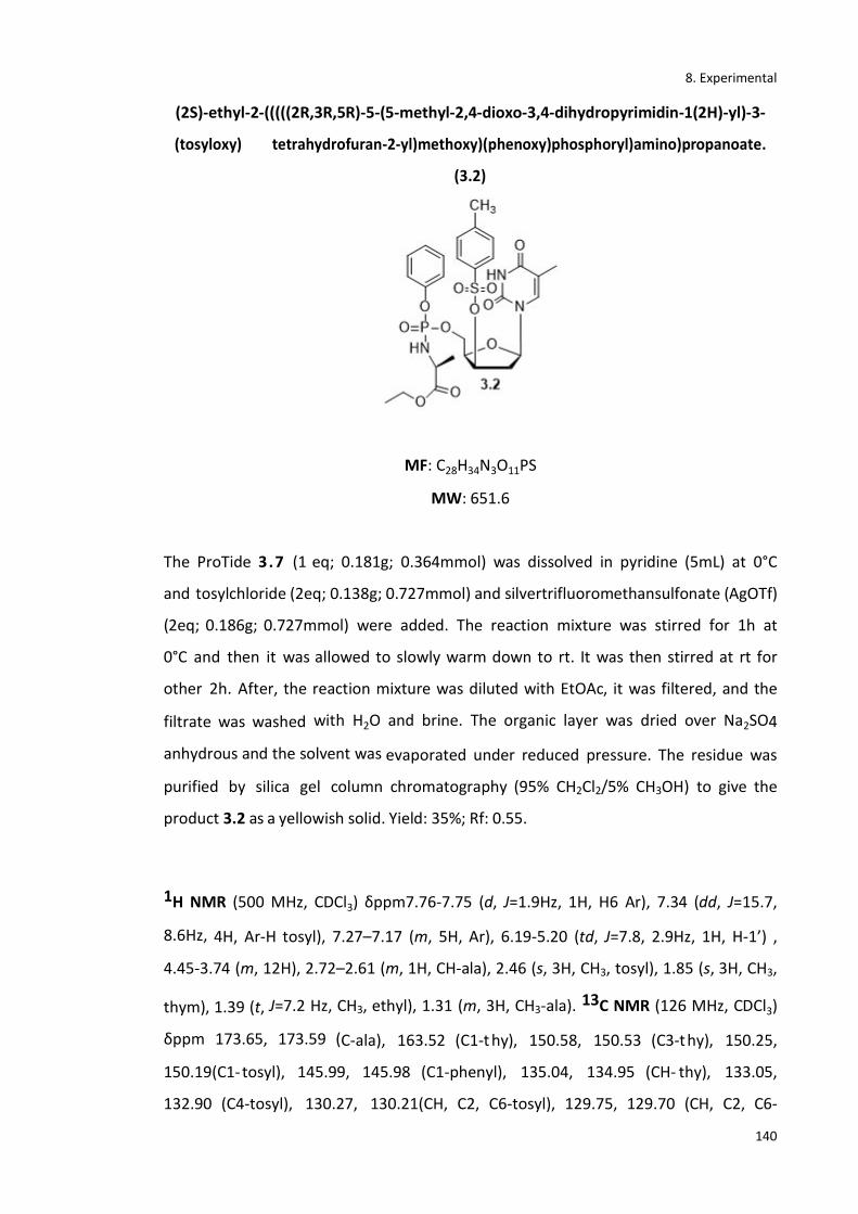
8. Experimental
140
(2S)-ethyl-2-(((((2R,3R,5R)-5-(5-methyl-2,4-dioxo-3,4-dihydropyrimidin-1(2H)-yl)-3-
(tosyloxy) tetrahydrofuran-2-yl)methoxy)(phenoxy)phosphoryl)amino)propanoate.
(3.2)
MF: C28H34N3O11PS
MW: 651.6
The ProTide 3.7 (1 eq; 0.181g; 0.364mmol) was dissolved in pyridine (5mL) at 0°C
and tosylchloride (2eq; 0.138g; 0.727mmol) and silvertrifluoromethansulfonate (AgOTf)
(2eq; 0.186g; 0.727mmol) were added. The reaction mixture was stirred for 1h at
0°C and then it was allowed to slowly warm down to rt. It was then stirred at rt for
other 2h. After, the reaction mixture was diluted with EtOAc, it was filtered, and the
filtrate was washed with H2O and brine. The organic layer was dried over Na2SO4
anhydrous and the solvent was evaporated under reduced pressure. The residue was
purified by silica gel column chromatography (95% CH2Cl2/5% CH3OH) to give the
product 3.2 as a yellowish solid. Yield: 35%; Rf: 0.55.
1H NMR (500 MHz, CDCl3) δppm7.76-7.75 (d, J=1.9Hz, 1H, H6 Ar), 7.34 (dd, J=15.7,
8.6Hz, 4H, Ar-H tosyl), 7.27–7.17 (m, 5H, Ar), 6.19-5.20 (td, J=7.8, 2.9Hz, 1H, H-1’) ,
4.45-3.74 (m, 12H), 2.72–2.61 (m, 1H, CH-ala), 2.46 (s, 3H, CH3, tosyl), 1.85 (s, 3H, CH3,
thym), 1.39 (t, J=7.2 Hz, CH3, ethyl), 1.31 (m, 3H, CH3-ala). 13C NMR (126 MHz, CDCl3)
δppm 173.65, 173.59 (C-ala), 163.52 (C1-t hy), 150.58, 150.53 (C3-thy), 150.25,
150.19(C1-tosyl), 145.99, 145.98 (C1-phenyl), 135.04, 134.95 (CH- thy), 133.05,
132.90 (C4-tosyl), 130.27, 130.21(CH, C2, C6-tosyl), 129.75, 129.70 (CH, C2, C6-
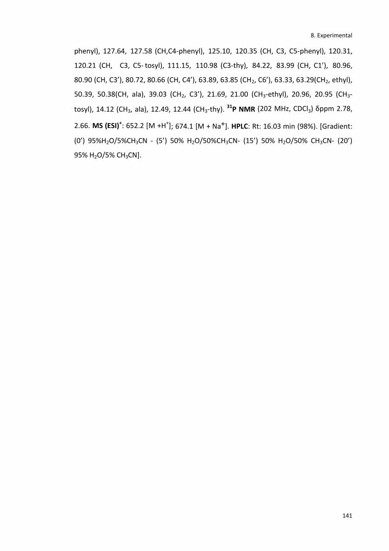
8. Experimental
141
phenyl), 127.64, 127.58 (CH,C4-phenyl), 125.10, 120.35 (CH, C3, C5-phenyl), 120.31,
120.21 (CH, C3, C5- tosyl), 111.15, 110.98 (C3-thy), 84.22, 83.99 (CH, C1’), 80.96,
80.90 (CH, C3’), 80.72, 80.66 (CH, C4’), 63.89, 63.85 (CH2, C6’), 63.33, 63.29(CH2, ethyl),
50.39, 50.38(CH, ala), 39.03 (CH2, C3’), 21.69, 21.00 (CH3-ethyl), 20.96, 20.95 (CH3-
tosyl), 14.12 (CH3, ala), 12.49, 12.44 (CH3-thy). 31P NMR (202 MHz, CDCl3) δppm 2.78,
2.66. MS (ESI)+: 652.2 [M +H+]; 674.1 [M + Na+]. HPLC: Rt: 16.03 min (98%). [Gradient:
(0’) 95%H2O/5%CH3CN - (5’) 50% H2O/50%CH3CN- (15’) 50% H2O/50% CH3CN- (20’)
95% H2O/5% CH3CN].
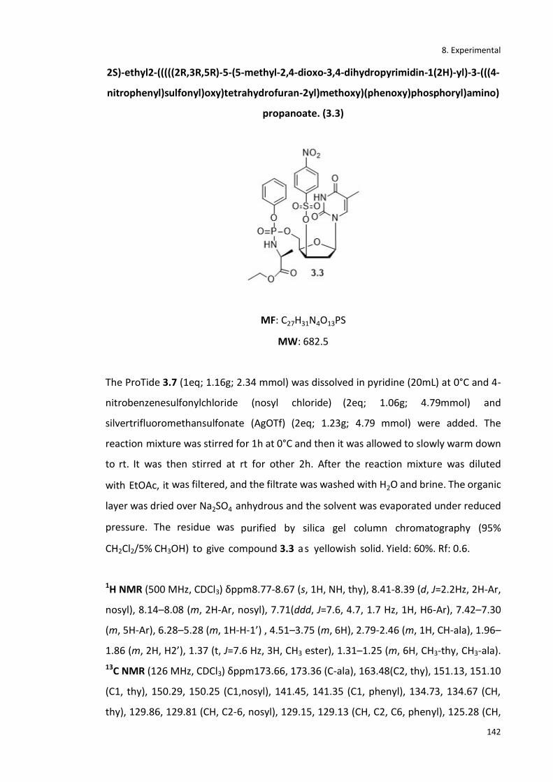
8. Experimental
142
2S)-ethyl2-(((((2R,3R,5R)-5-(5-methyl-2,4-dioxo-3,4-dihydropyrimidin-1(2H)-yl)-3-(((4-
nitrophenyl)sulfonyl)oxy)tetrahydrofuran-2yl)methoxy)(phenoxy)phosphoryl)amino)
propanoate. (3.3)
MF: C27H31N4O13PS
MW: 682.5
The ProTide 3.7 (1eq; 1.16g; 2.34 mmol) was dissolved in pyridine (20mL) at 0°C and 4-
nitrobenzenesulfonylchloride (nosyl chloride) (2eq; 1.06g; 4.79mmol) and
silvertrifluoromethansulfonate (AgOTf) (2eq; 1.23g; 4.79 mmol) were added. The
reaction mixture was stirred for 1h at 0°C and then it was allowed to slowly warm down
to rt. It was then stirred at rt for other 2h. After the reaction mixture was diluted
with EtOAc, it was filtered, and the filtrate was washed with H2O and brine. The organic
layer was dried over Na2SO4 anhydrous and the solvent was evaporated under reduced
pressure. The residue was purified by silica gel column chromatography (95%
CH2Cl2/5% CH3OH) to give compound 3.3 as yellowish solid. Yield: 60%. Rf: 0.6.
1H NMR (500 MHz, CDCl3) δppm8.77-8.67 (s, 1H, NH, thy), 8.41-8.39 (d, J=2.2Hz, 2H-Ar,
nosyl), 8.14–8.08 (m, 2H-Ar, nosyl), 7.71(ddd, J=7.6, 4.7, 1.7 Hz, 1H, H6-Ar), 7.42–7.30
(m, 5H-Ar), 6.28–5.28 (m, 1H-H-1’) , 4.51–3.75 (m, 6H), 2.79-2.46 (m, 1H, CH-ala), 1.96–
1.86 (m, 2H, H2’), 1.37 (t, J=7.6 Hz, 3H, CH3 ester), 1.31–1.25 (m, 6H, CH3-thy, CH3-ala).
13C NMR (126 MHz, CDCl3) δppm173.66, 173.36 (C-ala), 163.48(C2, thy), 151.13, 151.10
(C1, thy), 150.29, 150.25 (C1,nosyl), 141.45, 141.35 (C1, phenyl), 134.73, 134.67 (CH,
thy), 129.86, 129.81 (CH, C2-6, nosyl), 129.15, 129.13 (CH, C2, C6, phenyl), 125.28 (CH,

8. Experimental
143
C4, phe), 124.84, 124.77 (CH, C3, C5, phenyl), 120.15, 120.11 (CH, C3, C5, nosyl),
120.06, 120.02, 111.36, 111.23 (C, C3, thy), 84.14, 84.00 (CH, C1’), 80.58, 80.52 (CH,
C3’), 80.25, 80.18 (CH, C4’), 63.17, 63.14 (CH2, C5’), 62.82, 62.79(CH2, ethyl), 50.41,
50.20 (CH,ala), 39.18, 39.16 (CH2, C2’), 20.94, 20.90 (CH3, ethyl), 14.12, 14.11 (CH3, ala),
12.58, 12.56 (CH3, thy). 31P NMR (202 MHz, CDCl3) δppm2.75, 2.48. MS (ESI)+: 705.1 [M
+ Na+]. HPLC: Rt: 15.88 min (99%). *Gradient: (0’) 95%H2O/5%CH3CN - (5’) 50%
H2O/50%CH3CN- (15’) 50% H2O/50% CH3CN- (20’) 95% H2O/5% CH3CN].

8. Experimental
144
Tert-butyl 3-((2R,4R,5R)-5-(((((tert-butoxycarbonyl)((S)-1-ethoxy-1-oxopropan-2-
yl)amino)(phenoxy)phosphoryl)oxy)methyl)-4-(((4-
nitrophenyl)sulfonyl)oxy)tetrahydrofuran-2-yl)-5-methyl-2,6-dioxo-3,6-
dihydropyrimidine-1(2H)-carboxylate. (3.4)
MF: C37H47N4O17PS
MW: 882.83
The nosylate precursor (3.3) (1eq, 0.050g, 0.073mmol) was dissolved in pyridine (6mL)
at rt under nitrogen atmosphere. Diterbutyl dicarbonate (BOC2O) (1.3eq, 0.021ml,
0.020g, 0.095mmol) was added dropwise and the reaction was allowed to stir for 3
days. The crude product was then evaporated in vacuo and was purified by silica gel
column chromatography (CH2Cl2/CH3OH gradient from 100% CH2Cl2to 95% CH2Cl2) to
obtain the product 3.4 as a yellowish oil. Yield: 56%. Rf: 0.67 in 90% CH2Cl2/10%
CH3OH as TLC system.
1H NMR (500 MHz, CDCl3) δppm8.47–8.36 (d, 2H, J=2.2Hz, Ar, nosyl), 8.17–8.08 (m,
2H, Ar, nosyl), 7.38 (m, 1H, Ar), 7.32–7.24 (m, 4H), 7.17 (s, 1H, Ar), 6.31–6.24 (m, 1H-H-
1’), 4.51–3.75 (m, 7H), 2.78-2.43 (m, 1H, CH-Ala), 2.31–2.23 (m, 1H-H-2’), 2.01 (m, 3H,
CH3,thy), 1.53–1.49 (m, 9H, CH3, tert-butyl), 1.44 (m, 9H, CH3, tert-butyl), 1.38 (t,
J=7.6Hz, 3H, CH3, ester), 1.31 (m, 3H, CH3,ala). 13C NMR (126 MHz, CDCl3)
δppm175.50, 174.29 (C,ala), 161.32 (C2, thy), 150.13, 150.08 (C1, thy), 150.02, 150.00
(C1, nosyl) 143.51, 142.21 (C1, phenyl), 132.71, 132.23 (CH, thy), 130.68, 129.99 (CH,
C2-C6, nosyl), 129.34, 129.5 (CH, C2,C6, phenyl), 126.28 (CH, C4, phe), 125.79,
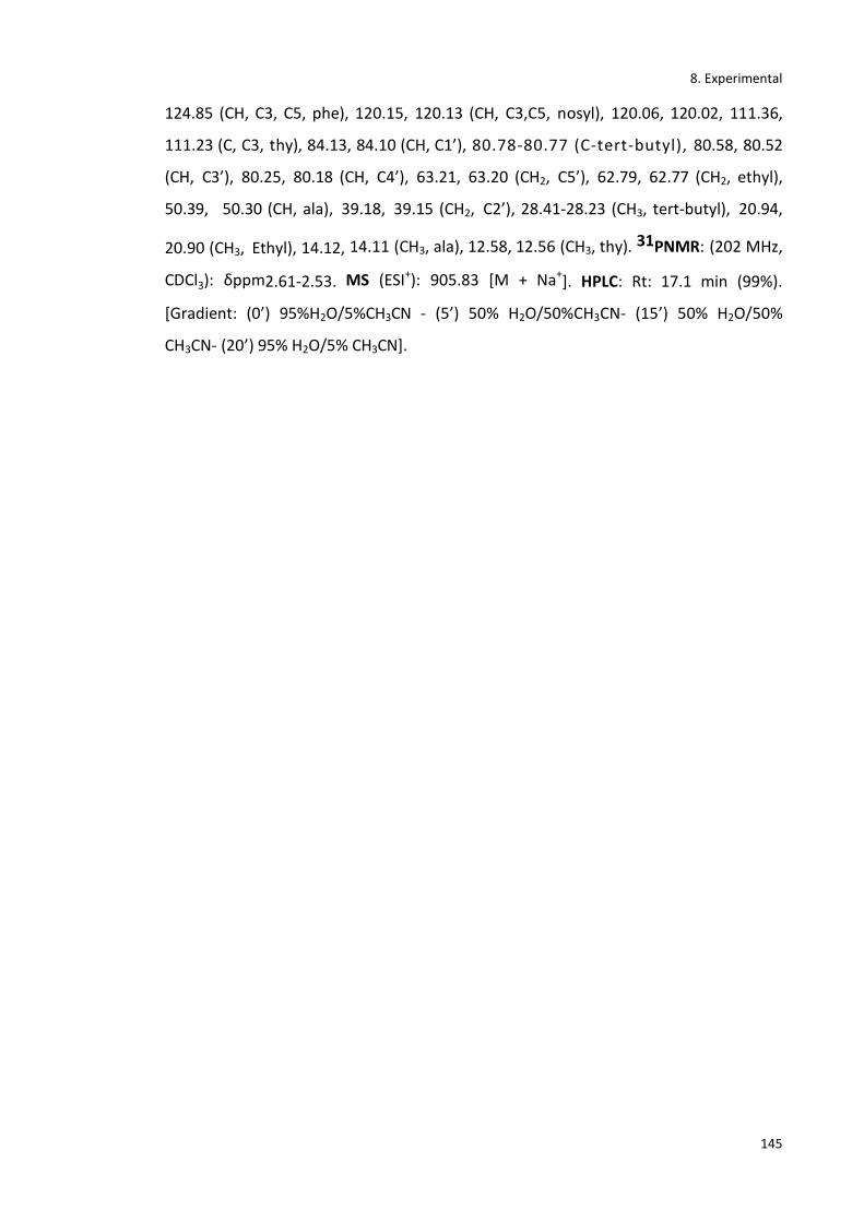
8. Experimental
145
124.85 (CH, C3, C5, phe), 120.15, 120.13 (CH, C3,C5, nosyl), 120.06, 120.02, 111.36,
111.23 (C, C3, thy), 84.13, 84.10 (CH, C1’), 80.78-80.77 (C-tert-butyl), 80.58, 80.52
(CH, C3’), 80.25, 80.18 (CH, C4’), 63.21, 63.20 (CH2, C5’), 62.79, 62.77 (CH2, ethyl),
50.39, 50.30 (CH, ala), 39.18, 39.15 (CH2, C2’), 28.41-28.23 (CH3, tert-butyl), 20.94,
20.90 (CH3, Ethyl), 14.12, 14.11 (CH3, ala), 12.58, 12.56 (CH3, thy). 31PNMR: (202 MHz,
CDCl3): δppm2.61-2.53. MS (ESI+): 905.83 [M + Na+]. HPLC: Rt: 17.1 min (99%).
*Gradient: (0’) 95%H2O/5%CH3CN - (5’) 50% H2O/50%CH3CN- (15’) 50% H2O/50%
CH3CN- (20’) 95% H2O/5% CH3CN].
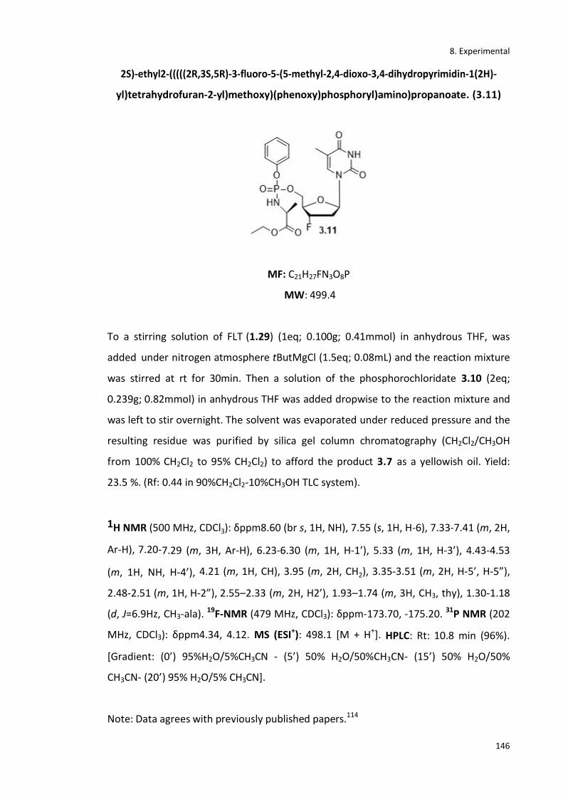
8. Experimental
146
2S)-ethyl2-(((((2R,3S,5R)-3-fluoro-5-(5-methyl-2,4-dioxo-3,4-dihydropyrimidin-1(2H)-
yl)tetrahydrofuran-2-yl)methoxy)(phenoxy)phosphoryl)amino)propanoate. (3.11)
MF: C21H27FN3O8P
MW: 499.4
To a stirring solution of FLT (1.29) (1eq; 0.100g; 0.41mmol) in anhydrous THF, was
added under nitrogen atmosphere tButMgCl (1.5eq; 0.08mL) and the reaction mixture
was stirred at rt for 30min. Then a solution of the phosphorochloridate 3.10 (2eq;
0.239g; 0.82mmol) in anhydrous THF was added dropwise to the reaction mixture and
was left to stir overnight. The solvent was evaporated under reduced pressure and the
resulting residue was purified by silica gel column chromatography (CH2Cl2/CH3OH
from 100% CH2Cl2 to 95% CH2Cl2) to afford the product 3.7 as a yellowish oil. Yield:
23.5 %. (Rf: 0.44 in 90%CH2Cl2-10%CH3OH TLC system).
1H NMR (500 MHz, CDCl3): δppm8.60 (br s, 1H, NH), 7.55 (s, 1H, H-6), 7.33-7.41 (m, 2H,
Ar-H), 7.20-7.29 (m, 3H, Ar-H), 6.23-6.30 (m, 1H, H-1’), 5.33 (m, 1H, H-3’), 4.43-4.53
(m, 1H, NH, H-4’), 4.21 (m, 1H, CH), 3.95 (m, 2H, CH2), 3.35-3.51 (m, 2H, H-5’, H-5”),
2.48-2.51 (m, 1H, H-2”), 2.55–2.33 (m, 2H, H2’), 1.93–1.74 (m, 3H, CH3, thy), 1.30-1.18
(d, J=6.9Hz, CH3-ala). 19F-NMR (479 MHz, CDCl3): δppm-173.70, -175.20. 31P NMR (202
MHz, CDCl3): δppm4.34, 4.12. MS (ESI+): 498.1 [M + H+]. HPLC: Rt: 10.8 min (96%).
*Gradient: (0’) 95%H2O/5%CH3CN - (5’) 50% H2O/50%CH3CN- (15’) 50% H2O/50%
CH3CN- (20’) 95% H2O/5% CH3CN].
Note: Data agrees with previously published papers.114

8. Experimental
147
Tert-butyl-3-((2R,4S,5R)-5-((((N-((S)-1-ethoxy-1-oxopropan-2-yl)-3,3-
dimethylbutanamido)(phenoxy)phosphoryl)oxy)methyl)-4-(fluoro-
18F)tetrahydrofuran-2-yl)-5-methyl-2,6-dioxo-3,6-dihydropyrimidine-1(2H)-
carboxylate. (3.12)
MF: C32H4518FN3O11P
MW: 696.70
Aqueous 18F (2.5 GBq), produced by the cyclotron by the nuclear reaction, was
trapped in a QMA cartridge before it was eluted with an aqueous solution of KHCO3 and
Kryptofix in CH3CN. The resulting 18F/KHCO3/Kryptofix complex was dried by co-
evaporation with CH3CN anhydrous (2x 1 mL) under reduced pressure and a stream of
nitrogen. A solution of the precursor 3.4 (20mg) in CH3CN anhydrous (1mL) was added
ad the reaction was stirred for 30 mins at 95°C. The resulting mixture was passed
through an alumina cartridge to finally obtain the radiolabelled product 3.12. The
reaction mixture was analyzed by analytical HPLC and radio TLC. (Analytical HPLC: (0’)
95%H2O/5%CH3CN - (5’) 50% H2O/50%CH3CN- (15’) 50% H2O/50%CH3CN- (20’)
95%H2O/5%CH3CN); Rt: 15 min.
CH3CNDMSO
Bkg 1
Region 1
Bkg 2
Region 2
Bkg 3
0:00 5:00 10:00 15:00 mm:ss
0.0
10000.0
20000.0
30000.0
Counts

8. Experimental
148
Ethyl ((((2R,3S,5R)-3-(fluoro-18F)-5-(5-methyl-2,4-dioxo-3,4-dihydropyrimidin-1(2H)-
yl)tetrahydrofuran-2-yl)methoxy)(phenoxy)phosphoryl)-L-alaninate. (2.1)
MF: C21H2718FN3O8P
MW: 498.43
The BOC protected [18F]-radiolabelled ProTide (3.12) was transferred into a V-vial. A
solution of 2N HCl (1mL) was added and it was allowed to stir for 10 mins. It was then
neutralised with NaOH 2N. The compound was then purified by semi-preparative HPLC
and was eluted after 35 minutes at a flow rate of 3.5 mL/min using 70% H2O/30%
MeCN as the mobile phase. The HPLC solvent was then removed from the mixture
under a stream of nitrogen. The radioactive product was taken up in saline and
subsequently flushed through a sterility filter to obtain a sterile and clean aqueous
solution of [18F]FLT ProTide (2.1). RCY of 15-30% (n=5, decay-corrected from end of
bombardment (EoB)), with high radiochemical purities (97%) and specific activities of
1800 mCi/μmol. The total synthesis time was 130 min after the end of bombardment
(EoB). The reaction mixture was analyzed by analytical HPLC and radio TLC.
(Analytical HPLC: (0’) 95%H2O/5%CH3CN - (5’) 50% H2O/50%CH3CN- (15’) 50%
H2O/50%CH3CN- (20’) 95%H2O/5%CH3CN); Rt: 10.8 min).

8. Experimental
149
8.3.2 Experimental section from chapter 4
2’deoxy-2’-α-F-5-iodouridine. (1.66)
MW: 372.1
MF: C9H10FIN2O4
To a solution of 2’-β -fluoro-2’-deoxyuridine (4.4) (2eq; 2g; 8.12mmol) in anhydrous
acetonitrile (50mL), iodine (1.2eq; 1.24g; 4.87mmol) and ceric ammonium nitrate
(CAN) (1eq; 2.23g; 4.062mmol) were added. The reaction mixture was stirred at 75°C
for 1h and it was then quenched with a saturated solution of Na2S2O3 and
concentrated. The residue was re-dissolved in ethyl acetate and washed twice with
brine. The organic layer was dried under MgSO4, filtered and concentrated in vacuo to
give compound 1.66 as a pale yellow solid. Yield: 60%. HPLC: Rt: 2.3 min (95%).
98%H2O/2%CH3CN.
1H-NMR (500 MHz, DMSO-d6): δppm11.69 (s, 1H, NH), 8.53 (s, 1H, 6-CH), 5.86 (d,
J=15.8Hz, 1H, 1'-CH), 5.60 (d, J=6.4Hz, 1H, 3'-OH), 5.39 (t, J=4.5Hz, 1H, 3'-CH), 5.04 (dd,
J=53.2, 4.1Hz, 1H, 2'-CH), 4.18 (ddd, J=23.4, 11.4, 7.2Hz, 1H, 4'-CH), 3.90 (d, J=8.2Hz,
1H, 5'-OH), 3.85–3.79 (m, 1H, 5'-CH), 3.63–3.58 (m, 1H, 5'-CH). 13C-NMR (125 MHz,
DMSO-d6): δppm167.88 (C=O), 165.01 (C=O), 145.02 (C-6), 125.19 (C-2'), 121.26 (C-1'),
115.81 (C-4'), 61.11 (C-5), 57.30 (C-3'), 45.87 (C-5'). 19F-NMR (470 MHz, DMSO-d6):
δppm-202.09.
Note: Data agrees with previously published papers.155

8. Experimental
150
Benzyl (chloro(phenoxy)phosphoryl)-L-alaninate. (4.5)
MW: 353.73
MF: C16H17ClNO4P
Compound 4.5 was prepared according to the standard procedure B. Anhydrous
triethylamine (2eq; 1.26mL; 0.918g; 9.08mmol), phenyl dichlorophosphate (3.9)(1eq;
0.678mL; 0.958 g; 4.54 mmol) and L-alanine benzyl ester hydrochloride salt ( 1eq;
1.50g; 4.54mmol) were reacted to afford compound 4.5 as a yellowish oil which wasn’t
further purified. Yield: 88%.
1H NMR (500MHz, CDCl3): δppm7.54–7.47 (m, 7H, Ar-H), 7.46–7.40 (m, 3H, Ar-H), 5.27
(d, J=8.4Hz, 2H, CH2-ester), 4.69 (d, J=9.9Hz, 1H, NH), 4.13 (dd, J=34.4, 29.8 Hz, 1H, CH-
ala), 1.52 (m, 3H, CH3-ala). 31P NMR (202 MHz, CDCl3): δppm8.03-7.75.
Note: Data agrees with previously published papers. 85,103,156

8. Experimental
151
Benzyl ((((2R,3R,4S,5R)-4-fluoro-3-hydroxy-5-(5-iodo-2,4-dioxo-3,4-dihydropyrimidin-
1(2H)-yl)tetrahydrofuran-2-yl)methoxy)(phenoxy)phosphoryl)-L-alaninate. (4.3)
MF: C25H26FIN3O9P
MW: 689.3
Compound 4.3 was prepared according to the standard procedure C. 1-(3-fluoro-4-
hydroxy-5-(hydromethyl)tetrahydrofuran-2-yl)-5-iodopyrimidine-2,4(1H,3H)-dione
(1.66) (1eq; 0.400g; 1.07mmol), NMI (5eq; 0.424mL; 0.439g; 5.35mmol), benzyl-2-
(chloro(phenoxy)phosphorylamino)propanoate (4.5)(3eq; 1.05g; 3.22mmol) were
reacted to obtain a residue that was purified by silica gel column chromatography
(CH2Cl2/CH3OH from 100% CH2Cl2 to 95% CH2Cl2) to obtain the product 4.3 as a yellowish
oil. Yield: 10%.
1H NMR (500 MHz, CDCl3): δppm10.59 (s, 1H, 3-NH), 7.89 (s, 1H, 6-CH), 7.53–7.48 (m,
2H, CH-phenyl), 7.45 (t, J=8Hz, 2H, CH-phenyl), 7.41-7.38 (m, 2H, CH-benz), 7,17-7.15
(m, 2H, CH-benz), 7.13 (t, 8Hz, 1H, CH-benzyl), 7.12 (t, J=7.4 Hz, 1H, CH-phenyl), 5.99
(m, 1H, 1'-CH), 5.79 (dd, J=47.6, 4.6 Hz, 1H, 2'-CH), 5.14 (m, 2H, CH2-benz), 4.90 (m, 2H,
5'-CH2), 4.39 (m, 1H, 4'-CH), 4.27 (m, 1H, 3'-CH), 4.19 (m, 1H, 3'-OH), 4.02 (m, 1H, NH-
ala), 3.99 (m, 1H, CH-ala), 1.29 (d, J=7.0Hz, 3H, CH3-ala). 13C NMR (125 MHz, CDCl3):
δppm 171.9 (C=O, ala), 168.12 (C-4), 144.18 (C-2), 149.0 (C-phenyl), 147.91 (C-
benzyl), 145.5, 145.1 (C-6), 128.31-121.48 (Ar-C), 94.3 (C-5), 92.10 (C-4'), 89.13 (C-2'),
83.6, 82.97 (C-1'), 81.08, 80.97 (C-ester), 69.61, 68.14 (C-3'), 67.23 (C-5'), 52.8 (CH-ala),
21.98 (CH3-ala). 19F NMR (470 MHz, CDCl3): δ-200.91, -201.15. 31P NMR (202 MHz,

8. Experimental
152
CDCl3): δppm3.90, 3.86. MS (ESI)+: 690.3 [M + H+]. HPLC: Rt: 13.4 min (97%). [Gradient:
(0’) 95%H2O/5%CH3CN - (5’) 50% H2O/50%CH3CN- (15’) 50% H2O/50% CH3CN- (20’)
95% H2O/5% CH3CN].

8. Experimental
153
(2R,3S,4R,5R)-5-((benzoyloxy)methyl)-3-(fluoro-18F)tetrahydrofuran-2,4-diyl
dibenzoate. (4.2)
MF: C26H2118FO7
MW: 463.4
Aqueous 18F (4.11 GBq), produced by the cyclotron by the nuclear reaction, was
trapped in a QMA cartridge before it was eluted with an aqueous solution of KHCO3 and
Kryptofix in CH3CN. The resulting 18F/KHCO3/Kryptofix complex was dried by co-
evaporation with CH3CN anhydrous (2x 1mL) under reduced pressure and a stream of
nitrogen. A solution of the triflate precursor of the sugar (4.1) (20 mg) in CH3CN
anhydrous (1 mL) was added and the reaction was stirred for 30 mins at 95°C. The
resulting mixture was passed through an alumina cartridge to finally obtain the
radiolabelled product that was used for next step without any further purification. The
reaction mixture was analyzed by analytical HPLC and radio TLC. (Analytical HPLC:
100% CH3CN; Rt: 8.3 min).
Note: Data agrees with previously published papers.155
0:00 5:00 10:00 15:00 mm:ss
0.0
5000.0
10000.0
15000.0
20000.0
25000.0
Counts

8. Experimental
154
5-iodo-2,4-bis((trimethylsilyl)oxy)pyrimidine. (4.7)
MF: C10H19IN2O2Si2
MW: 382.3
To a solution of 5-iodouracil (4.6) (1eq; 10mg; 0.042mmol) in dichloroethane
(500μL), hexamethyldisilazane (11.4eq; 100μL; 0.0774mg; 0.479mmol) and TMSOTf
(13.1eq; 100μl, 0.123mg; 0.549mmol) were added into a 4mL vial on a hot plate
placed into a hot cell. The mixture was stirred for 2h at 85°C. The crude mixture was
used directly for next step without further purification. The purity of the compound
was proved by analytical HPLC (88% H2O/12% CH3CN, Rt= 6.9 min) and LC-MS ([M+H+]:
383.2).
Note: Data agrees with previously published papers.155

8. Experimental
155
18FIAU
1-((2R,3S,4R,5R)-3-(fluoro-18F)-4-hydroxy-5-(hydroxymethyl)tetrahydrofuran-2-yl)-5-
iodopyrimidine-2,4(1H,3H)-dione. (1.16)
MF: C9H1018FN2O5
MW: 371.0
Compound synthesised with the E&Z modular lab (4.2), was delivered directly from
the unit to the vial containing 2-4-bis(trimethyl silyl)-5-iodo uracil (4.7). The mixture
was then heated at 85°C for 60 min. To this mixture, 0.5M of NaOMe in CH3OH (1mL)
was added and the reaction was stirred at 85°C for other 5 min. The precipitate was
reconstituted in water (1mL) and neutralized with 6N HCl. The reaction mixture was
then analyzed by analytical HPLC showing the formation of the 2 anomers α and β of
the 2’-deoxy-2’-fluoro-5-iodouridine. (Analytical HPLC: 98% H2O/2% CH3CN; Rt : α
anomer 2.1 min; β anomer 2.9 min). The anomeric mixture was then purified by semi-
preparative HPLC and the target compound (1.16) was eluted after 7.3mins at a flow
rate of 3.5mL/min using 20% CH3CN/ 80% H2O as the mobile phase to obtain the final
compound which was analysed by analytical HPLC. (Analytical HPLC: 98% H2O/2%
CH3CN; Rt: 2.1min).
Note: Data agrees with previously published papers.155
0:00 5:00 10:00 15:00 mm:ss
0.0
500.0
1000.0
1500.0
Counts
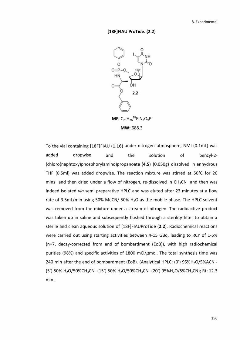
8. Experimental
156
[18F]FIAU ProTide. (2.2)
MF: C25H2618FIN3O9P
MW: 688.3
To the vial containing [18F]FIAU (1.16) under nitrogen atmosphere, NMI (0.1mL) was
added dropwise and the solution of benzyl-2-
(chloro(naphtoxy)phosphorylamino)propanoate (4.5) (0.050g) dissolved in anhydrous
THF (0.5ml) was added dropwise. The reaction mixture was stirred at 50°C for 20
mins and then dried under a flow of nitrogen, re-dissolved in CH3CN and then was
indeed isolated via semi preparative HPLC and was eluted after 23 minutes at a flow
rate of 3.5mL/min using 50% MeCN/ 50% H2O as the mobile phase. The HPLC solvent
was removed from the mixture under a stream of nitrogen. The radioactive product
was taken up in saline and subsequently flushed through a sterility filter to obtain a
sterile and clean aqueous solution of [18F]FIAUProTide (2.2). Radiochemical reactions
were carried out using starting activities between 4-15 GBq, leading to RCY of 1-5%
(n=7, decay-corrected from end of bombardment (EoB)), with high radiochemical
purities (98%) and specific activities of 1800 mCi/μmol. The total synthesis time was
240 min after the end of bombardment (EoB). (Analytical HPLC: (0’) 95%H2O/5%ACN -
(5’) 50% H2O/50%CH3CN- (15’) 50% H2O/50%CH3CN- (20’) 95%H2O/5%CH3CN); Rt: 12.3
min.

8. Experimental
157
0:00 5:00 10:00 15:00 mm:ss
0.0
100.0
200.0
300.0
400.0
Counts
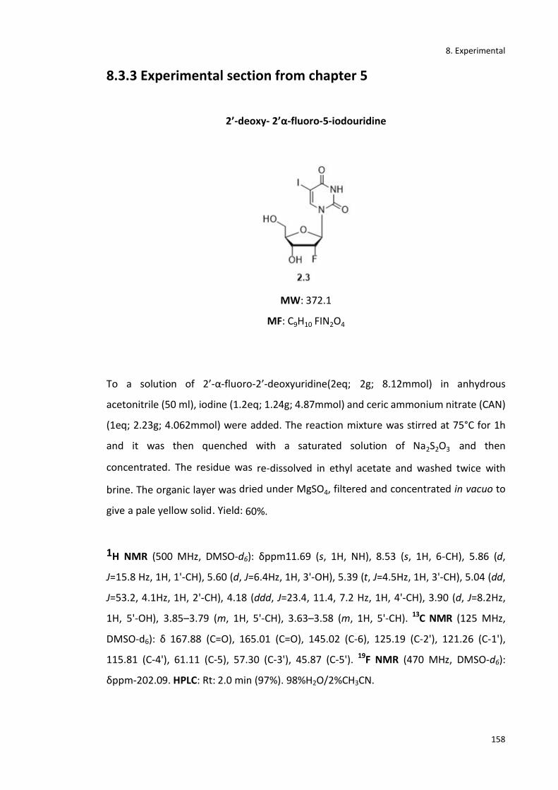
8. Experimental
158
8.3.3 Experimental section from chapter 5
2’-deoxy- 2’α-fluoro-5-iodouridine
MW: 372.1
MF: C9H10 FIN2O4
To a solution of 2’-α-fluoro-2’-deoxyuridine(2eq; 2g; 8.12mmol) in anhydrous
acetonitrile (50 ml), iodine (1.2eq; 1.24g; 4.87mmol) and ceric ammonium nitrate (CAN)
(1eq; 2.23g; 4.062mmol) were added. The reaction mixture was stirred at 75°C for 1h
and it was then quenched with a saturated solution of Na2S2O3 and then
concentrated. The residue was re-dissolved in ethyl acetate and washed twice with
brine. The organic layer was dried under MgSO4, filtered and concentrated in vacuo to
give a pale yellow solid. Yield: 60%.
1H NMR (500 MHz, DMSO-d6): δppm11.69 (s, 1H, NH), 8.53 (s, 1H, 6-CH), 5.86 (d,
J=15.8 Hz, 1H, 1'-CH), 5.60 (d, J=6.4Hz, 1H, 3'-OH), 5.39 (t, J=4.5Hz, 1H, 3'-CH), 5.04 (dd,
J=53.2, 4.1Hz, 1H, 2'-CH), 4.18 (ddd, J=23.4, 11.4, 7.2 Hz, 1H, 4'-CH), 3.90 (d, J=8.2Hz,
1H, 5'-OH), 3.85–3.79 (m, 1H, 5'-CH), 3.63–3.58 (m, 1H, 5'-CH). 13C NMR (125 MHz,
DMSO-d6): δ 167.88 (C=O), 165.01 (C=O), 145.02 (C-6), 125.19 (C-2'), 121.26 (C-1'),
115.81 (C-4'), 61.11 (C-5), 57.30 (C-3'), 45.87 (C-5'). 19F NMR (470 MHz, DMSO-d6):
δppm-202.09. HPLC: Rt: 2.0 min (97%). 98%H2O/2%CH3CN.
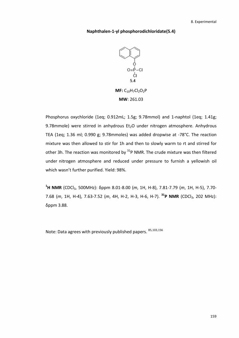
8. Experimental
159
Naphthalen-1-yl phosphorodichloridate(5.4)
MF: C10H7Cl2O2P
MW: 261.03
Phosphorus oxychloride (1eq; 0.912mL; 1.5g; 9.78mmol) and 1-naphtol (1eq; 1.41g;
9.78mmole) were stirred in anhydrous Et2O under nitrogen atmosphere. Anhydrous
TEA (1eq; 1.36 ml; 0.990 g; 9.78mmoles) was added dropwise at -78°C. The reaction
mixture was then allowed to stir for 1h and then to slowly warm to rt and stirred for
other 3h. The reaction was monitored by 31P NMR. The crude mixture was then filtered
under nitrogen atmosphere and reduced under pressure to furnish a yellowish oil
which wasn’t further purified. Yield: 98%.
1H NMR (CDCl3, 500MHz): δppm 8.01-8.00 (m, 1H, H-8), 7.81-7.79 (m, 1H, H-5), 7.70-
7.68 (m, 1H, H-4), 7.63-7.52 (m, 4H, H-2, H-3, H-6, H-7). 31P NMR (CDCl3, 202 MHz):
δppm 3.88.
Note: Data agrees with previously published papers. 85,103,156

8. Experimental
160
(S)-1-(neopentyloxy)-1-oxopropan-2-aminium – chloride. (5.6a)
MF: C8H18 Cl NO2
MW: 195.6
Compound 5.6a was prepared according to standard procedure A1. Thionyl chloride
(2eq; 1.63mL, 2.66g, 22.44mmol), neopentyl alcohol (15eq; 12.88mL; 10.11g;
0.168mol), L-alanine (1eq; 1g; 0.0112mol) were reacted to give the product 5.6a a
yellowish oil which wasn’t further purified. Yield: 57%.
1H NMR (500 MHz, CDCl3): δppm8.56 (s, 3H, NH3+), 4.92 (s, 2H, CH2-neopentyl), 3.92
(m, 1H, CH-ala), 1.39 (d, J=7.5 Hz, 3H, CH3-ala), 1.21 (d, J=6.0Hz, 9H, 3xCH3-neopentyl).
Note: Data agrees with previously published papers. 85,103,156

8. Experimental
161
(S)-1-(neopentyloxy)-1-oxopropan-2-aminium - 4-methylbenzenesulfonate. (5.6b)
MF: C15H25NO5S
MW: 331.4
Compound 5.6b was prepared according to standard procedure A2. L-alanine (1eq;
0.500g; 5.61mol), 2,2-dimethyl-1-propanol (15eq; 9.07mL; 7.41g; 0.084mol) and p-
toluenesulfonic acid (1.1eq; 1.17g; 6.17mmol) were reacted to give as a product a
white solid (5.6b) which wasn’t further purified. Yield: 83%.
1H NMR (500 MHz, CDCl3): δppm8.29 (s, 3H, NH3+), 7.49 (d, J=7.9Hz, 2H, CH-tosylate),
7.13 (d, J=7.7Hz, 2H, CH-tosylate), 3.89 (m, 1H, CH-ala), 3.06 (s, 2H, CH2-ester), 2.30 (s,
3H, CH3-tosylate), 0.94 (s, 3H, CH3-ala), 0.86–0.79 (m, 9H, 3xCH3-ester).
Note: Data agrees with previously published papers. 85,103,156

8. Experimental
162
(S)-1-isopropoxy-1-oxopropan-2-aminium chloride. (5.6c)
MF: C6H14ClNO2
MW: 167.6
Compound 5.6c was prepared according to standard procedure A1. Thionyl chloride
(2eq; 1.63mL, 2.66g, 22.44mmol), 2-propanol (isopropanol) (15eq; 12.88mL; 10.11g;
0.168mol) were reacted to give 5.6c as a white solid. Yield: 89%.
1H NMR (500 MHz, CDCl3): δppm8.66 (s, 3H, NH3+), 4.98 (dt, J=12.5, 6.2Hz, 1H, CH-
ester), 3.96 (m, 1H, CH-ala), 1.41 (d, J=7.2Hz, 3H, CH3-ala), 1.24 (d, J = 6 Hz, 6H, 2xCH3, i-
propyl).
Note: Data agrees with previously published papers. 85,103,156
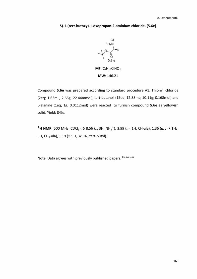
8. Experimental
163
S)-1-(tert-butoxy)-1-oxopropan-2-aminium chloride. (5.6e)
MF: C7H16ClNO2
MW: 146.21
Compound 5.6e was prepared according to standard procedure A1. Thionyl chloride
(2eq; 1.63mL, 2.66g, 22.44mmol), tert-butanol (15eq; 12.88mL; 10.11g; 0.168mol) and
L-alanine (1eq; 1g; 0.0112mol) were reacted to furnish compound 5.6e as yellowish
solid. Yield: 84%.
1H NMR (500 MHz, CDCl3): δ 8.56 (s, 3H, NH3+), 3.99 (m, 1H, CH-ala), 1.36 (d, J=7.1Hz,
3H, CH3-ala), 1.19 (s, 9H, 3xCH3, tert-butyl).
Note: Data agrees with previously published papers. 85,103,156

8. Experimental
164
(S)-1-isopropoxy-1-oxopropan-2-aminium 4-methylbenzenesulfonate. (5.6d)
MF: C13H24O5S
MW: 171.19
Compound 5.6d was prepared according to standard procedure A2. L-alanine (1eq;
0.500g; 5.61mol), isopropanol (15eq; 9.01mL; 7.39g; 0.081mol) and p-toluenesulfonic
acid (1.1eq; 1.17g; 6.17mmol) were used as reagents to give 5.6d as a white solid
which wasn’t further purified. Yield: 78%.
1H NMR (500 MHz, CDCl3): δppm8.34 (s, 3H, NH3+), 7.51 (d, J=7.8Hz, 2H, CH-tosylate),
7.09 (d, J=7.5Hz, 2H, CH-tosylate), 3.90 (m, 1H, CH-ala), 3.06 (m, 1H, CH-ester), 2.11 (s,
3H, CH3-tosylate), 1.11 (s, 3H, CH3-ala), 0.86–0.79 (d, J=7.2 Hz 2xCH3-ester).
Note: Data agrees with previously published papers. 85,103,156
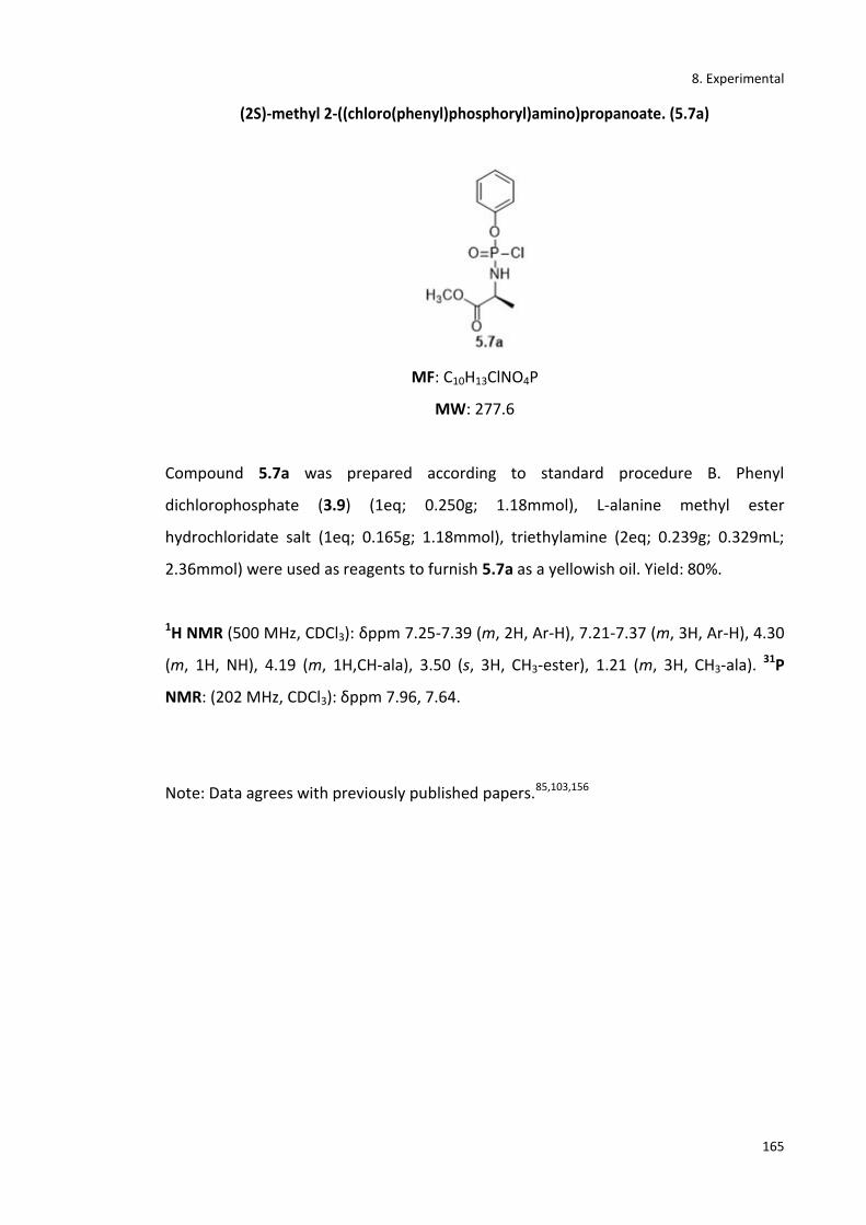
8. Experimental
165
(2S)-methyl 2-((chloro(phenyl)phosphoryl)amino)propanoate. (5.7a)
MF: C10H13ClNO4P
MW: 277.6
Compound 5.7a was prepared according to standard procedure B. Phenyl
dichlorophosphate (3.9) (1eq; 0.250g; 1.18mmol), L-alanine methyl ester
hydrochloridate salt (1eq; 0.165g; 1.18mmol), triethylamine (2eq; 0.239g; 0.329mL;
2.36mmol) were used as reagents to furnish 5.7a as a yellowish oil. Yield: 80%.
1H NMR (500 MHz, CDCl3): δppm 7.25-7.39 (m, 2H, Ar-H), 7.21-7.37 (m, 3H, Ar-H), 4.30
(m, 1H, NH), 4.19 (m, 1H,CH-ala), 3.50 (s, 3H, CH3-ester), 1.21 (m, 3H, CH3-ala). 31P
NMR: (202 MHz, CDCl3): δppm 7.96, 7.64.
Note: Data agrees with previously published papers.85,103,156
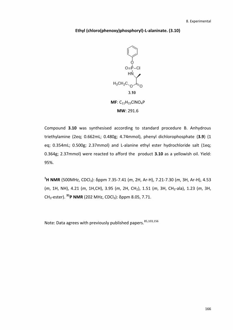
8. Experimental
166
Ethyl (chloro(phenoxy)phosphoryl)-L-alaninate. (3.10)
MF: C11H15ClNO4P
MW: 291.6
Compound 3.10 was synthesised according to standard procedure B. Anhydrous
triethylamine (2eq; 0.662mL; 0.480g; 4.74mmol), phenyl dichlorophosphate (3.9) (1
eq; 0.354mL; 0.500g; 2.37mmol) and L-alanine ethyl ester hydrochloride salt (1eq;
0.364g; 2.37mmol) were reacted to afford the product 3.10 as a yellowish oil. Yield:
95%.
1H NMR (500MHz, CDCl3): δppm 7.35-7.41 (m, 2H, Ar-H), 7.21-7.30 (m, 3H, Ar-H), 4.53
(m, 1H, NH), 4.21 (m, 1H,CH), 3.95 (m, 2H, CH2), 1.51 (m, 3H, CH3-ala), 1.23 (m, 3H,
CH3-ester). 31P NMR (202 MHz, CDCl3): δppm 8.05, 7.71.
Note: Data agrees with previously published papers.85,103,156

8. Experimental
167
Isopropyl(chloro(phenoxy)phosphoryl)-L-alaninate (5.7b)
MF: C12H17ClNO4P
MW: 305.69
Compound 5.7b was synthesised according to standard procedure B. Anhydrous
triethylamine (2eq; 1.08mL; 0.78g; 7.74mmol), phenyl dichlorophosphate (3.9) (1eq;
0.579 ml; 0.818 g; 3.87mmol) and L-alanine isopropyl ester hydrochloride salt (1eq;
0.650g; 3.87mmol) were reacted to afford 5.7b as a yellowish oil which wasn’t further
purified. Yield: 83%.
1H NMR (500 MHz, CDCl3): δppm 7.28 (m, 2H, CH-phenyl), 7.23 (m, 2H, CH-phenyl),
7.08 (m, 1H, CH-phenyl), 4.98 (m, 1H, CH-i-propyl), 4.37 (s, 1H, NH), 4.04 (m, 1H, CH-
ala), 1.30 (d, J=7.5, 3H, CH3- ala), 1.16 (dd, J=9.7, 3.5Hz, 6H, 2xCH3-ester). 31P NMR
(202MHz, CDCl3): δppm 8.18, 7.84.
Note: Data agrees with previously published papers. 85,103,156
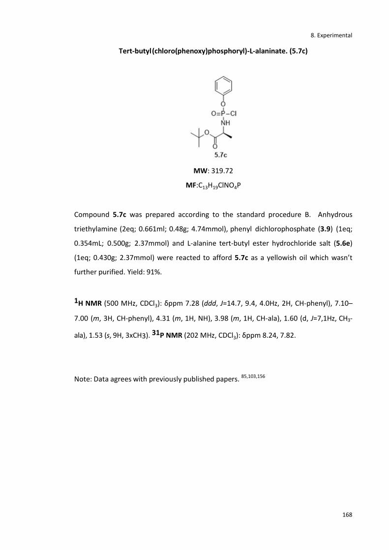
8. Experimental
168
Tert-butyl (chloro(phenoxy)phosphoryl)-L-alaninate. (5.7c)
MW: 319.72
MF:C13H19ClNO4P
Compound 5.7c was prepared according to the standard procedure B. Anhydrous
triethylamine (2eq; 0.661ml; 0.48g; 4.74mmol), phenyl dichlorophosphate (3.9) (1eq;
0.354mL; 0.500g; 2.37mmol) and L-alanine tert-butyl ester hydrochloride salt (5.6e)
(1eq; 0.430g; 2.37mmol) were reacted to afford 5.7c as a yellowish oil which wasn’t
further purified. Yield: 91%.
1H NMR (500 MHz, CDCl3): δppm 7.28 (ddd, J=14.7, 9.4, 4.0Hz, 2H, CH-phenyl), 7.10–
7.00 (m, 3H, CH-phenyl), 4.31 (m, 1H, NH), 3.98 (m, 1H, CH-ala), 1.60 (d, J=7,1Hz, CH3-
ala), 1.53 (s, 9H, 3xCH3). 31P NMR (202 MHz, CDCl3): δppm 8.24, 7.82.
Note: Data agrees with previously published papers. 85,103,156
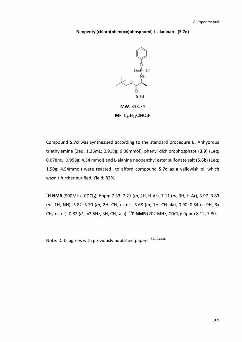
8. Experimental
169
Neopentyl(chloro(phenoxy)phosphoryl)-L-alaninate. (5.7d)
MW: 333.74
MF: C14H21ClNO4P
Compound 5.7d was synthesised according to the standard procedure B. Anhydrous
triethylamine (2eq; 1.26mL; 0.918g; 9.08mmol), phenyl dichlorophosphate (3.9) (1eq;
0.678mL; 0.958g; 4.54 mmol) and L-alanine neopenthyl ester sulfonate salt (5.6b) (1eq;
1.50g; 4.54mmol) were reacted to afford compound 5.7d as a yellowish oil which
wasn’t further purified. Yield: 82%.
1H NMR (500MHz, CDCl3): δppm 7.33–7.21 (m, 2H, H-Ar), 7.11 (m, 3H, H-Ar), 3.97–3.83
(m, 1H, NH), 3.82–3.70 (m, 2H, CH2-ester), 3.68 (m, 1H, CH-ala), 0.90–0.84 (s, 9H, 3x
CH3-ester), 0.82 (d, J=3.5Hz, 3H, CH3-ala). 31P NMR (202 MHz, CDCl3): δppm 8.12, 7.80.
Note: Data agrees with previously published papers. 85,103,156
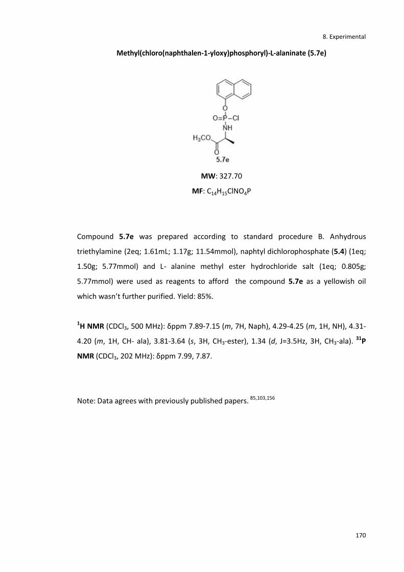
8. Experimental
170
Methyl(chloro(naphthalen-1-yloxy)phosphoryl)-L-alaninate (5.7e)
MW: 327.70
MF: C14H15ClNO4P
Compound 5.7e was prepared according to standard procedure B. Anhydrous
triethylamine (2eq; 1.61mL; 1.17g; 11.54mmol), naphtyl dichlorophosphate (5.4) (1eq;
1.50g; 5.77mmol) and L- alanine methyl ester hydrochloride salt (1eq; 0.805g;
5.77mmol) were used as reagents to afford the compound 5.7e as a yellowish oil
which wasn’t further purified. Yield: 85%.
1H NMR (CDCl3, 500 MHz): δppm 7.89-7.15 (m, 7H, Naph), 4.29-4.25 (m, 1H, NH), 4.31-
4.20 (m, 1H, CH- ala), 3.81-3.64 (s, 3H, CH3-ester), 1.34 (d, J=3.5Hz, 3H, CH3-ala). 31P
NMR (CDCl3, 202 MHz): δppm 7.99, 7.87.
Note: Data agrees with previously published papers. 85,103,156
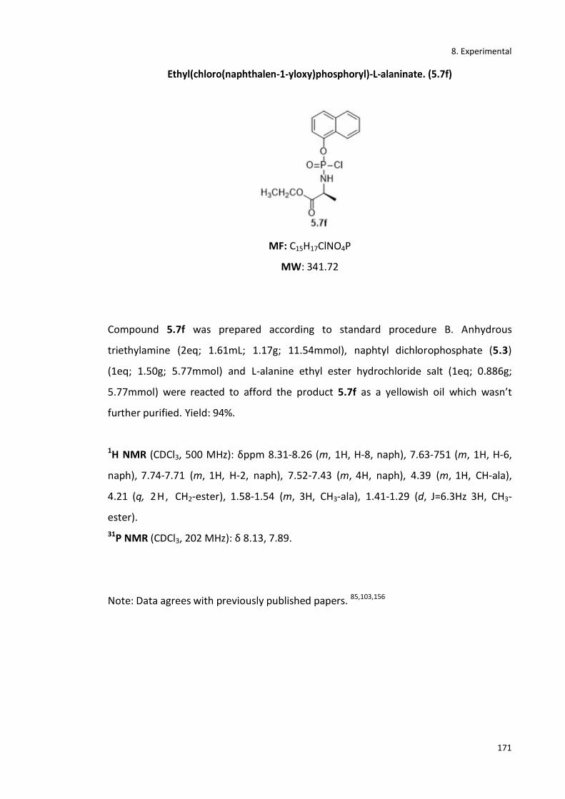
8. Experimental
171
Ethyl(chloro(naphthalen-1-yloxy)phosphoryl)-L-alaninate. (5.7f)
MF: C15H17ClNO4P
MW: 341.72
Compound 5.7f was prepared according to standard procedure B. Anhydrous
triethylamine (2eq; 1.61mL; 1.17g; 11.54mmol), naphtyl dichlorophosphate (5.3)
(1eq; 1.50g; 5.77mmol) and L-alanine ethyl ester hydrochloride salt (1eq; 0.886g;
5.77mmol) were reacted to afford the product 5.7f as a yellowish oil which wasn’t
further purified. Yield: 94%.
1H NMR (CDCl3, 500 MHz): δppm 8.31-8.26 (m, 1H, H-8, naph), 7.63-751 (m, 1H, H-6,
naph), 7.74-7.71 (m, 1H, H-2, naph), 7.52-7.43 (m, 4H, naph), 4.39 (m, 1H, CH-ala),
4.21 (q, 2H, CH2-ester), 1.58-1.54 (m, 3H, CH3-ala), 1.41-1.29 (d, J=6.3Hz 3H, CH3-
ester).
31P NMR (CDCl3, 202 MHz): δ 8.13, 7.89.
Note: Data agrees with previously published papers. 85,103,156
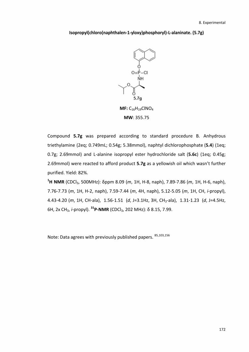
8. Experimental
172
Isopropyl(chloro(naphthalen-1-yloxy)phosphoryl)-L-alaninate. (5.7g)
MF: C16H19ClNO4
MW: 355.75
Compound 5.7g was prepared according to standard procedure B. Anhydrous
triethylamine (2eq; 0.749mL; 0.54g; 5.38mmol), naphtyl dichlorophosphate (5.4) (1eq;
0.7g; 2.69mmol) and L-alanine isopropyl ester hydrochloride salt (5.6c) (1eq; 0.45g;
2.69mmol) were reacted to afford product 5.7g as a yellowish oil which wasn’t further
purified. Yield: 82%.
1H NMR (CDCl3, 500MHz): δppm 8.09 (m, 1H, H-8, naph), 7.89-7.86 (m, 1H, H-6, naph),
7.76-7.73 (m, 1H, H-2, naph), 7.59-7.44 (m, 4H, naph), 5.12-5.05 (m, 1H, CH, i-propyl),
4.43-4.20 (m, 1H, CH-ala), 1.56-1.51 (d, J=3.1Hz, 3H, CH3-ala), 1.31-1.23 (d, J=4.5Hz,
6H, 2x CH3, i-propyl). 31P-NMR (CDCl3, 202 MHz): δ 8.15, 7.99.
Note: Data agrees with previously published papers. 85,103,156

8. Experimental
173
Benzyl(chloro(naphthalen-1-yloxy)phosphoryl)-L-alaninate (5.7h)
MF: C20H19ClNO4P
MW: 403.79
Compound 5.7h was prepared according to standard procedure B. Anhydrous
triethylamine (2eq; 0.749mL; 0.54g; 5.38mmol), naphtyl dichlorophosphate (5.3)
(1eq; 0.7g; 2.69mmol) and L-alanine benzyl ester hydrochloride salt (1eq; 0.45g;
2.69mmol) to furnish the product 5.7h. Yield: 80%
1H NMR (CDCl3, 500MHz): δppm 7.83-7.35 (m, 12H, naph and phenyl), 5.13-5.04 (m,
2H, CH2Ph), 4.41-4.31 (m, 1H, CHCH3), 1.51-1.46 (m, 3H, CH3.ala). 31P NMR (CDCl3,
202 MHz): δppm 8.14, 7.88.
Note: Data agrees with previously published papers. 85,103,156
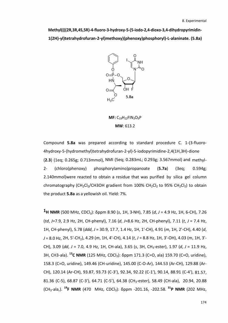
8. Experimental
174
Methyl((((2R,3R,4S,5R)-4-fluoro-3-hydroxy-5-(5-iodo-2,4-dioxo-3,4-dihydropyrimidin-
1(2H)-yl)tetrahydrofuran-2-yl)methoxy)(phenoxy)phosphoryl)-L-alaninate. (5.8a)
MF: C19H22FIN3O9P
MW: 613.2
Compound 5.8a was prepared according to standard procedure C. 1-(3-fluoro-
4hydroxy-5-(hydromethyl)tetrahydrofuran-2-yl)-5-iodopyrimidine-2,4(1H,3H)-dione
(2.3) (1eq; 0.265g; 0.713mmol), NMI (5eq; 0.283mL; 0.293g; 3.567mmol) and methyl-
2- (chloro(phenoxy) phosphorylamino)propanoate (5.7a) (3eq; 0.594g;
2.140mmol)were reacted to obtain a residue that was purified by silica gel column
chromatography (CH2Cl2/CH3OH gradient from 100% CH2Cl2 to 95% CH2Cl2) to obtain
the product 5.8a as a yellowish oil. Yield: 7%.
1H NMR (500 MHz, CDCl3): δppm 8.90 (s, 1H, 3-NH), 7.85 (d, J = 4.9 Hz, 1H, 6-CH), 7.26
(td, J=7.9, 2.9 Hz, 2H, CH-phenyl), 7.16 (d, J=8.6 Hz, 2H, CH-phenyl), 7.11 (t, J = 7.4 Hz,
1H, CH-phenyl), 5.78 (ddd, J = 30.9, 17.7, 1.4 Hz, 1H, 1'-CH), 4.91 (m, 1H, 2'-CH), 4.40 (d,
J = 8.0 Hz, 2H, 5'-CH2), 4.29 (m, 1H, 4'-CH), 4.14 (t, J = 8.8 Hz, 1H, 3'-OH), 4.03 (m, 1H, 3'-
CH), 3.09 (dd, J = 7.0, 4.9 Hz, 1H, CH-ala), 3.65 (s, 3H, CH3-ester), 1.97 (d, J = 11.9 Hz,
3H, CH3-ala). 13C NMR (125 MHz, CDCl3): δppm 171.3 (C=O, ala) 159.70 (C=O, uridine),
158.3 (C=O, uridine), 149.46 (CH-uridine), 145.00 (C-O-Ar), 144.53 (Ar-CH), 129.88 (Ar-
CH), 120.14 (Ar-CH), 93.87, 93.73 (C-3’), 92.34, 92.22 (C-1'), 90.14, 88.91 (C-4'), 81.57,
81.36 (C-5), 68.87 (C-3'), 64.71 (C-5’), 64.38 (CH3-ester), 58.49 (CH-ala), 20.94, 20.88
(CH3-ala,). 19F NMR (470 MHz, CDCl3): δppm -201.16, -202.58. 31P NMR (202 MHz,
8. Experimental
175
CDCl3): δppm 3.76, 3.30. MS (ESI)+: 636.0 [M+Na+]. HPLC: Rt: 10.1 min (97%).
*Gradient: (0’) 95%H2O/5%CH3CN - (5’) 50% H2O/50%CH3CN- (15’) 50% H2O/50%
CH3CN- (20’) 95% H2O/5% CH3CN].

8. Experimental
176
Ethyl-2-(((4-fluoro-3-hydroxy-5-(5-iodo-2,4-dioxo-3,4-dihydropyrimidin-1(2H)-
yl)tetrahydrofuran-2-yl)methoxy)(phenoxy)phosphoryl)-L-alaninate (5.8b)
MF: C20H24FIN3O9P
MW: 627.3
Compound 5.8b was prepared according to standard procedure C. 1-(3-fluoro-
4hydroxy-5-(hydromethyl)tetrahydrofuran-2-yl)-5-iodopyrimidine-2,4(1H,3H)-dione
(2.3) (1eq; 0.159g; 0.427mmol), NMI (5eq; 0.169mL; 0.175g; 2.138mmol) and ethyl-2-
(chloro(phenoxy) phosphorylamino)propanoate (3.10) (4.2eq; 0.524g; 1.79mmol) were
reacted to obtain a residue that was purified by silica gel column chromatography
(CH2Cl2/CH3OH gradient from 100% CH2Cl2 to 95% CH2Cl2) to obtain the product 5.8b
as a yellowish oil. Yield: 12%.
1H-NMR (500 MHz, CDCl3): δppm 9.82 (s, 1H, 3-NH), 7.85 (d, J=5.1 Hz, 1H, 6-CH), 7.26 –
7.21 (m, 2H, CH-phenyl), 7.16 (t, J=8.0 Hz, 2H, CH-phenyl), 7.07 (td, J=7.1, 3.3 Hz, 1H,
CH-phenyl), 5.78 (ddd, J=28.1, 17.8, 1.5 Hz, 1H, 1'-CH), 4.92 (ddd, J = 56.6, 52.5, 3.4 Hz,
1H, 2'-CH), 4.39 (d, 2H, 5'-CH2), 4.33 (m, 1H, 4'-CH), 4.25 (dd, J=11.7, 9.5 Hz, 1H, 3'-CH),
4.16 (t, J=6.7 Hz, 1H, 3'-OH), 4.05 (t, J=7.8 Hz, 2H, CH2-ester), 3.97 (m, 1H, NH-ala),
3.03 (dd, J=7.2, 4.6 Hz, 1H, CH-ala), 1.31 (m, 3H, CH3-ala), 1.15 (t, J=6.9 Hz, 3H, CH3-
ester). 13C-NMR (125 MHz, CDCl3): δppm 173.62 (C=O, ala), 160.29, 160.25 (C-4),
150.36 (C-2), 149.90, 149.88 (C-phenyl), 145.07, 144.63 (C-6), 129.83 - 120.25 (Ar-C),
93.87, 93.76 (C-5), 92.37, 92.25 (C-4'), 89.88, 89.05 (C-2'), 81.54, 81.35 (C-1'), 68.23 (C-

8. Experimental
177
3'), 65.08, 65.04 (C-5',), 61.85, 61.83 (CH2-ester), 53.46 (CH-ala), 20.90 (CH3-ala), 14.11
(CH3-ester). 19F NMR (470 MHz, CDCl3): δppm -201.12, -202.39. 31P-NMR (202 MHz,
CDCl3): δ 3.20, 3.48. MS(ESI)+: 628.03 [M+H+]. HPLC: Rt: 10.5 min (95%). *Gradient: (0’)
95%H2O/5%CH3CN - (5’) 50% H2O/50%CH3CN- (15’) 50% H2O/50% CH3CN- (20’) 95%
H2O/5% CH3CN.
8. Experimental
178
Isopropyl((((2R,3R,4S,5R)-4-fluoro-3-hydroxy-5-(5-iodo-2,4-dioxo-3,4-
dihydropyrimidin- 1(2H)-yl)tetrahydrofuran-2-yl)methoxy)(phenoxy)phosphoryl)-L-
alaninate. (5.8c)
MF: C21H26FIN3O9P
MW: 641.3
Compound 5.8c was prepared according to standard procedure C. 1-(3-fluoro-4-
hydroxy-5-(hydromethyl)tetrahydrofuran-2-yl)-5-iodopyrimidine-2,4(1H,3H)-dione
(2.3) (1eq; 0.250g; 0.672mmol), NMI (5eq; 0.266mL; 0.275g; 3.359mmol) and
isopropyl-2-(chloro(phenoxy) phosphorylamino)propanoate (5.7b) (3eq; 0.611g;
2.016mmol) were reacted to obtain a residue that was purified by silica gel column
chromatography (CH2Cl2/CH3OH gradient from 100% CH2Cl2 to 95% CH2Cl2) to obtain
the product 5.8c as a yellowish oil. Yield: 3%.
1H NMR (500 MHz, CDCl3): δppm 11.82 (s, 1H, 3-NH), 8.31 (d, J=4.9 Hz, 1H, 6-CH), 7.43–
7.21 (m, 2H, CH-phenyl), 7.18 (t, J=9.1 Hz, 2H, CH-phenyl), 7.07 (td, J=7.1, 3.9 Hz, 1H,
CH-phenyl), 6.07 (ddd, J=25.3, 15.3, 1.5 Hz, 1H, 1'-CH), 5.43 (ddd, J=54.3, 53.5, 4.4 Hz,
1H, 2'-CH), 4.98 (d, 2H, 5'-CH2), 4.71 (m, 1H, 4'-CH), 4.54 (dd, J=15.3, 9.4 Hz, 1H, 3'-CH),
4.12 (t, J=5.7 Hz, 1H, 3'-OH), 4.11 (m, 1H, CH-ester), 3.94 (m, 1H, NH-ala), 3.39 (d,
J=4.7Hz, 6H, 2x CH3-ester), 3.23 (dd, J=7.2, 4.6 Hz, 1H, CH-ala), 1.41 (m, 3H, CH3-ala).
13C NMR (125 MHz, CDCl3): δppm 171.61 (C=O, ala), 161.31 (C-4), 159.26 (C-2), 153.90,
152.18 (C-phenyl), 149.7, 148.69 (C-6), 131.23 -129.24 (Ar-C), 92.7(C-5), 92.14, 92.09
(C-4'), 90.03 (C-2'), 89.53, 87.41 (C-1'), 72.51 (C-3'), 71.03 (C-5'), 55.83 (CH-ester), 52.31

8. Experimental
179
(CH-ala), 21.23 (CH3-ala), 20.91 (CH3-ester). 19F NMR (470 MHz, CDCl3): δppm -207.09, -
205.19. 31P NMR (202 MHz, CDCl3): δppm 3.42, 3.29. MS (ESI)+ : 664.3 [M + Na+].
HPLC: Rt: 10.6 min (96%). *Gradient: (0’) 95%H2O/5%CH3CN - (5’) 50% H2O/50%CH3CN-
(15’) 50% H2O/50% CH3CN- (20’) 95% H2O/5% CH3CN].

8. Experimental
180
Neopentyl ((((2R,3R,4R,5R)-4-fluoro-3-hydroxy-5-(5-iodo-2,4-dioxo-3,4-
dihydropyrimidin-1(2H)-yl)tetrahydrofuran-2-yl)methoxy)(phenoxy)phosphoryl)-L-
alaninate. (5.8d)
MF: C23H30FIN3O9P
MW: 669.38
Compound 5.8d was obtained according to standard procedure C. 1-(3-fluoro-4-
hydroxy-5-(hydromethyl)tetrahydrofuran-2-yl)-5-iodopyrimidine-2,4(1H,3H)-dione
(2.3) (1eq; 0.293g; 0.79mmol), NMI (5eq; 0.309mL; 0.320g; 3.98mmol) and neopentyl-
2-(chloro(phenoxy) phosphorylamino)propanoate (5.7c) (3eq; 0.800g; 2.39mmol) were
used as reagents to obtain a crude that was purified by silica gel column
chromatography (CH2Cl2/CH3OH gradient from 100% CH2Cl2 to 95% CH2Cl2) to obtain
the product 5.8d as a yellowish oil. Yield: 3%.
1H NMR (500 MHz, CDCl3): δppm 9.76 (s, 1H, 3-NH), 7.70 (s, 1H, 6-CH), 7.27–7.22 (m,
2H, CH-phenyl), 7.14 (t, J=7.4 Hz, 2H, CH-phenyl), 7.07 (t, J=7.4 Hz, 1H, CH-phenyl), 5.87
(m, 1H, 1'-CH), 5.04 (dd, J=52.6, 4.6 Hz, 1H, 2'-CH), 4.40 (m, 2H, 5'-CH2), 4.35 (m, 1H,
4'-CH), 4.29 (m, 1H, 3'-CH), 4.18 (m, 1H, 3'-OH), 3.81 (m, 1H, NH-ala), 3.95 (s, CH2-ester)
3.39 (m, 1H, CH-ala), 1.38 (s, 9H, 3x CH3-ester), 1.27 (d, J=7.0 Hz, 3H, CH3-ala). 13C NMR
(125 MHz, CDCl3): δppm 172.73 (C=O, ala), 160.13, 160.08 (C-4), 150.80 (C-2), 149.68
(C-phenyl), 145.35, 145.31 (C-6), 129.77 - 120.11 (Ar-C), 92.03, 91.83 (C-5),91.51, 91.22
(C-4'), 89.25, 88.98 (C-2'), 82.36, 82.27 (C-1'), 82.02, 81.99 (CH2-ester), 69.65, 69.53 (C-
3'), 68.97, 68.84 (C-5'), 53.64, 53.58 (CH-ala), 27.92 (CH3-neopentyl), 21.10, 21.02 (CH3-
ala). 19F NMR (470 MHz, CDCl3): δppm -199.18, -203.16. 31P NMR (202 MHz, CDCl3):

8. Experimental
181
δppm 3.81, 3.74. MS (ESI)+: 692.5 [M + Na+]. HPLC: Rt: 11.1 min (95%). *Gradient: (0’)
95%H2O/5%CH3CN - (5’) 50% H2O/50%CH3CN- (15’) 50% H2O/50% CH3CN- (20’) 95%
H2O/5% CH3CN].

8. Experimental
182
Tert-butyl-2-(((4-fluoro-3-hydroxy-5-(5-iodo-2,4-dioxo-3,4-dihydropyrimidin-1(2H)-
yl)tetrahydrofuran-2-yl)methoxy)(phenoxy)phosphoryl)-L-alaninate. (5.8e)
MF: C22H28FIN3O9P
MW: 655.4
Compound 5.8e was prepared according to standard procedure C. 1-(3-fluoro-4-
hydroxy-5-(hydromethyl)tetrahydrofuran-2-yl)-5-iodopyrimidine-2,4(1H,3H)-dione
(2.3) (1eq; 0.271g; 0.729mmol), NMI (5eq; 0.289mL; 0.299g; 3.649mmol) and
tertbutyl-2- (chloro(phenoxy) phosphorylamino)propanoate (5.7d)(3eq; 0.700g;
2.189mmol) were used to obtain a crude that was purified by silica gel column
chromatography (CH2Cl2/CH3OH gradient from 100% CH2Cl2 to 95% CH2Cl2) to obtain
the product 5.8e as a yellowish oil. Yield: 6%.
1H NMR (500 MHz, CDCl3): δppm 10.31 (s, 1H, 3-NH), 8.59 (s, 1H, 6-CH), 7.51-8.43
(m, 2H, CH-phenyl), 7.29 (t, J=7.1 Hz, 2H, CH-phenyl), 7.13 (t, J=7.1 Hz, 1H, CH-phenyl),
6.01 (m, 1H, 1'-CH), 5.04 (dd, J = 49.3, 3.9 Hz, 1H, 2'-CH), 4.40 (m, 2H, 5'-CH2), 4.29 (m,
1H, 4'-CH), 4.23 (m, 1H, 3'-CH), 4.07 (m, 1H, 3'-OH), 3.96 (m, 1H, NH-ala), 3.27 (m, 1H,
CH-ala), 1.36 (s, 9H, 3x CH3-ester), 1.21 (d, J=7.0 Hz, 3H, CH3-ala). 13C NMR (125 MHz,
CDCl3): δppm 179.7 (C=O, ala), 159.13,159.0 (C-4), 156.2 (C-2), 148.21 (C-phenyl),
147.91, 147.43 (C-6), 125.21-124.92 (Ar-C), 98.32, 97.89 (C-5), 94.21, 93.97 (C-4'),
92.21, 92.19 (C-2'), 85.71, 84.35 (C-1'), 82.34, 81.76 (C-ester), 69.21, 68.72 (C-3'),
68.23, 68.19 (C-5'), 54.49, 53.48 (CH-ala), 25.09 (CH3-tert butyl), 23.65, 23.63 (CH3-
ala). 19F NMR (470 MHz, CDCl3): δppm -204.3, -203.91. 31P NMR (202 MHz, CDCl3):
δppm 3.91, 3.79. MS (ESI)+: 678.4 [M + Na+]. HPLC: Rt: 11.9 min (99%). *Gradient: (0’)

8. Experimental
183
95%H2O/5%CH3CN - (5’) 50% H2O/50%CH3CN- (15’) 50% H2O/50% CH3CN- (20’) 95%
H2O/5% CH3CN].

8. Experimental
184
Benzyl((((2R,3R,4S,5R)-4-fluoro-3-hydroxy-5-(5-iodo-2,4-dioxo-3,4-dihydropyrimidin-
1(2H)-yl)tetrahydrofuran-2-yl)methoxy)(phenoxy)phosphoryl)-L-alaninate. (5.8f)
MF: C25H26FIN3O9P
MW: 689.3
Compound 5.8f was prepared according to standard procedure C. 1-(3-fluoro-4-
hydroxy-5-(hydromethyl)tetrahydrofuran-2-yl)-5-iodopyrimidine-2,4(1H,3H)-dione
(2.3) (1eq; 0.200g; 0.537mmol), NMI (5eq; 0.212mL; 0.220g; 2.68mmol) and benzyl
(chloro(phenoxy)phosphoryl)-L-alaninate (4.5) (3eq; 0.570g; 1.61mmol)were reacted
to obtain a product that was purified by silica gel column chromatography
chromatography (CH2Cl2/CH3OH gradient from 100% CH2Cl2 to 95% CH2Cl2) to afford
the compound 5.8f as a yellowish oil. Yield: 11%.
1H NMR (500 MHz, CDCl3): δ 10.59 (s, 1H, 3-NH), 7.91 (s, 1H, 6-CH), 7.41–7.38 (m, 2H,
CH-phenyl), 7.32 (t, J=7.9 Hz, 2H, CH-phenyl), 7.37-7.35 (m, 2H, CH-benz), 7,25-7.23
(m, 2H, CH-benz) 7.22 (t, 7.9 Hz, 1H, CH-benzyl), 7.13 (t, J=7.2 Hz, 1H, CH-phenyl), 5.99
(m, 1H, 1'-CH), 5.42 (dd, J=49.6, 4.6 Hz, 1H, 2'-CH), 5.31 (m, 2H, CH2-benz), 4.68 (m, 2H,
5'-CH2), 4.52 (m, 1H, 4'- CH), 4.27 (m, 1H, 3'-CH), 4.11 (m, 1H, 3'-OH), 3.89 (m, 1H, NH-
ala), 3.71 (m, 1H, CH-ala), 1.26 (d, J=7.0 Hz, 3H, CH3-ala). 13C NMR (125 MHz, CDCl3):
δppm 171.7 (C=O, ala), 167.31 (C-4), 158.13 (C-phenyl), 155,43 (C-benzyl), 149.09,
149.92 (C-6), 145.65 (C-2), 129.98-120.32 (Ar-C), 92.61, 92.54 (C-5), 90.54,90.21(C-4'),
87.76, 86.99 (C-2'), 85.13, 83.51 (C-1’), 80.07, 80.03 (C-ester), 69.21, 68.52 (C-3'), 67.54

8. Experimental
185
(C-5’), 54.31 (CH-ala), 27.25 (CH3-ala). 19F NMR (470 MHz, CDCl3): δppm -200.79, -
200.12. 31P NMR (202 MHz, CDCl3): δppm 3.90, 3.87. MS (ESI)+: 690.3 [M + H+]. HPLC:
Rt: 13.2 min (98%). *Gradient: (0’) 95%H2O/5%CH3CN - (5’) 50% H2O/50%CH3CN- (15’)
50% H2O/50% CH3CN- (20’) 95% H2O/5% CH3CN].
8. Experimental
186
Methyl((((2R,3R,4S,5R)-4-fluoro-3-hydroxy-5-(5-iodo-2,4-dioxo-3,4-dihydropyrimidin-
1(2H)-yl)tetrahydrofuran-2-yl)methoxy)(naphthalen-1-yloxy)phosphoryl)-L-alaninate.
(5.8g)
MF: C23H24FIN3O9P
MW: 663.3
Compound 5.8g was synthesised according to standard procedure C. 1-(3-fluoro-4-
hydroxy-5-(hydromethyl)tetrahydrofuran-2-yl)-5-iodopyrimidine-2,4(1H,3H)-dione
(2.3) (1eq; 0.400g; 1.07mmol), NMI (5eq; 0.424mL; 0.439g; 5.35mmol) and methyl-2-
(chloro(naphthoxy) phosphorylamino)propanoate (5.7e) (3eq; 1.05g; 3.22mmol) were
reacted to obtain a product that was purified by silica gel column chromatography
(CH2Cl2/CH3OH gradient from 100% CH2Cl2 to 95% CH2Cl2) to afford the compound 5.8g
as a yellowish solid. Yield: 4%.
1H NMR (500 MHz, CDCl3): δppm 10.91 (d, J=41.4 Hz, 1H, 3-NH), 8.23 (d, J=4.9 Hz, 1H,
6-CH), 7.91 (m, 2H, CH, naph), 7.86-7.4 (m, 5H, CH-naph), 5.54 (ddd, J=34.9, 17.6, 1.3
Hz, 1H, 1'-CH), 4.90 (m, 1H,2'-CH), 4.60 (d, J=7.0 Hz, 2H, 5'-CH2), 4.31 (m, 1H, 4'-CH),
4.12 (t, J=8 Hz, 1H, 3'-OH), 4.13 (m, 1H, 3'-CH), 3.98 (s, 3H, CH3-ester) 3.87 (d, J=11.7
Hz, 1H, NH-ala), 3.11 (dd, J=8.0, 4.9 Hz, 1H, CH-ala), 1.28 (dd, J=10.1, 6.7 Hz, 3H, CH3-
ala),. 13C NMR (125 MHz, CDCl3): δ 163.10 (C=O, ala), 151.03 (C-naph), 145.13 (C-4),
145.01 (C-2), 144.19 (C-6), 128.13-119.91 (Ar-C), 91.73 (C-5), 92.91, 92.52 (C-4'), 91.19,
89.1 (C-2'), 81.12, 81.09 (C-1'), 65.37 (C-5'), 64.9, 64.31 (CH3-ester), 56.48 (CH-ala),
51.75 (C-3'), 39.11 (C-2'), 21.93, 20.99 (CH3-ala). 19F NMR (470 MHz, CDCl3): δppm -
201.10, -201.01. 31P NMR (202 MHz, CDCl3): δppm 3.75, 3.42. MS (ESI)+ : 664.3 [M +

8. Experimental
187
H+]. HPLC: Rt: 15.1 min (95%). *Gradient: (0’) 95%H2O/5%CH3CN - (5’) 50%
H2O/50%CH3CN- (15’) 50% H2O/50%CH3CN- (20’) 95% H2O/5%CH3CN].

8. Experimental
188
Ethyl ((((2R,3R,4S,5R)-4-fluoro-3-hydroxy-5-(5-iodo-2,4-dioxo-3,4-dihydropyrimidin-
1(2H)- yl)tetrahydrofuran-2-yl)methoxy)(naphthalen-1-yloxy)phosphoryl)-L-
alaninate. (5.8h)
MF: C24H26FIN3O9P
MW: 677.3
Compound 5.8h was obtained according to standard procedure C. 1-(3-fluoro-4-
hydroxy-5-(hydromethyl)tetrahydrofuran-2-yl)-5-iodopyrimidine-2,4(1H,3H)-dione
(2.3) (1eq, 0.400g, 1.07mmol), NMI (5 eq; 0.424mL; 0.439g; 5.35mmol) and ethyl-2-
(chloro(naphtoxy)phosphorylamino)propanoate (3eq; 1.098g; 3.22mmol) were used as
reagents to obtain a crude that was purified by silica gel column chromatography
(CH2Cl2/CH3OH gradient from 100% CH2Cl2 to 95% CH2Cl2) to afford the compound
5.8h as a yellowish oil. Yield: 5%.
1H NMR (500 MHz, CDCl3): δppm 11.9 (d, J=40.3 Hz, 1H, 3-NH), 8.19 (d, J=4.7 Hz, 1H, 6-
CH), 7.83 (m, 2H, CH, naph), 7.81-7.2 (m 5H, CH-naph), 5.5 (ddd, J=32.7, 12.3, 1.4 Hz,
1H, 1'-CH), 4.73 (m, 1H,2'-CH), 4.56 (d, J = 7.0 Hz, 2H, 5'-CH2), 4.21 (m, 1H, 4'-CH), 4.26
(m, 3'-OH), 4.14 (m, 2H, CH2-ester), 4.11 (m, 1H, 3'-CH), 3.89 (d, J = 10.7 Hz, 1H, NH-
ala), 3.21 (dd, J = 7.0, 4.9 Hz, 1H, CH-ala), 1.27 (d, J = 6.7 Hz, 3H, CH3-ala), 1.18 (t, J = 7.9
Hz, 3H, CH3-ester). 13C NMR (125 MHz, CDCl3): δppm 167.92 (C=O, ala), 159.03 (C-
naph), 144.3 (C-4), 143.9 (C-2), 144.19 (C- 6), 123.03 - 121.02 (Ar-C), 90.74 (C-5), 92.49
(C-4'), 91.21, 90.1 (C-2'), 83.1 (C-1'), 67.46 (C-5'), 62.9, 61.3 (CH3-ester), 61.9 (CH2-
ester), 54.93 (CH-ala), 50.31 (C-3'), 50.10 (C-2'), 22.15, 21.79 (CH3-ala). 19F NMR (470
MHz, CDCl3): δppm -201.39, -201.31. 31P NMR (202 MHz, CDCl3): δppm 3.74, 3.59. MS

8. Experimental
189
(ESI)+: 700.3 [M + Na+]. HPLC: Rt: 16.4 min (96%). *Gradient: (0’) 95%H2O/5%CH3CN -
(5’) 50% H2O/50%CH3CN- (15’) 50% H2O/50% CH3CN- (20’) 95% H2O/5% CH3CN].

8. Experimental
190
Tertbutyl((((2R,3R,4R,5R)-4-fluoro-3-hydroxy-5-(5-iodo-2,4-dioxo-3,4-
dihydropyrimidin-1(2H)-yl)tetrahydrofuran-2-yl)methoxy)(naphthalen-1-
yloxy)phosphoryl)-L-alaninate
MF: C26H30FIN3O9P
MW: 705.4
Compound 5.8i was synthesised according to standard procedure C. 1-(3-fluoro-4-
hydroxy-5-(hydromethyl)tetrahydrofuran-2-yl)-5-iodopyrimidine-2,4(1H,3H)-dione
(2.3) (1 eq; 0.158g; 0.426mmol) and NMI (5eq; 0.168mL; 0.175g; 0.213mmol) and
isopropyl-2-(chloro(naphtoxy)phosphorylamino)propanoate (5.7g) (3eq; 1.05g;
3.22mmol)were reacted to obtain a crude that was purified by silica gel column
chromatography (CH2Cl2/CH3OH gradient from 100% CH2Cl2 to 95% CH2Cl2) to afford
the compound 5.8i as a yellowish oil. Yield: 5%.
1H NMR (500 MHz, CDCl3): δppm 11.89 (d, J = 42.3 Hz, 1H, 3-NH), 8.21 (d, J = 5.1 Hz,
1H, 6-CH), 7.93 (m, 2H, CH, naph),7.21-7.20 (m, 5H, CH-naph), 5.43 (ddd, J = 34.5, 19.1,
1.9 Hz, 1H, 1'- CH), 4.89 (m, 1H, 2'-CH), 4.45 (d, J = 7.3 Hz, 2H, 5'-CH2), 4.19 (m, 1H, 4'-
CH), 4.09 (t, J = 7 Hz, 1H, 3'-OH), 4.10 (m, 1H, 3'-CH), 3.91 (d, J = 10.9 Hz, 1H, NH-ala),
3.87 (m, 1H, CH-i-propyl), 3.09 (dd, J = 7.0, 4.7 Hz, 1H, CH-ala), 1.33 (dd, J = 10.4, 4.1
Hz, 3H, CH3-ala), 1.19 (d, J = 7.9 Hz, 6H, 2x CH3- i-propyl). 13C NMR(125 MHz, CDCl3):
δppm 167.12 (C=O, ala), 161.02 (C-naph), 154.11 (C-4), 149.81 (C-2), 147.23 (C-6),
129.21 - 120.97 (Ar-C), 95.23 (C-5), 92.65, 92.34 (C-4'), 91.15, 88.76 (C-2'), 81.97, 81.54
(C-1'), 71.35 (C-5'), 68.9, 68.29 (CH-i-propyl), 55.38 (CH-ala), 50.29 (C-3'), 41.11 (C-2'),
27.88, 27.65 (CH3-ala,), 21.32-21.29 (CH3-i-propyl). 19F NMR (470 MHz, CDCl3): δppm -

8. Experimental
191
200.32, -200.11. 31P NMR (202 MHz, CDCl3): δppm 3.56, 3.12. MS (ESI)+: 728.5 [M +
Na+]. HPLC: Rt: 16.6 min (97%). *Gradient: (0’) 95%H2O/5%CH3CN - (5’) 50%
H2O/50%CH3CN- (15’) 50% H2O/50% CH3CN- (20’) 95% H2O/5% CH3CN].

8. Experimental
192
Benzyl ((((2R,3R,4R,5R)-4-fluoro-3-hydroxy-5-(5-iodo-2,4-dioxo-3,4-dihydropyrimidin-
1(2H)-yl)tetrahydrofuran-2-yl)methoxy)(naphthalen-1-yloxy)phosphoryl)-L-alaninate.
(5.8l)
MF: C26H30FIN3O9P
MW: 705.9
Compound 5.8l was prepared according to standard procedure C. 1-(3-fluoro-4-
hydroxy-5-(hydromethyl)tetrahydrofuran-2-yl)-5-iodopyrimidine-2,4(1H,3H)-dione
(2.3) (1eq; 0.427g; 1.14mmol), NMI (5eq; 0.454mL; 0.468g; 5.7mmol) and isopropyl-2-
(chloro(naphtoxy) phosphorylamino)propanoate (5.7g) (3eq; 1.05g; 3.22mmol) were
used to obtain a crude that was purified by silica gel column chromatography
(CH2Cl2/CH3OH gradient from 100% CH2Cl2 to 95% CH2Cl2) to afford the compound 5.8l as
a yellowish oil. Yield: 3%.
1H NMR (500 MHz, CDCl3): δppm 11.73 (d, J = 40.4 Hz, 1H, 3-NH), 8.64 (d, J = 4.7 Hz, 1H,
6-CH), 8.01 (m, 2H, CH, naph), 7.92-7.86 (m, 5H, CH-naph), 6.12 (ddd, J = 39.1, 14.3, 1.9
Hz, 1H, 1'- CH), 4.95 (m, 1H,2'-CH), 4.65 (d, J = 7.5 Hz, 2H, 5'-CH2), 4.22 (m, 1H, 4'-CH),
4.17 (t, J = 7.8 Hz, 1H, 3'-OH), 4.13 (m, 1H, 3'-CH), 3.92 (m, CH-i-propyl), 3.87 (d, J = 9.6
Hz, 1H, NH-ala), 3.64 (dd, J = 7.9, 4.7 Hz, 1H, CH-ala), 1.29 (dd, J = 10.5, 6.3 Hz, 3H, CH3-
ala), 1.21 (d, J = 7.9 Hz, 9H, 3x CH3-ester). 13C NMR(125 MHz, CDCl3): δppm 171.08 (C=O,
ala), 169.55 (C-naph), 155.17 (C-4), 153.00 (C-2), 149.23 (C-6), 131.82-129.27 (Ar-C),
99.07 (C-5), 95.76, 95.03 (C-4'), 91.09, 89.77 (C- 2'), 86.54, 87.09 (C-1'), 67.02 (C-5'), 64.1,
64.01 (CH-ester), 59.76 (CH-ala), 57.32 (C-3'), 39.87 (C-2'), 22.00, 20.15 (CH3-ala), 19.78,
19.54 (CH3-ester). 19F NMR (470 MHz, CDCl3): δppm -203.15, -202.65. 31P NMR (202
MHz, CDCl3): δ 3.91, 3.76. MS (ESI)+ : 706.6 [M + H+]. HPLC: Rt: 16.9 min (97%).

8. Experimental
193
*Gradient: (0’) 95%H2O/5%CH3CN - (5’) 50% H2O/50%CH3CN- (15’) 50% H2O/50%
CH3CN- (20’) 95% H2O/5% CH3CN].

8. Experimental
194
Benzyl ((((2R,3R,4S,5R)-4-fluoro-3-hydroxy-5-(5-iodo-2,4-dioxo-3,4-dihydropyrimidin-
1(2H)-yl)tetrahydrofuran-2-yl)methoxy)(phenoxy)phosphoryl)-L-alaninate. (4.3)
MF: C25H26FIN3O9P
MW: 689.3
Compound 4.3 was prepared according to the standard procedure C. 1-(3-fluoro-4-
hydroxy-5-(hydromethyl)tetrahydrofuran-2-yl)-5-iodopyrimidine-2,4(1H,3H)-dione
(1.66) (1eq; 0.400g; 1.07mmol), NMI (5eq; 0.424mL; 0.439g; 5.35mmol), benzyl-2-
(chloro(naphtoxy)phosphorylamino)
propanoate (4.5) (3eq; 1.05g; 3.22mmol) were reacted to obtain a residue that was
purified by silica gel column chromatography (CH2Cl2/CH3OH from 100% CH2Cl2 to 95%
CH2Cl2) to obtain the product 4.3 as a yellowish oil. Yield: 10%.
1H NMR (500 MHz, CDCl3): δppm10.59 (s, 1H, 3-NH), 7.89 (s, 1H, 6-CH), 7.53–7.48 (m,
2H, CH-phenyl), 7.45 (t, J=8Hz, 2H, CH-phenyl), 7.41-7.38 (m, 2H, CH-benz), 7,17-7.15
(m, 2H, CH-benz), 7.13 (t, 8Hz, 1H, CH-benzyl), 7.12 (t, J=7.4 Hz, 1H, CH-phenyl), 5.99
(m, 1H, 1'-CH), 5.79 (dd, J=47.6, 4.6 Hz, 1H, 2'-CH), 5.14 (m, 2H, CH2-benz), 4.90 (m, 2H,
5'-CH2), 4.39 (m, 1H, 4'-CH), 4.27 (m, 1H, 3'-CH), 4.19 (m, 1H, 3'-OH), 4.02 (m, 1H, NH-
ala), 3.99 (m, 1H, CH-ala), 1.29 (d, J=7.0Hz, 3H, CH3-ala). 13C NMR (125 MHz, CDCl3):
δppm 171.9 (C=O, ala), 168.12 (C-4), 144.18 (C-2), 149.0 (C-phenyl), 147.91 (C-benzyl),
145.5, 145.1 (C-6), 128.31-121.48 (Ar-C), 94.3 (C-5), 92.10 (C-4'), 89.13 (C-2'), 83.6,
82.97 (C-1'), 81.08, 80.97 (C-ester), 69.61, 68.14 (C-3'), 67.23 (C-5'), 52.8 (CH-ala),
21.98 (CH3-ala). 19F NMR (470 MHz, CDCl3): δppm-200.91, -201.15. 31P NMR (202 MHz,
8. Experimental
195
CDCl3): δppm3.90, 3.86. MS (ESI)+: 690.3 [M + H+]. HPLC: Rt: 13.4 min (98%). [Gradient:
(0’) 95%H2O/5%CH3CN - (5’) 50% H2O/50%CH3CN- (15’) 50% H2O/50% CH3CN- (20’)
95% H2O/5% CH3CN].
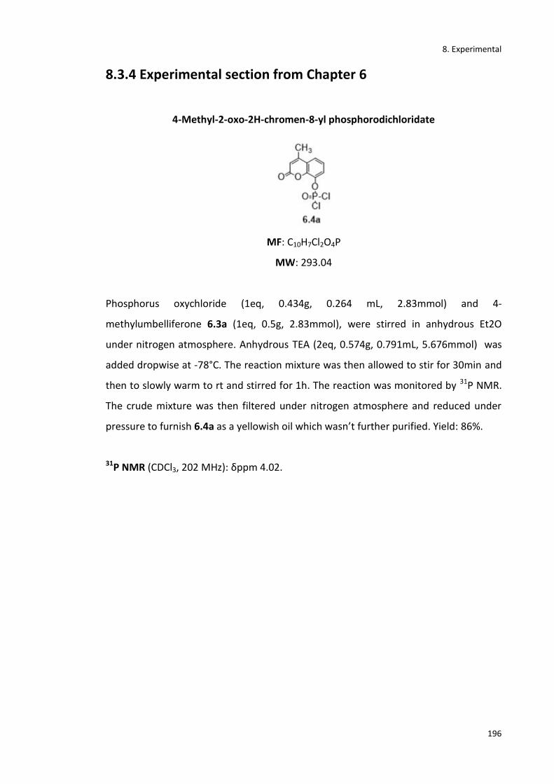
8. Experimental
196
8.3.4 Experimental section from Chapter 6
4-Methyl-2-oxo-2H-chromen-8-yl phosphorodichloridate
MF: C10H7Cl2O4P
MW: 293.04
Phosphorus oxychloride (1eq, 0.434g, 0.264 mL, 2.83mmol) and 4-
methylumbelliferone 6.3a (1eq, 0.5g, 2.83mmol), were stirred in anhydrous Et2O
under nitrogen atmosphere. Anhydrous TEA (2eq, 0.574g, 0.791mL, 5.676mmol) was
added dropwise at -78°C. The reaction mixture was then allowed to stir for 30min and
then to slowly warm to rt and stirred for 1h. The reaction was monitored by 31P NMR.
The crude mixture was then filtered under nitrogen atmosphere and reduced under
pressure to furnish 6.4a as a yellowish oil which wasn’t further purified. Yield: 86%.
31P NMR (CDCl3, 202 MHz): δppm 4.02.
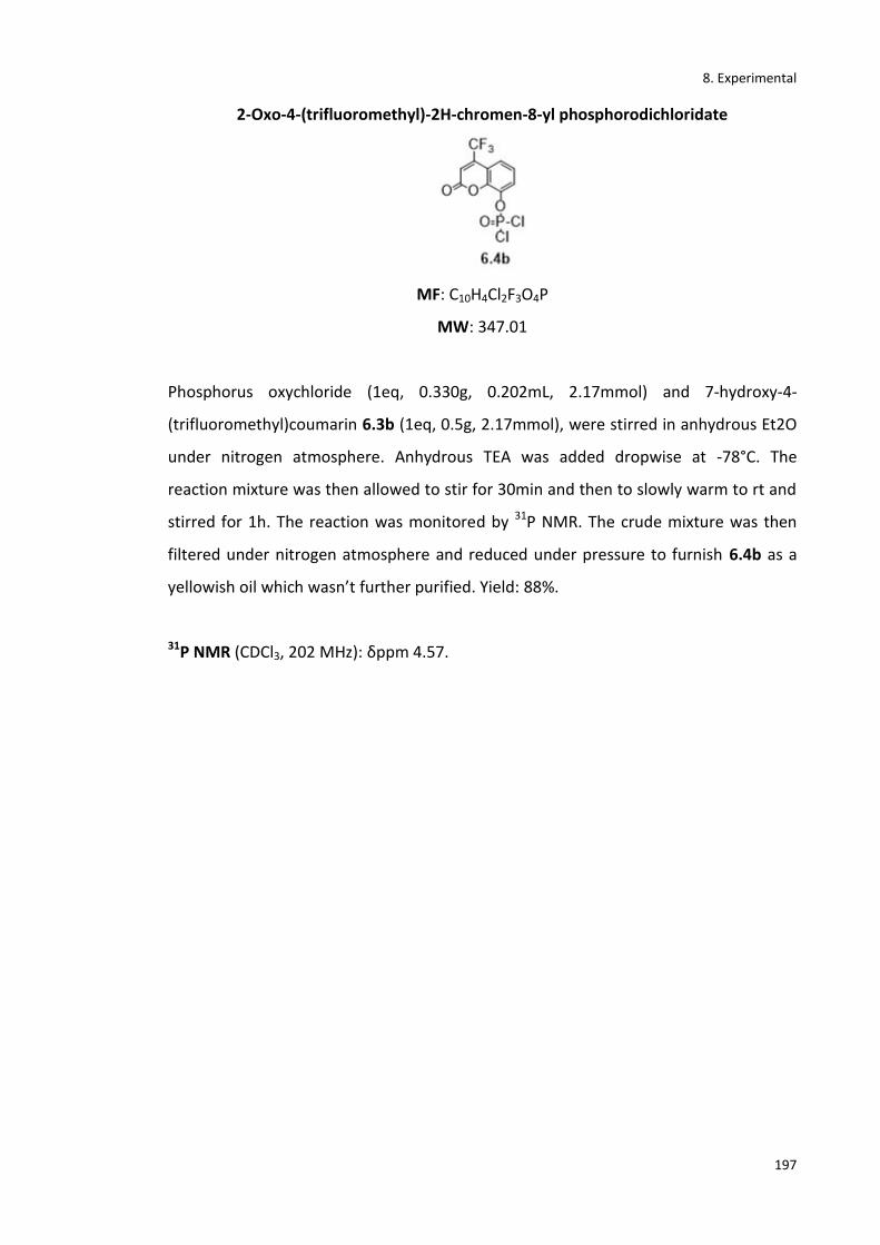
8. Experimental
197
2-Oxo-4-(trifluoromethyl)-2H-chromen-8-yl phosphorodichloridate
MF: C10H4Cl2F3O4P
MW: 347.01
Phosphorus oxychloride (1eq, 0.330g, 0.202mL, 2.17mmol) and 7-hydroxy-4-
(trifluoromethyl)coumarin 6.3b (1eq, 0.5g, 2.17mmol), were stirred in anhydrous Et2O
under nitrogen atmosphere. Anhydrous TEA was added dropwise at -78°C. The
reaction mixture was then allowed to stir for 30min and then to slowly warm to rt and
stirred for 1h. The reaction was monitored by 31P NMR. The crude mixture was then
filtered under nitrogen atmosphere and reduced under pressure to furnish 6.4b as a
yellowish oil which wasn’t further purified. Yield: 88%.
31P NMR (CDCl3, 202 MHz): δppm 4.57.
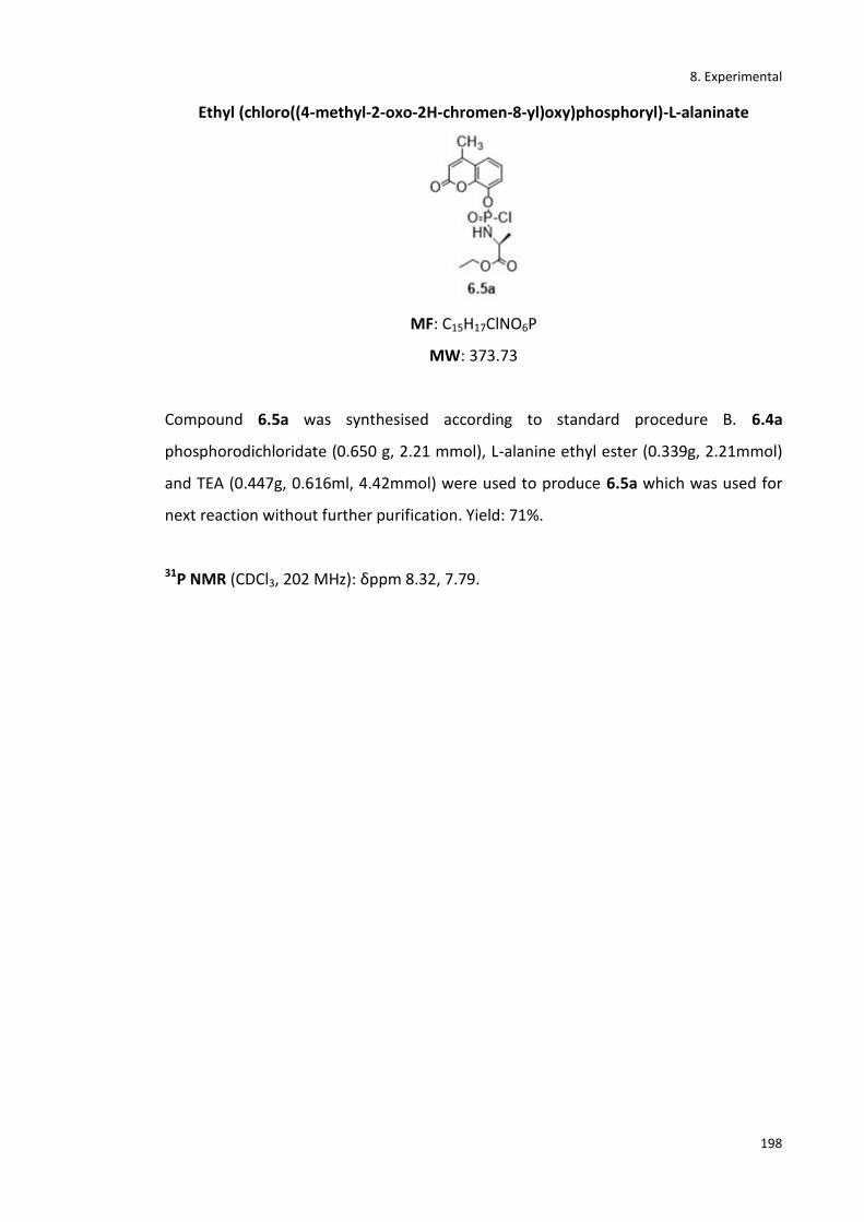
8. Experimental
198
Ethyl (chloro((4-methyl-2-oxo-2H-chromen-8-yl)oxy)phosphoryl)-L-alaninate
MF: C15H17ClNO6P
MW: 373.73
Compound 6.5a was synthesised according to standard procedure B. 6.4a
phosphorodichloridate (0.650 g, 2.21 mmol), L-alanine ethyl ester (0.339g, 2.21mmol)
and TEA (0.447g, 0.616ml, 4.42mmol) were used to produce 6.5a which was used for
next reaction without further purification. Yield: 71%.
31P NMR (CDCl3, 202 MHz): δppm 8.32, 7.79.
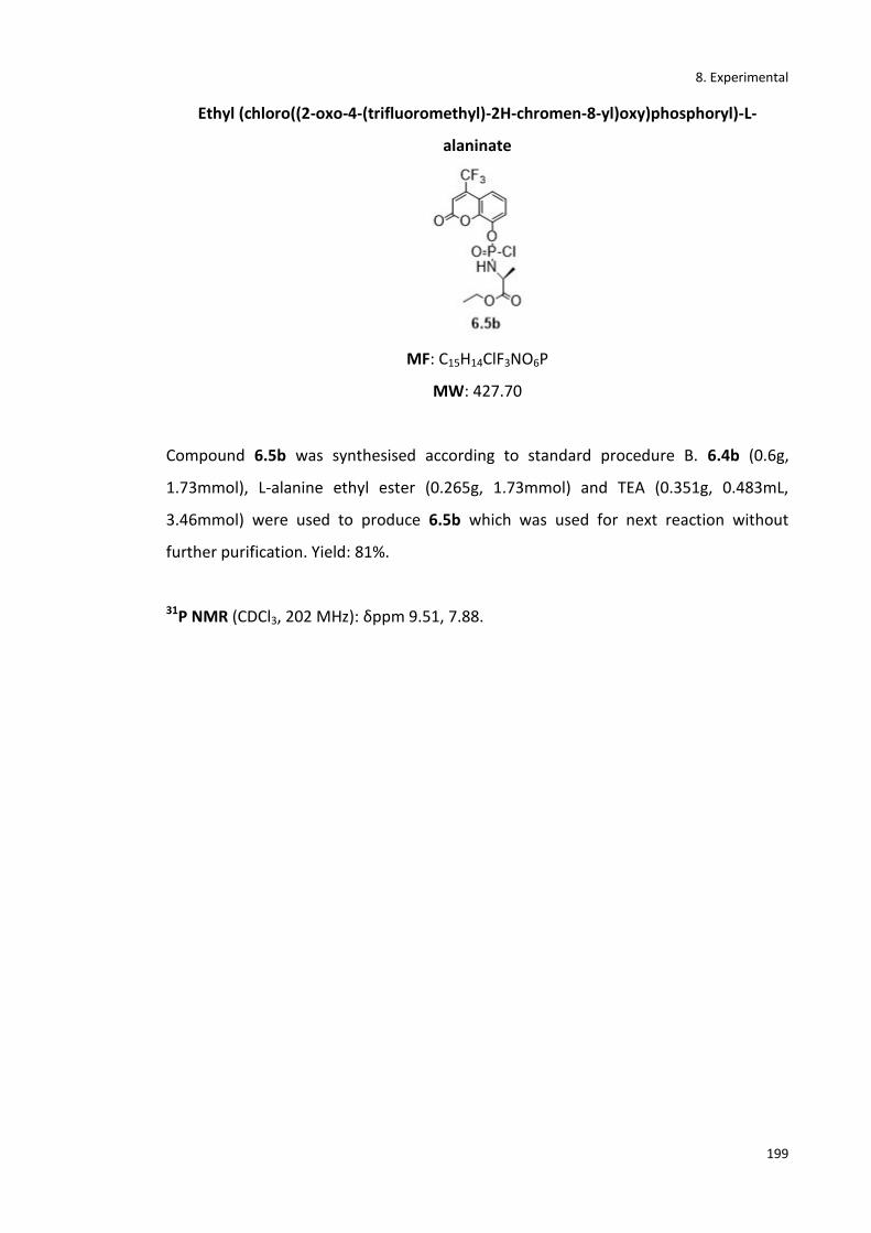
8. Experimental
199
Ethyl (chloro((2-oxo-4-(trifluoromethyl)-2H-chromen-8-yl)oxy)phosphoryl)-L-
alaninate
MF: C15H14ClF3NO6P
MW: 427.70
Compound 6.5b was synthesised according to standard procedure B. 6.4b (0.6g,
1.73mmol), L-alanine ethyl ester (0.265g, 1.73mmol) and TEA (0.351g, 0.483mL,
3.46mmol) were used to produce 6.5b which was used for next reaction without
further purification. Yield: 81%.
31P NMR (CDCl3, 202 MHz): δppm 9.51, 7.88.
8. Experimental
200
Ethyl ((((2R,3S,5R)-3-fluoro-5-(5-methyl-2,4-dioxo-3,4-dihydropyrimidin-1(2H)-
yl)tetrahydrofuran-2-yl)methoxy)((4-methyl-2-oxo-2H-chromen-8-
yl)oxy)phosphoryl)-L-alaninate. (6.6a)
MF: C25H29FN3O10P
MW: 581.49
Compound 6.6a was synthesised according to standard procedure D. FLT (1.29) (1eq,
0.146 g, 0.6 mmol), tBuMgCl (0.117 ml, 0.9 mmol) and 6.5a phosphorochloridate (2eq,
0.450 g, 1.2 mmol) were used to produce 6.6a. It was purified by silica gel coulm
chromatography (CH2Cl2/CH3OH from 100% CH2Cl2 to 95% CH2Cl2) to obtain a pale
white solid. Yield: 5 %.
1H-NMR (CDCl3, 500MHz): δppm 8.59, 8.53 (s, 1H, NH), 7.95-7.93 (m, 1H, ArH), 7.51,
7.49 (q, J =1.83 H; J = 1.6 Hz, 1H, ArH), 7.37-7.34 (m, 2H, ArH), 7.28-7.23 (m, 1H, H-1’),
6.97, 6.91, (t, J = 2, 8, 5 Hz, 1H, H-3’), 6.11-6.07 (m, 1H, ArH), 5.43, 5.36 (d, J = 1.8Hz,
2H, H-2’), 5.08-5.01 (m, 1H, H-4’), 4.99-4.88 (m, 2H, H-5’), 4.67-4.56 (m, 2H, CH2 ester),
4.06-3.89 (m, 2H, NH, CH-ala), 2.39 (d, J = 1.6 Hz, 3H, CH3-coum), 1.91, 1.85 (t, J = 1.6
Hz, 3H, CH3 thym), 1.41, 1.39 (d, J = 6.5 Hz, 3H, CH3 ala), 1.12, 1.11 (t, J = 7 Hz, 3H, CH3
ester). 13C NMR (CDCl3, 125MHz): δppm 175.83 (C=O-ala), 169.98, 167.43 (C=O-thym),
157.65, 157. 10 (C=O- coum), 153.78, 153.75 (ArC), 152.52, 152.43, 151.98, 151.76 (Ar-
C), 151.05 (C=O-thym), 139.18, 138.79 (ArCH), 135.64, 134.34 (C-3’), 131.21, 130.11 (C-
2’), 129.15, 127.54 (Ar-CH), 115.77 (Ar-C), 115.14, 114.61, 113.88, 113.43 (ArCH),
109.45, 109.21 (ArC), 107.11, 107.05, 89.43, 89.14 (C-1’), 83.21, 83.11 (C-4’), (71.88,
69.76, C-5’), 69.22, 69.10 (CH2 ester), 49.88, 49.73 (CH- ala), 29.31-29.25 (CH3-coum),
20.16 (CH3 ala), 19.43 (CH3-ester), 15.84, 14.34 (CH3-thym). 31P NMR (CDCl3, 202 MHz):

8. Experimental
201
δppm 3.01, 2.73. 19F NMR (470 MHz, CDCl3): δppm-175.2, -174.8. MS (ESI) +: 582.5
[M+H+].
8. Experimental
202
Ethyl ((((2R,3S,5R)-3-fluoro-5-(5-methyl-2,4-dioxo-3,4-dihydropyrimidin-1(2H)-
yl)tetrahydrofuran-2-yl)methoxy)((2-oxo-4-(trifluoromethyl)-2H-chromen-8-
yl)oxy)phosphoryl)-L-alaninate. (6.6b)
MF: C25H26F4N3O10P
MW: 635.46
Compound 6.6b was prepared according to standard procedure D. FLT (1.29) (1eq,
0.100g, 0.41mmol), tBuMgCl (0.08mL) and phosphorochloridate (0.349g, 0.82 mmol)
were used to produce Protide 6.6b that was purified by silica gel column
chromatography (CH2Cl2/CH3OH from 100% CH2Cl2 to 95% CH2Cl2) to obtain a pale
white solid. Yield: 11%.
1H-NMR (CDCl3, 500MHz): δppm 8.63, 8.61 (s, 1H, NH), 7.84-7.81 (m, 1H, ArH), 7.43,
7.41 (m, 1H, ArH), 7.25-7.19 (m, 2H, ArH), 7.19-7.18 (m, 1H, H-1’), 6.76, 6.75, (t, J = 1.6,
7, 4.3 Hz, 1H, H-3’), 6.03-6.00 (m, 1H, ArH), 5.64, 5.39 (d, J = 1.7 Hz, 2H, H-2’), 4.99-4.97
(m, 1H, H-4’), 4.49-4.43 (m, 2H, H-5’), 4.25-4.21 (m, 2H, CH2- ester), 4.11 (m, 1H, NH),
4.02 (m, 1H, CH-ala), 1.95, 1.89 (t, J = 1.5 Hz, 3H, CH3 thym), 1.54, 1.51 (d, J = 5 Hz, 3H,
CH3 ala), 1.21, 1.19 (t, J = 7 Hz, 3H, CH3 ester). 13C NMR (CDCl3, 125MHz): δppm 181.21
(C=O-ala), 165.98, 164.12 (C=O-thym), 159.65, 158. 10 (C=O- coum), 153.12, 153.01
(ArC), 152.76, 152.73, 151.86, 151.54 (Ar-C), 150.3 (C-CF3), 150.11 (C=O-thym), 141.18,
140.9 (ArCH), 139.64, 139.99 (C-3’), 137.11, 137.02 (C-2’), 131.53, 131.32 (Ar-CH),
121.99 (CF3), 118.23, 117.21, 117.10, 116.98 (ArCH), 110.32, 109.87 (ArC), 108.09,
108.04, 89.43, 89.23 (C-1’), 85.76, 85.11 (C-4’), 75.99, 68.71, (C-5’), 68.19, 67.99 (CH2
ester), 53.88, 53.53 (CH-ala), 20.99 (CH3-ala), 19.13 (CH3-ester), 15.99, 15.29 (CH3-

8. Experimental
203
thym). 31P NMR (CDCl3, 202 MHz): δppm: δ 3.12, 2.91. 19F NMR (CDCl3, 470MHz):
δppm - 64.78,-64.12,-174.3, -174.1. MS (ESI+): 658.2 [M+Na+].

9. Appendix
204
9. Appendix
1.18F counts against MBq standard curve. The activityof [18F]FIAU dissolved in PBS
was measured and used to make serial dilutions. The range of concentrations was
then measured using anautomatic gamma counter.
2. Raw data for standard curve
Sample
Counts/min Mean
MBq
KBq
A B
HEK 1 38029 33879 35954 0.001798 1.7977
HEK 2 33052 32571 32811.5 0.001641 1.640575
HEK 3 61659 52007 56833 0.002842 2.84165
TK-HEK1 133924 124467 129195.5 0.00646 6.459775
TK-HEK2 139935 139430 139682.5 0.006984 6.984125
TK-HEK3 145572 117115 131343.5 0.006567 6.567175

9. Appendix
205
3. Radioactivity measurements from cell washes. KBq of radioactivity from the
samples were generated using the standard curve in Figure 4.15
Sample
Counts KBq Total
Supernatan
t 1
Supernatan
t 2
Supernatan
t 1
Supernatan
t 2 Count KBq
TK- HEK1 2321157 192690 116.0579 9.6345
251384
7
125.692
4
TK-HEK2 2317760 205327 115.888 10.26635
252308
7
126.154
4
TK-HEK3 2315578 174083 115.7789 8.70415
248966
1
124.483
1
HEK1 2269951 129205 113.4976 6.46025
239915
6
119.957
8
HEK2 2271855 161629 113.5928 8.08145
243348
4
121.674
2
HEK3 2251568 144151 112.5784 7.20755
239571
9 119.786

9. Appendix
206
4. T-test to compare group means with the assumption of equal variances
HEK TK-HEK
Mean 0.1164 1.3396
St-Dev 0.0103 0.0859
Hypothesis:
HeKTK
HeKTK
H
H
:
:
1
0
Significance Level
α = 0.05
Pooled variance:
0037425.0233
0859.0130103.013 222
pS
0612.00037425.0 pS
Test Statistic:
48.24
3
1
3
100612
1164.03396.1
t
Degrees of Freedom:
4233 df
Critical value:
132.2ct
Compare and decide:
132.248.24 ctt Reject Ho

10. Bibliography
207
10. Bibliography
(1) Cole, E. L.; Stewart, M. N.; Littich, R.; Hoareau, R.; Scott, P. J. H. Radiosyntheses
Using Fluorine-18: The Art and Science of Late Stage Fluorination. Curr. Top.
Med. Chem. 2014, 14 (7), 875–900.
(2) Matthews, P. M.; Rabiner, E. A.; Passchier, J.; Gunn, R. N. Positron Emission
Tomography Molecular Imaging for Drug Development. Br. J. Clin. Pharmacol.
2012, 73 (2), 175–186.
(3) Rudin, M.; Weissleder, R. Molecular Imaging in Drug Discovery and
Development. Nat. Rev. Drug Discov. 2003, 2 (2), 123–131.
(4) Miller, P. W.; Long, N. J.; Vilar, R.; Gee, A. D. Synthesis of11C, 18F, 15O, and 13N
Radiolabels for Positron Emission Tomography. Angew. Chemie - Int. Ed. 2008,
47 (47), 8998–9033.
(5) Schlyer, D. J. PET Tracers and Radiochemistry. Ann. Acad. Med. Singapore 2004,
33 (2), 146–154.
(6) Jacobson, O.; Kiesewetter, D. O.; Chen, X. Fluorine-18 Radiochemistry, Labeling
Strategies and Synthetic Routes. Bioconjug. Chem. 2015, 26 (1), 1–18.
(7) Preshlock, S.; Tredwell, M.; Gouverneur, V. 18F-Labeling of Arenes and
Heteroarenes for Applications in Positron Emission Tomography. Chem. Rev.
2016, 116 (2), 719–766.
(8) van der Veldt, A. A. M.; Smit, E. F.; Lammertsma, A. A. Positron Emission
Tomography as a Method for Measuring Drug Delivery to Tumors in Vivo: The
Example of [11C]docetaxel. Front. Oncol. 2013, 3, 1–7.
(9) Daniels, S.; Tohid, S. F. M.; Velanguparackel, W.; Westwell, A. D. The Role and
Future Potential of Fluorinated Biomarkers in Positron Emission Tomography.
Expert Opin. Drug Discov. 2010, 5 (3), 291–304.
(10) Alauddin, M. M. Positron Emission Tomography (PET) Imaging with (18)F-Based

10. Bibliography
208
Radiotracers. Am. J. Nucl. Med. Mol. Imaging 2012, 2 (1), 55–76.
(11) Turkheimer, F. E.; Veronese, M.; Dunn, J. Experimental Design and Practical
Data Analysis in Positron Emission Tomography; London, 2014.
(12) Circular orbits | a2-level-level-revision, physics, fields-0, circular-orbits |
Revision World https://revisionworld.com/a2-level-level-revision/physics/fields-
0/circular-orbits (accessed Jan 1, 2018).
(13) Gómez-Vallejo, V.; Gaja, V.; Gona, K. B.; Llop, J. Nitrogen-13: Historical Review
and Future Perspectives. J. Labelled Comp. Radiopharm. 2014, 57 (4), 244–254.
(14) Bishop, A.; Satyamurthy, N.; Bida, G.; Hendry, G.; Phelps, M.; Barrio, J. R. Proton
Irradiation of [18O]O2: Production of [18F]F2 and [18F]F2 + [18F] OF2. Nucl.
Med. Biol. 1996, 23 (3), 189–199.
(15) Långström, B.; Antoni, G.; Gullberg, P.; Halldin, C.; Malmborg, P.; Någren, K.;
Rimland, A.; Svärd, H. Synthesis of L- and D-[Methyl-11C]methionine. J. Nucl.
Med. 1987, 28 (6), 1037–1040.
(16) Larsen, P.; Ulin, J.; Dahlstrøm, K.; Jensen, M. Synthesis of [11C]iodomethane by
Iodination of [11C]methane. Appl. Radiat. Isot. 1997, 48 (2), 153–157.
(17) Cai, L.; Lu, S.; Pike, V. W. Chemistry with [18F]Fluoride Ion. European J. Org.
Chem. 2008, 2008 (17), 2853–2873.
(18) Preshlock, S.; Tredwell, M.; Gouverneur, V. 18F-Labeling of Arenes and
Heteroarenes for Applications in Positron Emission Tomography. Chem. Rev.
2016, 116 (2), 719–766.
(19) Jacobson, O.; Kiesewetter, D. O.; Chen, X. Fluorine-18 Radiochemistry, Labeling
Strategies and Synthetic Routes. Bioconjug. Chem. 2015, 26 (1), 1–18.
(20) Saha, G. B. Basics of PET Imaging : Physics, Chemistry, and Regulations; Springer,
2010.
(21) Berridge, M. S.; Apana, S. M.; Hersh, J. M. Teflon Radiolysis as the Major Source
of Carrier in Fluorine-18. J. Label. Compd. Radiopharm. 2009, 52 (13), 543–548.
(22) Vallabhajosula, S. Molecular Imaging : Radiopharmaceuticals for PET and SPECT;
10. Bibliography
209
Springer-Verlag, 2009.
(23) FASTlab Multi-Tracer Platform - TRACERcenter Equipment - PET Radiopharmacy
- CATEGORIES http://www3.gehealthcare.com.sg/en-
gb/products/categories/pet-radiopharmacy/tracer_center_equipment/fastlab
(accessed Jan 1, 2018).
(24) L’Annunziata, M. F. Radiopharma instruments | Trasis http://www.trasis.com/
(accessed Jan 1, 2018).
(25) Scott, P. J. H.; Hockley, B. G. Radiochemical Synthesis. Volume I,
Radiopharmaceuticals for Positron Emission Tomography; Wiley, 2012.
(26) L’Annunziata, M. F. Handbook of Radioactivity Analysis; 2012.
(27) Lindegren, S.; Jensenb, H.; Jacobsson, L. A Radio-High-Performance Liquid
Chromatography Dual-Flow Cell Gamma-Detection System for on-Line
Radiochemical Purity and Labeling Efficiency Determination. J. Chromatogr. A
2014, 1337, 128–132.
(28) The Ionising Radiations Regulations 1999; Queen’s Printer of Acts of Parliament,
1999.
(29) Bixler, A.; Springer, G.; Lovas, R. Practical Aspects of Radiation Safety for Using
Fluorine-18. J. Nucl. Med. Technol. 1999, 27 (1), 14-6-9.
(30) He, Q.; Alfeazi, I.; Sadeghi, S. No-Carrier-Added Electrochemical Nucleophilic
Radiofluorination of Aromatics. J. Radioanal. Nucl. Chem. 2015, 303 (1), 1037–
1040.
(31) Jacobson, O.; Chen, X. PET Designated Flouride-18 Production and Chemistry.
Curr. Top. Med. Chem. 2010, 10 (11), 1048–1059.
(32) Kim, D. W.; Ahn, D. S.; Oh, Y. H.; Lee, S.; Kil, H. S.; Oh, S. J.; Lee, S. J.; Kim, J. S.;
Ryu, J. S.; Moon, D. H.; Chi, D. Y. A New Class of SN2 Reactions Catalyzed by
Protic Solvents: Facile Fluorination for Isotopic Labeling of Diagnostic Molecules.
J. Am. Chem. Soc. 2006, 128 (50), 16394–16397.
(33) Ganghua, T.; Xiaolan, T.; Fuhua, W.; Mingfang, W.; Baoyuan, L. A Facile and

10. Bibliography
210
Rapid Automated Synthesis of 3′-Deoxy-3′-[18F]fluorothymidine. Appl. Radiat.
Isot. 2010, 68 (9), 1734–1739.
(34) Cavaliere, A.; Probst, K. C.; Westwell, A. D.; Slusarczyk, M. Fluorinated
Nucleosides as an Important Class of Anticancer and Antiviral Agents. Future
Med. Chem. 2017, 9 (15), 1809–1833.
(35) Hollingworth, C.; Gouverneur, V. Transition Metal Catalysis and Nucleophilic
Fluorination. Chem. Commun. 2012, 48 (24), 2929.
(36) Kepe, V.; Moghbel, M. C.; Långström, B.; Zaidi, H.; Vinters, H. V; Huang, S.-C.;
Satyamurthy, N.; Doudet, D.; Mishani, E.; Cohen, R. M.; Høilund-Carlsen, P. F.;
Alavi, A.; Barrio, J. R. Amyloid-β Positron Emission Tomography Imaging Probes:
A Critical Review. J. Alzheimers. Dis. 2013, 36 (4), 613–631.
(37) Swallow, S. Fluorine in Medicinal Chemistry. Prog. Med. Chem. 2015, 54 (2), 65–
133.
(38) Buckingham, F.; Gouverneur, V. Asymmetric 18 F-Fluorination for Applications in
Positron Emission Tomography. Chem. Sci. 2016, 7 (3), 1645–1652.
(39) Hoover, A. J.; Lazari, M.; Ren, H.; Narayanam, M. K.; Murphy, J. M.; Van Dam, R.
M.; Hooker, J. M.; Ritter, T. A Transmetalation Reaction Enables the Synthesis of
[18F]5-Fluorouracil from [18F]Fluoride for Human PET Imaging. Organometallics
2016, 35 (7), 1008–1014.
(40) Purrington, S. T.; Kagen, B. S.; Patrick, T. B. Application of Elemental Fluorine in
Organic Synthesis. Chem. Rev. 1986, 86 (6), 997–1018.
(41) Casella, V.; Ido, T.; Wolf, A. P.; Fowler, J. S.; MacGregor, R. R.; Ruth, T. J.
Anhydrous F-18 Labeled Elemental Flurine for Radiopharmaceutical Preparation.
J. Nucl. Med. 1980, 21 (8), 750–757.
(42) Teare, H.; Robins, E. G.; Kirjavainen, A.; Forsback, S.; Sandford, G.; Solin, O.;
Luthra, S. K.; Gouverneur, V. Radiosynthesis and Evaluation of [18F]Selectfluor
Bis(triflate). Angew. Chemie Int. Ed. 2010, 49 (38), 6821–6824.
(43) Bauta, W.; Schulmeier, B. E.; Burke, B.; Puente, J. F.; Cantrell, W. R.; Lovett, D.;
Goebel, J.; Anderson, B.; Ionescu, D.; Ruichao, G. A New Process for
10. Bibliography
211
Antineoplastic Agent Clofarabine. Org. Proc. Res. Dev 2004, 8 (6), 889–896.
(44) Anderson, H.; Pillarsetty, N.; Cantorias, M.; Lewis, J. S. Improved Synthesis of 2’-
deoxy-2’-[18F]-Fluoro-1-Beta-D-Arabinofuranosyl-5-Iodouracil ([18F]-FIAU).
Nucl. Med. Biol. 2010, 37 (4), 439–442.
(45) Zhang, H.; Cantorias, M. V; Pillarsetty, N.; Burnazi, E. M.; Cai, S.; Lewis, J. S. An
Improved Strategy for the Synthesis of [18F]-Labeled Arabinofuranosyl
Nucleosides. Nucl. Med. Biol. 2012, 39 (8), 1182–1188.
(46) Shelton, J.; Lu, X.; Hollenbaugh, J. A.; Cho, J. H.; Amblard, F.; Schinazi, R. F.
Metabolism, Biochemical Actions, and Chemical Synthesis of Anticancer
Nucleosides, Nucleotides, and Base Analogs. Chem. Rev. 2016, 116 (23), 14379–
14455.
(47) Gallagher, B. M.; Fowler, J. S.; Gutterson, N. I.; MacGregor, R. R.; Wan, C. N.;
Wolf, A. P. Metabolic Trapping as a Principle of Oradiopharmaceutical Design:
Some Factors Resposible for the Biodistribution of [18F] 2-Deoxy-2-Fluoro-D-
Glucose. J. Nucl. Med. 1978, 19 (10), 1154–1161.
(48) Gambhir, S. S.; Czernin, J.; Schwimmer, J.; Silverman, D. H.; Coleman, R. E.;
Phelps, M. E. A Tabulated Summary of the FDG PET Literature. J. Nucl. Med.
2001, 42 (5 Suppl), 1S–93S.
(49) Adam, M. J. A Rapid, Stereoselective, High Yielding Synthesis of 2-Deoxy-2-
Fluoro-D-Hexopyranoses: Reaction of Glycals with Acetyl Hypofluorite. J. Chem.
Soc. Chem. Commun. 1982, 0 (13), 730.
(50) Shiue, C.-Y.; K.-C., A. P.; Wolf, A. P. A Rapid Synthesis of 2-Deoxy-2-Fluoro-D-
Glucose from Xenon Difluoride Suitable for Labelling with 18F. J. Label. Compd.
Radiopharm. 1983, 20 (2), 157–162.
(51) Hamacher, K.; Coenen, H. H.; Stocklin, G. Synthesis D-Glucose Using
Aminopolyether Supported Nucleophilic Substitution. J. Nucl. Med. 1986, 27
(February), 235–239.
(52) Yu, S. Review of F-FDG Synthesis and Quality Control. Biomed. Imaging Interv. J.
2006, 2 (4), e57.

10. Bibliography
212
(53) Pankiewicz, K. W. Fluorinated Nucleosides. Carbohydr. Res. 2000, 327 (1–2), 87–
105.
(54) Mangner, T. J.; Klecker, R. W.; Anderson, L.; Shields, A. F. Synthesis of 2′-Deoxy-
2′-[18F]fluoro-β-D-Arabinofuranosyl Nucleosides, [18F]FAU, [18F]FMAU,
[18F]FBAU and [18F]FIAU, as Potential PET Agents for Imaging Cellular
Proliferation. Nucl. Med. Biol. 2003, 30 (3), 215–224.
(55) Soloviev, D.; Lewis, D.; Honess, D.; Aboagye, E. [18F]FLT: An Imaging Biomarker
of Tumour Proliferation for Assessment of Tumour Response to Treatment. Eur.
J. Cancer 2012, 48 (4), 416–424.
(56) McKinley, E. T.; Ayers, G. D.; Smith, R. A.; Saleh, S. A.; Zhao, P.; Washington, M.
K.; Coffey, R. J.; Manning, H. C. Limits of [18F]-FLT PET as a Biomarker of
Proliferation in Oncology. PLoS One 2013, 8 (3), e58938.
(57) Tjuvajev, J. G.; Doubrovin, M.; Akhurst, T.; Cai, S.; Balatoni, J.; Alauddin, M. M.;
Finn, R.; Bornmann, W.; Thaler, H.; Conti, P. S.; Blasberg, R. G. Comparison of
Radiolabeled Nucleoside Probes (FIAU, FHBG, and FHPG) for PET Imaging of
HSV1-Tk Gene Expression. J. Nucl. Med. 2002, 43 (8), 1072–1083.
(58) Alauddin, M. M.; Shahinian, A.; Park, R.; Tohme, M.; Fissekis, J. D.; Conti, P. S.
Biodistribution and PET Imaging of [(18)F]-Fluoroadenosine Derivatives. Nucl.
Med. Biol. 2007, 34 (3), 267–272.
(59) Marik, J.; Ogasawara, A.; Martin-McNulty, B.; Ross, J.; Flores, J. E.; Gill, H. S.;
Tinianow, J. N.; Vanderbilt, A. N.; Nishimura, M.; Peale, F.; Pastuskovas, C.;
Greve, J. M.; van Bruggen, N.; Williams, S. P. PET of Glial Metabolism Using 2-
18F-Fluoroacetate. J. Nucl. Med. 2009, 50 (6), 982–990.
(60) Vingerhoets, F. J.; Schulzer, M.; Ruth, T. J.; Holden, J. E.; Snow, B. J.
Reproducibility and Discriminating Ability of Fluorine-18-6-Fluoro-L-Dopa PET in
Parkinson’s Disease. J. Nucl. Med. 1996, 37 (3), 421–426.
(61) Miller, P. W.; Long, N. J.; Vilar, R.; Gee, A. D. Synthesis of 11C, 18F, 15O, and 13N
Radiolabels for Positron Emission Tomography. Angew. Chem. Int. Ed. Engl.
2008, 47 (47), 8998–9033.

10. Bibliography
213
(62) Link, J. M.; Krohn, K. A.; Clark, J. C. Production of [11C]CH3I by Single Pass
Reaction of [11C]CH4 with I2. Nucl. Med. Biol. 1997, 24 (1), 93–97.
(63) Ametamey, S. M.; Honer, M.; Schubiger, P. A. Molecular Imaging with PET.
Chem. Rev. 2008, 108 (5), 1501–1516.
(64) da Silva, E. S.; Gómez-Vallejo, V.; Llop, J.; López-Gallego, F. Efficient Nitrogen-13
Radiochemistry Catalyzed by a Highly Stable Immobilized Biocatalyst. Catal. Sci.
Technol. 2015, 5 (5), 2705–2713.
(65) Tominaga, T.; Inoue, O.; Suzuki, K.; Yamasaki, T.; Hirobe, M. Synthesis of 13N-
Labelled Amines by Reduction of 13N-Labelled Amides. Int. J. Radiat. Appl.
Instrumentation. Part A. Appl. Radiat. Isot. 1986, 37 (12), 1209–1212.
(66) Martín, A.; SAN Sebastián, E.; Gómez-Vallejo, V.; Llop, J. Positron Emission
Tomograghy with [13N]ammonia Evidences Long-Term Cerebral Hyperperfusion
after 2h-Transient Focal Ischemia. Neuroscience 2012, 213, 47–53.
(67) Hoop, B.; Smith, T. W.; Burnham, C. A.; Correll, J. E.; Brownell, G. L.; Sanders, C.
A. Myocardial Imaging with 13 NH 4+ and a Multicrystal Positron Camera. J.
Nucl. Med. 1973, 14 (3), 181–183.
(68) Beaney, R.; Jones, T.; Lammertsma, A.; Mckenzie, C.; Halnan, K. Positron
Emission Tomography for in-Vivo Measurement of Regional Blood Flow, Oxygen
Utilisation, and Blood Volume in Patients with Breast Carcinoma. Lancet 1984,
323 (8369), 131–134.
(69) Al-Nahhas, A.; Win, Z.; Szyszko, T.; Singh, A.; Nanni, C.; Fanti, S.; Rubello, D.
Gallium-68 PET: A New Frontier in Receptor Cancer Imaging. Anticancer Res. 27
(6B), 4087–4094.
(70) Deri, M. A.; Zeglis, B. M.; Francesconi, L. C.; Lewis, J. S. PET Imaging with 89Zr:
From Radiochemistry to the Clinic. Nucl. Med. Biol. 2013, 40 (1), 3–14.
(71) Anderson, C. J.; Ferdani, R. Copper-64 Radiopharmaceuticals for PET Imaging of
Cancer: Advances in Preclinical and Clinical Research. Cancer Biother.
Radiopharm. 2009, 24 (4), 379–393.
(72) Kolb, H. C.; Finn, M. G.; Sharpless, K. B. Click Chemistry: Diverse Chemical

10. Bibliography
214
Function from a Few Good Reactions. Angew. Chem. Int. Ed. Engl. 2001, 40 (11),
2004–2021.
(73) Jones, T.; Rabiner, E. A.; PET Research Advisory Company. The Development,
Past Achievements, and Future Directions of Brain PET. J. Cereb. Blood Flow
Metab. 2012, 32 (7), 1426–1454.
(74) Matthews, P. M.; Rabiner, E. A.; Passchier, J.; Gunn, R. N. Positron Emission
Tomography Molecular Imaging for Drug Development. Br. J. Clin. Pharmacol.
2012, 73 (2), 175–186.
(75) Bergström, M.; Hargreaves, R. J.; Burns, H. D.; Goldberg, M. R.; Sciberras, D.;
Reines, S. A.; Petty, K. J.; Ögren, M.; Antoni, G.; Långström, B.; Eskola, O.;
Scheinin, M.; Solin, O.; Majumdar, A. K.; Constanzer, M. L.; Battisti, W. P.;
Bradstreet, T. E.; Gargano, C.; Hietala, J. Human Positron Emission Tomography
Studies of Brain Neurokinin 1 Receptor Occupancy by Aprepitant. Biol.
Psychiatry 2004, 55 (10), 1007–1012.
(76) Van Rompay, A. R.; Johansson, M.; Karlsson, A. Substrate Specificity and
Phosphorylation of Antiviral and Anticancer Nucleoside Analogues by Human
Deoxyribonucleoside Kinases and Ribonucleoside Kinases. Pharmacol. Ther.
2003, 100 (2), 119–139.
(77) Chu, C. K.; Baker, D. C.; Symposium on Nucleosides as Antitumor and Antiviral
Agents (1992 : San Francisco, C. . Nucleosides and Nucleotides as Antitumor and
Antiviral Agents.
(78) Van Rompay, A. R.; Johansson, M.; Karlsson, A.; Berg, J. M. (Jeremy M.;
Tymoczko, J. L.; Stryer, L.; Stryer, L. Biochemistry, 5th Edition; W.H. Freeman,
2002; Vol. 100.
(79) Balzarini, J.; Egberink, H.; Hartmann, K.; Cahard, D.; Vahlenkamp, T.; Thormar,
H.; De Clercq, E.; McGuigan, C. Antiretrovirus Specificity and Intracellular
Metabolism of 2’,3’ -Didehydro-2’,3’-dideoxythymidine (Stavudine) and Its 5’-
monophosphate Triester Prodrug So324. Mol. Pharmacol. 1996, 50 (5), 1207–
1213.

10. Bibliography
215
(80) Al-Madhoun, A. S.; Tjarks, W.; Eriksson, S. The Role of Thymidine Kinases in the
Activation of Pyrimidine Nucleoside Analogues. Mini Rev. Med. Chem. 2004, 4
(4), 341–350.
(81) Pradere, U.; Garnier-Amblard, E. C.; Coats, S. J.; Amblard, F.; Schinazi, R. F.
Synthesis of Nucleoside Phosphate and Phosphonate Prodrugs. Chem. Rev.
2014, 114 (18), 9154–9218.
(82) Mehellou, Y. The ProTides Boom. ChemMedChem 2016, 11 (11), 1–4.
(83) Mehellou, Y.; Rattan, H. S.; Balzarini, J. The ProTide Prodrug Technology: From
the Concept to the Clinic. J. Med. Chem. 2017, acs.jmedchem.7b00734.
(84) Serpi, M.; Madela, K.; Pertusati, F.; Slusarczyk, M. Synthesis of Phosphoramidate
Prodrugs: ProTide Approach. Curr. Protoc. Nucleic Acid Chem. 2013, Chapter 15.
(85) McGuigan, C.; Murziani, P.; Slusarczyk, M.; Gonczy, B.; Vande Voorde, J.;
Liekens, S.; Balzarini, J. Phosphoramidate ProTides of the Anticancer Agent FUDR
Successfully Deliver the Preformed Bioactive Monophosphate in Cells and
Confer Advantage over the Parent Nucleoside. J. Med. Chem. 2011, 54 (20),
7247–7258.
(86) Westwell, A. D. Fluorinated Pharmaceuticals: Advances in Medicinal Chemistry;
Future Science Ltd: London, UK, 2015.
(87) Isanbor, C.; O’Hagan, D. Fluorine in Medicinal Chemistry: A Review of Anti-
Cancer Agents. J. Fluor. Chem. 2006, 127 (3), 303–319.
(88) Ismail, F. M. D. Important Fluorinated Drugs in Experimental and Clinical Use. J.
Fluor. 2002, 118, 27–33.
(89) Park, B. K.; Kitteringham, N. R.; O’Neill, P. M. Metabolism of Fluorine-Containing
Drugs. Annu. Rev. Pharmacol. Toxicol. 2001, 41 (1), 443–470.
(90) Meier, C.; Knispel, T.; Marquez, V. E.; Siddiqui, M. A.; De Clercq, E.; Balzarini, J.
cycloSal-Pronucleotides of 2‘-Fluoro-Ara- and 2‘-Fluoro-Ribo-2‘,3‘-
Dideoxyadenosine as a Strategy to Bypass a Metabolic Blockade. J. Med. Chem.
1999, 42 (9), 1615–1624.
10. Bibliography
216
(91) Carreras, C. W.; Santi, D. V. The Catalytic Mechanism and Structure of
Thymidylate Synthase. Annu. Rev. Biochem. 1995, 64 (1), 721–762.
(92) Kaiyawet, N.; Rungrotmongkol, T.; Hannongbua, S. Effect of Halogen
Substitutions on dUMP to Stability of Thymidylate Synthase/dUMP/mTHF
Ternary Complex Using Molecular Dynamics Simulation. J. Chem. Inf. Model.
2013, 53 (6), 1315–1323.
(93) Malet-Martino, M.; Jolimaitre, P.; Martino, R. The Prodrugs of 5-Fluorouracil.
Curr. Med. Chem. Anticancer. Agents 2002, 2 (2), 267–310.
(94) Vande Voorde, J.; Liekens, S.; McGuigan, C.; Murziani, P. G. S.; Slusarczyk, M.;
Balzarini, J. The Cytostatic Activity of NUC-3073, a Phosphoramidate Prodrug of
5-Fluoro-2′-Deoxyuridine, Is Independent of Activation by Thymidine Kinase and
Insensitive to Degradation by Phosphorolytic Enzymes. Biochem. Pharmacol.
2011, 82 (5), 441–452.
(95) Ghazaly, E. A.; Slusarczyk, M.; McGuigan, C.; Harrison, D.; Blagden, S. P. Abstract
B46: NUC-3373: A Novel Pyrimidine Nucleotide Analogue That Overcomes Key
Cancer Drug Resistance Limiting Patient Survival. Mol. Cancer Ther. 2015, 14 (12
Supplement 2), B46–B46.
(96) Blagden, S. P.; Slusarczyk, M.; Serpi, M.; McGuigan, C.; Ghazaly, E. A. Abstract
CT028: First-in-Human Phase I Study of the Nucleotide Analogue NUC-3373
Designed to Overcome Fluoropyrimidine Drug Resistance Mechanisms. Cancer
Res. 2016, 76 (14 Supplement), CT028-CT028.
(97) Toschi, L.; Finocchiaro, G.; Bartolini, S.; Gioia, V.; Cappuzzo, F. Role of
Gemcitabine in Cancer Therapy. Futur. Oncol. 2005, 1 (1), 7–17.
(98) Eckel, F.; Schneider, G.; Schmid, R. M. Pancreatic Cancer: A Review of Recent
Advances. Expert Opin. Investig. Drugs 2006, 15 (11), 1395–1410.
(99) Kroep, J. R.; Loves, W. J. P.; van der Wilt, C. L.; Alvarez, E.; Talianidis, I.; Boven,
E.; Braakhuis, B. J. M.; van Groeningen, C. J.; Pinedo, H. M.; Peters, G. J.
Pretreatment Deoxycytidine Kinase Levels Predict in Vivo Gemcitabine
Sensitivity. Mol. Cancer Ther. 2002, 1 (6), 371–376.

10. Bibliography
217
(100) Heinemann, V.; Hertel, L. W.; Grindey, G. B.; Plunkett, W. Comparison of the
Cellular Pharmacokinetics and Toxicity of 2’,2’-difluorodeoxycytidine and 1-Beta-
D-Arabinofuranosylcytosine. Cancer Res. 1988, 48 (14), 4024–4031.
(101) Beumer, J. H.; Eiseman, J. L.; Parise, R. A.; Joseph, E.; Covey, J. M.; Egorin, M. J.
Modulation of Gemcitabine (2’,2’-Difluoro-2’-Deoxycytidine) Pharmacokinetics,
Metabolism, and Bioavailability in Mice by 3,4,5,6-Tetrahydrouridine. Clin.
Cancer Res. 2008, 14 (11), 3529–3535.
(102) Bender, D. M.; Bao, J.; Dantzig, A. H.; Diseroad, W. D.; Law, K. L.; Magnus, N. A.;
Peterson, J. A.; Perkins, E. J.; Pu, Y. J.; Reutzel-Edens, S. M.; Remick, D. M.;
Starling, J. J.; Stephenson, G. A.; Vaid, R. K.; Zhang, D.; McCarthy, J. R. Synthesis,
Crystallization, and Biological Evaluation of an Orally Active Prodrug of
Gemcitabine. J. Med. Chem. 2009, 52 (22), 6958–6961.
(103) Slusarczyk, M.; Lopez, M. H.; Balzarini, J.; Mason, M.; Jiang, W. G.; Blagden, S.;
Thompson, E.; Ghazaly, E.; McGuigan, C. Application of ProTide Technology to
Gemcitabine: A Successful Approach to Overcome the Key Cancer Resistance
Mechanisms Leads to a New Agent (NUC-1031) in Clinical Development. J. Med.
Chem. 2014, 57 (4), 1531–1542.
(104) Blagden, S. P. et all. Final Results of ProGem1, the First in-Human Phase I/II
Study of NUC-1031 in Patients with Solid Malignancies. | 2015 ASCO Annual
Meeting | Virtual Meeting | Meeting Library. J Clin Oncol. 2015, p suppl; abstr
2514.
(105) Ricci, F.; Tedeschi, A.; Morra, E.; Montillo, M. Fludarabine in the Treatment of
Chronic Lymphocytic Leukemia: A Review. Ther. Clin. Risk Manag. 2009, 5 (1),
187–207.
(106) Parker, W. B.; Cheng, Y. C. Inhibition of DNA Primase by Nucleoside
Triphosphates and Their Arabinofuranosyl Analogs. Mol. Pharmacol. 1987, 31
(2), 146–151.
(107) Huang, P.; Plunkett, W. Phosphorolytic Cleavage of 2-Fluoroadenine from 9-
Beta-D-Arabinofuranosyl-2-Fluoroadenine by Escherichia Coli. A Pathway for 2-
Fluoro-ATP Production. Biochem. Pharmacol. 1987, 36 (18), 2945–2950.

10. Bibliography
218
(108) Hallek, M.; Fischer, K.; Fingerle-Rowson, G.; Fink, A.; Busch, R.; Mayer, J.; Hensel,
M.; Hopfinger, G.; Hess, G.; von Grünhagen, U.; Bergmann, M.; Catalano, J.;
Zinzani, P.; Caligaris-Cappio, F.; Seymour, J.; Berrebi, A.; Jäger, U.; Cazin, B.;
Trneny, M.; Westermann, A.; Wendtner, C.; Eichhorst, B.; Staib, P.; Bühler, A.;
Winkler, D.; Zenz, T.; Böttcher, S.; Ritgen, M.; Mendila, M.; Kneba, M.; Döhner,
H.; Stilgenbauer, S.; International Group of Investigators; German Chronic
Lymphocytic Leukaemia Study Group. Addition of Rituximab to Fludarabine and
Cyclophosphamide in Patients with Chronic Lymphocytic Leukaemia: A
Randomised, Open-Label, Phase 3 Trial. Lancet 2010, 376 (9747), 1164–1174.
(109) Von Hoff, D. D. Phase I Clinical Trials with Fludarabine Phosphate. Semin. Oncol.
1990, 17 (5 Suppl 8), 33–38.
(110) Jordheim, L. P.; Durantel, D.; Zoulim, F.; Dumontet, C. Advances in the
Development of Nucleoside and Nucleotide Analogues for Cancer and Viral
Diseases. Nat. Rev. Drug Discov. 2013, 12 (6), 447–464.
(111) Daly, M. B.; Roth, M. E.; Bonnac, L.; Maldonado, J. O.; Xie, J.; Clouser, C. L.;
Patterson, S. E.; Kim, B.; Mansky, L. M. Dual Anti-HIV Mechanism of Clofarabine.
Retrovirology 2016, 13 (1), 20.
(112) Cleij, M. C.; Steel, C. J.; Brady, F. 3’-DEOXY-3’-*’*P+FLUOROTHYnIIDINE. 2001, 1
(1 ml), 871–873.
(113) Ghosn, J.; Quinson, A.-M.; Sabo, N.; Cotte, L.; Piketty, C.; Dorléacq, N.; Bravo,
M.-L.; Mayers, D.; Harmenberg, J.; Mårdh, G.; Valdez, H.; Katlama, C. Antiviral
Activity of Low-Dose Alovudine in Antiretroviral-Experienced Patients: Results
from a 4-Week Randomized, Double-Blind, Placebo-Controlled Dose-Ranging
Trial. HIV Med. 2007, 8 (3), 142–147.
(114) Velanguparackel, W.; Hamon, N.; Balzarini, J.; McGuigan, C.; Westwell, A. D.
Synthesis, Anti-HIV and Cytostatic Evaluation of 3′-Deoxy-3′- Fluorothymidine
(FLT) pro-Nucleotides. Bioorganic Med. Chem. Lett. 2014, 24 (10), 2240–2243.
(115) Klecker, R. W.; Katki, A. G.; Collins, J. M. Toxicity, Metabolism, DNA
Incorporation with Lack of Repair, and Lactate Production for 1-(2’-fluoro-2’-
deoxy-Beta-D-Arabinofuranosyl)-5-Iodouracil in U-937 and MOLT-4 Cells. Mol.
10. Bibliography
219
Pharmacol. 1994, 46 (6), 1204–1209.
(116) Cai, H.; Li, Z.; Conti, P. S. The Improved Syntheses of 5-Substituted 2’-
[18F]fluoro-2’-deoxy-Arabinofuranosyluracil Derivatives ([18F]FAU, [18F]FEAU,
[18F]FFAU, [18F]FCAU, [18F]FBAU and [18F]FIAU) Using a Multistep One-Pot
Strategy. Nucl. Med. Biol. 2011, 38 (5), 659–666.
(117) Ghosh, R. K.; Ghosh, S. M.; Chawla, S. Recent Advances in Antiretroviral Drugs.
Expert Opin. Pharmacother. 2011, 12 (1), 31–46.
(118) Frampton, J. E.; Perry, C. M. Emtricitabine: A Review of Its Use in the
Management of HIV Infection. Drugs 2005, 65 (10), 1427–1448.
(119) By, S.; Kates, D. E. Application for Inclusion of Emtricitabine On WHO Model List
of Essential Medicines. 2006.
(120) Pawlotsky, J. M.; Najera, I.; Jacobson, I. Resistance to Mericitabine, a Nucleoside
Analogue Inhibitor of HCV RNA-Dependent RNA Polymerase. Antiviral Therapy.
2012, pp 411–423.
(121) Summers, B. B.; Beavers, J. W. F.; Klibanov, O. M. Sofosbuvir, a Novel Nucleotide
Analogue Inhibitor Used for the Treatment of Hepatitis C Virus. J. Pharm.
Pharmacol. 2014, 66, 1653–1666.
(122) Cha, A.; Budovich, A. Sofosbuvir: A New Oral Once-Daily Agent for the
Treatment of Hepatitis C Virus Infection. P T 2014, 39 (5), 345–352.
(123) Bhatia, H. K.; Singh, H.; Grewal, N.; Natt, N. K. Sofosbuvir: A Novel Treatment
Option for Chronic Hepatitis C Infection. J. Pharmacol. Pharmacother. 2014, 5
(4), 278–284.
(124) Bullard-Feibelman, K. M.; Govero, J.; Zhu, Z.; Salazar, V.; Veselinovic, M.;
Diamond, M. S.; Geiss, B. J. The FDA-Approved Drug Sofosbuvir Inhibits Zika
Virus Infection. Antiviral Res. 2017, 137, 134–140.
(125) Lam, A. M.; Espiritu, C.; Bansal, S.; Micolochick Steuer, H. M.; Zennou, V.; Otto,
M. J.; Furman, P. A. Hepatitis C Virus Nucleotide Inhibitors PSI-352938 and PSI-
353661 Exhibit a Novel Mechanism of Resistance Requiring Multiple Mutations
within Replicon RNA. J. Virol. 2011, 85 (23), 12334–12342.

10. Bibliography
220
(126) Wang, C.; Song, Z.; Yu, H.; Liu, K.; Ma, X. Adenine: An Important Drug Scaffold
for the Design of Antiviral Agents. Acta Pharm. Sin. B 2015, 5 (5), 431–441.
(127) Couturier, O.; Luxen, A.; Chatal, J.-F.; Vuillez, J.-P.; Rigo, P.; Hustinx, R.
Fluorinated Tracers for Imaging Cancer with Positron Emission Tomography. Eur.
J. Nucl. Med. Mol. Imaging 2004, 31 (8), 1182–1206.
(128) Serpi, M.; Madela, K.; Pertusati, F.; Slusarczyk, M.; Serpi, M.; Madela, K.;
Pertusati, F.; Slusarczyk, M. Synthesis of Phosphoramidate Prodrugs: ProTide
Approach. In Current protocols in nucleic acid chemistry / edited by Serge L.
Beaucage ... [et al.]; John Wiley & Sons, Inc.: Hoboken, NJ, USA, 2013; Vol.
Chapter 15, p 15.5.1-15.5.15.
(129) Hassan, M. Z.; Osman, H.; Ali, M. A.; Ahsan, M. J. Therapeutic Potential of
Coumarins as Antiviral Agents. Eur. J. Med. Chem. 2016, 123, 236–255.
(130) Peck, M.; Pollack, H. A.; Friesen, A.; Muzi, M.; Shoner, S. C.; Shankland, E. G.;
Fink, J. R.; Armstrong, J. O.; Link, J. M.; Krohn, K. A. Applications of PET Imaging
with the Proliferation Marker [18F]-FLT. Q. J. Nucl. Med. Mol. Imaging 2015, 59
(1).
(131) Fletcher, S. The Mitsunobu Reaction in the 21 st Century. Org. Chem. Front.
2015, 2 (6), 739–752.
(132) Meyer, J. P.; Probst, K. C.; Trist, I. M. L.; Mcguigan, C.; Westwell, A. D.
Radiochemical Synthesis of 2’-[18F]-Labelled and 3’-[18F]-Labelled Nucleosides
for Positron Emission Tomography Imaging. J. Labelled Comp. Radiopharm.
2014, 57 (5), 333–337.
(133) Mitsunobu Reaction http://www.organic-
chemistry.org/namedreactions/mitsunobu-reaction.shtm (accessed Jan 2,
2018).
(134) Balagopala, M. I.; Ollapally, A. P.; Lee, H. J. An Improved Synthesis of
Azidothymidine. Nucleosides, Nucleotides and Nucleic Acids 1996, 15 (4), 899–
906.
(135) Miah, A.; Reese, C. B.; Song, Q.; Sturdy, Z.; Neidle, S.; Simpson, I. J.; Read, M.;

10. Bibliography
221
Rayner, E. 2′,3′-Anhydrouridine. A Useful Synthetic Intermediate. J. Chem. Soc.
Perkin Trans. 1 1998, 0 (19), 3277–3284.
(136) Turkman, N.; Gelovani, J. G.; Alauddin, M. M. A Novel Method for Stereospecific
Fluorination at the 2′-Arabino-Position of Pyrimidine Nucleoside: Synthesis of
[18F]-FMAU. J. Label. Compd. Radiopharm. 2010, 53 (13), 782–786.
(137) Yun, M.; Oh, S. J.; Ha, H.-J.; Ryu, J. S.; Moon, D. H. High Radiochemical Yield
Synthesis of 3’-deoxy-3’-*18F+fluorothymidine Using (5’-O-Dimethoxytrityl-2’-
deoxy-3’-O-Nosyl-Beta-D-Threo Pentofuranosyl)thymine and Its 3-N-BOC-
Protected Analogue as a Labeling Precursor. Nucl. Med. Biol. 2003, 30 (2), 151–
157.
(138) Alauddin, M. M. Nucleoside-Based Probes for Imaging Tumor Proliferation Using
Positron Emission Tomography. J. Label. Compd. Radiopharm. 2013, 56 (3–4),
237–243.
(139) Meyer, J.-P.; Probst, K. C.; Trist, I. M. L.; McGuigan, C.; Westwell, A. D. A Novel
Radiochemical Approach to 1-(2’-deoxy-2’-[18F]fluoro-β-D-
Arabinofuranosyl)cytosine (18F-FAC). J. Label. Compd. Radiopharm. 2014, 57
(11), 637–644.
(140) Wieland, D. M.; Tobes, M. C.; Manger, T. J. Analytical and Chromatographic
Techniques in Radiopharmaceutical Chemistry; Springer New York, 1986.
(141) Bhasin, S. K.; Gupta, R. Pharmaceutical Organic Chemistry.; Elsevier Health
Sciences APAC, 2012.
(142) PET Tracerlab FX/FX2
http://www3.gehealthcare.com/en/technical/molecular_imaging_-
_radiopharmaceutical/pet_tracerlab_fx (accessed Jan 2, 2018).
(143) Lao, Y.; Yang, C.; Zou, W.; Gan, M.; Chen, P.; Su, W. Quantification of Kryptofix
2.2.2 in [18F]fluorine-Labelled Radiopharmaceuticals by Rapid-Resolution Liquid
Chromatography. Nucl. Med. Commun. 2012, 33 (5), 498–502.
(144) Brichard, L.; Aigbirhio, F. I. An Efficient Method for Enhancing the Reactivity and
Flexibility of [ F ] Fluoride Towards Nucleophilic Substitution Using

10. Bibliography
222
Tetraethylammonium. European J. Org. Chem. 2014, 6145–6149.
(145) Qiu, X.-L.; Xu, X.-H.; Qing, F.-L. Recent Advances in the Synthesis of Fluorinated
Nucleosides. Tetrahedron 2010, 66 (4), 789–843.
(146) Alauddin, M. M. Nucleoside-Based Probes for Imaging Tumor Proliferation Using
Positron Emission Tomography. J. Labelled Comp. Radiopharm. 56 (3–4), 237–
243.
(147) Youn, H.; Chung, J.-K. Reporter Gene Imaging. Am. J. Roentgenol. 2013, 201 (2),
W206–W214.
(148) Strimbu, K.; Tavel, J. A. What Are Biomarkers? Curr. Opin. HIV AIDS 2010, 5 (6),
463–466.
(149) Neves, A. A.; Brindle, K. M. Assessing Responses to Cancer Therapy Using
Molecular Imaging. Biochim. Biophys. Acta 2006, 1766 (2), 242–261.
(150) Ponomarev, V. Nuclear Imaging of Cancer Cell Therapies. J. Nucl. Med. 2009, 50
(7), 1013–1016.
(151) Koehne, G.; Doubrovin, M.; Doubrovina, E.; Zanzonico, P.; Gallardo, H. F.;
Ivanova, A.; Balatoni, J.; Teruya-Feldstein, J.; Heller, G.; May, C.; Ponomarev, V.;
Ruan, S.; Finn, R.; Blasberg, R. G.; Bornmann, W.; Riviere, I.; Sadelain, M.;
O’Reilly, R. J.; Larson, S. M.; Gelovani Tjuvajev, J. G. Serial in Vivo Imaging of the
Targeted Migration of Human HSV-TK-Transduced Antigen-Specific
Lymphocytes. Nat. Biotechnol. 2003, 21 (4), 405–413.
(152) Ruggiero, A.; Thorek, D. L. J.; Guenoun, J.; Krestin, G. P.; Bernsen, M. R. Cell
Tracking in Cardiac Repair: What to Image and How to Image. Eur. Radiol. 2012,
22 (1), 189–204.
(153) Weir, J. P. Regulation of Herpes Simplex Virus Gene Expression. Gene 2001, 271
(2), 117–130.
(154) Miyagawa, T.; Gogiberidze, G.; Serganova, I.; Cai, S.; Balatoni, J. A.; Thaler, H. T.;
Ageyeva, L.; Pillarsetty, N.; Finn, R. D.; Blasberg, R. G. Imaging of HSV-Tk
Reporter Gene Expression: Comparison between [18F]FEAU, [18F]FFEAU, and
Other Imaging Probes. J. Nucl. Med. 2008, 49 (4), 637–648.

10. Bibliography
223
(155) Anderson, H.; Pillarsetty, N.; Cantorias, M.; Lewis, J. S. Improved Synthesis of 2’-
deoxy-2’-[18F]-Fluoro-1-Beta-D-Arabinofuranosyl-5-Iodouracil ([18F]-FIAU).
Nucl. Med. Biol. 2010, 37 (4), 439–442.
(156) Derudas, M.; Quintiliani, M.; Brancale, A.; Graciela, A.; Snoeck, R.; Balzarini, J.;
McGuigan, C. Evaluation of Novel Phosphoramidate ProTides of the 2′-Fluoro
Derivatives of a Potent Anti-Varicella Zoster Virus Bicyclic Nucleoside Analogue.
Antivir. Chem. Chemother. 2010, 21, 15–31.
(157) Sridharan, V.; Menendez, J. C. Cerium(IV) Ammonium Nitrate as a Catalyst in
Organic Synthesis. Chem. Rev. 2010, 110 (6), 3805–3849.
(158) Lepore, S. D.; Mondal, D. Recent Advances in Heterolytic Nucleofugal Leaving
Groups. Tetrahedron 2007, 63, 5103–5122.
(159) Douglas, P. K. Catalytic Stereoselective Glycosylation Process for Preparing 2’-
deoxy-2’,2’-difluoronucleosides and 2’-deoxy-2’-fluoronucleosides, 1993.
(160) Jimenez, J. C.; Tyson, D. R.; Dhar, S.; Nguyen, T.; Hamai, Y.; Bradshaw, R. A.;
Evans, G. R. D. Human Embryonic Kidney Cells (HEK-293 Cells): Characterization
and Dose-Response Relationship for Modulated Release of Nerve Growth Factor
for Nerve Regeneration. Plast. Reconstr. Surg. 2004, 113 (2), 605–610.
(161) Morin, K. W.; Duan, W.; Xu, L.; Zhou, A.; Moharram, S.; Knaus, E. E.; McEwan, A.
J. .; Wiebe, L. I. Cytotoxicity and Cellular Uptake of Pyrimidine Nucleosides for
Imaging Herpes Simplex Type-1 Thymidine Kinase (HSV-1 TK) Expression in
Mammalian Cells. Nucl. Med. Biol. 2004, 31 (5), 623–630.
(162) Gelderblom, H. R. Structure and Classification of Viruses; University of Texas
Medical Branch at Galveston, 1996.
(163) Murphy, F. A.; Nathanson, N. The Emergence of New Virus Diseases: An
Overview. Semin. Virol. 1994, 5 (2), 87–102.
(164) Wiwanitkit, V. Emerging Zika Virus Infection: What Should We Know? Asian Pac.
J. Trop. Biomed. 2016, 6 (7), 551–553.
(165) Bäck, A. T.; Lundkvist, A. Dengue Viruses - an Overview. Infect. Ecol. Epidemiol.
2013, 3.

10. Bibliography
224
(166) Gollnest, T.; de Oliveira, T. D.; Schols, D.; Balzarini, J.; Meier, C. Lipophilic
Prodrugs of Nucleoside Triphosphates as Biochemical Probes and Potential
Antivirals. Nat. Commun. 2015, 6, 8716.
(167) Stuyver, L. J.; Lostia, S.; Adams, M.; Judy, S.; Pai, B. S.; Grier, J.; Phillip, M.; Choi,
Y.; Chong, Y.; Choo, H.; Chu, C. K.; Otto, M. J.; Schinazi, R. F.; Analogues, Ј.;
Mathew, J. S.; Tharnish, P. M. Antiviral Activities and Cellular Toxicities of -
Didehydrocytidine Analogues Antiviral Activities and Cellular Toxicities of
Modified. 2002, 46 (12), 3854–3860.
(168) Petter Jordheim, L.; Durantel, D.; Zoulim, F.; Dumontet, C. Advances in the
Development of Nucleoside and Nucleotide Analogues for Cancer and Viral
Diseases. Nat. Publ. Gr. 2013, 12.
(169) McGuigan, C.; Bourdin, C.; Derudas, M.; Hamon, N.; Hinsinger, K.; Kandil, S.;
Madela, K.; Meneghesso, S.; Pertusati, F.; Serpi, M.; Slusarczyk, M.;
Chamberlain, S.; Kolykhalov, A.; Vernachio, J.; Vanpouille, C.; Introini, A.;
Margolis, L.; Balzarini, J. Design, Synthesis and Biological Evaluation of
Phosphorodiamidate Prodrugs of Antiviral and Anticancer Nucleosides. Eur. J.
Med. Chem. 2013, 70, 326–340.
(170) Griengl, H.; Wanek, E.; Schwarz, W.; Streicher, W.; Rosenwirth, B.; De Clercq, E.
2’-Fluorinated Arabinonucleosides of 5-(2-Haloalkyl)uracil: Synthesis and
Antiviral Activity. J Med Chem 1987, 30 (7), 1199–1204.
(171) Gentile, I.; Borgia, F.; Buonomo, A. R.; Castaldo, G.; Borgia, G. A Novel Promising
Therapeutic Option against Hepatitis C Virus: An Oral Nucleotide NS5B
Polymerase Inhibitor Sofosbuvir. Curr. Med. Chem. 2013, 20 (30), 3733–3742.
(172) Trials, C.; Manning, F. J.; Swartz, M.; Trials, C.; Isbn, M.; Pdf, T.; Press, N. A.;
Press, N. A.; Academy, N.; Academy, N.; Press, N. A. Review of the Fialuridine
(FIAU) Clinical Trials; 1995.
(173) McKenzie, R.; Fried, M. W.; Sallie, R.; Conjeevaram, H.; Di Bisceglie, A. M.; Park,
Y.; Savarese, B.; Kleiner, D.; Tsokos, M.; Luciano, C.; Pruett, T.; Stotka, J. L.;
Straus, S. E.; Hoofnagle, J. H. Hepatic Failure and Lactic Acidosis Due to
Fialuridine (FIAU), an Investigational Nucleoside Analogue for Chronic Hepatitis

10. Bibliography
225
B. N. Engl. J. Med. 1995, 333 (17), 1099–1105.
(174) McKenzie, R.; Fried, M. W.; Sallie, R.; Conjeevaram, H.; Di Bisceglie, A. M.; Park,
Y.; Savarese, B.; Kleiner, D.; Tsokos, M.; Luciano, C.; Pruett, T.; Stotka, J. L.;
Straus, S. E.; Hoofnagle, J. H. Hepatic Failure and Lactic Acidosis Due to
Fialuridine (FIAU), an Investigational Nucleoside Analogue for Chronic Hepatitis
B. N. Engl. J. Med. 1995, 333 (17), 1099–1105.
(175) Staschke, K. A.; Colacino, J. M. Priming of Duck Hepatitis B Virus Reverse
Transcription in Vitro: Premature Termination of Primer DNA Induced by the 5’-
triphosphate of Fialuridine. J. Virol. 1994, 68 (12), 8265–8269.
(176) Colacino, J. M.; Malcolm, S. K.; Jaskunas, S. R. Effect of Fialuridine on Replication
of Mitochondrial DNA in CEM Cells and in Human Hepatoblastoma Cells in
Culture. Antimicrob. Agents Chemother. 1994, 38 (9), 1997–2002.
(177) Cui, L.; Yoon, S.; Schinazi, R. F.; Sommadossi, J. P. Cellular and Molecular Events
Leading to Mitochondrial Toxicity of 1-(2-Deoxy-2-Fluoro-1-Beta-D-
Arabinofuranosyl)-5-Iodouracil in Human Liver Cells. J. Clin. Invest. 1995, 95 (2),
555–563.
(178) Sikka, V.; Chattu, V. K.; Popli, R. K.; Galwankar, S. C.; Kelkar, D.; Sawicki, S. G.;
Stawicki, S. P.; Papadimos, T. J. The Emergence of Zika Virus as a Global Health
Security Threat: A Review and a Consensus Statement of the INDUSEM Joint
Working Group (JWG). J. Glob. Infect. Dis. 2016, 8 (1), 3–15.
(179) Mcguigan, C.; Salgado\, A.; Yarnold ’, C.; Harries, T. Y.; De Clercq2, E.; Balzarlnl, J.
’Short Communication Novel Nucleoside Phosphoramidates as Inhibitors of HIV:
Studies on the Stereochemical Requirements of the Phosphoramidate Amino
Acid. Antivir. Chem. Chemother. 1996, 7 (4), 184–188.
(180) McGuigan, C.; Cahard, D.; Salgado, A.; De Clercq, E.; Balzarini, J.
Phosphoramidates as Potent Prodrugs of Anti-HIV Nucleotides: Studies in the
Amino Region. Antivir. Chem. Chemother. 1996, 7 (1), 31–36.
(181) McGuigan, C.; Tsang, H. W.; Cahard, D.; Turner, K.; Velazquez, S.; Salgado, A.;
Bidois, L.; Naesens, L.; De Clercq, E.; Balzarini, J. Phosphoramidate Derivatives of
10. Bibliography
226
d4T as Inhibitors of HIV: The Effect of Amino Acid Variation. Antiviral Res. 1997,
35 (3), 195–204.
(182) Knaggs, M. H.; McGuigan, C.; Harris, S. A.; Heshmati, P.; Cahard, D.; Gilbert, I. H.;
Balzarini, J. A QSAR Study Investigating the Effect of L-Alanine Ester Variation on
the Anti-HIV Activity of Some Phosphoramidate Derivatives of d4T. Bioorg. Med.
Chem. Lett. 2000, 10 (18), 2075–2078.
(183) Elder, D. P.; Delaney, E.; Teasdale, A.; Eyley, S.; Reif, V. D.; Jacq, K.; Facchine, K.
L.; Oestrich, R. S.; Sandra, P.; David, F. The Utility of Sulfonate Salts in Drug
Development. J. Pharm. Sci. 2010, 99 (7), 2948–2961.
(184) WHO | Zika Virus. WHO 2018.
(185) Kruse, C. A.; Mitchell, D. H.; Kleinschmidt-DeMasters, B. K.; Franklin, W. A.;
Morse, H. G.; Spector, E. B.; Lillehei, K. O. Characterization of a Continuous
Human Glioma Cell Line DBTRG-05MG: Growth Kinetics, Karyotype, Receptor
Expression, and Tumor Suppressor Gene Analyses. In Vitro Cell. Dev. Biol. 28A
(9–10), 609–614.
(186) Vecchi, C.; Montosi, G.; Pietrangelo, A. Huh-7: A Human “hemochromatotic” Cell
Line. Hepatology 2010, 51 (2), 654–659.
(187) Gosert, R.; Egger, D.; Lohmann, V.; Bartenschlager, R.; Blum, H. E.; Bienz, K.;
Moradpour, D. Identification of the Hepatitis C Virus RNA Replication Complex in
Huh-7 Cells Harboring Subgenomic Replicons. J. Virol. 2003, 77 (9), 5487–5492.
(188) Chan, J. F.-W.; Yip, C. C.-Y.; Tsang, J. O.-L.; Tee, K.-M.; Cai, J.-P.; Chik, K. K.-H.;
Zhu, Z.; Chan, C. C.-S.; Choi, G. K.-Y.; Sridhar, S.; Zhang, A. J.; Lu, G.; Chiu, K.; Lo,
A. C.-Y.; Tsao, S.-W.; Kok, K.-H.; Jin, D.-Y.; Chan, K.-H.; Yuen, K.-Y. Differential Cell
Line Susceptibility to the Emerging Zika Virus: Implications for Disease
Pathogenesis, Non-Vector-Borne Human Transmission and Animal Reservoirs.
Emerg. Microbes Infect. 2016, 5 (8), e93–e93.
(189) Mumtaz, N.; Jimmerson, L. C.; Bushman, L. R.; Kiser, J. J.; Aron, G.; Reusken, C. B.
E. M.; Koopmans, M. P. G.; van Kampen, J. J. A. Cell-Line Dependent Antiviral
Activity of Sofosbuvir against Zika Virus. Antiviral Res. 2017, 146, 161–163.

10. Bibliography
227
(190) Kherlopian, A. R.; Song, T.; Duan, Q.; Neimark, M. A.; Po, M. J.; Gohagan, J. K.;
Laine, A. F. A Review of Imaging Techniques for Systems Biology. BMC Syst. Biol.
2008, 2 (1), 74.
(191) Puaux, A.-L.; Ong, L. C.; Jin, Y.; Teh, I.; Hong, M.; Chow, P. K. H.; Golay, X.;
Abastado, J.-P. A Comparison of Imaging Techniques to Monitor Tumor Growth
and Cancer Progression in Living Animals. Int. J. Mol. Imaging 2011, 2011,
321538.
(192) Liu, S.; Conti, P. S.; Li, Z. PET/Fluorescence Imaging: An Opportunity to Integrate
Diagnosis with Surgery. Omi. J. Radiol. 2013, 2 (1).
(193) Shraddha, B.; Swati, D.; Vilasrao, K. Concise Review on Fluorescence
Spectroscopy – A Commonly Utilized Technique in Study of Drugs-Protein
Interaction. Int. J. pharma Sci. Res. 2015, 6, 927–933.
(194) Wysocki, L. M.; Lavis, L. D. Advances in the Chemistry of Small Molecule
Fluorescent Probes. Curr. Opin. Chem. Biol. 2011, 15 (6), 752–759.
(195) Venkata Sairam, K.; Gurupadayya, B. M.; Chandan, R. S.; Nagesha, D. K.;
Vishwanathan, B. A Review on Chemical Profile of Coumarins and Their
Therapeutic Role in the Treatment of Cancer. Curr. Drug Deliv. 2016, 13 (2),
186–201.
(196) Das, N.; Dhanawat, M.; Dash, B.; Nagarwal, R. C.; Shrivastava, S. K. Codrug: An
Efficient Approach for Drug Optimization. Eur. J. Pharm. Sci. 2010, 41 (5), 571–
588.
(197) Christie, R. M.; Lui, C.-H. Studies of Fluorescent Dyes: Part 2. An Investigation of
the Synthesis and Electronic Spectral Properties of Substituted 3-(2′-
Benzimidazolyl)coumarins. Dye. Pigment. 2000, 47 (1–2), 79–89.
(198) Hassan, M. Z.; Osman, H.; Ali, M. A.; Ahsan, M. J. Therapeutic Potential of
Coumarins as Antiviral Agents. Eur. J. Med. Chem. 2016, 123, 236–255.
(199) Lavis, L. D.; Raines, R. T. Bright Building Blocks for Chemical Biology. ACS Chem.
Biol. 2014, 9 (4), 855–866.
(200) Torres, F. C.; Brucker, N.; Andrade, S. F.; Kawano, D. F.; Garcia, S. C.; Poser, G. L.
10. Bibliography
228
von; Eifler-Lima, V. L. New Insights into the Chemistry and Antioxidant Activity of
Coumarins. Curr. Top. Med. Chem. 2014, 14 (22), 2600–2623.
(201) Penta, S. Advances in Structure and Activity Relationship of Coumarin
Derivatives.
(202) Kashman, Y.; Gustafson, K. R.; Fuller, R. W.; Cardellina, J. H.; McMahon, J. B.;
Currens, M. J.; Buckheit, R. W.; Hughes, S. H.; Cragg, G. M.; Boyd, M. R. The
Calanolides, a Novel HIV-Inhibitory Class of Coumarin Derivatives from the
Tropical Rainforest Tree, Calophyllum Lanigerum. J. Med. Chem. 1992, 35 (15),
2735–2743.
(203) Xu, Z. Q.; Flavin, M. T.; Jenta, T. R. Calanolides, the Naturally Occurring Anti-HIV
Agents. Curr. Opin. Drug Discov. Devel. 2000, 3 (2), 155–166.
(204) Sancho, R.; Márquez, N.; Gómez-Gonzalo, M.; Calzado, M. A.; Bettoni, G.; Coiras,
M. T.; Alcamí, J.; López-Cabrera, M.; Appendino, G.; Muñoz, E. Imperatorin
Inhibits HIV-1 Replication through an Sp1-Dependent Pathway. J. Biol. Chem.
2004, 279 (36), 37349–37359.
(205) Kaushik-Basu, N.; Bopda-Waffo, A.; Talele, T. T.; Basu, A.; Costa, P. R. R.; da Silva,
A. J. M.; Sarafianos, S. G.; Noël, F. Identification and Characterization of
Coumestans as Novel HCV NS5B Polymerase Inhibitors. Nucleic Acids Res. 2008,
36 (5), 1482–1496.
(206) Nakamura, T.; Funahashi, M.; Takagaki, K.; Munakata, H.; Tanaka, K.; Saito, Y.;
Endo, M. Effect of 4-Methylumbelliferone on Cell-Free Synthesis of Hyaluronic
Acid. Biochem. Mol. Biol. Int. 1997, 43 (2), 263–268.
(207) Nagy, N.; Kuipers, H. F.; Frymoyer, A. R.; Ishak, H. D.; Bollyky, J. B.; Wight, T. N.;
Bollyky, P. L. 4-Methylumbelliferone Treatment and Hyaluronan Inhibition as a
Therapeutic Strategy in Inflammation, Autoimmunity, and Cancer. Front.
Immunol. 2015, 6, 123.
(208) Kosaki, R.; Watanabe, K.; Yamaguchi, Y. Overproduction of Hyaluronan by
Expression of the Hyaluronan Synthase Has2 Enhances Anchorage-Independent
Growth and Tumorigenicity. Cancer Res. 1999, 59 (5), 1141–1145.

10. Bibliography
229
(209) National Institutes of Health. National Institutes of Health Consensus
Development Conference Statement: Management of Hepatitis C: 2002--June
10-12, 2002. Hepatology 2002, 36 (5 Suppl 1), S3-20.
(210) Li, X.; Zeng, H.; Wang, P.; Lin, L.; Liu, L.; Zhen, P.; Fu, Y.; Lu, P.; Zhu, H.
Reactivation of Latent HIV-1 in Latently Infected Cells by Coumarin Compounds:
Hymecromone and ScoparoneReactivation of Latent HIV-1 in Latently Infected
Cells by Coumarin Compounds: Hymecromone and Scoparone. Curr. HIV Res.
2016, 14 (6), 484–490.
(211) Harada, K.; Kubo, H.; Tomigahara, Y.; Nishioka, K.; Takahashi, J.; Momose, M.;
Inoue, S.; Kojima, A. Coumarins as Novel 17β-Hydroxysteroid Dehydrogenase
Type 3 Inhibitors for Potential Treatment of Prostate Cancer. Bioorg. Med.
Chem. Lett. 2010, 20 (1), 272–275.
(212) Hakamata, W.; Tamura, S.; Hirano, T.; Nishio, T. Multicolor Imaging of
Endoplasmic Reticulum-Located Esterase As a Prodrug Activation Enzyme. ACS
Med. Chem. Lett. 2014, 5 (4), 321–325.
(213) Mattoo, B. N. Absorption and Fluorescence Spectra of Coumarins. Trans.
Faraday Soc. 1956, 52 (0), 1184.
(214) Lau, W. M.; White, A. W.; Gallagher, S. J.; Donaldson, M.; McNaughton, G.;
Heard, C. M. Scope and Limitations of the Co-Drug Approach to Topical Drug
Delivery. Curr. Pharm. Des. 2008, 14 (8), 794–802.
(215) Al-Masoudi, N. A.; Al-Salihi, N. J.; Marich, Y. A.; Markus, T. Synthesis, and
Fluorescence Properties of Coumarin and Benzocoumarin Derivatives
Conjugated Pyrimidine Scaffolds for Biological Imaging Applications. J. Fluoresc.
2015, 25 (6), 1847–1854.
(216) Tang, G.; Tang, X.; Wen, F.; Wang, M.; Li, B. A Facile and Rapid Automated
Synthesis of 3’-deoxy-3’-[(18)F]fluorothymidine. Appl. Radiat. Isot. 2010, 68 (9),
1734–1739.

10. Bibliography
230