Embed Size (px)
Citation preview

Radiographic Identification of the PrimaryMedial Knee Structures
By Coen A.Wijdicks, MSc, Chad J. Griffith, BS, Robert F. LaPrade, MD, PhD, Steinar Johansen, MD,Adam Sunderland, MSc, Elizabeth A. Arendt, MD, and Lars Engebretsen, MD, PhD
Investigation performed at the Orthopaedic Biomechanics Laboratory, Department of Orthopaedic Surgery, University of Minnesota,Minneapolis, Minnesota, and the Orthopaedic Center, Ulleval University Hospital and Faculty of Medicine, University of Oslo, Oslo, Norway
Background: Radiographic landmarks for medial knee attachment sites during anatomic repairs or reconstructionsare unknown. If identified, they could assist in the preoperative evaluation of structure location and allow for postoperativeassessment of reconstruction tunnel placement.
Methods: Radiopaquemarkerswere implanted into the femoral and tibial attachments of the superficialmedial collateralligament and the femoral attachments of the posterior oblique andmedial patellofemoral ligaments of eleven fresh-frozen,nonpaired cadaveric knee specimens. Both anteroposterior and lateral radiographsweremade. Structureswere assessedwithin quadrants formed by the intersection of reference lines projected on the lateral radiographs. Quantitative mea-surements were performed by three independent examiners. Intraobserver reproducibility and interobserver reliabilitywere determined with use of intraclass correlation coefficients.
Results: The overall intraclass correlation coefficients for intraobserver reproducibility and interobserver reliability were0.996 and 0.994, respectively. On the anteroposterior radiographs, the attachment sites of the superficial medialcollateral ligament, posterior oblique ligament, andmedial patellofemoral ligament were 30.5 ± 2.4 mm, 34.8 ± 2.7mm,and 42.3 ± 2.1 mm from the femoral joint line, respectively. On the lateral femoral radiographs, the attachment of thesuperficial medial collateral ligament was 6.0 ± 0.8 mm from the medial epicondyle and was located in the anterodistalquadrant. The attachment of the posterior oblique ligament was 7.7 ± 1.9 mm from the gastrocnemius tubercle and waslocated in the posterodistal quadrant. The attachment of the medial patellofemoral ligament was 8.9 ± 2.0 mm from theadductor tubercle andwas located in the anteroproximal quadrant. On the lateral tibial radiographs, the proximal and distaltibial attachments of the superficial medial collateral ligament were 15.9 ± 5.2 and 66.1 ± 3.6 mm distal to the tibialinclination, respectively.
Conclusions: The attachment locations of the main medial knee structures can be qualitatively and quantitativelycorrelated to osseous landmarks and projected radiographic lines, with close agreement among examiners.
Clinical Relevance: The present study identifies medial knee structure attachment sites with use of radiographiclandmarks and thus allows for reliable preoperative and postoperative assessments of surgical repairs and reconstructionsof the main medial knee structures.
Injuries to the medial (tibiofemoral) knee structures, col-lectively called the medial collateral ligament, are the mostcommon knee ligament injuries1-6. Previous studies have
demonstrated that the superficial medial collateral ligamentand the posterior oblique ligament are the main stabilizingstructures of the medial tibiofemoral joint7-15. In addition, the
medial patellofemoral ligament is the main medial stabilizer ofthe patellofemoral joint16, and its femoral attachment is locatedin close proximity to the femoral attachments of the medialknee stabilizers15.
A review of the literature provides numerous qualita-tive4,8,11,17-21 and quantitative15 gross anatomic descriptions of
Disclosure: In support of their research for or preparation of this work, one or more of the authors received, in any one year, outside funding or grants inexcess of $10,000 from the Research Council of Norway, grant #175047/D15, and Health East Norway, grant #10703604. Neither they nor a memberof their immediate families received payments or other benefits or a commitment or agreement to provide such benefits from a commercial entity. Nocommercial entity paid or directed, or agreed to pay or direct, any benefits to any research fund, foundation, division, center, clinical practice, or othercharitable or nonprofit organization with which the authors, or a member of their immediate families, are affiliated or associated.
521
COPYRIGHT ! 2009 BY THE JOURNAL OF BONE AND JOINT SURGERY, INCORPORATED
J Bone Joint Surg Am. 2009;91:521-9 d doi:10.2106/JBJS.H.00909
To learn more about this study, please click here: http://drlaprade.com

these medial knee structures; however, there is a lack of es-tablished and validated radiographic descriptions of the medialknee anatomy. Understanding both the gross and radiographicanatomy is important for the surgical treatment of medial kneeinjuries. In revision surgery cases, the frequent presence of het-erotopic ossification after injury 22 or obliteration of the normalosseous anatomy by previous surgical hardware or tunnels canmake identification of the normal medial knee attachment sites
very difficult, and radiographic guidelines could assist with theinterpretation of medial structure location on these radiographs.In addition, quantitative assessment of the radiographic locationof structures would assist in the evaluation of postoperativeplacement of these structures. Thus, radiographic orientationshould be an important adjunct to utilize for the preoperativeplanning and postoperative assessment of repairs or recon-structions of medial knee structures.
Fig. 1
A and B: Illustrations demonstrating the placement of radiopaque markers for the medial knee structure attachment sites (spheres at attachmentsites and cut T-pins at osseous landmarks). MPFL = medial patellofemoral ligament, POL = posterior oblique ligament, and sMCL = superficialmedial collateral ligament.
Fig. 2
Illustration (left) and anteroposterior knee radiograph (right) demonstrating the placement of the reference line intersecting the distalmostpoints of the lateral and medial femoral condyles. A = adductor tubercle, B = medial patellofemoral ligament (MPFL) attachment,C = gastrocnemius tubercle, D = posterior oblique ligament (POL) attachment, E = superficial medial collateral ligament (sMCL) attachment,and F = medial epicondyle.
522
THE JOURNAL OF BONE & JOINT SURGERY d J B J S .ORG
VOLUME 91-A d NUMBER 3 d MARCH 2009RADIOGRAPHIC IDENTIF ICAT ION OF THE PRIMARY
MEDIAL KNEE STRUCTURES
To learn more about this study, please click here: http://drlaprade.com

The purpose of the present study was to qualitatively andquantitatively define radiographic landmarks for the locationsof the femoral and tibial attachments of the superficial medial
collateral ligament and the femoral attachment sites of themedial patellofemoral ligament and posterior oblique ligament.Our hypothesis was that a standardized radiographic mea-
Fig. 3
Illustration (left) and anteroposterior knee radiograph (right) demonstrating the placement of a reference line crossing the most proximalaspects of the lateral and medial tibial plateaus. A = direct arm of semimembranosus muscle (DASM) attachment, B = proximal tibialsuperficial medial collateral ligament (sMCL) attachment, and C = distal tibial superficial medial collateral ligament attachment.
Fig. 4
Illustration (left) and lateral knee radiograph (right) demonstrating the placement of the femoral reference lines. Line 1 was drawnas an extension of the posterior femoral cortex, and line 2 was drawn perpendicular to line 1 and passed through the posteriorportion of the Blumensaat line. The numbers 1 through 4 in the radiograph indicate quadrants of the lateral aspect of the distal partof the femur. A = adductor tubercle, B = medial patellofemoral ligament (MPFL) attachment, C = gastrocnemius tubercle, D =
posterior oblique ligament (POL) attachment, E = superficial medial collateral ligament (sMCL) attachment, F = medial epicondyle,quadrant 1 = anteroproximal, quadrant 2 = posteroproximal, quadrant 3 = anterodistal, and quadrant 4 = posterodistal.
523
THE JOURNAL OF BONE & JOINT SURGERY d J B J S .ORG
VOLUME 91-A d NUMBER 3 d MARCH 2009RADIOGRAPHIC IDENTIF ICAT ION OF THE PRIMARY
MEDIAL KNEE STRUCTURES
To learn more about this study, please click here: http://drlaprade.com
surement method would reproducibly describe the locationsof these important medial knee attachment sites in referenceto each other, to projected radiographic reference lines, and toother osseous and soft-tissue medial knee landmarks.
Materials and MethodsSpecimen Preparation
Eleven fresh-frozen, nonpaired cadaveric knees from donorswho had had a mean age of 72.2 years (range, forty-five to
eighty-nine years) and no evidence of previous ligament injury,large osteophytes, or prior disease were utilized for the presentstudy. The sartorius, gracilis, and semitendinosus muscles andtendons were isolated and detached from their tibial attach-ments. Deeper dissection isolated the direct arm of the semi-membranosus, themedial epicondyle, the adductor tubercle, thegastrocnemius tubercle, and the osseous attachments of the su-perficial medial collateral ligament, posterior oblique ligament,and medial patellofemoral ligament. The soft tissues were thenremoved from six attachments: (1) the femoral attachment of thesuperficial medial collateral ligament, (2) the proximal tibial at-tachment of the superficial medial collateral ligament (describedas the point where the anterior arm of the semimembranosuscrosses deep to the superficial medial collateral ligament)15, (3)the distal tibial attachment of the superficial medial collateralligament, (4) the femoral attachment of the posterior obliqueligament, (5) the femoral attachment of the medial patellofemo-ral ligament, and (6) the tibial attachment of the direct arm ofthe semimembranosus. A 2-mm stainless steel sphere (Small
Parts, Miami Lakes, Florida) was embedded into the center ofeach attachment site by placing the spherewithin the centerof anosteochondral transfer device (OATS system; Arthrex, Naples,Florida) that corresponded to the diameter of the attachment siteof the respective structure and by using a smallmallet to drive thesphere into the subchondral bone. The sharp ends of 1-mm-diameter T-pins (Advantus, Jacksonville, Florida), cut to ap-proximately 5 mm in length, were embedded flush with thecortical bone surface at the centers of the medial epicondyle,the adductor tubercle, and the gastrocnemius tubercle (Fig. 1).
Image Collection and MeasurementsWith use of a fluoroscopy C-arm (OEC Miniview 6800 MobileImaging System; GE OEC Medical Systems, Salt Lake City,Utah), the locations of the previously identified landmarkswere captured on true anteroposterior and lateral radiographs.The knees were positioned at 30" of flexion for lateral imagesand in full extension for anteroposterior images. True anter-oposterior radiographs were made by superimposing the an-terior and posterior margins of the tibial plateaus on eachother and positioning the tibial eminences in the center of thefemoral condyles23. True lateral radiographs were made bysuperimposing the posterior aspects of the medial and lateralfemoral condyles on each other. A 1 · 1-cm radiopaque gridwas included on all radiographs to correct for any magnifica-tion disparity between specimens.
Measurements were collected with use of a picture archiv-ing and communication system (PACS)program(Imagecast; IDX
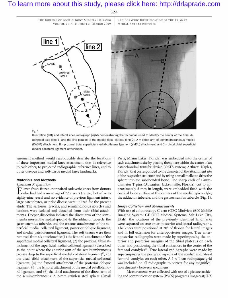
Fig. 5
Illustration (left) and lateral knee radiograph (right) demonstrating the technique used to identify the center of the tibial di-aphyseal axis (line 1) and the line parallel to the medial tibial plateau (line 2). A = direct arm of semimembranosus muscle(DASM) attachment, B = proximal tibial superficial medial collateral ligament (sMCL) attachment, and C = distal tibial superficialmedial collateral ligament attachment.
524
THE JOURNAL OF BONE & JOINT SURGERY d J B J S .ORG
VOLUME 91-A d NUMBER 3 d MARCH 2009RADIOGRAPHIC IDENTIF ICAT ION OF THE PRIMARY
MEDIAL KNEE STRUCTURES
To learn more about this study, please click here: http://drlaprade.com
Systems [division of GE Healthcare], Burlington, Vermont). Dis-tances between structure attachmentsweremeasured to the centerof the radiopaque markers, while measurements to the medialepicondyle, gastrocnemius tubercle15, and adductor tubercle weremade to the blunt ends of the T-pins, which were, as previouslystated, flush with the edge of the osseous cortex.
Interstructure distances as well as perpendicular distancesbetween structures and reference lines were measured by threeindependent examinerswithdiffering experience levels, includinga research fellow (Examiner 1 [C.A.W.]), a fourth-year medicalstudent (Examiner 2 [C.J.G.]), and a board-certified orthopaedicsurgeon (Examiner 3 [R.F.L.]). All measurements were repeatedafter an interval of at least two weeks in order to limit any recallbias. Each observer was blinded to the others’ readings.
On the anteroposterior radiographs, the positions offemoral landmarks were measured perpendicular to a refer-ence line drawn tangential to the most distal aspect of thefemoral condyles (Fig. 2). Similarly, tibial landmarks weremeasured perpendicular to a reference line intersecting themost proximal aspects of the medial and lateral tibial plateaus(Fig. 3). On the lateral radiographs, femoral structures weremeasured both in relation to a reference line projected distallyalong the posterior femoral cortex and also in relation to areference line that intersected the posterior aspect of theBlumensaat line and was perpendicular to the posterior fem-oral cortex extension line (Fig. 4)24. The locations of the tibial
attachments on the lateral radiographs were determined bymeasuring the distances between the structures and a referenceline defining the diaphyseal axis of the tibia25. Two circles (oneproximal circle, which was immediately distal to the tibial tu-bercle, and one distal circle, which was approximately 3 cmdistal to the first circle) were digitally created in the PACSprogram such that the anterior and posterior tibial diaphysealcortices were tangential to the circumferences of the circles. Aline drawn intersecting the centers of both circles representedthe diaphyseal axis. Finally, a line that represented the tibialslope was drawn parallel to the anterior and posterior edges ofthe medial tibial plateau on the lateral radiographs (Fig. 5).
Statistical AnalysisStatistical analysis was performed to examine intraobserverreproducibility and interobserver reliability. Single-measure intra-class correlation coefficients were calculated to determine varia-bility within and among observers for measurement groups26.Intraclass correlation coefficients of >0.75 were consideredexcellent27.
Source of FundingIn support of the present study, grants from the ResearchCouncil of Norway (grant #175047/D15) and Health EastNorway (grant #10703604) were used to pay for the salaries oflaboratory personnel, University of Minnesota overhead ex-penses, supplies, illustrations, cadaveric knees, travel costs for theNorwegian authors, and other such expenses related to the study.
Results
All measurements were made in reference to the centers ofthe structures’ attachment sites and are reported as the av-
erage and the standard deviation. For the purposes of thepresent study, we defined positions anterior or proximal to the
TABLE I Quantitative Relationships of Medial Knee Structures
to Landmarks and Reference Lines on Femoral
Anteroposterior Radiograph
Relationship Distance* (mm)
Distance from femoral attachment ofsuperficial medial collateral ligament to:
Femoral condylar line 30.5 ± 2.4Medial epicondyle 2.8 ± 1.1Adductor tubercle 17.9 ± 0.4Gastrocnemius tubercle 11.0 ± 2.5Posterior oblique ligament 5.9 ± 2.1Medial patellofemoral ligament 12.2 ± 2.4
Distance from femoral attachment ofposterior oblique ligament to:
Femoral condylar line 34.8 ± 2.7Medial epicondyle 7.0 ± 2.0Adductor tubercle 13.3 ± 1.1Gastrocnemius tubercle 6.6 ± 1.6Medial patellofemoral ligament 7.7 ± 1.3
Distance from femoral attachment ofmedial patellofemoral ligament to:
Femoral condylar line 42.3 ± 2.1Medial epicondyle 13.3 ± 2.4Adductor tubercle 6.2 ± 1.5Gastrocnemius tubercle 3.1 ± 1.0
*The values are given as the mean and the standard deviation.
TABLE II Quantitative Relationships of Medial Knee Structures
to Landmarks and Reference Lines on Tibial
Anteroposterior Radiograph
Relationship Distance* (mm)
Distance from proximal tibial attachment ofsuperficial medial collateral ligament to:
Tibial plateau line 211.2 ± 3.5Distal attachment of superficial medialcollateral ligament
50.4 ± 5.0
Distance from distal tibial attachment ofsuperficial medial collateral ligament to:
Tibial plateau line 260.1 ± 5.5
Distance from direct arm ofsemimembranosus muscle to:
Tibial plateau line 210.9 ± 3.7
*The values are given as the mean and the standard deviation.Negative values indicate a position posterior or distal in relation tothe reference line.
525
THE JOURNAL OF BONE & JOINT SURGERY d J B J S .ORG
VOLUME 91-A d NUMBER 3 d MARCH 2009RADIOGRAPHIC IDENTIF ICAT ION OF THE PRIMARY
MEDIAL KNEE STRUCTURES
To learn more about this study, please click here: http://drlaprade.com

reference lines as having positive values andpositionsposteriorordistal to the reference lines as having negative values.With regardto the anteroposterior radiographs, the line drawn parallel to thedistal aspect of the femoral condyleswas referred to as the femoralcondylar line and the line that crossed the proximal aspect of thetibial plateaus was referred to as the tibial plateau line.
Results on the lateral femoral radiographs were qualita-tivelydescribed according to their locationwith a quadrant25. Thequadrants were created by two intersecting lines, one along theposterior femoral cortex and one perpendicular to the posteriorfemoral cortex extension reference line and passing through theposterior aspect of the Blumensaat line. The four quadrantscreated were the (1) anteroproximal, (2) posteroproximal,(3) anterodistal, and (4) posterodistal quadrants (Fig. 4).
Superficial Medial Collateral LigamentAnteroposterior RadiographsThe femoral attachment of the superficial medial collateralligament was 2.8 ± 1.1 mm proximal to the medial epicondyle,
17.9 ± 0.4 mm distal to the adductor tubercle, and 11.0 ± 2.5mm distal to the gastrocnemius tubercle (Table I). The femoralattachment of the superficial medial collateral ligament was5.9 ± 2.1 mm distal to the femoral posterior oblique ligamentattachment point and 12.2 ± 2.4 mm distal to the femoralmedial patellofemoral ligament attachment point. In addition,the femoral attachment of the superficial medial collateralligament was 30.5 ± 2.4 mm proximal to the femoral joint line(Table I).
The proximal tibial attachment of the superficial medialcollateral ligamentwas 11.2± 3.5mmdistal to the tibial joint line.The distal tibial attachment of the superficial medial collateralligament was 60.1 ± 5.5 mm distal to the tibial joint line and50.4± 5.0mmdistal to the proximal tibial attachment (Table II).
Lateral RadiographsQualitatively, the femoral attachment of the superficial medialcollateral ligament was located in the anterodistal quadrant(Fig. 4). Quantitatively, it was 6.0 ± 0.8 mm from the medialepicondyle, 20.7 ± 2.9 mm from the adductor tubercle, and17.5 ± 2.7 mm from the gastrocnemius tubercle (Table III).The femoral attachment of the superficial medial collateralligament was 12.9 ± 3.0 mm from the femoral attachment ofthe posterior oblique ligament and 14.2 ± 2.6 mm from thefemoral attachment of the medial patellofemoral ligament.With regard to the osseous reference lines, it was 8.6 ± 3.6 mmanterior to the posterior femoral cortex extension line and11.0 ± 2.3 mm distal to the reference line perpendicular to theposterior femoral cortex and intersecting the posterior aspectof the Blumensaat line.
TABLE III Quantitative Relationships of Medial Knee Structures
to Landmarks and Reference Lines on Femoral Lateral
Radiograph
Relationship Distance* (mm)
Distance from femoral attachment ofsuperficial medial collateral ligament to:
Posterior femoral cortex extension line 8.6 ± 3.6Perpendicular line to Blumensaat line† 211.0 ± 2.3Medial epicondyle 6.0 ± 0.8Adductor tubercle 20.7 ± 2.9Gastrocnemius tubercle 17.5 ± 2.7Posterior oblique ligament 12.9 ± 3.0Medial patellofemoral ligament 14.2 ± 2.6
Distance from femoral attachment ofposterior oblique ligament to:
Posterior femoral cortex extension line 22.4 ± 4.4Perpendicular line to Blumensaat line† 25.6 ± 2.8Medial epicondyle 18.1 ± 2.8Adductor tubercle 16.4 ± 1.3Gastrocnemius tubercle 7.7 ± 1.9Medial patellofemoral ligament 14.2 ± 1.9
Distance from femoral attachment ofmedial patellofemoral ligament to:
Posterior femoral cortex extension line 8.8 ± 5.3Perpendicular line to Blumensaat line† 2.6 ± 2.1Medial epicondyle 15.9 ± 3.2Adductor tubercle 8.9 ± 2.0Gastrocnemius tubercle 12.5 ± 3.0
*The values are given as the mean and the standard deviation.Negative values indicate a position posterior or distal in relation tothe reference line. †Reference line perpendicular to the posteriorfemoral cortex extension reference line intersecting the posterioraspect of the Blumensaat line.
TABLE IV Quantitative Relationships of Medial Knee Structures
to Landmarks and Reference Lines on Tibial Lateral
Radiograph
Relationship Distance* (mm)
Distance from proximal tibial attachment ofsuperficial medial collateral ligament to:
Diaphyseal axis of tibia 226.5 ± 5.8Tibial slope line 215.9 ± 5.2Distal attachment of superficialmedial collateral ligament
51.7 ± 4.6
Direct arm of semimembranosus muscle 15.9 ± 3.0
Distance from distal tibial attachment ofsuperficial medial collateral ligament to:
Diaphyseal axis of tibia 211.8 ± 3.2Tibial slope line 266.1 ± 3.6
Distance from direct arm ofsemimembranosus muscle to:
Diaphyseal axis of tibia 43.0 ± 2.5Tibial slope line 13.9 ± 3.5
*The values are given as the mean and the standard deviation.Negative values indicate a position posterior or distal in relation tothe reference line.
526
THE JOURNAL OF BONE & JOINT SURGERY d J B J S .ORG
VOLUME 91-A d NUMBER 3 d MARCH 2009RADIOGRAPHIC IDENTIF ICAT ION OF THE PRIMARY
MEDIAL KNEE STRUCTURES
To learn more about this study, please click here: http://drlaprade.com

The proximal tibial attachment of the superficial medialcollateral ligament was located 51.7 ± 4.6 mm from the distaltibial attachment of the superficial medial collateral ligamentand 15.9 ± 3.0 mm from the attachment of the direct arm ofthe semimembranosus muscle (Table IV). Overall, it was26.5 ± 5.8 mm posterior to the tibial diaphyseal axis and 15.9 ±5.2 mm distal to the tibial slope line. The distal tibial attach-ment of the superficial medial collateral ligament was 11.8 ±3.2 mm posterior to the tibial diaphyseal axis line and 66.1 ±3.6 mm distal to the tibial slope line.
Posterior Oblique LigamentAnteroposterior RadiographsThe femoral attachment of the posterior oblique ligament was34.8 ± 2.7 mm proximal to the femoral joint line. It was 7.0 ±2.0 mm proximal to the medial epicondyle, 13.3 ± 1.1 mmdistal to the adductor tubercle, 6.6 ± 1.6 mm distal to thegastrocnemius tubercle, and 7.7 ± 1.3 mm distal to the medialpatellofemoral ligament attachment point (Table I).
Lateral RadiographsQualitatively, the femoral attachment of the posterior obliqueligament was located in the posterodistal quadrant (Fig. 4).Quantitatively, the posterior oblique ligament attachment was18.1 ± 2.8 mm from the medial epicondyle, 16.4 ± 1.3 mmfrom the adductor tubercle, 7.7 ± 1.9 mm from the gastroc-
nemius tubercle, and 14.2 ± 1.9 mm from the medial pa-tellofemoral ligament attachment site. With regard to thereference lines, the posterior oblique ligament attachmentwas 2.4 ± 4.4 mm posterior to the posterior femoral cortexextension line and 5.6 ± 2.8 mm distal to the perpendicularline at the posterior aspect of the Blumensaat line (TableIII).
Medial Patellofemoral LigamentAnteroposterior RadiographsThe femoral attachment of the medial patellofemoral ligamentwas 42.3 ± 2.1 mm proximal to the femoral joint line. It was13.3± 2.4mmproximal to themedial epicondyle, 6.2± 1.5mmdistal to the adductor tubercle, and 3.1 ± 1.0 mm proximal tothe gastrocnemius tubercle (Table I).
Lateral RadiographsQualitatively, the medial patellofemoral ligament femoral at-tachment was in the anteroproximal quadrant (Fig. 4). Quanti-tatively, the medial patellofemoral ligament was 15.9 ± 3.2 mmfrom the medial epicondyle, 8.9 ± 2.0 mm from the adductortubercle, and 12.5 ± 3.0 mm from the gastrocnemius tubercle.With regard to the femoral reference lines, it was 8.8 ± 5.3 mmanterior to the posterior femoral cortex extension line and 2.6 ±2.1 mm proximal to the perpendicular line at the posterioraspect of the Blumensaat line (Table III).
TABLE V Quantitative Radiographic Measurement Comparison of Medial Knee Structures to Previously Defined Gross Anatomy
Measurements
Distance* (mm)
Relationship Current Study† LaPrade et al.15‡
Lateral radiographDistance from femoral attachment of superficial medial collateral ligament to:Medial epicondyle 6.0 ± 0.8 6.4 (3.9 to 9.6)Posterior oblique ligament 12.9 ± 3.0 11.1 (5.7 to 14.9)
Distance from femoral attachment of posterior oblique ligament to:Medial epicondyle 18.1 ± 2.8 16.5 (11.2 to 19.0)
Distance from attachment of medial patellofemoral ligament to:Medial epicondyle 15.9 ± 3.2 14.2 (6.9 to 18.8)
Anteroposterior radiographDistance from femoral attachment of superficial medial collateral ligament to:Femoral condylar line 30.5 ± 2.4 26.8 (13.1 to 32.2)
Distance from proximal tibial attachment of superficial medial collateral ligament to:Tibial plateau line 211.2 ± 3.5 212.2 (28.8 to 215.3)
Distance from distal tibial attachment of superficial medial collateral ligament to:Tibial plateau line 260.1 ± 5.5 261.2 (252.4 to 270.5)Proximal tibial attachment of superficial medial collateral ligament 51.7 ± 4.6 49.2 (41.7 to 55.9)
Distance from direct arm of semimembranosus to:Tibial plateau line 210.9 ± 3.7 212.1 (29.2 to 214.9)
*Negative values indicate a position posterior or distal in relation to the reference line. †The values are given as the mean and the standarddeviation. ‡The values are given as the mean, with the range in parentheses.
527
THE JOURNAL OF BONE & JOINT SURGERY d J B J S .ORG
VOLUME 91-A d NUMBER 3 d MARCH 2009RADIOGRAPHIC IDENTIF ICAT ION OF THE PRIMARY
MEDIAL KNEE STRUCTURES
To learn more about this study, please click here: http://drlaprade.com

Data AnalysisIntraobserver intraclass correlation coefficients were 0.996,0.995, and 0.997 for Examiners 1, 2, and 3, respectively. Theoverall combined intraobserver intraclass correlation coef-ficient was 0.996, which demonstrates a high likelihood thatthe examiners were able to consistently draw accurate referencelines and to obtain reproducible measurements between thesemedial knee structures. Interobserver reliability was assessedbetween each of the examiners in the first and second trials aswell as for both trials combined. There was no significantdifference between examiners for either trial state. The overallinterobserver intraclass correlation coefficient for the com-bined trial was 0.994, which indicates that persons not involvedwith this study would have a high probability of measuringsimilar distances between these medial knee structures on thesame radiographs.
Discussion
Reconstructions of chronic medial knee injuries have beenassociated with varying success rates, and one reason for
this inconsistency might be imprecise reattachment of therepaired or reconstructed structures. We found that we wereable to consistently assess, both qualitatively and quantitatively,the anatomic attachment sites of the superficial medial collat-eral ligament, the posterior oblique ligament, and the femoralinsertion of the medial patellofemoral ligament on standardradiographs. These attachment sites were correlated with knownradiographic locations on the distal part of the femur andproximal part of the tibia and with standard radiographicreference lines. All examiners were able to reproducibly andaccurately measure the distances between attachment sites andthe projected reference lines despite a variability in experiencelevel, as evidenced by the high interobserver and intraobserverintraclass correlation coefficients. This observation is importantbecause it demonstrates that these guidelines can be utilizedby surgeons unfamiliar with the complex anatomy associatedwith chronic or revision medial knee injuries. The qualitativedescriptions of the femoral attachment sites on the lateral ra-diographs are important because they allow these results tobe applied to both large and small knees. Furthermore, thequalitative descriptions also can be an effective method for in-traoperative verification of tunnel placement on fluoroscopyor plain radiographs, similar to the common intraoperativepractice for radiographic confirmation of tibial tunnel locationduring posterior cruciate ligament reconstruction28.
Our results compare well with those of one previous studyin which the investigators quantitatively determined the anatomiclocations of these attachment sites and osseous landmarks15. Forexample, on anteroposterior radiographs, the distances from thefemoral and tibial attachments of the superficial medial collateralligament to the femoral and tibial joint lines, and the distancefrom the attachment of the direct arm of the semimembranosustendon to the tibial joint line, are very close between the twostudies (Table V). Furthermore, comparison of the results of thetwo studies with regard to the distances between the medial epi-condyle and the superficial medial collateral ligament, the poste-
rior oblique ligament, and the medial patellofemoral ligament onlateral radiographs yielded measurement differences of £1.8 mm.These slight differences in measurements may be due to the factthat the previous study utilized three-dimensional distances be-tween structures, whereas radiographs only allow measurement oftwo-dimensional distances. Overall, we believe that the similarmeasurements between the two studies further validate the ra-diographic findings of the present study.
A qualitative review of the structure attachment sites re-veals that, on the anteroposterior radiographs, the superficialmedial collateral ligament had themost distal femoral attachmentof the investigated structures, with the posterior oblique ligamentattaching slightly proximal to it. The medial patellofemoral lig-ament was the most proximal soft-tissue attachment on themedial femoral condyle and was located approximately 1.5times the distance from the femoral condylar line in compari-son with the attachment of the superficial medial collateralligament. On the lateral radiographs, evaluation of the femorallandmarks of the medial knee structures revealed that theposterior oblique ligament was located slightly posterior andwas the closest structure to the posterior femoral cortex ex-tension line whereas the superficial medial collateral ligamentwas located anterior to this reference line and nearly equidistantfrom this line as the medial patellofemoral ligament was. Inaddition, both the posterior oblique ligament and the superfi-cial medial collateral ligament were located distal to the per-pendicular line through the posterior aspect of the Blumensaatline.
We found similarities between our study and the studyby Schottle et al.24 in terms of the qualitative location of thefemoral attachment of the medial patellofemoral ligament. Inboth studies, the femoral attachment of the medial patellofem-oral ligament was found to be located anterior to the posteriorfemoral cortex extension line and proximal to the perpendicularline at the posterior aspect of the Blumensaat line. However, wedid find quantitative differences between these studies. In thepresent study, we found that themedial patellofemoral ligamentwas located an average of 8.8 mm anterior to the posteriorfemoral cortex extension line and 2.6 mm proximal to theperpendicular line through the posterior aspect of the Blu-mensaat line, which correlates with the femoral location of themedial patellofemoral ligament in a previously publishedquantitative anatomic study15. In the study by Schottle et al.,this attachment was reported to be located 1.3 mm anteriorand 5.5 mm proximal, respectively, to these projected lines,which, on the basis of the previous quantitative anatomicstudy15, is defined as the location of the adductor tubercle.However, Schottle et al. did not utilize a mechanism of spec-imen calibration (i.e., a 1 · 1-cm calibration grid), nor did theyperform intraclass or interclass correlations between exam-iners to validate their readings.
One of the limitations of the present study was that itinvolved a small number of specimens (n = 11); however, thenumber of specimens was greater than the number of specimens(n = 8) in a study investigating the radiographic location of thefemoral attachment of the medial patellofemoral ligament24. In
528
THE JOURNAL OF BONE & JOINT SURGERY d J B J S .ORG
VOLUME 91-A d NUMBER 3 d MARCH 2009RADIOGRAPHIC IDENTIF ICAT ION OF THE PRIMARY
MEDIAL KNEE STRUCTURES
To learn more about this study, please click here: http://drlaprade.com

addition, our specimens were from donors who had been olderthan the usual ages of patients who undergo medial knee sur-gery, but the soft-tissue attachment sites and osseous landmarksof interest do not vary with age and our specimens had noevidence of previous injury or large osteophyte formation. Theage range in the present study also compares favorably with thatin a study that defined the anatomy of the medial part of theknee15. After analysis of our data, we can conclude that ourmeasurements were highly correlative. Furthermore, joint-spacenarrowing resulting from mild osteoarthritis, not observable ongross examination, did not affect the results as no measurementswere made across the joint line.
In conclusion, we believe that the attachment locationsof the main medial knee structures can be qualitatively andquantitatively correlated to osseous landmarks and projectedradiographic lines, which will allow for more consistent radio-graphic assessments of anatomic repairs and reconstructions.Utilization of these radiographic guidelines will allow for im-proved identification of the medial knee structure attachment
sites and for preoperative, intraoperative, and postoperativeassessments of surgical repairs and reconstructions of the mainmedial knee structures. nNOTE: The authors thank Conrad Lindquist for his contributions to the present study.
Coen A. Wijdicks, MScChad J. Griffith, BSRobert F. LaPrade, MD, PhDAdam Sunderland, MScElizabeth A. Arendt, MDDepartment of Orthopaedic Surgery, University of Minnesota,2450 Riverside Avenue South, R200, Minneapolis, MN 55454.E-mail address for R.F. LaPrade: [email protected]
Steinar Johansen, MDLars Engebretsen, MD, PhDOrthopaedic Center, Ulleval University Hospital and Faculty of Medicine,University of Oslo, 0407 Oslo, Norway
References
1. Fetto JF, Marshall JL. Medial collateral ligament injuries of the knee: a rationalefor treatment. Clin Orthop Relat Res. 1978;132:206-18.
2. Najibi S, Albright JP. The use of knee braces, part 1: prophylactic knee braces incontact sports. Am J Sports Med. 2005;33:602-11.
3. Grood ES, Noyes FR, Butler DL, Suntay WJ. Ligamentous and capsular restraintspreventing straight medial and lateral laxity in intact human cadaver knees. J BoneJoint Surg Am. 1981;63:1257-69.
4. Hughston JC. The importance of the posterior oblique ligament in repairs ofacute tears of the medial ligaments in knees with and without an associatedrupture of the anterior cruciate ligament. Results of long-term follow-up. J BoneJoint Surg Am. 1994;76:1328-44.
5. Kannus P. Long-term results of conservatively treated medial collateral ligamentinjuries of the knee joint. Clin Orthop Relat Res. 1988;226:103-12.
6. LaPrade RF. The medial collateral ligament complex and the posterolateralaspect of the knee. In: Arendt EA, editor. Orthopaedic knowledge update. Sportsmedicine 2. Rosemont, IL: American Academy of Orthopaedic Surgeons; 1999. p327-40.
7. Amis AA, Firer P, Mountney J, Senavongse W, Thomas NP. Anatomy and bio-mechanics of the medial patellofemoral ligament. Knee. 2003;10:215-20.
8. Brantigan OC, Voshell AF. The tibial collateral ligament: its function, its bursae,and its relation to the medial meniscus. J Bone Joint Surg Am. 1943;25:121-31.
9. Muller W. The knee: form, function, and ligament reconstruction. New York:Springer; 1983. p 8-75.
10. Robinson JR, Bull AM, Amis AA. Structural properties of the medial collateralligament complex of the human knee. J Biomech. 2005;38:1067-74.
11. Robinson JR, Sanchez-Ballester J, Bull AM, Thomas R de W, Amis AA. Theposteromedial corner revisited. An anatomical description of the passive restrain-ing structures of the medial aspect of the human knee. J Bone Joint Surg Br.2004;86:674-81.
12. Slocum DB, Larson RL. Rotatory instability of the knee. Its pathogenesis and aclinical test to demonstrate its presence. J Bone Joint Surg Am. 1968;50:211-25.
13. Warren LA, Marshall JL, Girgis F. The prime static stabilizer of the medial sideof the knee. J Bone Joint Surg Am. 1974;56:665-74.
14. Wymenga AB, Kats JJ, Kooloos J, Hillen B. Surgical anatomy of the medialcollateral ligament and the posteromedial capsule of the knee. Knee Surg SportsTraumatol Arthrosc. 2006;14:229-34.
15. LaPrade RF, Engebretsen AH, Ly TV, Johansen S, Wentorf FA, Engebretsen L.The anatomy of the medial part of the knee. J Bone Joint Surg Am. 2007;89:2000-10.
16. Bicos J, Fulkerson JP, Amis A. Current concepts review: the medial patellofem-oral ligament. Am J Sports Med. 2007;35:484-92.
17. Haimes JL, Wroble RR, Grood ES, Noyes FR. Role of the medial structures inthe intact and anterior cruciate ligament-deficient knee. Limits of motion in thehuman knee. Am J Sports Med. 1994;22:402-9.
18. Kaplan EB. Some aspects of functional anatomy of the human knee joint. ClinOrthop Relat Res. 1962;23:18-29.
19. Last RJ. Some anatomical details of the knee joint. J Bone Joint Surg Br.1948;30:683-8.
20. Warren LF, Marshall JL. The supporting structures and layers on themedial side of the knee: an anatomical analysis. J Bone Joint Surg Am. 1979;61:56-62.
21. Fischer RA, Arms SW, Johnson RJ, Pope MH. The functional relationship of theposterior oblique ligament to the medial collateral ligament of the human knee. AmJ Sports Med. 1985;13:390-7.
22. Mendes LF, Pretterklieber ML, Cho JH, Garcia GM, Resnick DL, Chung CB.Pellegrini-Stieda disease: a heterogeneous disorder not synonymous with ossifi-cation/calcification of the tibial collateral ligament-anatomic and imaging investi-gation. Skeletal Radiol. 2006;35:916-22.
23. Mazzuca SA, Brandt KD, Dieppe PA, Doherty M, Katz BP, Lane KA. Effect ofalignment of the medial tibial plateau and x-ray beam on apparent progression ofosteoarthritis in the standing anteroposterior knee radiograph. Arthritis Rheum.2001;44:1786-94.
24. Schottle PB, Schmeling A, Rosenstiel N, Weiler A. Radiographic landmarks forfemoral tunnel placement in medial patellofemoral ligament reconstruction. AmJ Sports Med. 2007;35:801-4.
25. Pietrini SD, LaPrade RF, Griffith CJ, Wijdicks CA, Ziegler CG. Quantification ofposterolateral knee radiographic landmarks. Am J Sports Med. In press.
26. Landis JR, Koch GG. The measurement of observer agreement for categoricaldata. Biometrics. 1977;33:159-74.
27. Shrout PE, Fleiss JL. Intraclass correlations: uses in assessing rater reliability.Psychol Bulletin. 1979;86:420-8.
28. Sekiya JK, West RV, Ong BC, Irrgang JJ, Fu FH, Harner CD. Clinical outcomesafter isolated arthroscopic single-bundle posterior cruciate ligament reconstruction.Arthroscopy. 2005;21:1042-50.
529
THE JOURNAL OF BONE & JOINT SURGERY d J B J S .ORG
VOLUME 91-A d NUMBER 3 d MARCH 2009RADIOGRAPHIC IDENTIF ICAT ION OF THE PRIMARY
MEDIAL KNEE STRUCTURES
To learn more about this study, please click here: http://drlaprade.com

![TheInfluenceofPartialKneeReplacementDesignson ...Medial knee OA causes severe knee pain and knee stiff-ness, reduces knee function, and leads to disability [1, 8]. The most common](https://img.pdfslide.net/doc/110x75/5e7d90350c36be371f219f33/theiniuenceofpartialkneereplacementdesignson-medial-knee-oa-causes-severe.jpg)