Embed Size (px)
Citation preview
SM Journal of Urology
Gr upSM
How to cite this article Winter A, Engels S, Süykers M, Henke RP and Wawroschek F. Radioisotope Guided Sentinel Lymph Node Dissection in Prostate Cancer: Rate of Lymph Node Involvement Depending on Preoperative Tumor Characteristics in More Than 2,100 Patients. SM J Urol. 2015; 1(1): 1002.
https://dx.doi.org/10.36876/smju.1002
OPEN ACCESS
ISSN: 2574-8017
IntroductionPLND is still the gold standard for LN staging in clinically localized prostate cancer. The
diagnostic accuracy of available imaging procedures is quite inferior to the histological verification of LN metastases. The LN status is a crucial prognostic factor in prostate cancer. Presence and extension of LNI is associated with an increased risk of systemic dissemination and progression of the disease. Moreover, the role of PLND as a therapeutic intervention has been the focus of renewed interest. Several reports indicate that PLND improves biochemical relapse-free survival, especially in patients with minimal lymphatic dissemination [1,2]. LNI prevalence is directly related to the number of dissected LNs and extent of the PLND [3,4]. There is general consensus that an ePLND achieves the highest staging accuracy. However, the rate of complications rises along with the number of LNs removed [5-7].
Because of therapeutic consequences, the high expenditure of time and the increased complication rate of the ePLND and due to the low detection rate of limited PLND procedures, in 1999, Wawroschek et al. started to transfer techniques and concepts of the SLN identification in other tumor entities to prostate cancer [8,9].
The clinical impact of the so-called SLN is based on the phenomenon of a primary lymphatic filter level of the tumor beyond further lymphatic spread into the adjacent LNs takes place, suggesting that a negative SLN excludes lymphatic metastasis. The sentinel technique in prostate cancer differs from those in other tumor entities. In breast cancer, malignant melanoma and penile cancer [10], a well-directed peritumoral injection is only placed to observe the lymphatic drainage of
Research Article
Radioisotope Guided Sentinel Lymph Node Dissection in Prostate Cancer: Rate of Lymph Node Involvement Depending on Preoperative Tumor Characteristics in More Than 2,100 PatientsAlexander Winter1*, Svenja Engels1, Marie-Christin Süykers1, Rolf-Peter Henke2 and Friedhelm Wawroschek1
1University Hospital for Urology, Klinikum Oldenburg, School of Medicine and Health Sciences, Carl von Ossietzky University, Oldenburg, Oldenburg, Germany2Institute of Pathology Oldenburg, Oldenburg, Germany
Article Information
Received date: Apr 17, 2015 Accepted date: Oct 01, 2015 Published date: Oct 08, 2015
*Corresponding author
Alexander Winter, University Hospital for Urology, Klinikum Oldenburg, School of Medicine and Health Sciences, Carl von Ossietzky University Oldenburg, Rahel-Straus-Str. 10, 26133 Oldenburg, Germany, Tel: +49 0 441 403 2302; Fax: +49 0 441 403 2303; Email: [email protected]
Distributed under Creative Commons CC-BY 4.0
Keywords Prostate cancer; Sentinel lymph node dissection; Lymphadenectomy; Lymph node involvement
Abbreviations ICG: Indocyanine Green; LN: Lymph Node; LNI: Lymph Node Involvement; PLND: Pelvic Lymph Node Dissection; PSA: Prostate Specific Antigen; SLN: Sentinel Lymph Node; sPLND: sentinel Pelvic Lymph Node Dissection; ePLND: extended Pelvic Lymph Node Dissection
Article DOI 10.36876/smju.1002
Abstract
Background: Extended pelvic lymph node dissection is the gold standard for lymph node staging in prostate cancer. Sentinel lymph node dissection has replaced extended lymphadenectomy in several tumors. The aim of the study was to stratify the rate of lymph node involvement in prostate cancer patients undergoing radio guided sentinel lymph node dissection depending on preoperative tumor characteristics.
Methods: We analyzed 2,102 prostate cancer patients (median age 67 years, IQR 62-71 years) who underwent radioisotope guided sentinel lymphadenectomy and retropubic radical prostatectomy between January 2005 and February 2015 in a retrospective single center study. Median prostate specific antigen was 7.8 ng/ml (IQR 5.5-12.7 ng/ml). 99mTechnetium nanocolloid (ca. 200 MBq) was transrectally injected into the prostate. A few hours later scintigraphy was carried out. Sentinel lymph nodes were intraoperatively detected using a gamma probe. The rate of lymph node invasion was analyzed for D’Amico risk groups and in relation to biopsy Gleason scores.
Results: The median number of lymph nodes removed was 10 (IQR 7-13). Overall, 19.3% of patients (n=405) had lymph node involvement; 2.9% (n=18) in low, 15.6% (n=139) in intermediate and 42.0% (n=248) in high risk disease. 64 (6.5%) of 984 patients with Gleason score ≤6 prostate cancer were lymph node positive; 20.6% (n=154) or 44.2% (n=84) in patients with Gleason score 7 (3+4; 4+3) and 57.5% (n=103) in Gleason score ≥8 prostate cancer.
Conclusion: We present the largest study on sentinel lymph node dissection in prostate cancer patients until now. The rate of lymph node invasion was higher in the examined sentinel collective than expected according to extended lymphadenectomy series. These results demonstrate the reliability and high sensitivity of sentinel lymphadenectomy for the detection of lymph node metastases in prostate cancer patients.
Citation: Winter A, Engels S, Süykers M, Henke RP and Wawroschek F. Radioisotope Guided Sentinel Lymph Node Dissection in Prostate Cancer: Rate of Lymph Node Involvement Depending on Preoperative Tumor Characteristics in More Than 2,100 Patients. SM J Urol. 2015; 1(1): 1002.
https://dx.doi.org/10.36876/smju.1002
Page 2/4
Gr upSM Copyright Winter A
the tumor and the first draining LN. In prostate cancer, which occurs commonly multifocal, it is unknown with absolute certainty from which part of the organ the metastatic spread originates. Therefore, the goal of prostate lymph scintigraphy must be the imaging of all primary draining LNs of the prostate, under which the SLN of cancer also exist. Wawroschek et al. stated that all pre-operatively and intraoperatively identified gamma radiation-active nodes would be defined as SLN [8,9]. The same working group has shown that the most frequent location of SLN was the external and internal iliac region (32.3%; 30.9%), followed by the obturator fossa (26.5%), the presacral (5.7%), and other regions [11].
Presently, different new tracers, such as the near-infrared fluorescent dye ICG in connection with robotic [12], laparoscopic [13], and open radical prostatectomies [14], and magnetic nanoparticles [15] are being tested for marking and intraoperative detection of SLNs. In all of these studies the intra-prostatic sentinel tracer injection did not cause any problems to the operation.
Since 2005 the Oldenburg working group has gained experience in more than 2,100 prostate cancer cases who underwent a radio guided sPLND together with a radical retropubic prostatectomy. In this study we present the largest study on radio guided sPLND in prostate cancer patients until now. We stratify the rate of LNI in prostate cancer patients undergoing sPLND depending on preoperative tumor characteristics.
Materials and MethodsPatients
A total of 2,113 consecutive prostate cancer patients (cT1, cT2, and cT3) were identified, who underwent sPLNDs in combination with radical retropubic prostatectomy carried out by four highly experienced surgeons in a single center between January 2005 and February 2015. Patients with incomplete clinical information for PSA, clinical stage, or biopsy Gleason score (n=11; 0.5%) were excluded. The final sample included 2,102 patients. All patients had been informed verbally and in writing about a sPLND and radical retropubic prostatectomy and signed a consent form.
SPLND technique
The sPLND technique was applied as described by Wawroschek et al. [9]. 99mTechnetium nanocolloid was transrectally injected 24 hours before surgery into the prostate under ultrasound guidance. Three injections were done per prostate lobe. Activity attained about 100 MBq per lobe and total injection volume was about 1.2 ml. A few hours after injection, scintigraphy was carried out.
The radioactivity of the LNs was intraoperatively measured using two different gamma probe systems (C-Trak System, Care Wise, MorganHill, CA, USA; Crystal Probe SG04, Crystal Photonics GmbH, Berlin, Germany). The two systems were used separately and have comparable characteristics (e.g. sensitivity) [16]. LNs identified as SLNs by the gamma probe were dissected. For surgical reasons, LNs other than SLNs directly adjoining and adhering to SLNs were also removed, if an in-situ separation was not possible. Furthermore, in the case of SLNs in the obturator fossa area, the surrounding non-radioactive lymphatic tissue of the fossa was also dissected. However,
lymphatic tissue of the fossa was not resected, if no SLN existed in the fossa area.
In prostate cancer, the radiation exposure to the operating surgeons due to SLN detection in the operating room (ca. 2.3 µSv) is comparable to the small exposure in breast cancer [17]. Therefore, the staff in the operating room is not to be considered as occupationally exposed to radiation (critical value: 1 mSv / year).
Histopathological examination
All LNs were initially cut in 3 mm transverse sections, routinely processed and completely embedded in paraffin; 4–5 µm thick sections were stained with hematoxylin-eosin. Selected cases of serial sections were analysed. Immunohistochemistry with a pancytokeratin antibody (AE1/AE3) was carried out to confirm or exclude metastatic spread in rare cases with inconclusive conventional histology.
Measurement
The rate of patients with LNI was analyzed in total as well as for D’Amico risk groups: low-risk (PSA ≤10ng/ml and/or Gleason sore <6 and/or ≤cT2a), intermediate-risk (PSA 10–20 ng/ml and/or Gleason score 7 and/or cT2b) and high-risk prostate cancer (PSA >20 ng/ml and/or Gleason score 8–10 and/or ≥T2c).The rate of patients with LNI was also calculated under consideration of the biopsy Gleason score.
ResultsTable 1 lists the summary of patient characteristics and details to
the incidence of LNI depending on preoperative and postoperative tumour stage and Gleason sum. The median number of LNs removed was 10 (IQR 7–13). The median number of positive LNs per patient was 2 (IQR 1–3). Overall, 19.3% of patients (n=405) had LNI.
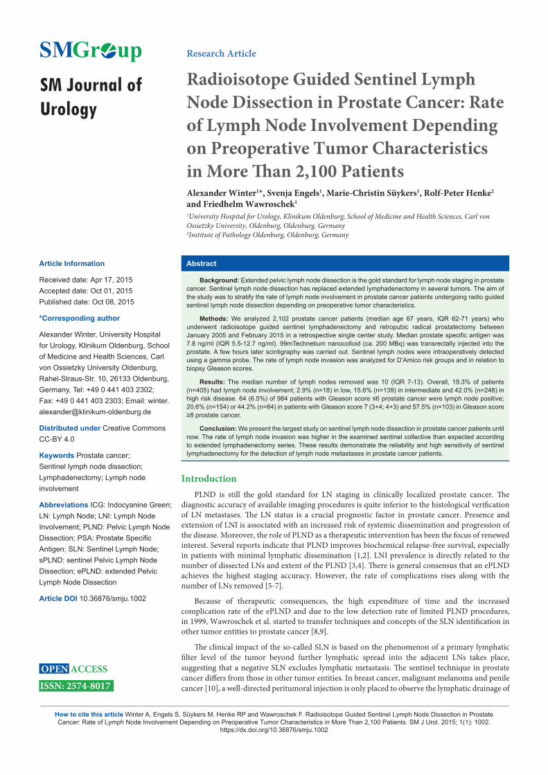
The incidence of LNI patients depending on D’Amico risk groups ranged from 2.9% (low risk) to 42.0% in high risk prostate cancer patients. The distribution of the risk groups of the examined population can be found in Figure 1.
In consideration of the biopsy Gleason score, 6.5% of the patients with Gleason score ≤6 were LN positive. Regarding the patients with
Figure 1: Incidence of patients with lymph node involvement depending on preoperative tumor characteristics (D’Amico risk groups) and distribution of risk groups of the examined collective. GS: Gleason score.
Citation: Winter A, Engels S, Süykers M, Henke RP and Wawroschek F. Radioisotope Guided Sentinel Lymph Node Dissection in Prostate Cancer: Rate of Lymph Node Involvement Depending on Preoperative Tumor Characteristics in More Than 2,100 Patients. SM J Urol. 2015; 1(1): 1002.
https://dx.doi.org/10.36876/smju.1002
Page 3/4
Gr upSM Copyright Winter A
Gleason score 7 (3+4, respectively 4+3), 20.6% and 44.2% were LN positive. Patients with Gleason score ≥8 prostate cancer showed LN metastases in 57.5% of cases. Table 2 shows the number of patients, LNs and LN metastases in relation to biopsy Gleason score in detail.
DiscussionThere is general consensus that extended PLND achieves the
highest accuracy for LN staging in prostate cancer patients. However, the staging benefit of ePLND is accompanied by the potential risk of morbidity. Therefore, sentinel guided LN dissection has replaced extended lymphadenectomy in several tumors. Different studies show that ePLND increase the risk of morbidity in radical prostatectomy. In three of these studies, the complications of PLND increased significantly with the number of dissected LN [5-7].
SPLND has been introduced in some centers to solve this dilemma. In these studies, a high staging accuracy and a low morbidity of sPLND has been shown [9]. In a meta-analysis [18] the pooled detection rate of sPLND was 93.8% with a pooled sensitivity rate of 94%. In the largest study [19] conducted, falsely detected negative results (non-SLN metastases found in the absence of SLN metastases) were found in less than 6% of the cases. In the present study, more LNI patients were detected by sPLND than expected according to the data of ePLND series (Table 3).
In order to find a compromise between a reliable LN staging and the potential morbidity of the PLND guidelines recommend candidates for extended PLND as patients with intermediate or high risk prostate cancer [24,25]. However, there is no consensus on the risk-level of a LNI that would be the ideal cut-off. For instance, the National Comprehensive Cancer Network deems a cut-off acceptable if it leads to waiving 50% of the PLNDs prior to radical prostatectomy at the expense of proof or removal of LN metastases in 12% of the cases with LNI [26]. The EAU guidelines suggest that PLND might
be spared in patients with <5% risk of LNI calculated by a nomogram based on extended PLND [20,22]. We have not yet defined a cut-off for choosing a sPLND. In view of the low morbidity of sPLNDs in combination with the high sensitivity of proof of metastases, we question the ability to define a cut-off. One should also note that especially patients with minimal LNI appear to benefit from removal of lymph node metastases [27].
On the other side, the sentinel approach is also subject to limitations. One problem with this technique is that when LNs are fully metastasized or lymph pathways are blocked, the afferent lymph will be directed to other LNs/ non-sentinel LNs [28]. These nodes will not be positive on SLN imaging, resulting in false negative findings. The false negative rate was shown to correlate with the Gleason score. Patients with a high-risk disease could thus have both positive SLNs and positive non-SLNs [29]. If the goal in such cases is to remove all pelvic LN metastases, high risk patients have the option of undergoing a combination of a sPLND and an ePLND. As such, the possibility of an ePLND overlooking a part of the LN metastases, possibly in the pre-sacral region, is overcome by being able to detect it through the sPLND. Reportedly, Joniau et al. [30] did not detect 13% of metastatic LNs by applying only an ePLND.
The radioisotope guided sentinel technique also has specific drawbacks. This procedure depends on the availability of nuclear medicine and radio tracers, which in recent years have been problematic because of cutbacks in production. Furthermore, the use of radioisotope exposes patients and healthcare workers to radiation and is heavily controlled by legislation. This calls for the development of new, simpler, radiation-free, but accurate methods, for SLN marking and intraoperative detection - especially those that a urologist can apply independently without the complicated and problematic logistics associated with nuclear medicine. Presently, different tracers, such as the near-infrared fluorescent dye ICG, are being tested to mark SLNs, especially in connection with robotic [15] and laparoscopic [16] radical prostatectomies, and super- paramagnetic iron oxid nanoparticles in the open procedure [15] with promising results. In the last mentioned feasibility study we have shown that a radiation-free, magnetometer guided sPLND procedure is simple and can be performed alone by a urologist. In the case of
Overalln= 2102
pN0n= 1697 (80.7 %)
pN1n= 405 (19.3 %)
Median age at surgery in yrs (IQR) 67 (62 – 71) 67 (61 – 71) 68 (63 – 71)
Median total PSA ng/ml (IQR) 7.8 (5.5 – 12.7) 7.1 (5.3 – 10.9) 12.3 (7.8 – 20.7)
Median No. of LN removed (IQR) 10 (7 – 13) 10 (7 – 13) 12 (9 – 14,5)
Median No. of positive LN (IQR) - 0 (-) 2 (1 – 3)
T-category (%)T1cT2T3
1141 (54.3)918 (43.7)
43 (2.0)
1036 (61.0)652 (38.4)
9 (0.5)
105 (25.9)266 (65.7)
34 (8.4)Biopsy Gleason sum (%)≤ 67≥ 8
984 (46.8)939 (44.7)179 (8.5)
920 (54.2)701 (41.3)
76 (4.5)
64 (15.8)238 (58.8)103 (25.4)
Postoperative Gleason sum (%)≤ 67≥ 8
345 (16.4)1548 (73.6)
209 (9.9)
342 (20.2)1280 (75.4)
75 (4.4)
3 (0.7)268 (66.2)134 (33.1)
Pathologic stagepT2apT2bpT2cpT3apT3bpT4
191 (9.1)38 (1.8)
1084 (51.6)408 (19.4)327 (15.6)
54 (2.6)
186 (11.0)37 (2.2)
1044 (61.5)298 (17.6)117 (6.9)15 (0.9)
5 (1.2)1 (0.2)
40 (9.9)110 (27.2)210 (51.9)
39 (9.6)
Table 1: Patient characteristics.
Gleason score
Patients (n)
pN1 Patients (%)
Lymph nodes (n)
pN1 Lymph nodes (%)
≤ 6 984 64 (6.5) 9866 137 (1.4)7 (3+4) 749 154 (20.6) 8169 377 (4.6)7 (4+3) 190 84 (44.2) 2168 221 (10.2)≥ 8 179 103 (57.5) 2338 310 (13.3)
Table 2: Number of patients, lymph nodes and lymph node metastases in relation to biopsy Gleason score.
LiteraturPrevalence of LN metastases Number of patients PLND
method% n
Briganti et al. [20] 11.0 602 ePLNDGodoy et al. [21] 5.2 4,176 ePLNDBriganti et al. [22] 8.3 588 ePLNDAbdollha et al. [23] 13.8 5,274 ePLNDWinter et al. 19.4 2,102 sPLND
Table 3: Prevalence of lymph node metastases in extended lymphadenecomy and radioguided sPLND series in comparison.
Citation: Winter A, Engels S, Süykers M, Henke RP and Wawroschek F. Radioisotope Guided Sentinel Lymph Node Dissection in Prostate Cancer: Rate of Lymph Node Involvement Depending on Preoperative Tumor Characteristics in More Than 2,100 Patients. SM J Urol. 2015; 1(1): 1002.
https://dx.doi.org/10.36876/smju.1002
Page 4/4
Gr upSM Copyright Winter A
fluorescense labeling of SLNs it hast to be consider that this approach is principally restricted by the limited tissue penetration of near-infrared fluorescence signals (< 1 cm) and the functional properties of the currently used free ICG [31]. Functionally, ICG is a rapidly clearing lymphatic perfusions marker, which does not remain in the SLNs like the radiocolloid and/or is not absorbed there like the latter by macrophages. Coupled procedures (ICG-99mTechnetium nanocolloid) are either quite extensive or again involve radioactivity.
ConclusionThis analysis represents the largest study on SLN dissection in
prostate cancer patients until now. Compared with the results of ePLND series, the higher rate of LN positive patients particularly in the low and intermediate risk groups underpins the sensitivity of the sentinel approach. This data and the promising results of studies with new and radiation-free tracers that can be used by an urologist alone, speak clearly to the future viability of the sentinel technology in prostate cancer.
References
1. Withrow DR, DeGroot JM, Siemens DR, Groome PA. Therapeutic value of lymph node dissection at radical prostatectomy: a population-based case-cohort study. BJU Int. 2011; 108: 209-216.
2. Schumacher MC, Burkhard FC, Thalmann GN, Fleischmann A, Studer UE. Good outcome for patients with few lymph node metastases after radical retropubic prostatectomy. Eur Uro. 2008; l54: 344-352.
3. Bader P, Burkhard FC, Markwalder R, Studer UE. Is a limited lymph node dissection an adequate staging procedure for prostate cancer? J Urol. 2002; 168: 514-518.
4. Heidenreich A, Ohlmann CH, Polyakov S. Anatomical extent of pelvic lymphadenectomy in patients undergoing radical prostatectomy. Eur Urol. 2007; 52: 29-37.
5. Briganti A, Chun FK, Salonia A, Suardi N, Gallina A, Da Pozzo LF, et al. Complications and other surgical outcomes associated with extended pelvic lymphadenectomy in men with localized prostate cancer. Eur Urol. 2006; 50: 1006-1013.
6. Musch M, Klevecka V, Roggenbuck U, Kroepfel D. Complications of pelvic lymphadenectomy in 1,380 patients undergoing radical retropubic prostatectomy between 1993 and 2006. J Urol. 2008; 179: 923-928.
7. Winter A, Vogt C, Weckermann D, Wawroschek F. Complications of pelvic lymphadenectomy in clinically localised prostate cancer: different techniques in comparison and dependency on the number of removed lymph nodes. Aktuelle Urol. 2011; 42: 179-183.
8. Wawroschek F, Vogt H, Weckermann D, Wagner T, Harzmann R. The sentinel lymph node concept in prostate cancer-first results of gamma probe-guided sentinel lymph node identification. Eur Urol. 1999; 36: 595-600.
9. Wawroschek F, Vogt H, Weckermann D, Wagner T, Hamm M, Harzmann R. Radioisotope guided pelvic lymph node dissection for prostate cancer. J Urol. 2001; 166: 1715-1719.
10. Cabanas RM. An approach for the treatment of penile carcinoma. Cancer. 1977; 39: 456-466.
11. Wawroschek F, Hamm M, Weckermann D, Vogt H, Harzmann R. Lymph node staging in clinically localized prostate cancer. Urol Int. 2003; 71: 129-135.
12. Manny TB, Patel M, Hemal AK. Fluorescence-enhanced robotic radical prostatectomy using real-time lymphangiography and tissue marking with percutaneous injection of unconjugated indocyanine green: the initial clinical experience in 50 patients. Eur Urol. 2014; 65: 1162-1168.
13. Jeschke S, Lusuardi L, Myatt A, Hruby S, Pirich C, Janetschek G. Visualisation of the lymph node pathway in real time by laparoscopic radioisotope- and fluorescence-guided sentinel lymph node dissection in prostate cancer staging. Urology, 2012; 80: 1080-1086.
14. Yuen K, Miura T, Sakai I, Kiyosue A, Yamashita M. Intraoperative fluorescence imaging for detection of sentinel lymph nodes and lymphatic vessels during open prostatectomy using indocyanine green. J Urol. 2015.
15. Winter A, Woenkhaus J, Wawroschek F. A novel method for intraoperative sentinel lymph node detection in prostate cancer patients using superparamagnetic iron oxide nanoparticles and a handheld magnetometer: the initial clinical experience. Ann Surg Oncol. 2014; 21: 4390-4396.
16. Wengenmair H, Kopp J, Sciuk J. Quality criteria, minimal requirements and future developments. In: Munz D.L.: The sentinel lymph node concept in oncology. Zuckerschwerdt Verlag: München, Bern, Wien, New York. 2001; 68-80.
17. Brenner W, Ostertag H, Peppert E, Czech N, Kampen WU, Muhle C, et al. Radiation exposure to the personnel in the operating room and in the pathology due to SLN detection with Tc-99m-nanocolloid in breast cancer patients. Nuklearmedizin. 2000; 39: 142-145.
18. Sadeghi R, Tabasi KT, Bazaz SM, Kakhki VR, Massoom AF, Gholami H, et al. Sentinel node mapping in the prostate cancer. Meta-analysis. Nuklearmedizin. 2011; 50: 107-115.
19. Holl G, Dorn R, Wengenmair H, Weckermann D, Sciuk J. Validation of sentinel lymph node dissection in prostate cancer: experience in more than 2,000 patients. Eur J Nucl Med Mol Imaging. 2009; 36: 1377-1382.
20. Briganti A, Chun FK, Salonia A, Gallina A, Farina E, Da Pozzo LF, et al. Validation of a nomogram predicting the probability of lymph node invasion based on the extent of pelvic lymphadenectomy in patients with clinically localized prostate cancer. BJU Int. 2006; 98: 788-793.
21. Godoy G, Chong KT, Cronin A, Vickers A, Laudone V, Touijer K, et al. Extent of pelvic lymph node dissection and the impact of standard template dissection on nomogram prediction of lymph node involvement. Eur Urol. 2011; 60: 195-201.
22. Briganti A, Larcher A, Abdollah F, Capitanio U, Gallina A, Suardi N, et al. Updated nomogram predicting lymph node invasion in patients with prostate cancer undergoing extended pelvic lymph node dissection: the essential importance of percentage of positive cores. Eur Urol. 2012; 61: 480-487.
23. Abdollah F, Suardi N, Gallina A, Bianchi M, Tutolo M, Passoni N, et al. Extended pelvic lymph node dissection in prostate cancer: a twenty year audit in a single center. Ann Oncol. 2013; 24: 1459-1466.
24. Leitlinienprogramm Onkologie (Deutsche Krebsgesellschaft, Deutsche Krebshilfe, AWMF) (2014) Interdisziplinäre Leitlinie der Qualität S3 zur Früherkennung, Diagnose und Therapie der verschiedenen Stadien des Prostatakarzinoms. Kurzversion 3.1 AWMF-Register-Nummer 043/022OL. 2015.
25. Mottet N, Bellmunt J, Briers E, van den Bergh RCN, Bolla M. Guidelines on prostate cancer. European Association of Urology. 2015.
26. Mohler J, Bahnson RR, Boston B, Busby JE, D’Amico A, Eastham JA, et al. NCCN clinical practice guidelines in oncology: prostate cancer. J Natl Compr Canc Netw. 2010; 8: 162-200.
27. Briganti A, Karnes JR, Da Pozzo LF, Cozzarini C, Gallina A, Suardi N, et al. Two positive nodes represent a significant cut-off value for cancer specific survival in patients with node positive prostate cancer. A new proposal based on a two-institution experience on 703 consecutive N+ patients treated with radical prostatectomy, extended pelvic lymph node dissection and adjuvant therapy. Eur Urol. 2009; 55: 261-270.
28. Morgan-Parkes JH. Metastases: mechanisms, pathways, and cascades. AJR Am J Roentgenol. 1995; 164: 1075-1082.
29. Weckermann D, Dorn R, Holl G, Wagner T, Harzmann R. Limitations of radioguided surgery in high-risk prostate cancer. Eur Urol. 2007; 51: 1549-1556.
30. Joniau S, Van den Bergh L, Lerut E, Deroose CM, Haustermans K, Oyen R, et al. Mapping of pelvic lymph node metastases in prostate cancer. Eur Urol. 2013; 63: 450-458.
31. Van den Berg NS, van Leeuwen FW, van der Poel HG. Fluorescence guidance in urologic surgery. Curr Opin Urol. 2012; 22: 109-120.