Embed Size (px)
Citation preview
RADIOISOTOPE KNEE JOINT SCANS IN HAEMOPHILIA
AND CHRISTMAS DISEASE
CHARLES D. FORBES, WILLIAM R. GREIG, COLIN R. M. PRENTICE, GLASGOW, ScoTLANI,
and
GEORGE P. MCNICOL,* LEEDS, ENGLAND
From the LJnilersitv Departineizi of Medicine, Glasgow Royal Infirmary
As early as I 892 K#{246}nigshowed that the recurrent haemarthroses of haemophilia resulted
in progressive joint destruction. This aspect of haemophilia has been studied by many
investigators (Freund 1925; Key 1932; Collins 1949; DePalma and Cotter l956b; Marion,
Favre-Gilly, Picault and Gauthier 1965). Observations have also been made ofjoint abnormalities
in haemophilic dogs (Swanton 1957, 1959). Nevertheless, there is still doubt about the
mechanisms underlying the acute and chronic joint changes. In acute haemarthrosis it is
thought that bleeding starts in the synovial and subsynovial tissues of the villi with formation
of sInall haematomata, which become confluent and rupture into the joint cavity to produce
the sudden severe pain and immobility (Swanton 1957, 1959). Likewise it is thought that it is
recurrent intra-articular bleeds which lead to chronic arthropathy, hypertrophv ofthe synovium
and adherence of adjacent villi progressing to produce joints with a thickened hyperaemic
synovium and fibrosis of the subsynovial tissues. After these changes are established
degenerative changes occur in the adjacent cartilage and bone.
We have investigated the changes in acute and chronic haernophilic arthropathy using
radioisotope joint scanning. Intravenous technetium (9ImTc) with a half-life of six hours is
an ideal isotope forjoint scanning (Laneet 1968) and has been used to study otherjoint diseases.
The scans, which consist of a pictorial image of the isotope localisation in and around the
joint, show changes in regional blood flow at the time of study and are abnormal during
acute inflammation (Weiss, Maxfield, Munson and Hidalgo 1965, 1966; Whaley, Pack, Boyle,
Dick, Downie, Buchanan and Gillespie 1968; McCarty, Polcyn and Collins 1970). While
visualisation of the isotope in an inflamed joint reflects mainly increased vascularity of the
synovial membrane and otherjoint tissues (Alarcon-Segovia, Trujeque, Tovar and Adame 1967;
Whaley et a!. 1968), part of the localisation of the radioactivity may be due directly to the
synovium binding the technetium (Green and Hays 1969; McCarty, Polcyn, Collins and
Gottschalk 1970; Mowat, Disney and Vaughan 1971). For these reasons joint scans are
abnormal in a variety of inflammatory joint diseases, such as rheumatoid arthritis, gout,
psoriatic arthritis, acute pseudo-gout and experimental synovitis, but are not characteristically
specific for one type of inflammatory change. Nevertheless, as McCarty and his colleagues
have shown, the joint scan may be much more abnormal than the degree of symptoms and
signs would suggest, and because they can be repeated at intervals and do give an index of
blood flow in a joint, we thought it might be helpful to use joint scanning to measure the extent
of the synovial lesion in the joints of haemophiliacs. In addition we have compared the scans
taken during acute haemarthrosis with those taken in quiescent chronic haemophilic
arthropathy. Other assessments included the scan abnormalities in relation to the number of
previous bleeding episodes and to the clinical assessment of the joint abnormality. The present
study was confined to the knee joints, where scanning is optimum for clarity and comparative
work.
* Professor of Medicine, Leeds General Infirmary.
468 TIlE JOURNAL OF BONE AND JOINT SURGERY
RADIOISOTOPE KNEE JOINT SCANS IN HAEMOPHILIA AND CHRISTMAS DISEASE 469
METHODS AND PATIENTS STUDIED
To obtain the knee-joint scans, five millicuries of technetium, as sodium pertechnetate
(NaTcO4), were given as a single bolus intravenously to each patient and the knee-joint scans
commenced twenty-five minutes later as suggested by Whaley and his colleagues. The image
of the radioactivity distribution in the joint was obtained using a rectilinear scintiscanner
(Selo Model DS4/4) which has two five-inch detectors. Scans were performed of both knees,
first in the antero-posterior and then in the lateral position. The display was in colour, the
line spacing and the scan speed being adjusted for each patient, joint and position. The time
taken for each knee was approximately fifteen minutes.
Thirty-five patients were included in this study, ranging in age from eleven to sixty years,
and informed consent was obtained from all parents or subjects. Twenty-eight had classical
haemophilia and seven had Christmas disease. There was no clinical evidence of any other
variety ofjoint disease and the anti-nuclear factor and Rose-Waaler tests were negative.
Classification of the clinical severity of haemophilia was described by Biggs and Macfarlane
(1962). Patients were graded as “severe” cases if they had suffered repeated haemarthroses
with serious crippling and deep tissue haemorrhages from little or no provocation, as
“moderate” cases if they had sustained few haemarthroses with no serious crippling or
occasional muscle haematomas, and as “mild” cases if they had no haemarthrosis or other
spontaneous bleeding and merely gave a history of abnormal bleeding after definite injury.
Thus eighteen of our patients were classified as “severe”, six as “moderate” and eleven as
“mild” cases.
In each patient specific clinical assessment of the knee joints was carried out and each
of the features was arbitrarily graded from 0-3 ; these features were pain, effusion, pyrexia,
crepitus and degree of contracture. This we have called the “signs score”. The number of
previous bleeding episodes into the kneejoints was also graded from 0-3, a score ofO recording
no previous haemarthrosis, 1 one haemarthrosis, 2 less than one haemarthrosis per year and
3 more than one haemarthrosis per year; this we have called the “incidence score”. Six of
the thirty-five patients were seen with acute haemarthroses on a background of previous
episodes ofjoint bleeding, and fifteen with chronic joint disease of varying degrees of severity.
At the time of examination two patients had subacute disease ; this category was defined as a
swollen, non-tender joint with a normal range of movement which had not been the seat of
an acute bleed for at least six months. Twelve patients gave no previous history of joint
bleeds and had normal knee joints on clinical examination. In six of the patients clinical
examination and scanning was repeated as they recovered from or developed an acute joint
bleed.
The radioisotope scan colour images were examined by one observer (C. D. F.). Various
classifications of the images were considered, but it was ultimately decided that the most
practical method should be simple and visual. Accordingly, the group of scans from each
knee joint was examined and then said to be normal if it were obviously similar to knee joint
scans from controls in other studies (group 0). In the normal knee scan there is very little
isotope uptake. If the scans were obviously abnormal in width, depth and degree of isotope
uptake, with or without irregular localisation, they were said to be very abnormal (group 3).
Very abnormal scans show a high isotope uptake. Scans which were obviously abnormal but
not grossly so were counted as grade 2. Slightly abnormal scans were grade I . Examples of
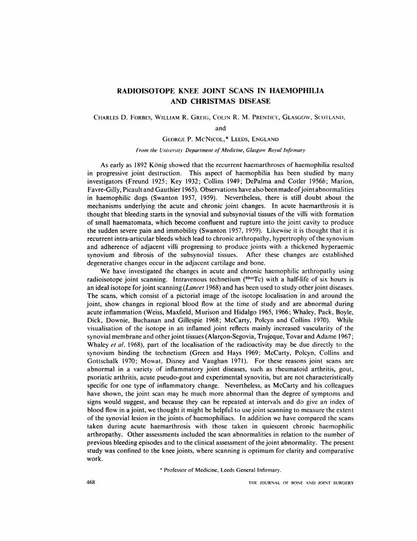
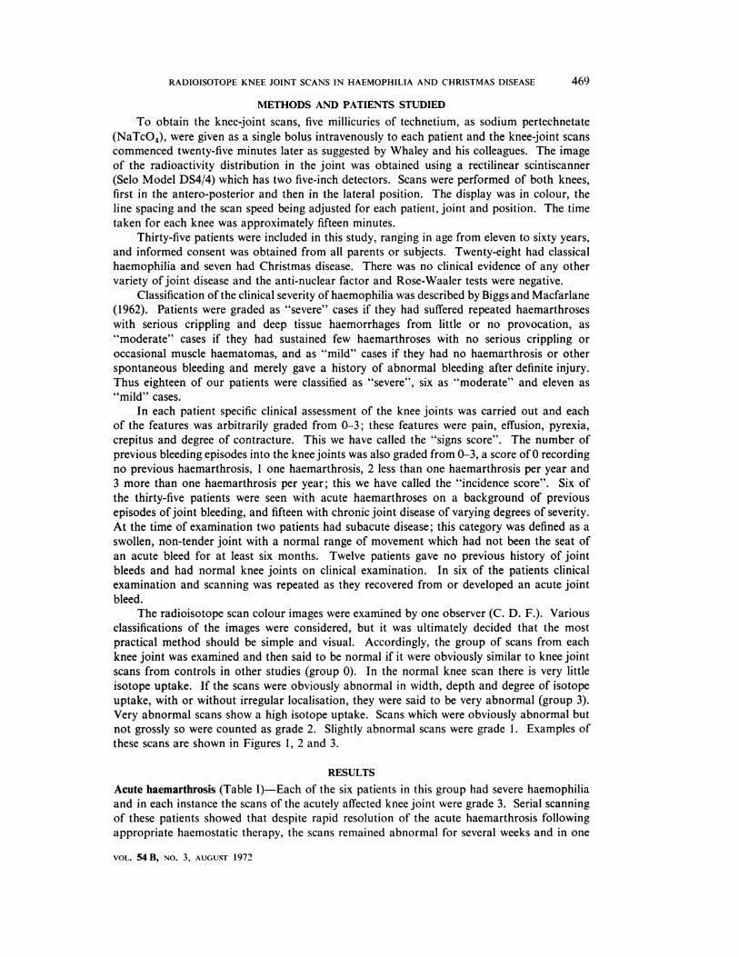
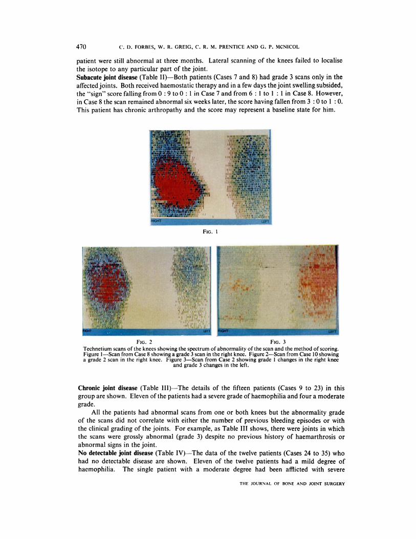
these scans are shown in Figures 1 , 2 and 3.
RESULTS
Acute haemarthrosis (Table 1)-Each of the six patients in this group had severe haemophilia
and in each instance the scans of the acutely affected knee joint were grade 3. Serial scanning
of these patients showed that despite rapid resolution of the acute haemarthrosis following
appropriate haemostatic therapy, the scans remained abnormal for several weeks and in one
VOL. 54 B, NO. 3, AUGUST 1972
I
FIG. 1
FIG. 2 FIG. 3
Technetium scans of the knees showing the spectrum of abnormality of the scan and the method of scoring.Figure I-Scan from Case 8 showing a grade 3 scan in the right knee. Figure 2-Scan from Case 10 showinga grade 2 scan in the right knee. Figure 3-Scan from Case 2 showing grade I changes in the right knee
and grade 3 changes in the left.
470 C. D. FORBES, W. R. GREIG, C. R. M. PRENTICE AND G. P. MCNICOL
THE JOURNAL OF BONE AND JOINT SURGERY
patient were still abnormal at three months. Lateral scanning of the knees failed to localise
the isotope to any particular part of the joint.
Subacute joint disease (Table 11)-Both patients (Cases 7 and 8) had grade 3 scans only in the
affectedjoints. Both received haemostatic therapy and in a few days thejoint swelling subsided,
the “sign” score falling from 0 : 9 to 0 : 1 in Case 7 and from 6 : 1 to 1 : 1 in Case 8. However,
in Case 8 the scan remained abnormal six weeks later, the score having fallen from 3 : 0 to 1 : 0.
This patient has chronic arthropathy and the score may represent a baseline state for him.
Chronic joint disease (Table 111)-The details of the fifteen patients (Cases 9 to 23) in this
group are shown. Eleven ofthe patients had a severe grade ofhaemophilia and four a moderate
grade.
All the patients had abnormal scans from one or both knees but the abnormality grade
of the scans did not correlate with either the number of previous bleeding episodes or with
the clinical grading of the joints. For example, as Table III shows, there were joints in which
the scans were grossly abnormal (grade 3) despite no previous history of haemarthrosis or
abnormal signs in the joint.
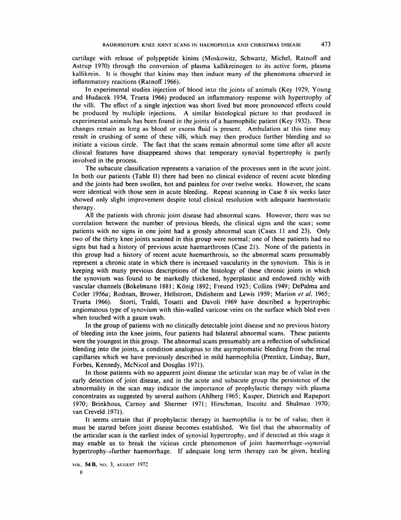
No detectable joint disease (Table IV)-The data of the twelve patients (Cases 24 to 35) who
had no detectable disease are shown. Eleven of the twelve patients had a mild degree of
haemophilia. The single patient with a moderate degree had been afflicted with severe
RADIOISOTOPE KNEE JOINT SCANS IN HAEMOPHILIA AND CHRISTMAS DISEASE 471
paralytic poliomyelitis as a child and had never borne weight on the joints of the lower limbs;
however, he had sustained multiple other bleedings consistent with this degree of defect.
Despite the absence ofjoint signs, four of the patients had abnormal scans (grade 2 or 3).
DISCUSSION
One of the earliest, most frequent and most disabling manifestations of haemophitia is
acute haemarthrosis, the kneejoint being most frequently affected (Ghormley and Clegg 1948,
Fonio and Buhler 1952, DePalma and Cotler 1956b, Jordan 1958, Webb and Dixon 1960).
Haemorrhages into the joints start to appear as soon as the child begins to walk and Kerr
TABLE I
THE F1NDING IN SIX PATIENTS WITH ACUTE HAEMARTHROSIS
� � Incidence ofCase Age � � .� bleeding score � Sl�ns score Scan
number � (years) even y ________��____ ---�______________________� � Right � Left � Right � Left Right � Left
1 � 24 � Severe � 3 � 3 � I I � 4 3 0
2 � 17 � Severe � 3 3 4 � 12 1 3
3 14 Severe 3 � 3 0� 8� 0 3
4 22 Severe� 3 � 3 � 3 � 11 1 3
5 30 Severe 3 3 4 12 1 3
6 60 Severe 3 3 12 4 3 2
TABLE II
THE FINDING IN Two PATIENTS WITH SUBACUTE JOINT DISEASE
Casenumber
Age Severity(years)�
Incidence ofbleeding score Signs score Scan��Right Left Right Left Right Left
7
8
14 Moderate
21 Severe
�
0 1 0 0 0 3
3 2 6 1 30
�
(1963) has shown that 80 per cent of children under ten years of age with severe haemophilia
have abnormal knee joints. The etiology of these changes is poorly understood, but the joint
changes seen in haemophilic dogs (Swanton 1957, 1959) are similar to those in humans
(DePalma and Cotter 1956a). In our study all patients who presented with acute haemarthroses
had radiological evidence of chronic degenerative joint disease as a result of multiple previous
acute haemarthroses. As the study demonstrates, the scan abnormality particularly matches
the clinical severity in acute haemarthrosis. However, with appropriate therapy the joint signs
resolved within forty-eight hours, but serial scans remained abnormal for several weeks
suggesting a persistent increase of the local synovial blood flow throughout the knee.
The etiology of the signs in the joint in acute haemophilic haemarthrosis is not known,
but it has been suggested that pain is due to capsular distension and that intra-articular
bleeding will continue until the intrasynovial pressure exceeds that of the capillaries and
arterioles of the bleeding site. The signs are those of acute inflammation and are probably
the result of free blood in the joint cavity. Free blood is probably influenced by articular
VOL. MB, NO. 3, AUGUST 1972
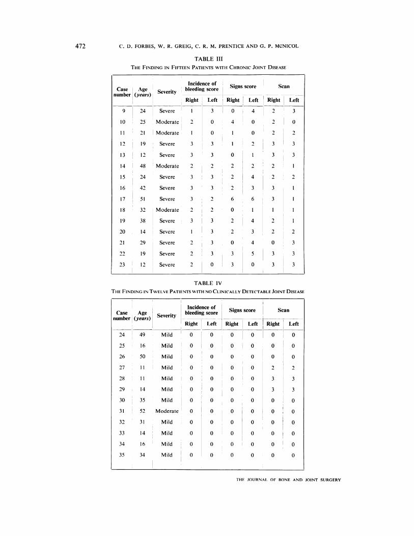
TABLE III
THE FINDING IN FIFTEEN PATIENTS WITH CHRONIC JOINT DISEASE
Case � Age � Severit � bleedingscore � Signs score � Scannumber � (years) � � � �
� � Right Left Right � Left �_Right_� Left
9 � 24 Severe � I 3 0 � 4 � 2 � 3
10 � 25 � Moderate � 2 0 4 � 0 � 2 � 0
11 � 21 � Moderate � 1 0 1 0 � 2 2
l2� I9Severe 3 3 1 � 2 � 3 3
13 � 12 Severe 3 � 3 0 � 1 3 3
14 � 48 � Moderate 2 � 2 � 2 2 � 2
IS � 24 Severe 3 � 3 � 2 4 2
�: � �: Moderate 2 � 2 � 0 � 1 � I
19 38 Severe � 3 � 3 2 � 4 2
20 14 Severe 1 � 3 2 3 � 2
21 29 Severe 2 � 3 0 4 0
: :: :::::::2
3
3
3
TABLE Iv
THE FINDING IN TWELVE PATIENTS WITH NO CLINICALLY DETECTABLE JOINT DISEASE
49 Mild
25 16 Mild
26 50 Mild
27 II Mild
28 II Mild
29 14 Mild
30 35 Mild
31 52 Moderate
32 31 Mild
33 14 Mild
34 16 Mild
35 34 Mild
0
0
0
0
0
0
0
0
0
0
0
0
0
0
0
0
0
0
0
0
0
0
0
0
0
0
0
0
0
0
0
0
0
0
0
0
0
0
0
0
0
0
0
0
0
0
0
0
0
3
3
0
0
0
0
0
0
0
0
3
3
0
0
0
0
0
0
472 C. D. FORBES, W. R. GREIG, C. R. M. PRENTICE AND G. P. McNICOL
THF JOURNAL OF BONE AND JOINT SURGERY
RADIOISOTOPE KNEE JOINT SCANS IN 1-IAEMOPHILIA AND CHRISTMAS DISEASE 473
cartilage with release of polypeptide kinins (Moskowitz, Schwartz, Michel, Ratnoff and
Astrup 1970) through the conversion of plasma kallikreinogen to its active form, plasma
kallikrein. It is thought that kinins may then induce many of the phenomena observed in
inflammatory reactions (Ratnoff 1966).
In experimental studies injection of blood into the joints of animals (Key 1929, Young
and Hudacek 1954, Trueta 1966) produced an inflammatory response with hypertrophy of
the villi. The effect of a single injection was short lived but more pronounced effects could
be produced by multiple injections. A similar histological picture to that produced in
experimental animals has been found in thejoints ofa haemophilic patient (Key 1932). These
changes remain as long as blood or excess fluid is present. Ambulation at this time may
result in crushing of some of these villi, which may then produce further bleeding and so
initiate a vicious circle. The fact that the scans remain abnormal some time after all acute
clinical features have disappeared shows that temporary synovial hypertrophy is partly
involved in the process.
The subacute classification represents a variation of the processes seen in the acute joint.
In both our patients (Table II) there had been no clinical evidence of recent acute bleeding
and the joints had been swollen, hot and painless for over twelve weeks. However, the scans
were identical with those seen in acute bleeding. Repeat scanning in Case 8 six weeks later
showed only slight improvement despite total clinical resolution with adequate haemostatic
therapy.
All the patients with chronic joint disease had abnormal scans. However, there was no
correlation between the number of previous bleeds, the clinical signs and the scan ; some
patients with no signs in one joint had a grossly abnormal scan (Cases 1 1 and 23). Only
two of the thirty knee joints scanned in this group were normal ; one of these patients had no
signs but had a history of previous acute haemarthroses (Case 21). None of the patients in
this group had a history of recent acute haemarthrosis, so the abnormal scans presumably
represent a chronic state in which there is increased vascularity in the synovium. This is in
keeping with many previous descriptions of the histology of these chronic joints in which
the synovium was found to be markedly thickened, hyperplastic and endowed richly with
vascular channels (Bokelmann 1881 ; Konig 1892; Freund 1925; Collins 1949; DePalma and
Cotler 1956a; Rodnan, Brower, Hellstrom, Didisheim and Lewis 1959; Marion et a!. 1965;
Trueta 1966). Storti, Traldi, Tosatti and Davoli 1969 have described a hypertrophic
angiomatous type of synovium with thin-walled varicose veins on the surface which bled even
when touched with a gauze swab.
In the group of patients with no clinically detectable joint disease and no previous history
of bleeding into the knee joints, four patients had bilateral abnormal scans. These patients
were the youngest in this group. The abnormal scans presumably are a reflection of subclinical
bleeding into the joints, a condition analogous to the asymptomatic bleeding from the renal
capillaries which we have previously described in mild haemophilia (Prentice, Lindsay, Barr,
Forbes, Kennedy, McNicol and Douglas 1971).
In those patients with no apparent joint disease the articular scan may be of value in the
early detection of joint disease, and in the acute and subacute group the persistence of the
abnormality in the scan may indicate the importance of prophylactic therapy with plasma
concentrates as suggested by several authors (Ahlberg 1965 ; Kasper, Dietrich and Rapaport
1970; Brinkhous, Carnoy and Shermer 1971 ; Hirschman, ltscoitz and Shulman 1970;
vanCreveld 1971).
It seems certain that if prophylactic therapy in haemophilia is to be of value, then it
must be started before joint disease becomes established. We feel that the abnormality of
the articular scan is the earliest index of synovial hypertrophy, and if detected at this stage it
may enable us to break the vicious circle phenomenon of joint haemorrhage-�synovial
hypertrophy-�further haemorrhage. If adequate long term therapy can be given, healing
VOL. 54 B, NO. 3, AUGUST 1972
F

474 C. D. FORBES, W. R. GREIG, C. R. M. PRENTICE AND G. P. MCNICOL
THE JOURNAL OF BONE AND JOINT SURGERY
could proceed, perhaps with resolution of the synovial changes. if prophylactic therapy is
attempted when there is established bone disease the results are poor (van Creveld, Hoedemaeker,
Kingma and Wagenvoort 1971).
Recently, synovectomy has been carried out in patients with chronic haemophilic joint
disease (Storti et a!. 1969). The preliminary results seem encouraging but as yet no long
term follow-up is available. The articular scan should prove of value in the selection of patients
for this operation and in the assessment of the short and long term results. Articular scanning
may also prove of value in the assessment of steroids and antifibrinolytic agents in the
treatment of acute joint bleeds.
SUMMARY
I . in thirty-five patients, twenty-eight with classical haemophilia and seven with Christmas
disease, arthropathy of the knee of various grades has been investigated by radioisotope
scanning after intravenous injection of technetium, 99mTc.
2. The abnormality of the colour scan particularly matches the clinical severity in acute
haemarthrosis.
3. in patients with no clinically apparent joint disease the scan may be of value in the early
detection of involvement.
4. The possible value of articular scanning in the selection of patients for treatment and
in the assessment of the short and long term results is discussed.
REFERENCES
AHLBERG, A. (1965): Haemophilia in Sweden. VII Incidence, Treatment and Prophylaxis ofArthropathy andother Musculo-skeletal Manifestations of Haemophilia A and B. Acta Orthopaedica Scandinavica,
Supplementum 77.
ALAR#{231}ON-SEGOVIA, D., TRUJEQUE, M., TOVAR, E., ADAME, M. A. (1967): Scintillation Scanning of Joints with
Technetium 99m. (Abstract) Arthritis and Rheumatism, 10, 262.
ANON (1968): Editorial. Lance,’, 2, 131.BIGG5, R., and MACFARLANE, R. G. (1962): Human Blood Coagulation and its Disorders. Third Edition.
Oxford : Blackwell Scientific Publications.BOKELMANN, W. (1 881) : Ueber die Natur u,zd Bedeutung der haemophilen Gelenkaffectionen. Gottingen:
W. F. K#{228}stner.
BRINKHOUS, K. M., CARNOY,� Ch. N., and SHERMER, R. W. (1971): Use of High-Potency Antihemophilic
Concentrates in Prophylaxis. Thrombosis et diathesis haemorrhagica, Supplement 43, p. 139.
COLLINS, D. H. (1949): Haemophilia. In The Pathology of Articular and Spinal Diseases, p. 225. London:Edward Arnold & Co.
CREVELD, S. van (1969): Prophylaxis of Joint Haemorrhages in Haemophilia. Ada Haematologica, 41, 206.
CREVELD, S. van (1971): Prophylaxis of Joint Haemorrhages in Haemophilia. Acta Haematologica, 45, 120.
CREVELD, S. van, HOEDEMAEKER, Ph. J., KINGMA, M. J., and WAGENVOORT, C. A. (1971): Degeneration of
Joints in Haemophiliacs under Treatment by Modern Methods. Journal of Bone and Joint Surgery,
53-B, 296.
DEPALMA, A. F., and COTLER, J. M. (1956a): Haemophilic Arthropathy. Archives ofSurgery, 72, 247.
DEPALMA, A. F., and COTLER, J. M. (1956b): Hemophilic Arthropathy. Clinical Orthopaedics, 8, 163.
DICK, W. C., NEUFELD, R. R., PRENTICE, A. G., WOODBURN, A., WHALEY, K., NUKI, G., and BUCHANAN, W. W.
(1970): Measurement ofJoint Inflammation: A Radioisotopic Method. Annals ofthe Rheumatic Diseases,
29, 135.
FoNlo, A., and BUHLER, W. (1952): Die rontgenologische Darstellung des Blutergelenkes an Hand von 136Gelenkaufnahmen der Fonioschen Sammlung. Radiologia Clinica, 21, 316.
FREUND, E. (1925): Die Gelenkerkrankung der Bluter. Virchows Archiv f#{252}rpaihologische Anatomie und
Physiologie und f#{252}rklinische Medizin, 256, 158.GHORMLEY, R. K., and CLEGG, R. S. (1948): Bone and Joint Changes in Hemophilia. Journal of Bone and
Joint Surgery, 30-A, 589.
GREEN, F. A., and HAYS, M. T. (1969): Joint Scanning: Mechanism and Application. Arthritis and Rheumatism,
12, 299.
HIRSCHMAN, R. J., ITSCOITZ, S. B., and SHULMAN, N. R. (1970): Prophylactic Treatment of Factor VIIIDeficiency. Blood, 35, 189.
JORDAN, H. H. (1958): Hemophilic Arthropathies. Springfield, lllinois: Charles C. Thomas.
RADIOISOTOPE KNEE JOINT SCANS IN HAEMOPHILIA AND CHRISTMAS DISEASE 475
KASPER, C. K., DIETRICH, S. L.,and RAPAPORT, S. I. (1970): Hemophilia Prophylaxis with Factor VIII Concentrate.
Archives of Internal Medicine, 125, 1004.
KERR, C. B. (1963): The Management ofHaemophilia. Glebe: AustralasianMedical Publishing Co. Ltd.
KEY, J. A. (1929): Experimental Arthritis. Journal ofBone and Joint Surgery, 11, 705.
KEY, J. A. (1932): Hemophilic Arthritis (Bleeder’s Joints). Annals ofSurgery, 95, 198.KONIG, F. (1892): Die Gelenkerkrankungen bei Blutern mit besonderer BerUcksichtigung der Diagnose.
Sammiung klinischer Vortrage von R. von Volkmann, N.F. Chfrurgie, nr. 11, 233.
MCCARTY, D. J., POLCYN, R. E., COLLINS, P. A., and GOTFSCHALK, A.(1970): 99m Technetium Scintiphotographyin Arthritis. I. Technic and Interpretation. Arthritis and Rheumatism, 13, 11.
MCCARTY, D. J., POLCYN, R. E., and COLLINS, P. A. (1970): 99m Technetium Scintiphotography in Arthritis.
II. Its Nonspecificity and Clinical and Roentgenographic Correlations in Rheumatoid Arthritis. Arthritis
and Rheumatism, 13, 21.
MARION, J., FAVRE-GILLY, J., PICAULT, Ch., and GAUTHIER, G. (1965) : Ddcouverte d’une h#{233}mophilie it l’occasion
d’une intervention pour synovite villeuse du coude. H#{233}mostase, 5, 69.
MosKowrrz, R. W., SCHWARTZ, H. J., MICHEL, R., RATNOFF, 0. D., and ASTRUP, T. (1970): Generation of
Kinin-like Agents by Chondroitin Sulfate, Heparin, Chitin Sulfate and Human Articular Cartilage;Possible Pathophysiologic Implications. Journal ofLaboratory and Clinical Medicine, 76, 790.
MOWAT, A. G., DISNEY, T. F., and VAUGHAN, J. H. (1971): Articular Scanning and External Counting in
Experimental Synovitis in the Guinea Pig. Annals ofthe Rheumatic Diseases, 30, 183.
PRENTICE, C. R. M., LINDSAY, R. M., BARR, R. D., FORBES, C. D., KENNEDY, A. C., MCNICOL, G. P., and
DOUGLAS, A. S. (1971): Renal Complications in Haemophilia and Christmas Disease. Quarterly Journal
ofMedicine, 40, 47.RATNOFF, 0. D. (1966): The Biology and Pathology of the Initial Stages of Blood Coagulation. Progress in
Hematology, 5, 204.RODNAN, G. P., BROWER, T. D., HELLSTROM, H. R., DIDISHEIM, P., and LEWIS, J. H. (1959): Post Mortem
Examination of an Elderly Severe Hemophiliac, with Observations on the Pathologic Findings in
Hemophilic Joint Disease. Arthritis and Rheumatism, 2, 152.
STORTI, E., TRALDI, A., TOSATFI, E., and DAVOLI, P. G. (1969): Synovectomy, a New Approach to Haemophilic
Arthropathy. Acta Haematologica, 41, 193.STORTI, E., TRALDI, A., TOSATFI, E., and DAVOLI, P. G. (1969): La sinoviectomia nell’emofilia. Gaz:ette
sanitaria (Milano), 40, 229.
SWANTON, M. C. (1957): The Pathology of Hemarthrosis in Hemophilia. In Hemophilia and Hemophiioid
Diseases, p. 219. Edited by K. M. Brinkhous. Chapel Hill: University of North Carolina Press.SWANTON, M. C. (1959): Hemophilic Arthropathy in Dogs. Laboratory Investigation, 8, 1269.
TRUETA, J. (1966): The Orthopaedic Management of Patients with Haemophilia and Christmas Disease. In
Treatment of Haemophilia and other Coagulation Disorders, p. 279. Edited by R. Biggs and R. G.Macfarlane. Oxford : Blackwell Scientific Publications.
WEBB, J. B., and DIXON, A. St. J. (1960): Haemophilia and Haemophilic Arthropathy. Annals ofihe Rheumatic
Diseases, 19, 143.WEISS, T. E., MAXFIELD, W. S., MURISON, P. J., and HIDALGO, J. U. (1965): lodinated Human Serum Albumin
(1.131) Localisation Studies of Rheumatoid Arthritis Joints by Scintillation Scanning. Arthritis and
Rheumatism, 8, 976.
WEISS, T. E., MAXFIELD, W. S., MURISON, P. J., and HJDALGO, J. U. (1966): Scintillation Scanning in RheumatoidArthritis. Southern Medical Journal, 59, 484.
WHALEY, K., PACK, A. I., BOYLE, J. A., DICK, W. C., DOWNIE, W. W., BUCHANAN, W. W., and GILLESPIE, F. C.
(1968): The Articular Scan in Patients with Rheumatoid Arthritis: A Possible Method of QuantitatingJoint Inflammation using Radio-technetium. Clinical Science, 35, 547.
YOUNG, J. M., and HUDACEK, A. G. (1954): Experimental Production of Pigmented Villonodular Synovitis in
Dogs. American Journal ofPathology, 30, 799.
VOL. 54 B, NO. 3, AUGUST 1972