Embed Size (px)
Citation preview
Jared Kesselheim, HMS 2005Gillian Lieberman, MD January, 2004
Radiological Evaluation of Acute Ischemic Stroke
Jared Kesselheim, HMS IIIGillian Lieberman, MD
2
Jared Kesselheim, HMS 2005Gillian Lieberman, MD
Agenda
Background: StrokePatient L and the Neuroimaging of Acute Ischemic Stroke: CT and MRINeuroanatomy review: Circle of WillisFrontiers in Neuroimaging of Acute Ischemic StrokeTake Home Points
3
Jared Kesselheim, HMS 2005Gillian Lieberman, MD
Agenda
Background: StrokePatient L and the Neuroimaging of Acute Ischemic Stroke: CT and MRINeuroanatomy review: Circle of WillisFrontiers in Neuroimaging of Acute Ischemic StrokeTake Home Points
4
Jared Kesselheim, HMS 2005Gillian Lieberman, MD
Stroke
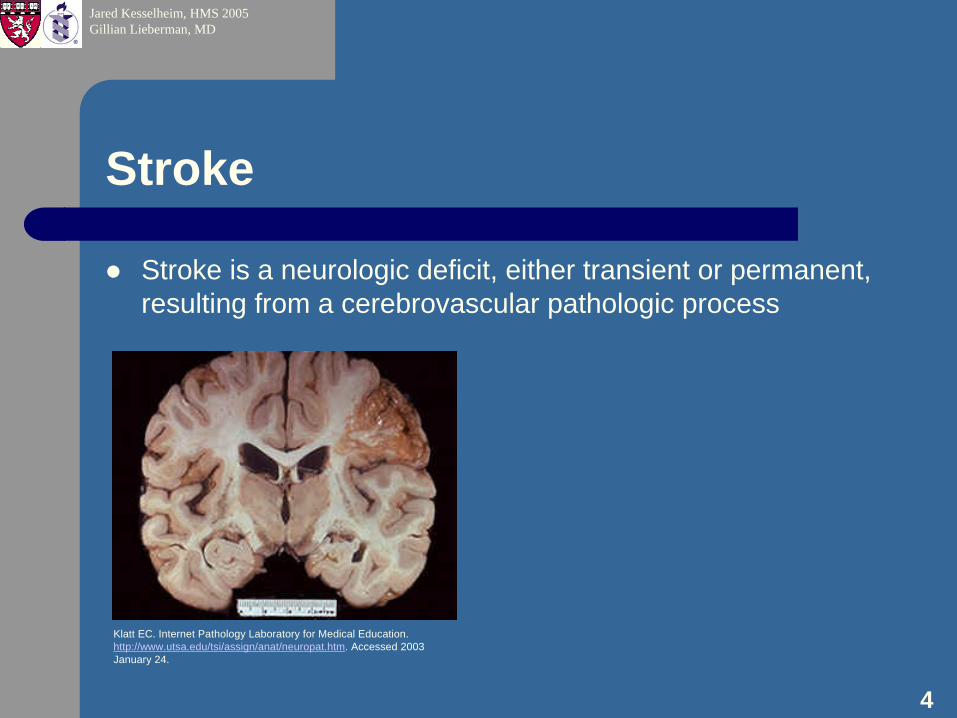
Stroke is a neurologic deficit, either transient or permanent, resulting from a cerebrovascular pathologic process
Klatt EC. Internet Pathology Laboratory for Medical Education. http://www.utsa.edu/tsi/assign/anat/neuropat.htm. Accessed 2003 January 24.
5
Jared Kesselheim, HMS 2005Gillian Lieberman, MD
Impact of Stroke
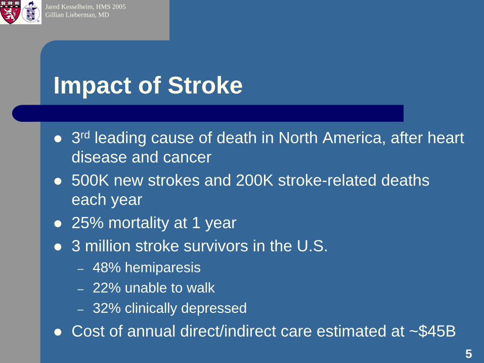
3rd leading cause of death in North America, after heart disease and cancer500K new strokes and 200K stroke-related deaths each year25% mortality at 1 year3 million stroke survivors in the U.S.
– 48% hemiparesis– 22% unable to walk– 32% clinically depressed
Cost of annual direct/indirect care estimated at ~$45B
6
Jared Kesselheim, HMS 2005Gillian Lieberman, MD
Etiologies of Stroke
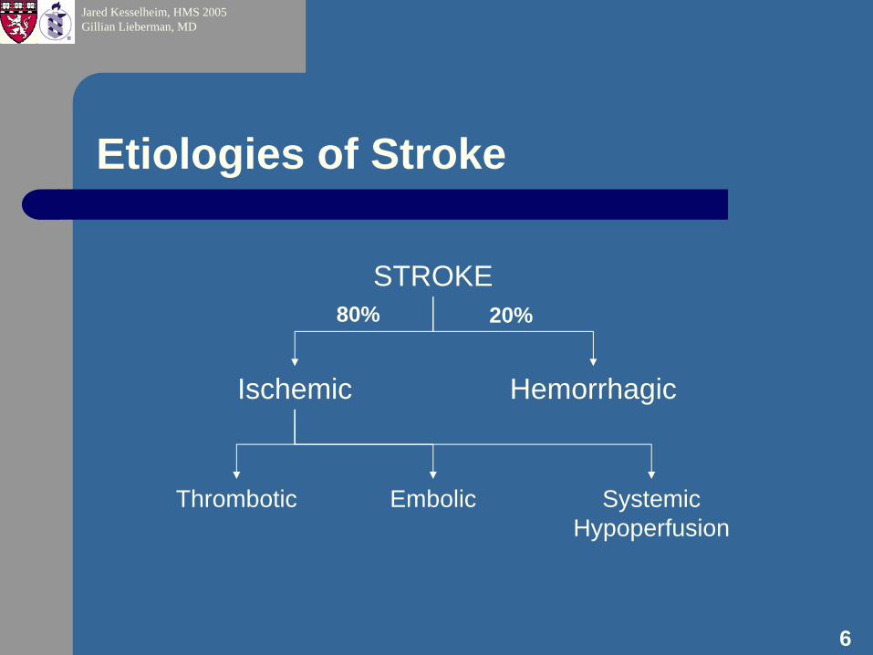
STROKE
Ischemic Hemorrhagic
80% 20%
Thrombotic Systemic Hypoperfusion
Embolic
7
Jared Kesselheim, HMS 2005Gillian Lieberman, MD
Early Diagnosis of Acute Ischemic Stroke is Critical
NINDS (National Institute of Neurological Disorders and Stroke) Stroke Study demonstrated morbidity benefit from intravenous tPA treatment for acute ischemic stroke within 3 hours of onset of symptomstPA intervention from 3-6 hours after onset of symptoms is controversial
8
Jared Kesselheim, HMS 2005Gillian Lieberman, MD
Agenda
Background: StrokePatient L and the Neuroimaging of Acute Ischemic Stroke: CT and MRINeuroanatomy review: Circle of WillisFrontiers in Neuroimaging of Acute Ischemic StrokeTake Home Points
9
Jared Kesselheim, HMS 2005Gillian Lieberman, MD
Patient: Ms. L.
CC: New onset L-sided hemiparesisHPI: 91 y.o. female with hx of severe dementia and atrial fibrillation presents with decreased movement of her left side noticed by her caretaker.Meds: Diltiazem, Raloxifene, Vit B12PE: VSS, No spontaneous movements of left upper or lower extremitiesWhat imaging should be performed?
10
Jared Kesselheim, HMS 2005Gillian Lieberman, MD
Goals of Imaging in Acute Stroke
1. Exclude hemorrhage as etiology of deficit2. Evaluate the extent of brain injury3. Identify any specific vascular lesion
responsible for the event
11
Jared Kesselheim, HMS 2005Gillian Lieberman, MD
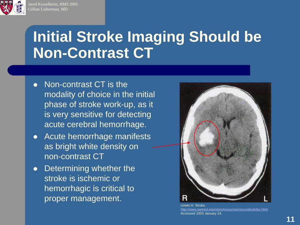
Initial Stroke Imaging Should be Non-Contrast CT
Non-contrast CT is the modality of choice in the initial phase of stroke work-up, as it is very sensitive for detecting acute cerebral hemorrhage.Acute hemorrhage manifests as bright white density on non-contrast CTDetermining whether the stroke is ischemic or hemorrhagic is critical to proper management.
Unwin H. Stroke. http://www.swmed.edu/stars/resources/neurodisslides.html. Accessed 2003 January 24.
12
Jared Kesselheim, HMS 2005Gillian Lieberman, MD
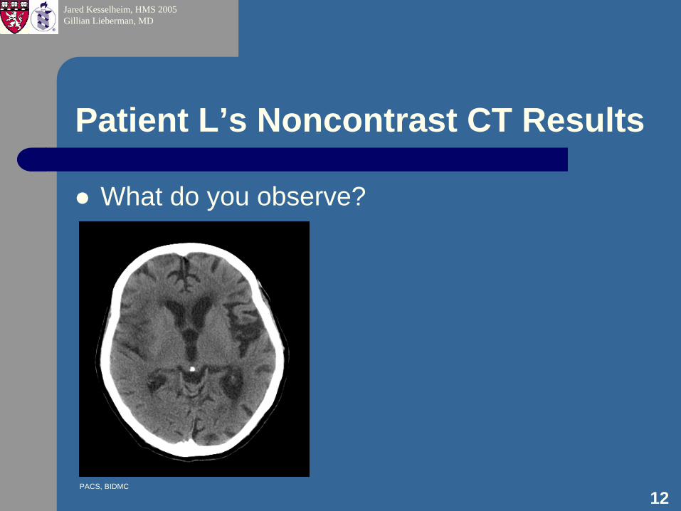
Patient L’s Noncontrast CT Results
What do you observe?
PACS, BIDMC
13
Jared Kesselheim, HMS 2005Gillian Lieberman, MD
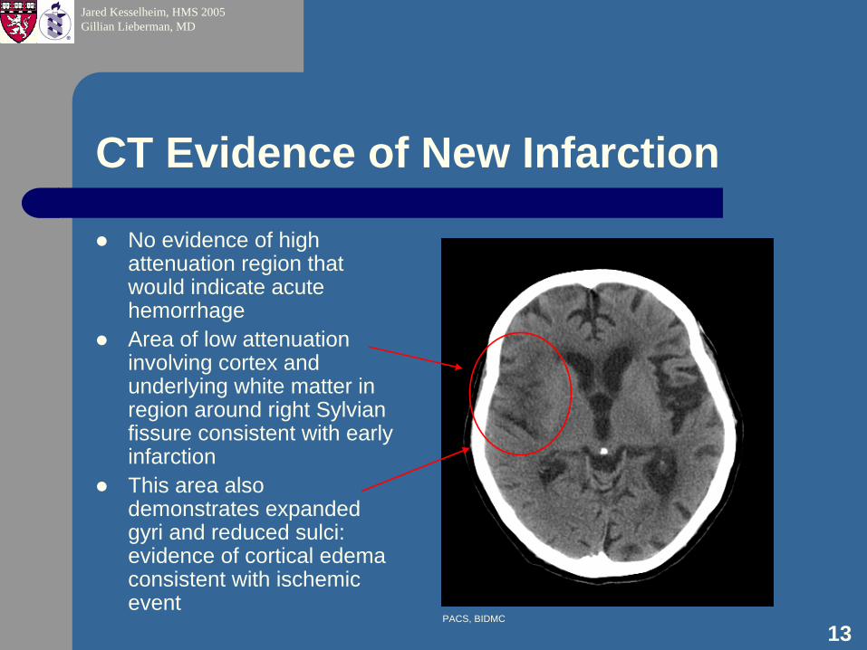
CT Evidence of New Infarction
No evidence of high attenuation region that would indicate acute hemorrhageArea of low attenuation involving cortex and underlying white matter in region around right Sylvianfissure consistent with early infarctionThis area also demonstrates expanded gyri and reduced sulci: evidence of cortical edema consistent with ischemic event
PACS, BIDMC
14
Jared Kesselheim, HMS 2005Gillian Lieberman, MD
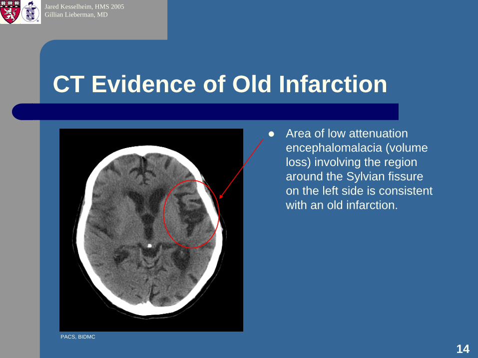
CT Evidence of Old Infarction
Area of low attenuation encephalomalacia (volume loss) involving the region around the Sylvian fissure on the left side is consistent with an old infarction.
PACS, BIDMC

15
Jared Kesselheim, HMS 2005Gillian Lieberman, MD
MRI Was Performed…
Technique: Axial T1W, T2 FLAIR and DWI imaging were performedGadolinium contrast was NOT used – no need for contrast in MRI evaluation of stroke. Much more useful in MRI evaluation of inflammatory, infectious, and neoplastic processes
16
Jared Kesselheim, HMS 2005Gillian Lieberman, MD
MRI vs CT for Detecting Ischemic Stroke
MRI is more sensitive than non-contrast CT for detecting the early changes of acute ischemic stroke
– Studies of CT sensitivity for early signs of cerebral infarction have yielded results from 56% to 81%
– A study comparing DWI MRI with CT in patients imaged within 6 hours of stroke-like symptoms yielded a 45% sensitivity for CT and >95% sensitivity for DWI MRI
– DWI MRI has been shown to be able to detect changes related to brain ischemia within 15-30 minutes of the ischemic insult.
17
Jared Kesselheim, HMS 2005Gillian Lieberman, MD
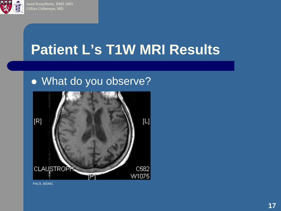
Patient L’s T1W MRI Results
What do you observe?
PACS, BIDMC
18
Jared Kesselheim, HMS 2005Gillian Lieberman, MD
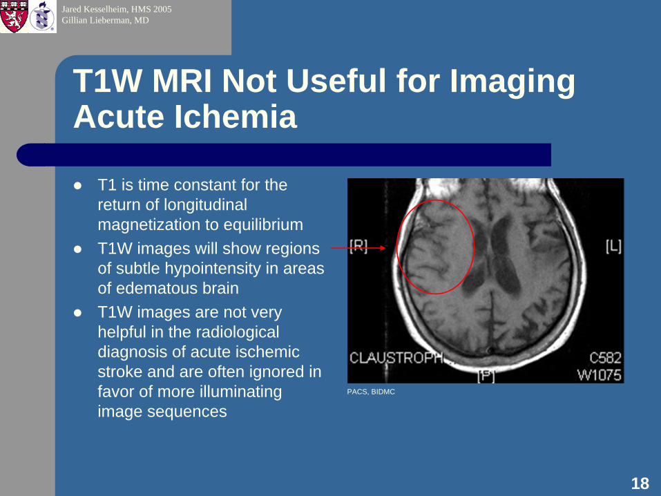
T1W MRI Not Useful for Imaging Acute Ichemia
T1 is time constant for the return of longitudinal magnetization to equilibriumT1W images will show regions of subtle hypointensity in areas of edematous brainT1W images are not very helpful in the radiological diagnosis of acute ischemic stroke and are often ignored in favor of more illuminating image sequences
PACS, BIDMC
19
Jared Kesselheim, HMS 2005Gillian Lieberman, MD
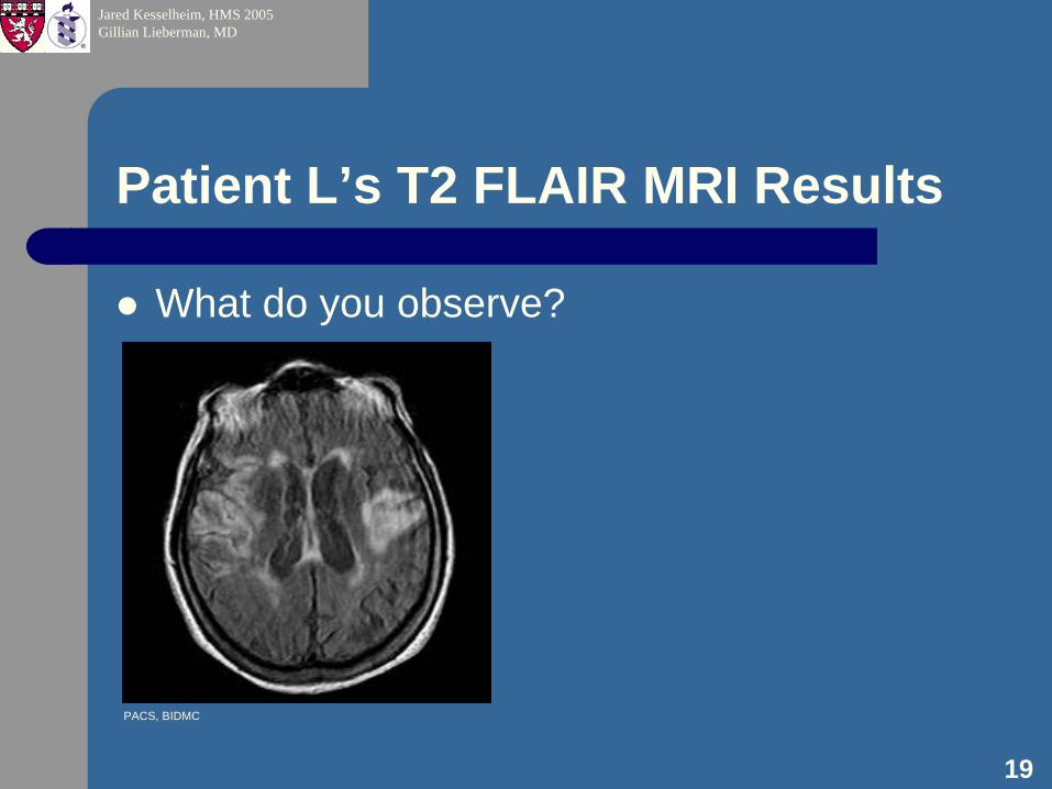
Patient L’s T2 FLAIR MRI Results
What do you observe?
PACS, BIDMC
20
Jared Kesselheim, HMS 2005Gillian Lieberman, MD
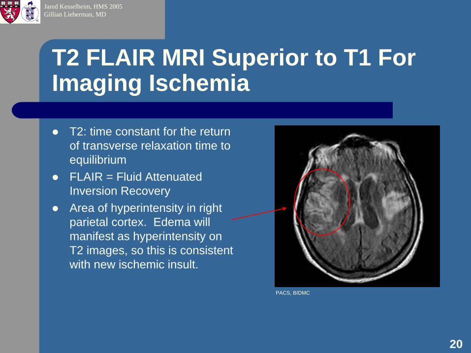
T2 FLAIR MRI Superior to T1 For Imaging Ischemia
T2: time constant for the return of transverse relaxation time to equilibriumFLAIR = Fluid Attenuated Inversion RecoveryArea of hyperintensity in right parietal cortex. Edema will manifest as hyperintensity on T2 images, so this is consistent with new ischemic insult.
PACS, BIDMC

21
Jared Kesselheim, HMS 2005Gillian Lieberman, MD
T2 FLAIR Poor at Differentiating Old vs New Ischemic Event
Area of hyperintensity in left parietal cortex. In this region of old infarction, the hyperintensity is indicative of gliosis, the glial proliferation in response to brain insults such as infarction which manifests as hyperintensity on T2 images
PACS, BIDMC
22
Jared Kesselheim, HMS 2005Gillian Lieberman, MD
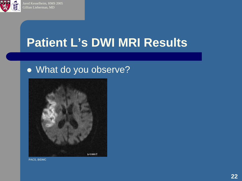
Patient L’s DWI MRI Results
What do you observe?
PACS, BIDMC
23
Jared Kesselheim, HMS 2005Gillian Lieberman, MD
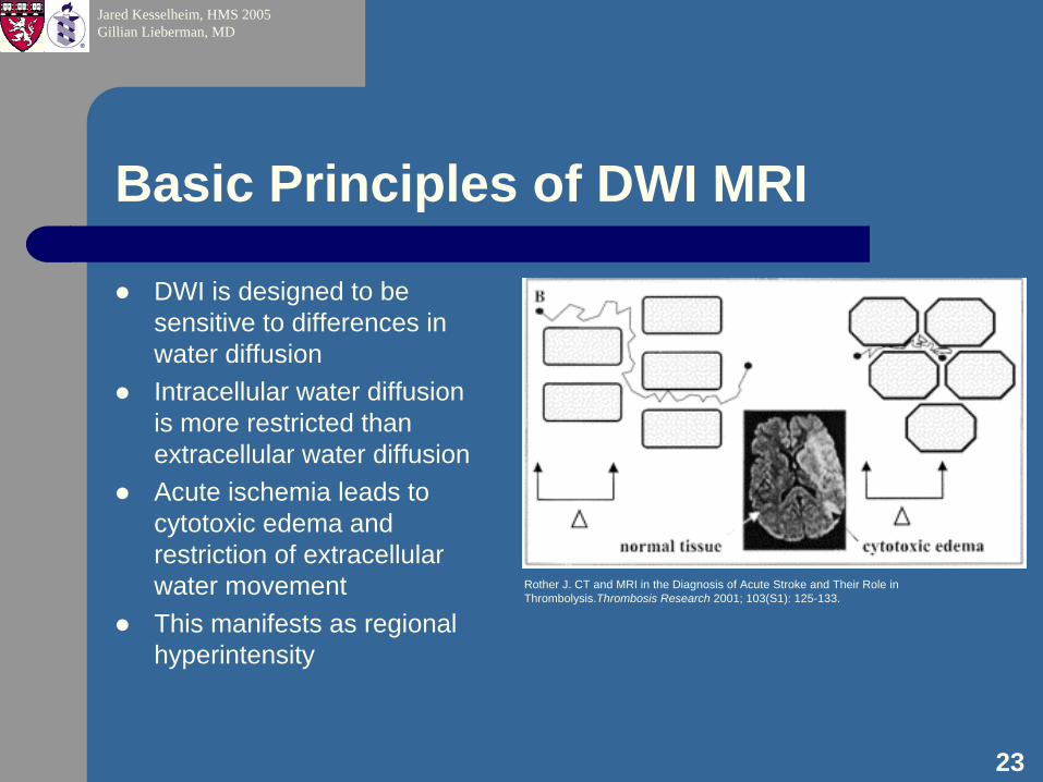
Basic Principles of DWI MRI
DWI is designed to be sensitive to differences in water diffusionIntracellular water diffusion is more restricted than extracellular water diffusionAcute ischemia leads to cytotoxic edema and restriction of extracellular water movementThis manifests as regional hyperintensity
Rother J. CT and MRI in the Diagnosis of Acute Stroke and Their Role in Thrombolysis.Thrombosis Research 2001; 103(S1): 125-133.
24
Jared Kesselheim, HMS 2005Gillian Lieberman, MD
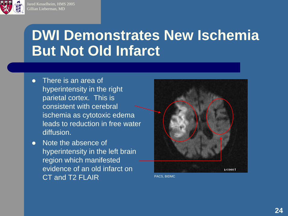
DWI Demonstrates New Ischemia But Not Old Infarct
There is an area of hyperintensity in the right parietal cortex. This is consistent with cerebral ischemia as cytotoxic edema leads to reduction in free water diffusion.Note the absence of hyperintensity in the left brain region which manifested evidence of an old infarct on CT and T2 FLAIR PACS, BIDMC
25
Jared Kesselheim, HMS 2005Gillian Lieberman, MD
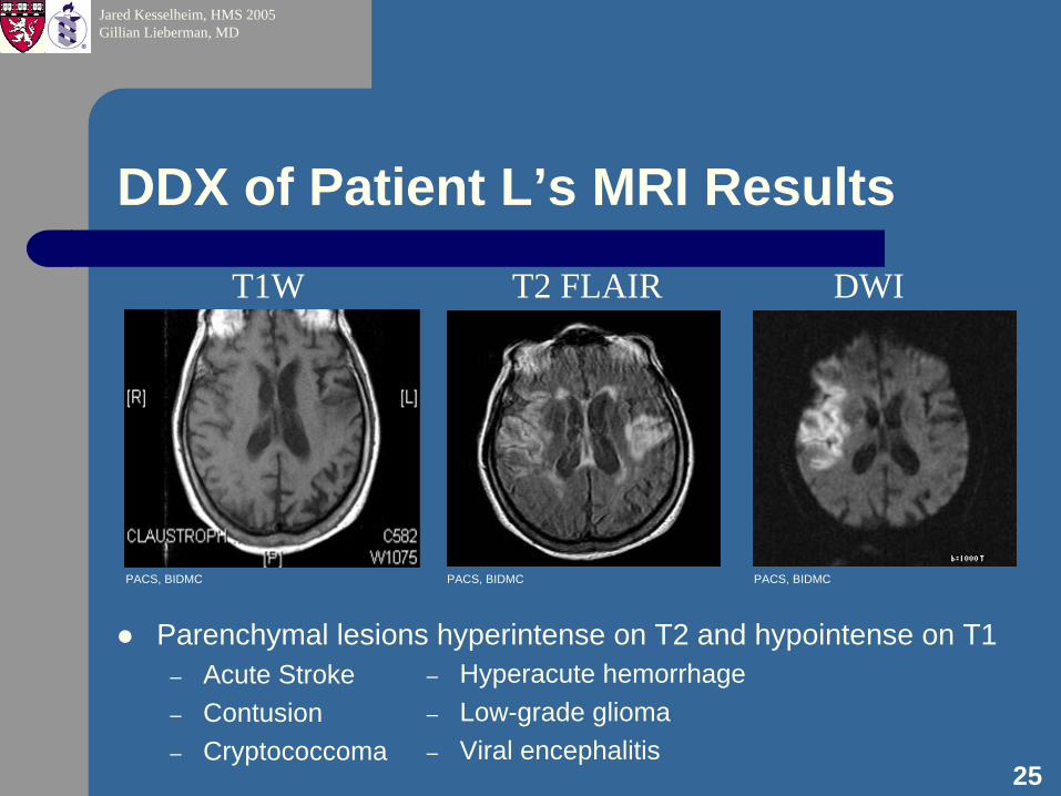
DDX of Patient L’s MRI Results
Parenchymal lesions hyperintense on T2 and hypointense on T1– Acute Stroke– Contusion– Cryptococcoma
T1W T2 FLAIR DWI
– Hyperacute hemorrhage– Low-grade glioma– Viral encephalitis
PACS, BIDMC PACS, BIDMCPACS, BIDMC
26
Jared Kesselheim, HMS 2005Gillian Lieberman, MD
Diagnosis: Acute Ischemic Stroke
What does the territory of the ischemic area tell us about the likely location of the stroke?Territory of ischemic stroke implicates the right middle cerebral arteryMRA of the Circle of Willis was performed to help assess flow abnormalities in the right MCA
27
Jared Kesselheim, HMS 2005Gillian Lieberman, MD
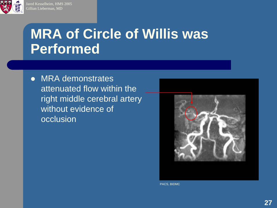
MRA of Circle of Willis was Performed
MRA demonstrates attenuated flow within the right middle cerebral artery without evidence of occlusion
PACS, BIDMC
28
Jared Kesselheim, HMS 2005Gillian Lieberman, MD
Agenda
Background: StrokePatient L and the Neuroimaging of Acute Ischemic Stroke: CT and MRINeuroanatomy review: Circle of WillisFrontiers in Neuroimaging of Acute Ischemic StrokeTake Home Points
29
Jared Kesselheim, HMS 2005Gillian Lieberman, MD
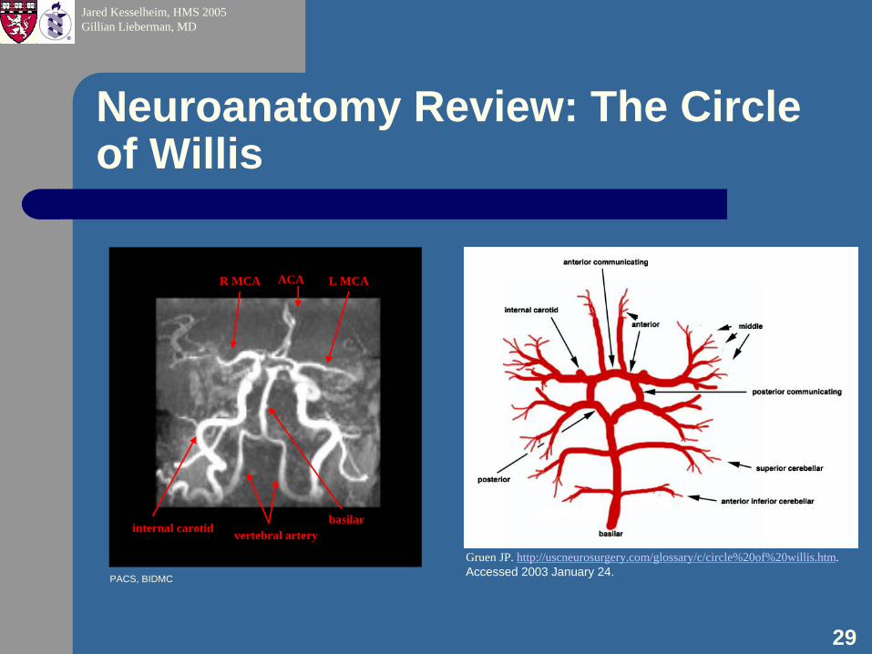
Neuroanatomy Review: The Circle of Willis
Gruen JP. http://uscneurosurgery.com/glossary/c/circle%20of%20willis.htm. Accessed 2003 January 24.
vertebral arteryinternal carotid
ACAR MCA
basilar
L MCA
PACS, BIDMC
30
Jared Kesselheim, HMS 2005Gillian Lieberman, MD
Agenda
Background: StrokePatient L and the Neuroimaging of Acute Ischemic Stroke: CT and MRINeuroanatomy review: Circle of WillisFrontiers in Neuroimaging of Acute Ischemic StrokeTake Home Points
31
Jared Kesselheim, HMS 2005Gillian Lieberman, MD
Frontiers
Multiple MRI and CT protocols are being evaluated in acute stroke patients for their capability to:– Detect tissue at risk of infarction– Predict prognosis– Identify subpopulations that would benefit from
acute thrombolytic treatment even beyond the three hour window
32
Jared Kesselheim, HMS 2005Gillian Lieberman, MD
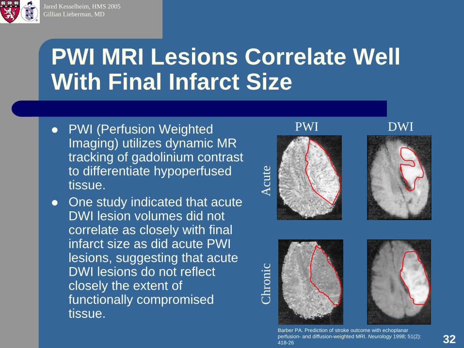
PWI MRI Lesions Correlate Well With Final Infarct Size
PWI (Perfusion Weighted Imaging) utilizes dynamic MR tracking of gadolinium contrast to differentiate hypoperfusedtissue.One study indicated that acute DWI lesion volumes did not correlate as closely with final infarct size as did acute PWI lesions, suggesting that acute DWI lesions do not reflect closely the extent of functionally compromised tissue.
PWI DWI
Acu
teC
hron
ic
Barber PA. Prediction of stroke outcome with echoplanar perfusion- and diffusion-weighted MRI. Neurology 1998; 51(2): 418-26
33
Jared Kesselheim, HMS 2005Gillian Lieberman, MD
Agenda
Background: StrokePatient L and the Neuroimaging of Acute Ischemic Stroke: CT and MRINeuroanatomy review: Circle of WillisFrontiers in Neuroimaging of Acute Ischemic StrokeTake Home Points
34
Jared Kesselheim, HMS 2005Gillian Lieberman, MD
Take Home Points
Early diagnosis of acute ischemia is critical as it guides therapeutic intervention with tPANon-contrast CT is the first imaging modality. It is excellent at detecting hemorrhagic stroke, but has low sensitivity for the early changes of acute brain ischemiaT2 and DWI MRI are very sensitive for early brain ischemia which manifest as hyperintense lesionsStudies are underway to define MRI and CT protocols that can better predict prognosis and identify salvageable brain tissue
35
Jared Kesselheim, HMS 2005Gillian Lieberman, MD
ReferencesBarber PA. Prediction of stroke outcome with echoplanar perfusion- and diffusion-weighted MRI. Neurology1998; 51(2): 418-26./SLIDE#32Gonzalez RG, Schaefer PW, Buonanno FS, Schwamm LH, Budzik RF, Rordorf G, Wang B, Sorensen AG, Koroshetz WJ. Diffusion-weighted MR imaging: diagnostic accuracy in patients imaged within 6 hours of stroke symptom onset. Radiology 1999; 210(1):155-62.Gruen JP. http://uscneurosurgery.com/glossary/c/circle%20of%20willis.htm. Accessed 2003 January 24./SLIDE#29Klatt EC. Internet Pathology Laboratory for Medical Education. http://www.utsa.edu/tsi/assign/anat/neuropat.htm. Accessed 2003 January 24./SLIDE#4Moseley ME, Kucharczyk J, Mintorovitch J, Cohen Y, Kurhanewicz J, Derugin N, Asgari H, Norman D. Diffusion-weighted MR imaging of acute stroke: correlation with T2-weighted and magnetic susceptibility-enhanced MR imaging in cats. Am J Neuroradiol 1990; 11(3):423-9.The National Institute of Neurological Disorders and Stroke rt-PA Stroke Study Group. Tissue plasminogenactivator for acute ischemic stroke. N Engl J Med 1995;333(24):1581-7.Rother J. CT and MRI in the Diagnosis of Acute Stroke and Their Role in Thrombolysis.Thrombosis Research2001; 103(S1): 125-133./SLIDE#23Unwin H. Stroke. http://www.swmed.edu/stars/resources/neurodisslides.html. Accessed 2003 January 24./SLIDE#11

36
Jared Kesselheim, HMS 2005Gillian Lieberman, MD
Acknowledgements
Barbara Appignani, MDSteve Reddy, MDGillian Lieberman, MDLarry Barbaras, WebmasterPamela LepkowskiFellow BI-Deaconess Core Radiology Students