Embed Size (px)
Citation preview

Radiology WorkshopExtremities
Andrew Haims
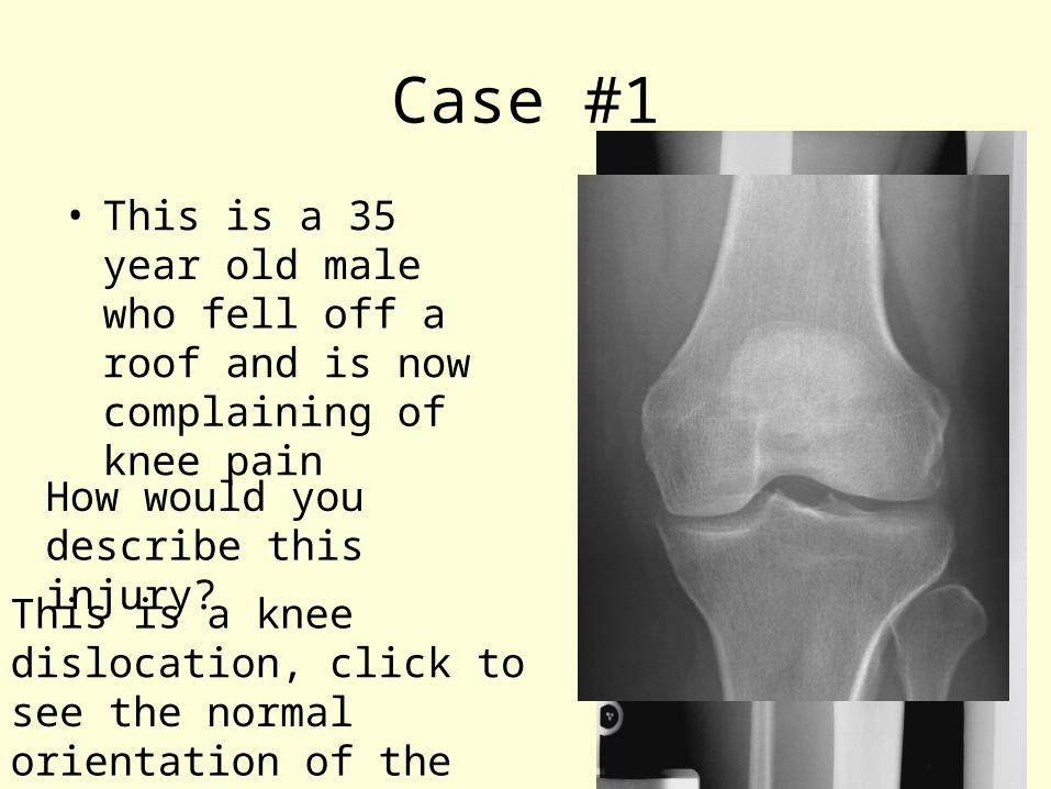
Case #1
• This is a 35 year old male who fell off a roof and is now complaining of knee pain
How would you describe this injury?
This is a knee dislocation, click to see the normal orientation of the tibia and the femur
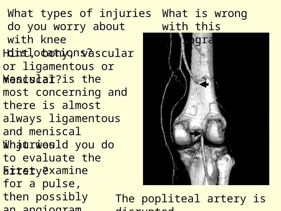
What types of injuries do you worry about with knee dislocations?
Hint, bony, vascular or ligamentous or meniscal?Vascular is the most concerning and there is almost always ligamentous and meniscal injuriesWhat would you do to evaluate the artery?First examine for a pulse, then possibly an angiogram
What is wrong with this angiogram?
The popliteal artery is disrupted
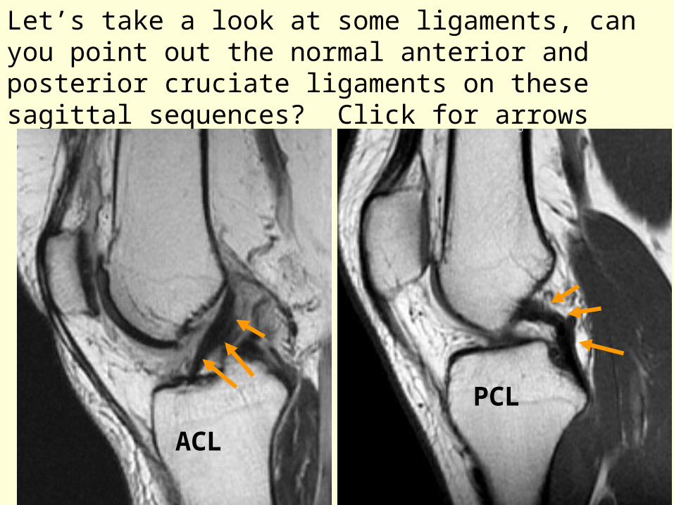
Let’s take a look at some ligaments, can you point out the normal anterior and posterior cruciate ligaments on these sagittal sequences? Click for arrows
ACL
PCL
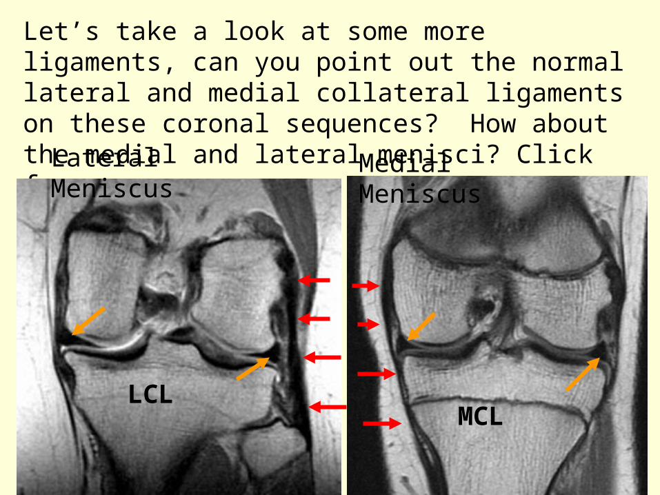
Let’s take a look at some more ligaments, can you point out the normal lateral and medial collateral ligaments on these coronal sequences? How about the medial and lateral menisci? Click for arrows
LCLMCL
Lateral Meniscus Medial Meniscus
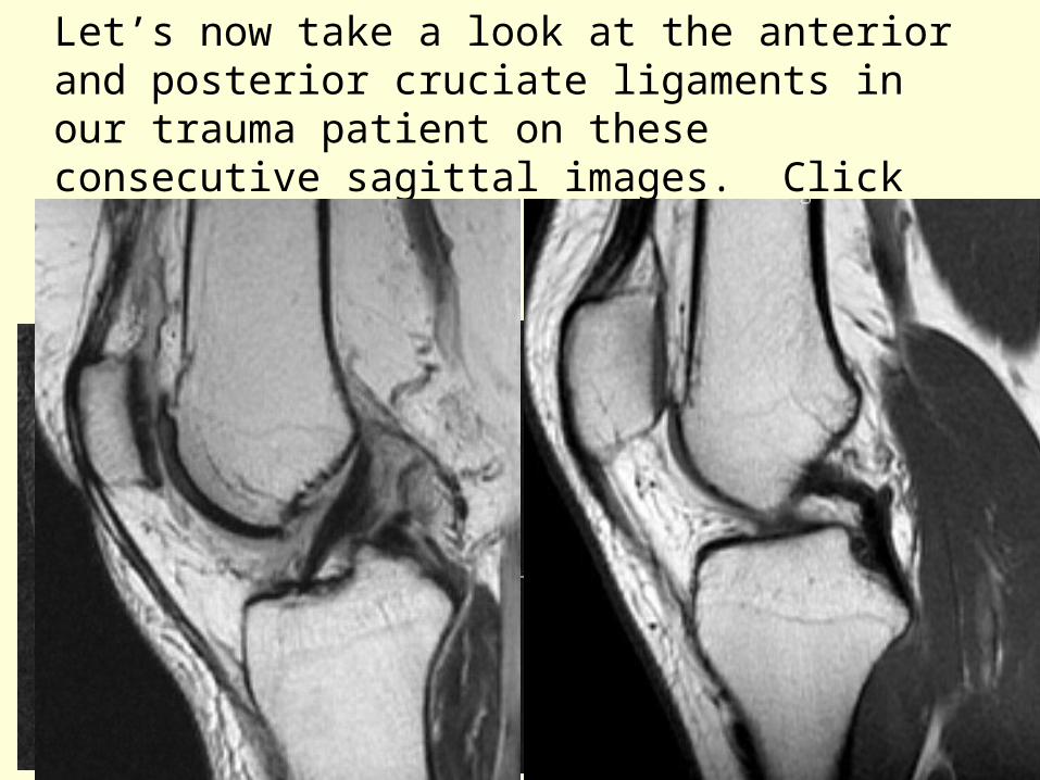
Let’s now take a look at the anterior and posterior cruciate ligaments in our trauma patient on these consecutive sagittal images. Click for another look at the normals.These are both torn off their femoral attachments
ACL stump
PCL stump
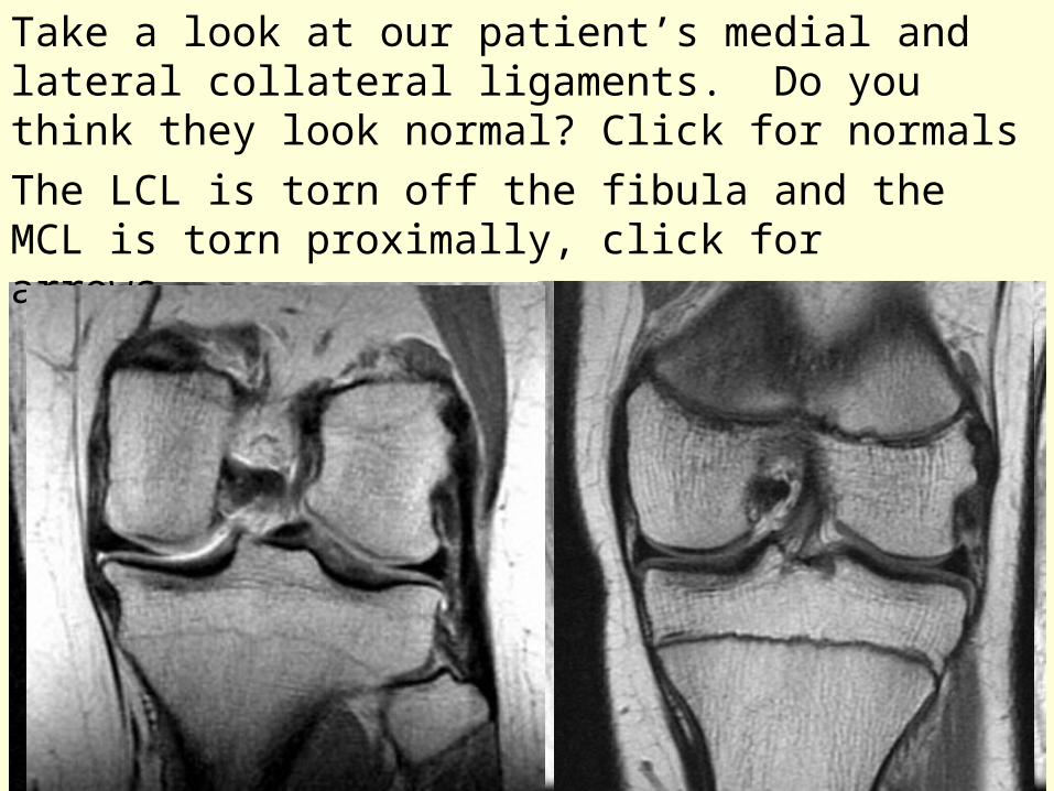
Take a look at our patient’s medial and lateral collateral ligaments. Do you think they look normal? Click for normals
The LCL is torn off the fibula and the MCL is torn proximally, click for arrows.
LCL stump
MCL disrupted fibers
Case 2
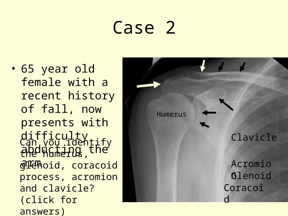
• 65 year old female with a recent history of fall, now presents with difficulty abducting the arm
Can you identify the humerus, glenoid, coracoid process, acromion and clavicle? (click for answers)
Humerus
CoracoidGlenoidAcromion
Clavicle
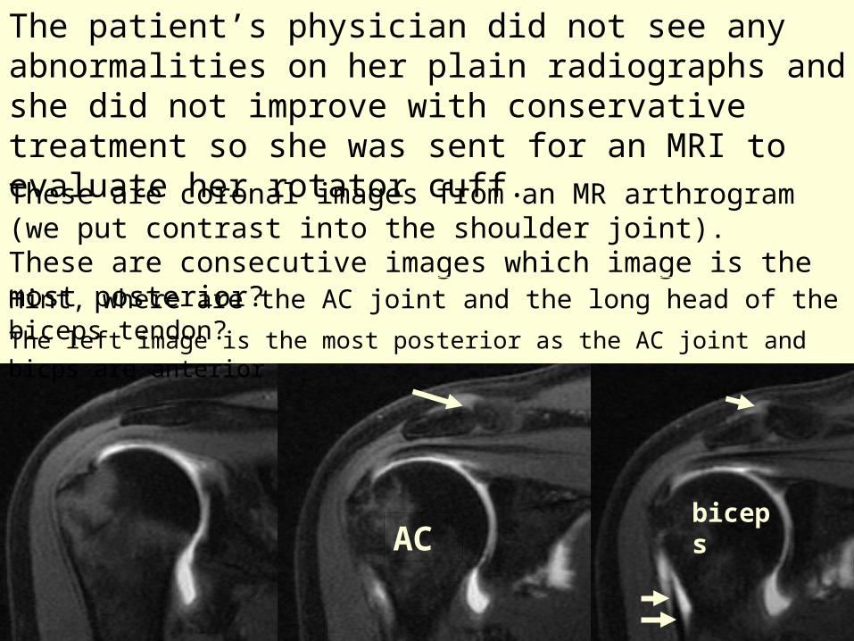
The patient’s physician did not see any abnormalities on her plain radiographs and she did not improve with conservative treatment so she was sent for an MRI to evaluate her rotator cuff.
These are coronal images from an MR arthrogram (we put contrast into the shoulder joint). These are consecutive images which image is the most posterior?Hint, where are the AC joint and the long head of the biceps tendon?
ACbiceps
The left image is the most posterior as the AC joint and bicps are anterior

Now take a look at the rotator cuff tendons, which one do we see on the posterior image and which one do we see on the most anterior 2 images?
Infraspinatus tendon Supraspinatus tendon
These are the most common rotator cuff tendons to tear. There is no tear in this patient. Where would you expect to see contrast if there was a tear? Click to see a tear
Contrast above the cuff tendons
tear
contrast

What are the other 2 rotator cuff tendons (we already discussed the supraspinatus and infraspinatus tendons?
Teres minor and subscapularis tendons, Can you find them on the provided axial images?
ANTERIOR
POSTERIOR
Teres Minor
Subscapularis
By the attachment of these muscles to the humerus can you determine their function
Subscap=internal rotation
Teres minor=external rotation
Can you identify the anterior and posterior labrum
Anterior labrum
Posterior labrum

Now for the diagnosis: on this is a coronal fat-suppressed T2 weighted image do you see an abnormality?
There is extensive edema in the greater tuberosity of the humerus consistent with a nondisplaced fracture.Try and think why this would cause supra and infraspinatus symptoms without a rotator cuff tearThe supra and infraspinatus tendons attach to the greater tuberosity
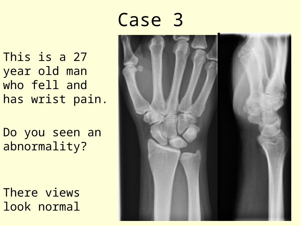
Case 3
This is a 27 year old man who fell and has wrist pain.
Do you seen an abnormality?
There views look normal
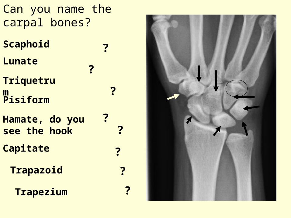
Can you name the carpal bones?
?Scaphoid
?Lunate
?Triquetrum
?
Pisiform
?Hamate, do you see the hook
?Capitate
?Trapazoid
?Trapezium

This patient subsequently underwent an MRI of the wrist to evaluate the etiology of his pain. Can you identify the radial and ulnar arteries on these axial sequences. Hint, palpate your own
pulses to determine where to look
DORSAL
VOLAR
Radial artery Ulnar artery
Can you find the median nerve
Radius
Ulna
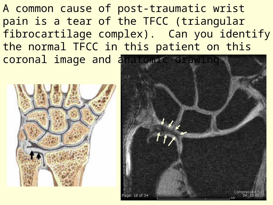
A common cause of post-traumatic wrist pain is a tear of the TFCC (triangular fibrocartilage complex). Can you identify the normal TFCC in this patient on this coronal image and anatomic drawing.
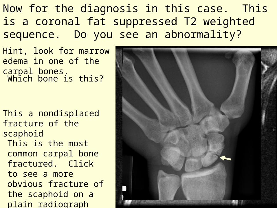
Now for the diagnosis in this case. This is a coronal fat suppressed T2 weighted sequence. Do you see an abnormality?
Hint, look for marrow edema in one of the carpal bones.
This a nondisplaced fracture of the scaphoid
Which bone is this?
This is the most common carpal bone fractured. Click to see a more obvious fracture of the scaphoid on a plain radiograph

The End