Embed Size (px)
Citation preview
Radiosynthesis and Biodistribution of 99mTc-Trimethoprim: A Novel Radiolabeled Antibiotic for Bacterial Infection Imaging Using
Experimental Animals
Hasan DEMİROĞLU 1 Gökçen TOPAL 2 Yasemin PARLAK 2 Fikriye Gül GÜMÜŞER 2
Elgin Uluer TÜRKÖZ 3 Volkan TEKİN 4 Buket ATEŞ 1 Perihan ÜNAK 4 Uğur AVCIBAŞI 11 Department of Chemistry, Faculty of Art and Science, Manisa Celal Bayar University, TR-45140 Manisa - TURKEY2 Department of Nuclear Medicine, Manisa Celal Bayar University, Faculty of Medicine, TR-45140 Manisa - TURKEY3 Department of Histology and Embryology, Manisa Celal Bayar University, Faculty of Medicine, TR-45140 Manisa - TURKEY4 Institute of Nuclear Sciences, Ege University, Department of Nuclear Applications, TR-35100 Bornova, İzmir - TURKEY
Article Code: KVFD-2017-19052 Received: 24.11.2017 Accepted: 28.01.2018 Published Online: 28.01.2018
How to Cite This Article
Demiroğlu H, Topal G, Parlak Y, Gümüşer FG, Türköz EU, Tekin V, Ateş B, Ünak P, Avcıbaşı U: Radiosynthesis and biodistribution of 99mTc-trimethoprim: A novel radiolabeled antibiotic for bacterial infection imaging using experimental animals. Kafkas Univ Vet Fak Derg, 24 (3): 393-400, 2018 (Article in Press). DOI: 10.9775/kvfd.2017.19052
AbstractIn the present article, we focused on the radiolabeling and evaluation of 99mTc–TMH complex as a potential candidate for infection imaging in vivo. For this; Trimethoprim (TMH) used to treat bacterial infections was investigated to label with 99mTc. Labeling was performed using thin (II) chloride as a reducing agent at room temperature for 1 h and radiochemical analysis involved thin layer radiochromatography (TLRC) and high pressure liquid radiochromatograpy (HPLRC) methods. The stability of labeled antibiotic was checked in the presence of rat blood serum at 37ºC up to 180 min. The maximum radiolabeling yield was found to be 96±2% and remained constant at more than 85±1% even in rat serum for 180 min after radiolabeling. Static image of 99mTc-TMH in male rats demonstrated that important radiation signals are present in the infected site at first glance in 30 min. After 30 min the uptake of the 99mTc-TMH as ID/g% in the infected muscle (INM) and normal muscle (NM) of the rats were 7.5±1.5% and 5.00±1.2%, respectively. In the INM/NM ratio a desirable behavior was observed as the values for the INM/NM increased up to 10.6. 99mTc-TMH prepared with high yield is able to localize well in the bacterially infected muscle of the rats. As a result, 99mTc-TMH may be developed as a radiopharmaceutical agent to distinguish infection from inflammation by nuclear imaging.
Keywords: Trimethoprim, 99mTc, Infection imaging agent, S. aureus
99mTc-Trimethoprim’in Radyosentezi ve Biyodağılımı: Deney Hayvanları Kullanılarak Bakteriyel Enfeksiyon Görüntüleme İçin Yeni Bir
Radyoişaretli Antibiyotik
ÖzBu çalışmada in vivo enfeksiyon görüntüleme için potansiyel bir aday olarak 99mTc-TMH’ı değerlendirdik ve radyosentezi üzerine odaklandık. Bunun için bakterilerden kaynaklı enfeksiyonların tedavisinde kullanılan Trimethoprim (TMH) 99mTc ile işaretlenmiştir. İşaretlenme oda sıcaklığında 1 saat süreyle bir indirgenme ajanı olan Sn(II)Cl2 kullanılarak gerçekleştirilmiş ve ince tabaka radyokromatografi (TLRC) ve yüksek basınçlı sıvı radyokromatografisi (HPLRC) metodlarını içeren radyokimyasal analizler yapılmıştır. İşaretli antibiyotiğin sıçan kan serumundaki kararlılığı 37ºC’de 180. dk.’ya kadar kontrol edilmiştir. Maksimum bağlanma verimi %96±2 olarak bulunmuş ve işaretli antibiyotiğin işaretlenmeden sonra 180 dk süresi içinde sıçan serumunda %85±1’in üzerindeki bir oranda sabit kaldığı görülmüştür. Dişi sıçanlardaki 99mTc-TMH’ın statik görüntüsü ilk 30 dk içinde enfeksiyon bölgesinde işaretli antibiyotiğin önemli bir radyasyon sinyali verdiğini göstermiştir. 30 dk sonra 99mTc-TMH’ın enfekte kas (INM) ve normal kas (NM) daki tutulum değerleri sırasıyla %7.5±1.5 ve %5.00±1.2 dir. INM/NM oranı 10.6 değerine kadar attığı için INM/NM oranında makul bir artış gözlenmiştir. Yüksek verimle hazırlanmış 99mTc-TMH bakteriler tarfından enfekte edilmiş sıçan kaslarında lokalize olabilmektedir. Sonuç olarak 99mTc-TMH nükleer görüntülemede kullanılabilecek bir radyofarmasötik olarak geliştirilebilir.
Anahtar sözcükler: Trimethoprim, 99mTc, Enfeksiyon Görüntüleme Ajanı, S. aureus
İletişim (Correspondence) +90 236 2013157 Fax: +90 236 2412158 [email protected]; [email protected]
Research ArticleKafkas Univ Vet Fak Derg24 (3): 393-400, 2018DOI: 10.9775/kvfd.2017.19052
KafKas Universitesi veteriner faKUltesi Dergisi
JoUrnal Home-Page: http://vetdergikafkas.orgonline sUbmission: http://submit.vetdergikafkas.org
394Radiosynthesis and Biodistribution ...
INTRODUCTION
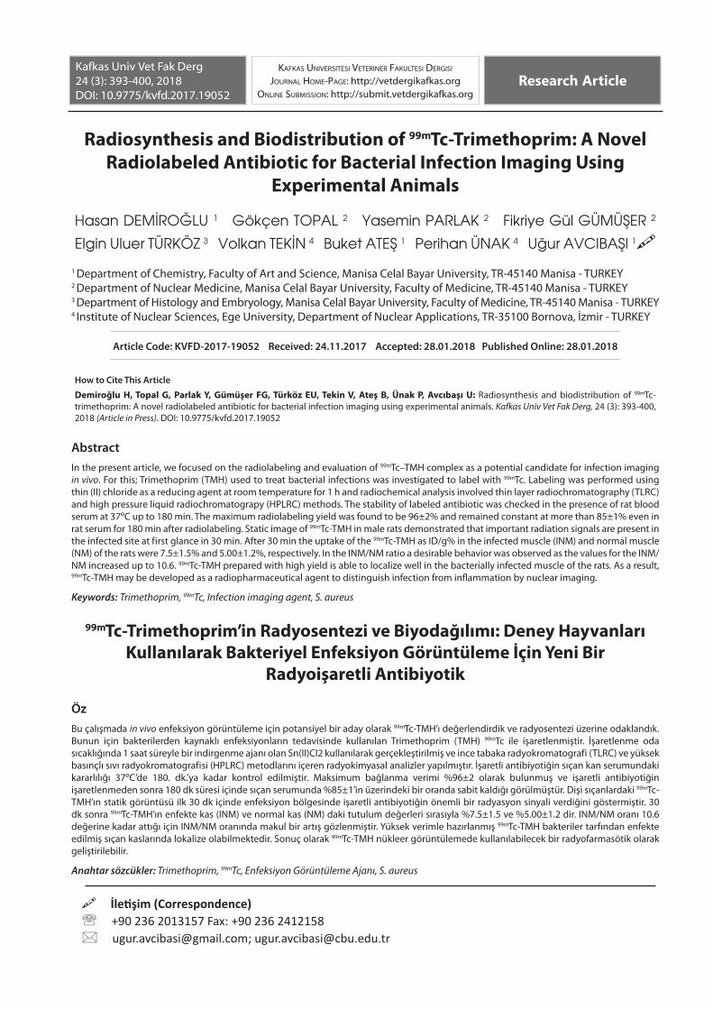
Trimethoprim (TMP) given in Fig. 1 is frequently used as human medicines to treat bronchitis and urinary tract infections. In most circumstance, they are used in combination as chemical therapy agents and growth promoters in veterinary [1-15].
In the early stages of infectious diseases, the diagnostic roles of the currently used techniques, such as ultra-sound technology (UST), computerized tomography (CT) and magnetic resonance imaging (MRI), have proven unsatisfactory. However, nuclear medicine scintigraphy (NMS) has shown promise in the precise and early diagnosis of infection and its discrimination from inflammation. NMS has considerably improved the rate of accurate diagnosis and to a large extent decreased the chances of incorrect identification [16,17]. Trimethoprim is rapidly absorbed following oral administration. Trimethoprim exists in the blood as unbound, protein-bound and metabolized forms. Trimethoprim is metabolized in vitro to 11 different metabolites, of which, five are glutathione adducts and six are oxidative metabolites, including the major metabolites, 1- and 3-oxides and the 3- and 4-hydroxy derivatives [18].
Technetium-99m (99mTc)-labeled radiopharmaceuticals have further potentiated the diagnostic capabilities of NMS in the topical decade specifically for the diagnosis of deep tissue infection and its discrimination from inflammation. Recently, some novel specific infection radiotracers have been developed, including ours, for localization and dis- crimination of infection from inflammation. The effective-ness of these radiopharmaceuticals has encouraged the development of novel and more specific agents [19-26].
A novel approach using bacterially binding radiolabeled
antibiotics was evaluated with different researchers. 99mTc-labeled ciprofloxacin, alafosfalin, enrofloxacin, gatifloxacin, cefepime, ceftriaxone, levofloxacin, and cefotaxime were evaluated as an infection imaging agent [27].
Uyaroğlu et al.[28] have reported that the 99mTc-SMX would be developed as a radiopharmaceutical agent to distinguish infection from inflammation by nuclear imaging.
In this study, TMH was labeled with technetium-99m pertechnetate using SnCl2·2H2O as reducing agent. The labeling efficiency depends on ligand/reductant ratio, pH, and volume of reaction mixture. Radiochemical purity and stability of 99mTc-TMH was determined by thin layer radiochromatography (TLRC) and HPLRC. Biodistribution and scintigraphy studies of 99mTc-TMP were performed in a model of bacterial infection in Albino Wistar rats. A significantly higher accumulation of 99mTc-TMH was seen at sites of S. aureus infected animals.
MATERIAL and METHODSAll chemicals and solvents were used without further purification. TMH, stannous tartrate and other chemicals were purchased from Sigma-Aldrich (USA). 99mTc was eluted from the molybdenum-99 (99Mo)/99mTc generator from Eczacıbaşı Monrol (Turkey) in the Nuclear Medicine Department of Manisa Celal Bayar University. Dose Calibrator (Atomlab 100, Biodex Medical Systems) was used for counting radioactive samples. Staphylococcus aureus (SA) bacteria were obtained from Pharmaceutical Microbiology Department of Manisa Celal Bayar University. The Animal Ethics Committee of the Manisa Celal Bayar University gave approval for the animal experiments (Number: 28, 26/04/2016 dated).
Radiolabeling of TMH with 99mTc
In the labeling of TMH with 99mTc, 100 μg (100 μL) of TMP was added to a vial. About 5 μg (50 μL) of SnCl2.2H2O was added into the vial as a reducing agent and pH of this mixture was adjusted to 10 by using 1.0 M of NaOH solution. Then, 37MBq (1 mCi) of Na99mTcO4 was added into the vial and incubated at room temperature for 1 h.
Quality Control Studies
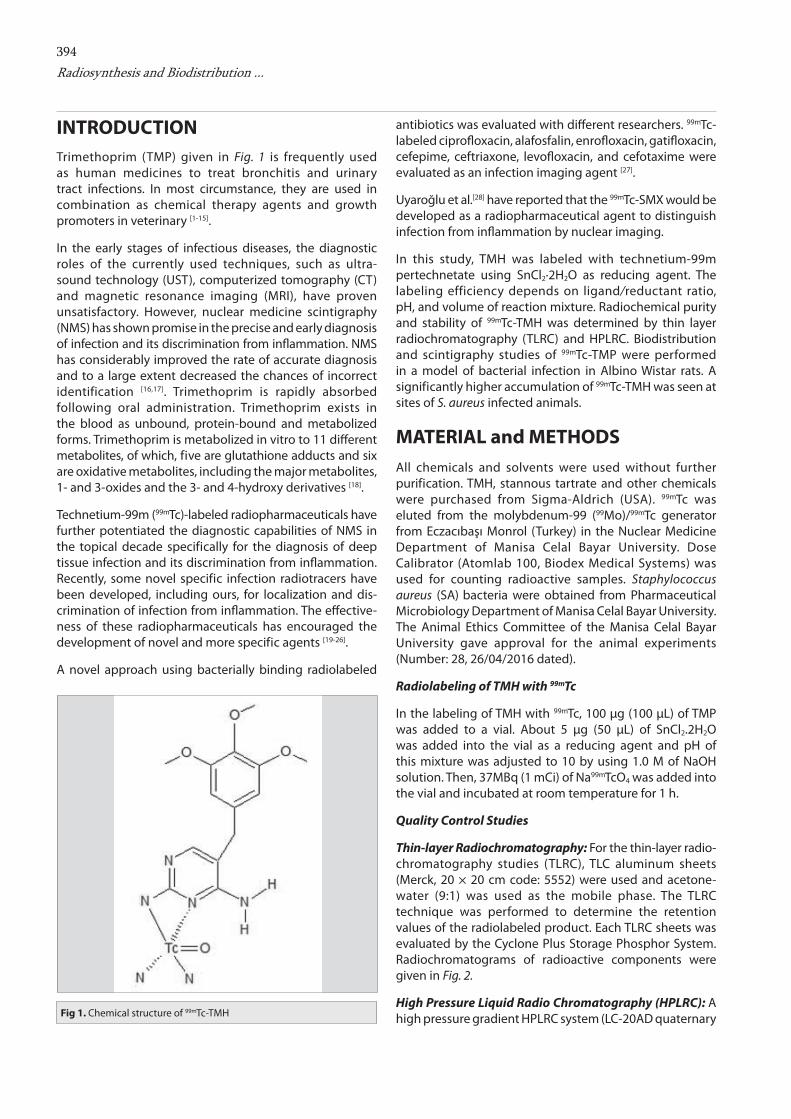
Thin-layer Radiochromatography: For the thin-layer radio- chromatography studies (TLRC), TLC aluminum sheets (Merck, 20 × 20 cm code: 5552) were used and acetone-water (9:1) was used as the mobile phase. The TLRC technique was performed to determine the retention values of the radiolabeled product. Each TLRC sheets was evaluated by the Cyclone Plus Storage Phosphor System. Radiochromatograms of radioactive components were given in Fig. 2.
High Pressure Liquid Radio Chromatography (HPLRC): A high pressure gradient HPLRC system (LC-20AD quaternary Fig 1. Chemical structure of 99mTc-TMH
395
DEMİROĞLU, TOPAL, PARLAK, GÜMÜŞERTÜRKÖZ, TEKİN, ATEŞ, ÜNAK, AVCIBAŞI
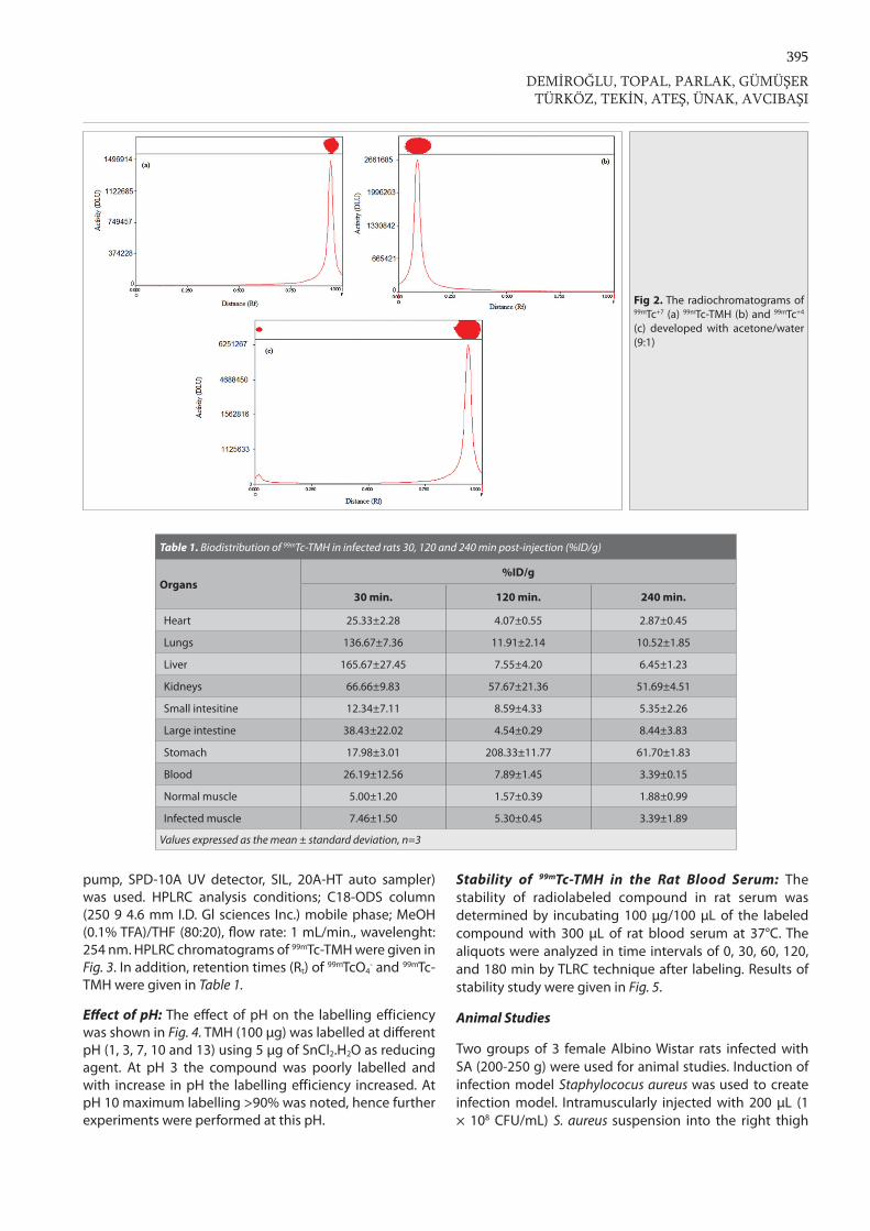
pump, SPD-10A UV detector, SIL, 20A-HT auto sampler) was used. HPLRC analysis conditions; C18-ODS column (250 9 4.6 mm I.D. Gl sciences Inc.) mobile phase; MeOH (0.1% TFA)/THF (80:20), flow rate: 1 mL/min., wavelenght: 254 nm. HPLRC chromatograms of 99mTc-TMH were given in Fig. 3. In addition, retention times (Rt) of 99mTcO4
- and 99mTc-TMH were given in Table 1.
Effect of pH: The effect of pH on the labelling efficiency was shown in Fig. 4. TMH (100 μg) was labelled at different pH (1, 3, 7, 10 and 13) using 5 μg of SnCl2.H2O as reducing agent. At pH 3 the compound was poorly labelled and with increase in pH the labelling efficiency increased. At pH 10 maximum labelling >90% was noted, hence further experiments were performed at this pH.
Stability of 99mTc-TMH in the Rat Blood Serum: The stability of radiolabeled compound in rat serum was determined by incubating 100 μg/100 μL of the labeled compound with 300 μL of rat blood serum at 37°C. The aliquots were analyzed in time intervals of 0, 30, 60, 120, and 180 min by TLRC technique after labeling. Results of stability study were given in Fig. 5.
Animal Studies
Two groups of 3 female Albino Wistar rats infected with SA (200-250 g) were used for animal studies. Induction of infection model Staphylococus aureus was used to create infection model. Intramuscularly injected with 200 μL (1 × 10⁸ CFU/mL) S. aureus suspension into the right thigh
Table 1. Biodistribution of 99mTc-TMH in infected rats 30, 120 and 240 min post-injection (%ID/g)
Organs%ID/g
30 min. 120 min. 240 min.
Heart 25.33±2.28 4.07±0.55 2.87±0.45
Lungs 136.67±7.36 11.91±2.14 10.52±1.85
Liver 165.67±27.45 7.55±4.20 6.45±1.23
Kidneys 66.66±9.83 57.67±21.36 51.69±4.51
Small intesitine 12.34±7.11 8.59±4.33 5.35±2.26
Large intestine 38.43±22.02 4.54±0.29 8.44±3.83
Stomach 17.98±3.01 208.33±11.77 61.70±1.83
Blood 26.19±12.56 7.89±1.45 3.39±0.15
Normal muscle 5.00±1.20 1.57±0.39 1.88±0.99
Infected muscle 7.46±1.50 5.30±0.45 3.39±1.89
Values expressed as the mean ± standard deviation, n=3
Fig 2. The radiochromatograms of 99mTc+7 (a) 99mTc-TMH (b) and 99mTc+4 (c) developed with acetone/water (9:1)
396Radiosynthesis and Biodistribution ...
muscle. Then the infection was allowed to develop for 48 h.
Scintigraphic Imaging Studies
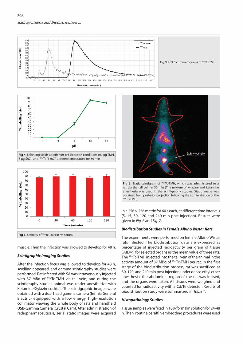
After the infection focus was allowed to develop for 48 h, swelling appeared, and gamma scintigraphy studies were performed. Rat infected with SA was intravenously injected with 37 MBq of 99mTc-TMH via tail vein, and during the scintigraphy studies animal was under anesthetize with Ketamine/Xylazin cocktail. The scintigraphic images were obtained with a dual head gamma camera (Infinia General Electric) equipped with a low energy, high-resolution collimator viewing the whole body of rats and handheld USB-Gamma Camera (Crystal Cam). After administration of radiopharmaceuticals, serial static images were acquired
in a 256 × 256 matrix for 60 s each, at different time intervals (5, 15, 30, 120 and 240 min post-injection). Results were given in Fig. 6 and Fig. 7.
Biodistribution Studies in Female Albino Wistar Rats
The experiments were performed on female Albino Wistar rats infected. The biodistribution data are expressed as percentage of injected radioactivity per gram of tissue (%ID/g) for selected organs as the mean value of three rats. The 99mTc-TMH injected into the tail vein of the animal in the activity amount of 37 MBq of 99mTc-TMH per rat. In the first stage of the biodistribution process, rat was sacrificed at 30, 120, and 240 min post injection under dense ethyl ether anesthesia, the abdominal region of the rat was incised, and the organs were taken. All tissues were weighed and counted for radioactivity with a Cd/Te detector. Results of biodistribution study were summarized in Table 1.
Histopathology Studies
Tissue samples were fixed in 10% formalin solution for 24-48 h. Then, routine paraffin embedding procedures were used
Fig 3. HPLC chromatograms of 99mTc-TMH
Fig 4. Labelling yields at different pH. Reaction condition: 100 µg TMH, 5 µg SnCl2 and 99mTc (1 mCi) at room temperature for 60 min
Fig 5. Stability of 99mTc-TMH in rat serum
Fig 6. Static scintigram of 99mTc-TMH, which was administered to a rat via the tail vein in 30 min (The mixture of xylazine and ketamine anesthesia was used in the scintigraphy studies. Static image was obtained from posterior projection following the administration of the 99mTc-TMH)
397
and 5 µm thick sections were cut from embedded tissues.
Paraffin Embedding Procedure
Fixed tissues washed with tap water for one night in order to remove the fixative solution. Then they were dehydrated through a graded ethanol series (60%, 70%, 80% and 95%), cleared in xylene and embedded in paraffin. Sections with 5 µm thickness were cut by rotary microtome (RM 2135, Leica) and were stained with hematoxylin-eosin for histochemical analyses.
Hematoxylin-Eosin Staining Procedure
Sections dewaxed under 60ºC overnight were immersed in xylene for 1 h and then rehydrated through a graded series of ethanol (95%, 80%, 70%, and 60%) for 2 min in each concentration and they were then washed in tap water for 5 min. Sections were stained with hematoxylin (01562E, Surgipath, Bretton, Peter Borough, Cambridge Shire) and washed in tap water for 5 min. Then sections were stained with eosin (01602E, Surgipath, Bretton, Peter Borough, Cambridge Shire) and washed in tap water for 5 min. After sections dehydrated with 80% and 95% alcohol and immersed in xylene and mounted with entellan (UN 1866, Merck, Darmstadt, Germany).
Statistical Analysis
The calculation of means and standard deviations were made on Microsoft Excel. One-way analysis of variance was used to determine statistical significance. Differences at the 95% confidence level (P<0.05) were considered significant.
RESULTS
Radiochemical purity and stability of 99mTc-TMH complex were assessed by thin layer and HPLRC methods. In thin layer chromatography using Acetone-water (9:1) as the solvent, free pertechnetate moved with the solvent front (Rf = 0.9), while 99mTc-TMH and reduced hydrolyzed technetium remained at the origin as seen in Fig. 2. Reduced hydrolyzed technetium was determined by using ethanol: water: ammonium hydroxide mixture (2:5:1) or 5 N NaOH as the mobile phase, where reduced hydrolyzed technetium remains at the origin (Rf = 0) while other species migrate with the solvent front (Rf=1). The radiochemical yield was determined as 96±2%. Also, radiochemical purity of 99mTc-THH was confirmed by HPLRC as seen in Fig. 3. Retention times of radio components, TcO4
- and 99mTc-TMH, were found to be 4.105±0.158 and 9.202±0.102 (min), respectively.
Effect of pH
As shown in Fig. 4, at pH 1 the labeling yield of 99mTc-TMH complex was equal to 0 and this yield increased with increasing the pH of the reaction mixture where pH 10 gave the maximum labeling yield of 96%. By increasing the pH greater than 10, the labeling yield remained constant.
Stability Test
As seen in Fig. 5, incubation of the preparation containing 99mTc-TMH in normal serum for 180 min at 37ºC resulted in a small decrease in the yield of 99mTc-TMH (85±2%) determined by TLRC.
DEMİROĞLU, TOPAL, PARLAK, GÜMÜŞERTÜRKÖZ, TEKİN, ATEŞ, ÜNAK, AVCIBAŞI
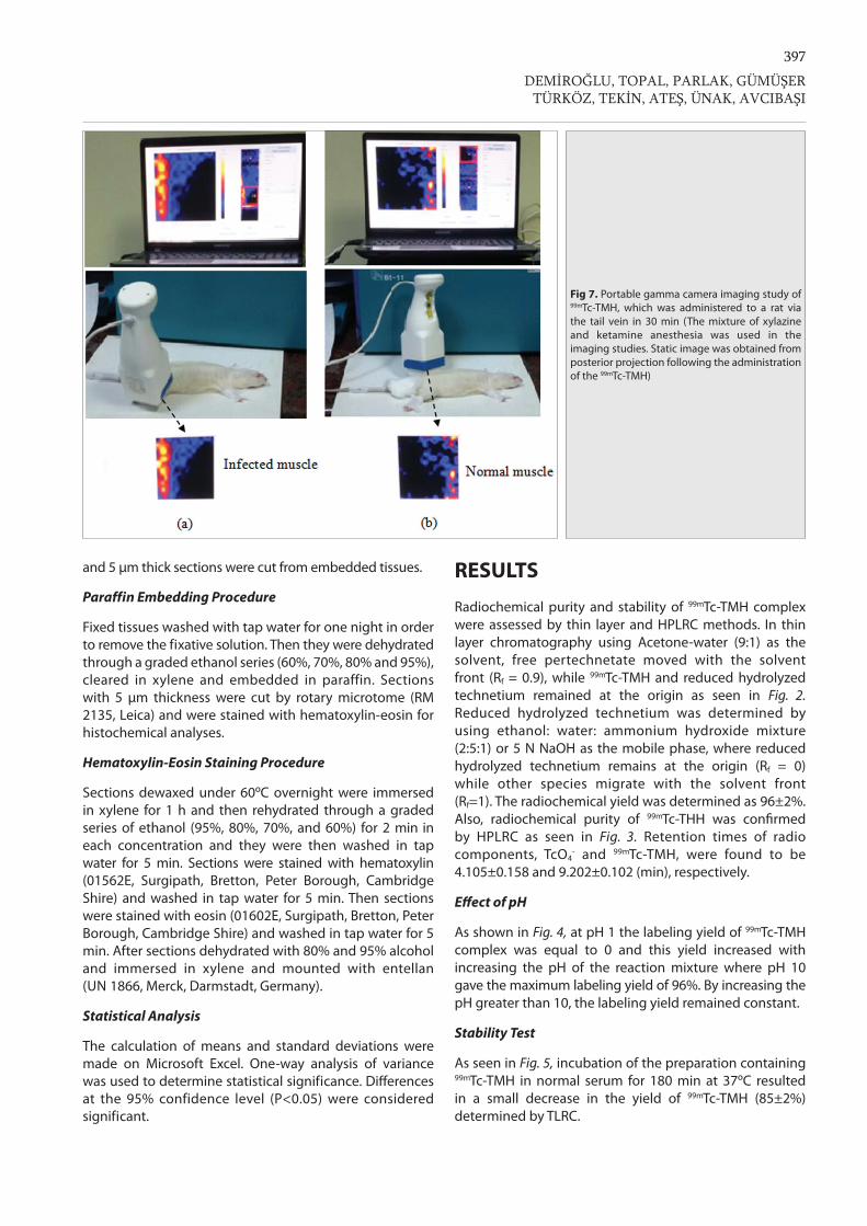
Fig 7. Portable gamma camera imaging study of 99mTc-TMH, which was administered to a rat via the tail vein in 30 min (The mixture of xylazine and ketamine anesthesia was used in the imaging studies. Static image was obtained from posterior projection following the administration of the 99mTc-TMH)
398Radiosynthesis and Biodistribution ...
Imaging Studies
After the administration of 99mTc-TMH to rats, static images were obtained by using a dual head gamma camera (Infinia General Electric) and handheld USB-Gamma Camera (CrystalCam). Static image of 99mTc-TMH in male rats demonstrated that important radiation signals are present in the infected site at first glance
in 30 min as seen in Fig. 6 and Fig. 7.
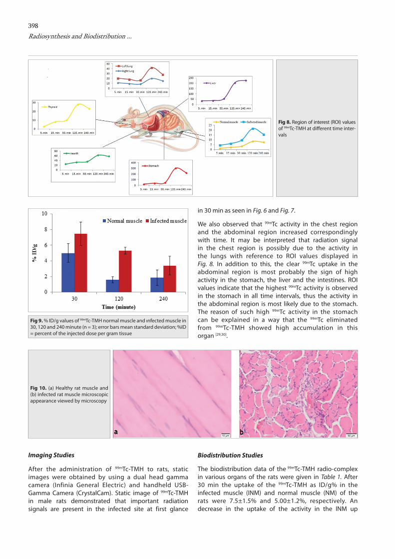
We also observed that 99mTc activity in the chest region and the abdominal region increased correspondingly with time. It may be interpreted that radiation signal in the chest region is possibly due to the activity in the lungs with reference to ROI values displayed in Fig. 8. In addition to this, the clear 99mTc uptake in the abdominal region is most probably the sign of high activity in the stomach, the liver and the intestines. ROI values indicate that the highest 99mTc activity is observed in the stomach in all time intervals, thus the activity in the abdominal region is most likely due to the stomach. The reason of such high 99mTc activity in the stomach can be explained in a way that the 99mTc eliminated from 99mTc-TMH showed high accumulation in this organ [29,30].
Biodistribution Studies
The biodistribution data of the 99mTc-TMH radio-complex in various organs of the rats were given in Table 1. After 30 min the uptake of the 99mTc-TMH as ID/g% in the infected muscle (INM) and normal muscle (NM) of the rats were 7.5±1.5% and 5.00±1.2%, respectively. An decrease in the uptake of the activity in the INM up
Fig 9. % ID/g values of 99mTc-TMH normal muscle and ınfected muscle in 30, 120 and 240 minute (n = 3); error bars mean standard deviation; %ID = percent of the injected dose per gram tissue
Fig 8. Region of interest (ROI) values of 99mTc-TMH at different time inter-vals
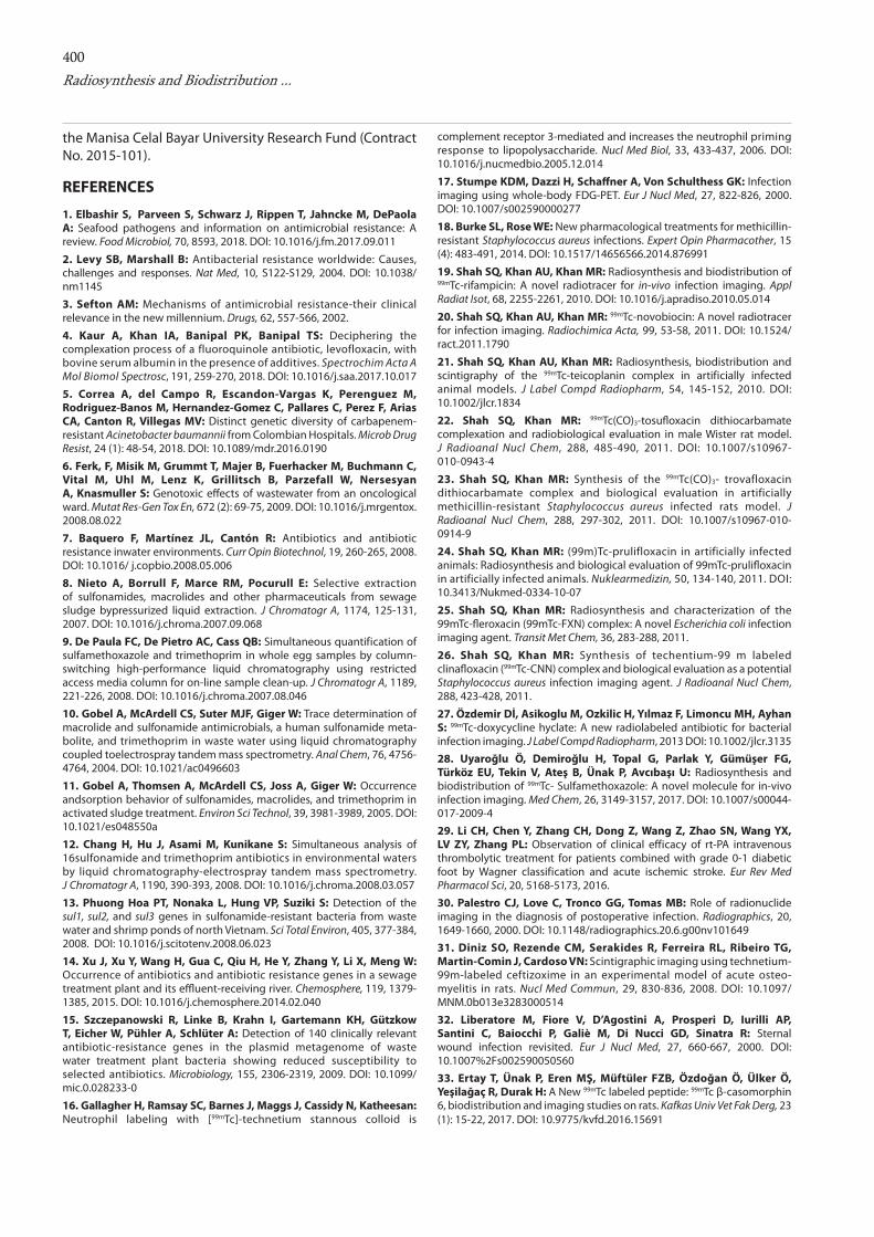
Fig 10. (a) Healthy rat muscle and (b) infected rat muscle microscopic appearance viewed by microscopy
a b
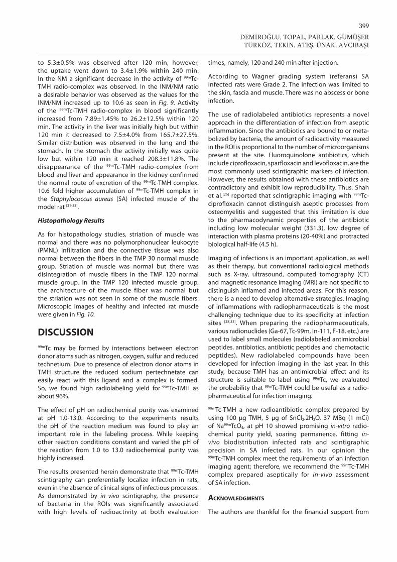
399
to 5.3±0.5% was observed after 120 min, however, the uptake went down to 3.4±1.9% within 240 min. In the NM a significant decrease in the activity of 99mTc-TMH radio-complex was observed. In the INM/NM ratio a desirable behavior was observed as the values for the INM/NM increased up to 10.6 as seen in Fig. 9. Activity of the 99mTc-TMH radio-complex in blood significantly increased from 7.89±1.45% to 26.2±12.5% within 120 min. The activity in the liver was initially high but within 120 min it decreased to 7.5±4.0% from 165.7±27.5%. Similar distribution was observed in the lung and the stomach. In the stomach the activity initially was quite low but within 120 min it reached 208.3±11.8%. The disappearance of the 99mTc-TMH radio-complex from blood and liver and appearance in the kidney confirmed the normal route of excretion of the 99mTc-TMH complex. 10.6 fold higher accumulation of 99mTc-TMH complex in the Staphylococcus aureus (SA) infected muscle of the model rat [31-33].
Histopathology Results
As for histopathology studies, striation of muscle was normal and there was no polymorphonuclear leukocyte (PMNL) infiltration and the connective tissue was also normal between the fibers in the TMP 30 normal muscle group. Striation of muscle was normal but there was disintegration of muscle fibers in the TMP 120 normal muscle group. In the TMP 120 infected muscle group, the architecture of the muscle fiber was normal but the striation was not seen in some of the muscle fibers. Microscopic images of healthy and infected rat muscle were given in Fig. 10.
DISCUSSION99mTc may be formed by interactions between electron donor atoms such as nitrogen, oxygen, sulfur and reduced technetium. Due to presence of electron donor atoms in TMH structure the reduced sodium pertechnetate can easily react with this ligand and a complex is formed. So, we found high radiolabeling yield for 99mTc-TMH as about 96%.
The effect of pH on radiochemical purity was examined at pH 1.0-13.0. According to the experiments results the pH of the reaction medium was found to play an important role in the labeling process. While keeping other reaction conditions constant and varied the pH of the reaction from 1.0 to 13.0 radiochemical purity was highly increased.
The results presented herein demonstrate that 99mTc-TMH scintigraphy can preferentially localize infection in rats, even in the absence of clinical signs of infectious processes. As demonstrated by in vivo scintigraphy, the presence of bacteria in the ROIs was significantly associated with high levels of radioactivity at both evaluation
times, namely, 120 and 240 min after injection.
According to Wagner grading system (referans) SA infected rats were Grade 2. The infection was limited to the skin, fascia and muscle. There was no abscess or bone infection.
The use of radiolabeled antibiotics represents a novel approach in the differentiation of infection from aseptic inflammation. Since the antibiotics are bound to or meta-bolized by bacteria, the amount of radioactivity measured in the ROI is proportional to the number of microorganisms present at the site. Fluoroquinolone antibiotics, which include ciprofloxacin, sparfloxacin and levofloxacin, are the most commonly used scintigraphic markers of infection. However, the results obtained with these antibiotics are contradictory and exhibit low reproducibility. Thus, Shah et al.[26] reported that scintigraphic imaging with 99mTc-ciprofloxacin cannot distinguish aseptic processes from osteomyelitis and suggested that this limitation is due to the pharmacodynamic properties of the antibiotic including low molecular weight (331.3), low degree of interaction with plasma proteins (20-40%) and protracted biological half-life (4.5 h).
Imaging of infections is an important application, as well as their therapy, but conventional radiological methods such as X-ray, ultrasound, computed tomography (CT) and magnetic resonance imaging (MRI) are not specific to distinguish inflamed and infected areas. For this reason, there is a need to develop alternative strategies. Imaging of inflammations with radiopharmaceuticals is the most challenging technique due to its specificity at infection sites [28,33]. When preparing the radiopharmaceuticals, various radionuclides (Ga-67, Tc-99m, In-111, F-18, etc.) are used to label small molecules (radiolabeled antimicrobial peptides, antibiotics, antibiotic peptides and chemotactic peptides). New radiolabeled compounds have been developed for infection imaging in the last year. In this study, because TMH has an antimicrobial effect and its structure is suitable to label using 99mTc, we evaluated the probability that 99mTc-TMH could be useful as a radio-pharmaceutical for infection imaging.
99mTc-TMH a new radioantibiotic complex prepared by using 100 μg TMH, 5 μg of SnCl2.2H2O, 37 MBq (1 mCi) of Na99mTcO4, at pH 10 showed promising in-vitro radio-chemical purity yield, soaring permanence, fitting in-vivo biodistribution infected rats and scintigraphic precision in SA infected rats. In our opinion the 99mTc-TMH complex meet the requirements of an infection imaging agent; therefore, we recommend the 99mTc-TMH complex prepared aseptically for in-vivo assessment of SA infection.
Acknowledgments
The authors are thankful for the financial support from
DEMİROĞLU, TOPAL, PARLAK, GÜMÜŞERTÜRKÖZ, TEKİN, ATEŞ, ÜNAK, AVCIBAŞI
400Radiosynthesis and Biodistribution ...
the Manisa Celal Bayar University Research Fund (Contract No. 2015-101).
REFERENCES
1. Elbashir S, Parveen S, Schwarz J, Rippen T, Jahncke M, DePaola A: Seafood pathogens and information on antimicrobial resistance: A review. Food Microbiol, 70, 8593, 2018. DOI: 10.1016/j.fm.2017.09.011
2. Levy SB, Marshall B: Antibacterial resistance worldwide: Causes, challenges and responses. Nat Med, 10, S122-S129, 2004. DOI: 10.1038/nm1145
3. Sefton AM: Mechanisms of antimicrobial resistance-their clinical relevance in the new millennium. Drugs, 62, 557-566, 2002.
4. Kaur A, Khan IA, Banipal PK, Banipal TS: Deciphering the complexation process of a fluoroquinole antibiotic, levofloxacin, with bovine serum albumin in the presence of additives. Spectrochim Acta A Mol Biomol Spectrosc, 191, 259-270, 2018. DOI: 10.1016/j.saa.2017.10.017
5. Correa A, del Campo R, Escandon-Vargas K, Perenguez M, Rodriguez-Banos M, Hernandez-Gomez C, Pallares C, Perez F, Arias CA, Canton R, Villegas MV: Distinct genetic diversity of carbapenem-resistant Acinetobacter baumannii from Colombian Hospitals. Microb Drug Resist, 24 (1): 48-54, 2018. DOI: 10.1089/mdr.2016.0190
6. Ferk, F, Misik M, Grummt T, Majer B, Fuerhacker M, Buchmann C, Vital M, Uhl M, Lenz K, Grillitsch B, Parzefall W, Nersesyan A, Knasmuller S: Genotoxic effects of wastewater from an oncological ward. Mutat Res-Gen Tox En, 672 (2): 69-75, 2009. DOI: 10.1016/j.mrgentox. 2008.08.022
7. Baquero F, Martínez JL, Cantón R: Antibiotics and antibiotic resistance inwater environments. Curr Opin Biotechnol, 19, 260-265, 2008. DOI: 10.1016/ j.copbio.2008.05.006
8. Nieto A, Borrull F, Marce RM, Pocurull E: Selective extraction of sulfonamides, macrolides and other pharmaceuticals from sewage sludge bypressurized liquid extraction. J Chromatogr A, 1174, 125-131, 2007. DOI: 10.1016/j.chroma.2007.09.068
9. De Paula FC, De Pietro AC, Cass QB: Simultaneous quantification of sulfamethoxazole and trimethoprim in whole egg samples by column-switching high-performance liquid chromatography using restricted access media column for on-line sample clean-up. J Chromatogr A, 1189, 221-226, 2008. DOI: 10.1016/j.chroma.2007.08.046
10. Gobel A, McArdell CS, Suter MJF, Giger W: Trace determination of macrolide and sulfonamide antimicrobials, a human sulfonamide meta-bolite, and trimethoprim in waste water using liquid chromatography coupled toelectrospray tandem mass spectrometry. Anal Chem, 76, 4756-4764, 2004. DOI: 10.1021/ac0496603
11. Gobel A, Thomsen A, McArdell CS, Joss A, Giger W: Occurrence andsorption behavior of sulfonamides, macrolides, and trimethoprim in activated sludge treatment. Environ Sci Technol, 39, 3981-3989, 2005. DOI: 10.1021/es048550a
12. Chang H, Hu J, Asami M, Kunikane S: Simultaneous analysis of 16sulfonamide and trimethoprim antibiotics in environmental waters by liquid chromatography-electrospray tandem mass spectrometry. J Chromatogr A, 1190, 390-393, 2008. DOI: 10.1016/j.chroma.2008.03.057
13. Phuong Hoa PT, Nonaka L, Hung VP, Suziki S: Detection of the sul1, sul2, and sul3 genes in sulfonamide-resistant bacteria from waste water and shrimp ponds of north Vietnam. Sci Total Environ, 405, 377-384, 2008. DOI: 10.1016/j.scitotenv.2008.06.023
14. Xu J, Xu Y, Wang H, Gua C, Qiu H, He Y, Zhang Y, Li X, Meng W: Occurrence of antibiotics and antibiotic resistance genes in a sewage treatment plant and its effluent-receiving river. Chemosphere, 119, 1379-1385, 2015. DOI: 10.1016/j.chemosphere.2014.02.040
15. Szczepanowski R, Linke B, Krahn I, Gartemann KH, Gützkow T, Eicher W, Pühler A, Schlüter A: Detection of 140 clinically relevant antibiotic-resistance genes in the plasmid metagenome of waste water treatment plant bacteria showing reduced susceptibility to selected antibiotics. Microbiology, 155, 2306-2319, 2009. DOI: 10.1099/mic.0.028233-0
16. Gallagher H, Ramsay SC, Barnes J, Maggs J, Cassidy N, Katheesan: Neutrophil labeling with [99mTc]-technetium stannous colloid is
complement receptor 3-mediated and increases the neutrophil priming response to lipopolysaccharide. Nucl Med Biol, 33, 433-437, 2006. DOI: 10.1016/j.nucmedbio.2005.12.014
17. Stumpe KDM, Dazzi H, Schaffner A, Von Schulthess GK: Infection imaging using whole-body FDG-PET. Eur J Nucl Med, 27, 822-826, 2000. DOI: 10.1007/s002590000277
18. Burke SL, Rose WE: New pharmacological treatments for methicillin-resistant Staphylococcus aureus infections. Expert Opin Pharmacother, 15 (4): 483-491, 2014. DOI: 10.1517/14656566.2014.876991
19. Shah SQ, Khan AU, Khan MR: Radiosynthesis and biodistribution of 99mTc-rifampicin: A novel radiotracer for in-vivo infection imaging. Appl Radiat Isot, 68, 2255-2261, 2010. DOI: 10.1016/j.apradiso.2010.05.014
20. Shah SQ, Khan AU, Khan MR: 99mTc-novobiocin: A novel radiotracer for infection imaging. Radiochimica Acta, 99, 53-58, 2011. DOI: 10.1524/ract.2011.1790
21. Shah SQ, Khan AU, Khan MR: Radiosynthesis, biodistribution and scintigraphy of the 99mTc-teicoplanin complex in artificially infected animal models. J Label Compd Radiopharm, 54, 145-152, 2010. DOI: 10.1002/jlcr.1834
22. Shah SQ, Khan MR: 99mTc(CO)3-tosufloxacin dithiocarbamate complexation and radiobiological evaluation in male Wister rat model. J Radioanal Nucl Chem, 288, 485-490, 2011. DOI: 10.1007/s10967- 010-0943-4
23. Shah SQ, Khan MR: Synthesis of the 99mTc(CO)3- trovafloxacin dithiocarbamate complex and biological evaluation in artificially methicillin-resistant Staphylococcus aureus infected rats model. J Radioanal Nucl Chem, 288, 297-302, 2011. DOI: 10.1007/s10967-010-0914-9
24. Shah SQ, Khan MR: (99m)Tc-prulifloxacin in artificially infected animals: Radiosynthesis and biological evaluation of 99mTc-prulifloxacin in artificially infected animals. Nuklearmedizin, 50, 134-140, 2011. DOI: 10.3413/Nukmed-0334-10-07
25. Shah SQ, Khan MR: Radiosynthesis and characterization of the 99mTc-fleroxacin (99mTc-FXN) complex: A novel Escherichia coli infection imaging agent. Transit Met Chem, 36, 283-288, 2011.
26. Shah SQ, Khan MR: Synthesis of techentium-99 m labeled clinafloxacin (99mTc-CNN) complex and biological evaluation as a potential Staphylococcus aureus infection imaging agent. J Radioanal Nucl Chem, 288, 423-428, 2011.
27. Özdemir Dİ, Asikoglu M, Ozkilic H, Yılmaz F, Limoncu MH, Ayhan S: 99mTc-doxycycline hyclate: A new radiolabeled antibiotic for bacterial infection imaging. J Label Compd Radiopharm, 2013 DOI: 10.1002/jlcr.313528. Uyaroğlu Ö, Demiroğlu H, Topal G, Parlak Y, Gümüşer FG, Türköz EU, Tekin V, Ateş B, Ünak P, Avcıbaşı U: Radiosynthesis and biodistribution of 99mTc- Sulfamethoxazole: A novel molecule for in-vivo infection imaging. Med Chem, 26, 3149-3157, 2017. DOI: 10.1007/s00044-017-2009-429. Li CH, Chen Y, Zhang CH, Dong Z, Wang Z, Zhao SN, Wang YX, LV ZY, Zhang PL: Observation of clinical efficacy of rt-PA intravenous thrombolytic treatment for patients combined with grade 0-1 diabetic foot by Wagner classification and acute ischemic stroke. Eur Rev Med Pharmacol Sci, 20, 5168-5173, 2016.30. Palestro CJ, Love C, Tronco GG, Tomas MB: Role of radionuclide imaging in the diagnosis of postoperative infection. Radiographics, 20, 1649-1660, 2000. DOI: 10.1148/radiographics.20.6.g00nv10164931. Diniz SO, Rezende CM, Serakides R, Ferreira RL, Ribeiro TG, Martin-Comin J, Cardoso VN: Scintigraphic imaging using technetium-99m-labeled ceftizoxime in an experimental model of acute osteo-myelitis in rats. Nucl Med Commun, 29, 830-836, 2008. DOI: 10.1097/MNM.0b013e328300051432. Liberatore M, Fiore V, D’Agostini A, Prosperi D, Iurilli AP, Santini C, Baiocchi P, Galiè M, Di Nucci GD, Sinatra R: Sternal wound infection revisited. Eur J Nucl Med, 27, 660-667, 2000. DOI: 10.1007%2Fs00259005056033. Ertay T, Ünak P, Eren MŞ, Müftüler FZB, Özdoğan Ö, Ülker Ö, Yeşilağaç R, Durak H: A New 99mTc labeled peptide: 99mTc β-casomorphin 6, biodistribution and imaging studies on rats. Kafkas Univ Vet Fak Derg, 23 (1): 15-22, 2017. DOI: 10.9775/kvfd.2016.15691


![Radiosynthesis and Bioconjugation of [18 F]FPy5yne - Triumf](https://img.pdfslide.net/doc/110x75/6203aba2da24ad121e4c1a6b/radiosynthesis-and-bioconjugation-of-18-ffpy5yne-triumf.jpg)


![Efficient Radiosynthesis of a [18F]-Phosphonium Salt](https://img.pdfslide.net/doc/110x75/6259435e41a07670ef292b68/efficient-radiosynthesis-of-a-18f-phosphonium-salt-.jpg)