Embed Size (px)
Citation preview
Available online at www.sciencedirect.com
www.elsevier.com/locate/asr
ScienceDirect
Advances in Space Research 57 (2016) 2385–2395
Raman–Mossbauer–XRD studies of selected samples from‘‘Los Azulejos” outcrop: A possible analogue for assessing the
alteration processes on Mars
E.A. Lalla a,b,⇑, A. Sanz-Arranz b, G. Lopez-Reyes b, A. Sansano b, J. Medina b,D. Schmanke c, G. Klingelhoefer c, J.A. Rodrıguez-Losada d, J. Martınez-Frıas e, F. Rull b
aExtreme Light Infrastructure – Nuclear Physics (ELI-NP), Horia Hulubei National Institute of Physics and Nuclear Engineering (IFIN HH),
30 Reactorului Street, P.O. Box MG-6, 077125 Magurele, jud. Ilfov, RomaniabUnidad Asociada UVa-CSIC al Centro de Astrobiologıa, Edificio INDITI, Parque Tecnologico de Boecillo, 47151 Boecillo, Valladolid, Spain
c Institut fur Anorganische Chemie und Analytische Chemie, Johannes Gutenberg, Universitat Mainz, 55099 Mainz, GermanydDepartamento de Biologıa Animal, Edafologıa y Geologıa, Universidad de la Laguna, 38200 San Cristobal de La Laguna, Santa Cruz de Tenerife,
SpaineDinamica Terrestre y Observacion de la Tierra, Instituto de Geociencias, IGEO (CSIC-UCM), Facultad de Ciencias Geologicas, Ciudad
Universitaria, 28040 Madrid, Spain
Received 27 April 2015; received in revised form 7 March 2016; accepted 8 March 2016Available online 16 March 2016
Abstract
The outcrop of ‘‘Los Azulejos” is visible at the interior of the Canadas Caldera in Tenerife Island (Spain). It exhibits a great variety ofalteration processes that could be considered as terrestrial analogue for several geological processes on Mars. This outcrop is particularlyinteresting due to the content of clays, zeolite, iron oxides, and sulfates corresponding to a hydrothermal alteration catalogued as ‘‘Azule-jos” type alteration. A detailed analysis by portable and laboratory Raman systems as well as other different techniques such as X-raydiffraction (XRD) and Mossbauer spectroscopy has been carried out (using twin-instruments from Martian lander missions: Mossbauerspectrometer MIMOS-II from the NASA-MER mission of 2001 and the XRD diffractometer from the NASA-MSL Curiosity mission of2012). The mineral identification presents the following mineral species: magnetite, goethite, hematite, anatase, rutile, quartz, gregoryite,sulfate (thenardite and hexahydrite), diopside, feldspar, analcime, kaolinite and muscovite. Moreover, the in-situ Raman and Micro-Raman measurements have been performed in order to compare the capabilities of the portable system specially focused for the nextESA Exo-Mars mission. The mineral detection confirms the sub-aerial alteration on the surface and the hydrothermal processes bythe volcanic fluid circulations in the fresh part. Therefore, the secondary more abundant mineralization acts as the color agent of therocks. Thus, the zeolite–illite group is the responsible for the bluish coloration, as well as the feldspars and carbonates for the whitishand the iron oxide for the redish parts. The XRD system was capable to detect a minor proportion of pyroxene, which is not visible byRaman and Mossbauer spectroscopy due to the ‘‘Azulejos” alteration of the parent material on the outcrop. On the other hand, Moss-bauer spectroscopy was capable of detecting different types of iron-oxides (Fe3+/2+-oxide phases). These analyses emphasize the strengthof the different techniques and the working synergy of the three different techniques together for planetary space missions.� 2016 COSPAR. Published by Elsevier Ltd. All rights reserved.
Keywords: Raman; Mars; Alteration processes; Mineralogy; Volcanic analogues
http://dx.doi.org/10.1016/j.asr.2016.03.014
0273-1177/� 2016 COSPAR. Published by Elsevier Ltd. All rights reserved.
⇑ Corresponding author at: Extreme Light Infrastructure – Nuclear Physics (ELI-NP), Horia Hulubei National Institute of Physics and NuclearEngineering (IFIN HH), 30 Reactorului Street, P.O. Box MG-6, 077125 Magurele, jud. Ilfov, Romania. Tel.: +40 214042300.
E-mail address: [email protected] (E.A. Lalla).
2386 E.A. Lalla et al. / Advances in Space Research 57 (2016) 2385–2395
1. Introduction
Several volcanic places have been used as possible terres-trial analogues taking into account the volcanic activitiesand the huge variety of geological processes discovered onMars heretofore (Chevrier andMathe, 2007) such asHawai-ian Island, Marion Island, among others (Graham et al.,2015; Prinsloo et al., 2011). Considering the previousresearch, one of the most important alteration processes ofvolcanic materials on Martian surface are related to thehydrothermal processes (Merle et al., 2010). All the informa-tion yielded from the different NASA missions such as theMars Exploration Rover (MER, (Rayl, 2014)) and theMarsScience Laboratory-Curiosity (MSL, (Kuhn, 2015a)) haveimproved the understanding of the geological diversity ofthe planet. However, the possibility of establishing new ter-restrial environments as analogue test sites for future spacemissions such as ESA-ExoMars mission (Bost et al., 2015;Courreges-Lacoste et al., 2007; Rull-Perez and Martinez-Frias, 2006) and the future NASA mission in 2020(Grossman, 2013) is needed. In this regard, new terrestrialvolcanic analogue environments can provide newdatawhichcould be used for the interpretation of the geological historyof Mars and the data collected by the different future spacemissions (Bishop et al., 2004; Lalla, 2014; Prinsloo et al.,2011). Special attention has to be paid to the mineralogybecause it is the best indicator of the physical–chemicalgeo-processes and the paragenetic assemblages. Thus, themineral diversity of new terrestrial analogues could allowthe scientific community to increase the knowledge aboutthe geological processes throughout the history of Mars(Mouginis-Mark andRobinson, 1992). Examining theMar-tian mineralogy, it has been observed that it is as rich as theEarthmineralogy showing secondarymineralization such asoxides, phyllosilicates, sulfates, carbonates, zeolites andclays (Chevrier and Mathe, 2007; Lalla et al., 2010; Minittiand Hamilton, 2010; Ruff, 2004). Secondary and accessoryminerals have a paragenetic origin from hydrothermal reac-tions with the sub-surface fluids, water alteration, and sub-aerial processes. Nevertheless, there is a controversy aboutthe detailed processes occurred on Mars, especially withthe chemically weathered basalt (Mahaney et al., 2012).The use of natural samples from basaltic terrestrial ana-logues have been selected to perform future analysis onMarsrelevant samples (with which the technology can be testedand improved).
It has been demonstrated that Tenerife Island is an areaof reference for carrying out research and technologicalstudies with planetary and astrogeological implications(Lalla et al., 2015). Several places of the island have beenselected considering the fluid–rock interactions caused bythe weathering processes, the submarine and sub-aerialalteration, hydrothermalism and the geomorphological fea-tures (Lalla et al., 2015a,b)
The main motivation of this paper is to study and regis-ter a complete spectroscopic analysis (mainly by Ramanspectroscopy) of the selected outcrop corresponding to
‘‘Los Azulejos”, which exhibits visible alterations fromthe original rocks (called ‘‘Azulejos” alteration), and whoseresults could be used as potential model substances for theoriginal altered material on Mars. On the other hand, thedata collected using twin-instrument from present andfuture different space missions in terrestrial analoguesmay contribute for the next generation objectives in spaceexploration. Finally, this study is also focused to improvethe strength of the Raman spectroscopy for space explo-ration and to complement the results with the capabilitiesof the XRD and Mossbauer techniques.
2. Geological setting
The Canadas Caldera, at the central part of TenerifeIsland, as may indicated by (Marti and Gudmundsson,2000) was formed by several collapse episodes of the Cana-das central volcanic edifice during several highly explosiveeruptions of phonolitic magmas (see Fig. 1). From the geo-chemical point of view, the Canadas edifice has been and isalso affected by active hydrothermal and fumaloric activi-ties as well as CO2 diffuse emission (Villasante-Marcoset al., 2014), this has acted after the Caldera formation.The combination of the mechanical and geochemical pro-cesses produced a heterogeneous volcanic system, wherereally interesting specific outcrops could become a terres-trial analogues for instrument testing and contribute tothe science development of new space missions. In thisregard, one of the most interesting macro-structural forma-tion is the called the outcrop of ‘‘Los Azulejos” (Fig. 1),that forms a part of the Caldera wall. One of the mostimpressive features at this outcrop is the variety of the dif-ferent processes involved (i.e. hydrothermal processes,argilization and parent mineralogical alteration) (Galindoet al., 2005; Villasante-Marcos et al., 2014). Galindoet al., 2005, have described these geological features indi-cating that the faults within ‘‘Los Azulejos” structure affectthe igneous materials of the Canadas edifice by the inclinedand sub-vertical sheets intrusions formation. In addition, ithas been reported a variable displacement of the fault isreported in (Galindo et al., 2005). They also point out thatthe kinetic indicators such as the offset of stratigraphic lay-ers, shear-cleavage structures in mylonitic foliation, amongothers, in combination with the CO2 active diffuse degas-sing activities and the hydrothermal activities cause a hugevariety of mineralogical species. The colored outcrop of‘‘Los Azulejos” presents bluish, greenish, whitish, and yel-lowish colors (Fig. 1). The bluish to greenish degradationcorresponds to a combination of analcime and clay miner-als such as smectite and illite. On the other hand, yellowishand blank coloration can be observed on the fumarolicstructure and the argilization of the parent minerals. Thesecolorations present really interesting formations like mush-rooms textures and veins circulation with sulfates and ironoxides mineral species (Bustillo, 1989; Galindo et al., 2005).The colors in the alteration correspond to a process termed‘‘Los Azulejos” type alteration and it is also present on
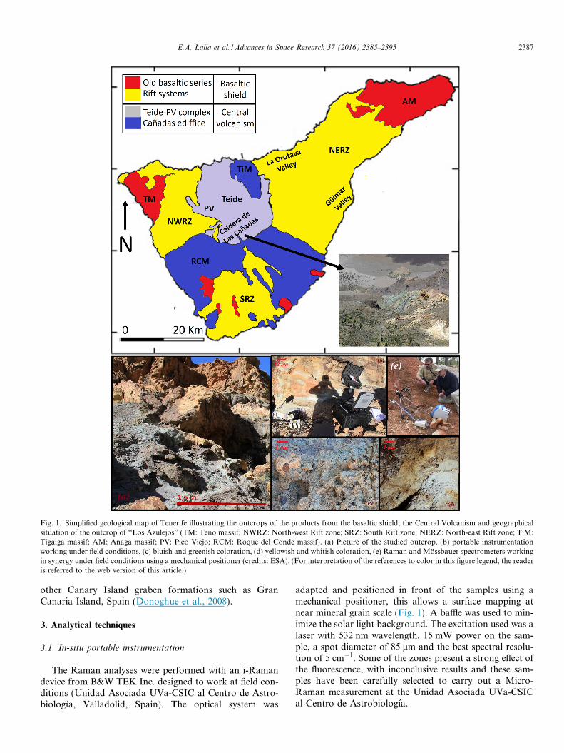
Fig. 1. Simplified geological map of Tenerife illustrating the outcrops of the products from the basaltic shield, the Central Volcanism and geographicalsituation of the outcrop of ‘‘Los Azulejos” (TM: Teno massif; NWRZ: North-west Rift zone; SRZ: South Rift zone; NERZ: North-east Rift zone; TiM:Tigaiga massif; AM: Anaga massif; PV: Pico Viejo; RCM: Roque del Conde massif). (a) Picture of the studied outcrop, (b) portable instrumentationworking under field conditions, (c) bluish and greenish coloration, (d) yellowish and whitish coloration, (e) Raman and Mossbauer spectrometers workingin synergy under field conditions using a mechanical positioner (credits: ESA). (For interpretation of the references to color in this figure legend, the readeris referred to the web version of this article.)
E.A. Lalla et al. / Advances in Space Research 57 (2016) 2385–2395 2387
other Canary Island graben formations such as GranCanaria Island, Spain (Donoghue et al., 2008).
3. Analytical techniques
3.1. In-situ portable instrumentation
The Raman analyses were performed with an i-Ramandevice from B&W TEK Inc. designed to work at field con-ditions (Unidad Asociada UVa-CSIC al Centro de Astro-biologıa, Valladolid, Spain). The optical system was
adapted and positioned in front of the samples using amechanical positioner, this allows a surface mapping atnear mineral grain scale (Fig. 1). A baffle was used to min-imize the solar light background. The excitation used was alaser with 532 nm wavelength, 15 mW power on the sam-ple, a spot diameter of 85 lm and the best spectral resolu-tion of 5 cm�1. Some of the zones present a strong effect ofthe fluorescence, with inconclusive results and these sam-ples have been carefully selected to carry out a Micro-Raman measurement at the Unidad Asociada UVa-CSICal Centro de Astrobiologıa.
2388 E.A. Lalla et al. / Advances in Space Research 57 (2016) 2385–2395
The Mossbauer spectra were collected with a copy of theMiniaturized Mossbauer spectrometer MIMOS II spec-trometer from the past NASA-MER-mission (AK Klingel-hoefer, Mars Mossbauer Group, Mainz, Germany). Thesystem has a Co57/Rh source with an intensity of about50 mCi. The measurements have been performed at roomtemperature and without sample preparation in a backscat-tering geometry.
The XRD system for the measurement was the TerraXRD diffractometer instrument (based on the MSL-CheMin concept, available at the Unidad Asociada UVa-CSIC with a detector of 1024 � 256 pixels, 2D peltiercooled CCD camera for XRD with a source of cobalt X-ray tube, working at 30 kV and 300 lA. For the XRD anal-ysis, a preparation was necessary: powdering a minimumpart of the samples (from 2 to 4 mg) and sieving with agranulometry lower than 150 lm. The XRD measurementand analysis were obtained using a 0.25� 2h� FWHM reso-lution, a 2h� spectral range of 5–55� and 200 accumulationswith an exposition time of 15 s. The mineral identificationsoftware used was the Xpowder12 (Martin-Ramos, 2004).
3.2. Laboratory instrumentation
The micro-Raman mineralogical characterization of thesamples was performed with a micro-Raman system fromthe Unidad Asociada UVa-CSIC. The system is composedby a microscope Nikon Eclipse E600 coupled to a spec-trometer KOSI Holospec f/1.8i, with best spectral resolu-tion of 5 cm�1, illuminated by a laser REO LSRP-3501He–Ne (632.8 nm wavelength). The detector was a CCDAndor DV420A-OE-130. The maximum laser power usedon the sample was 14 mW with a minimum spot diameterof 15 lm. The Raman mapping of the bulk surface of thesample was done by the micro-Raman Prior Proscan IImotorized stage in automatic mode in order to detect thedifferent compositional mineralization. However, the opti-mum recording conditions were obtained by varying thelaser power, microscope objective, and the confocal spotsize (XY instrument) as required for the different samples.The spectra have been directly acquired on the samplematerial without any sample preparation.
The conventional X-ray diffraction analyses were carriedout at University of Valladolid, Spain with a XRD diffrac-tometer Philips PW1710 equipped with an automatic diver-gent slit graphite monochromator and Cu-anode.Experimental conditions: CuKa radiation, k = 0.154 nm, anickel filter, an aluminum sample-holder, 40 kV generatorvoltage, generator current 30 mA with a relation intensityof 0.5 (a1/a2), and angle range (2h�) from 5� to 70�. The stepssize applied is 0.02� and the identification has been doneusing Match! Program system, the crystallography OpenDatabase (COD), the ICDD System (International Centrefor Diffraction Data) in PDF-2 (Power Diffraction Files),and the JCPDS (Joint Committee on Powder diffractionStandards).
4. Results
4.1. Sample recollection and description
The samples from the different places of the in-situ anal-ysis have been collected according to the coloration andcharacterized by the laboratory instrumentation at theUnidad Asociada UVa-CSIC and at Mars MossbauerGroup, Mainz, Germany. Moreover, the selected samplespresent different grain size, color, morphological character-istics, and stratigraphic position which are visually distinc-tive to the human and under microscope magnification. Atotal of 14 samples have been collected using a hammer andthe blade of knife carefully stored in plastic bags to avoid/minimize possible contamination. Table 1 presents the pic-tures, cataloguing, the general Raman analysis and XRDof each sample collection.
4.2. Raman analysis
The in-situ Raman analysis can be observed in Fig. 2and the laboratory measurement is presented in Fig. 3.Moreover, the complete mineral species are depicted onTable 1. The identification of the mineral species has beendone considering the following references: Fe-oxides (Jubband Allen, 2010; Rull et al., 2007), Ti-oxides (Lukacevicet al., 2012; Sekiya et al., 2001), Si-oxides (Karwowskiet al., 2013; Zotov et al., 1999), carbonates (Buzgar andApopei, 2009; Koura et al., 1996), sulfates (Buzgar et al.,2009; Chio et al., 2005), silicates (Freeman et al., 2008),zeolites (Chen et al., 2007; Frost et al., 2014) and clays(Frost et al., 2001; Haley et al., 1982; Martens et al.,2002) (see Table 2). Also, the RUFF database and the Uni-dad Asociada spectra collection have been used for theidentification (Downs et al., 2015).
The iron oxides detected on the outcrop correspond tohematite, goethite, and magnetite. The detection has beendone considering the principal active Raman vibrationsof each mineral. The vibrations exhibited by the magnetitewere at 670, 550, and 300 cm�1 approximately which canbe assigned to the following modes A1g, T2g, and Eg vibra-tional modes (Jubb and Allen, 2010; Rull et al., 2007). Inthe case of the hematite, the Raman principal vibrationsmodes are produced at 225 (A1g), 245 (Eg), 291 (Eg), 410(Eg), 500 (A1g), and 611 (Eg) cm�1 with the magnon at1321 (2Eu) cm
�1. The goethite presents a combination ofseveral vibrations at 244 (Eg), 299 (Eg), 385 (Eg), 480(A1g), 550 (A1g), and 681 (Eg) cm
�1 (Hanesch, 2009). TheTi-oxides detected on the different samples correspond toanatase and rutile. According to the factor group analysis,the anatase has six Raman active modes (A1g + 2B1g + 3-Eg) allowed to appear at 145 (Eg), 200 (Eg), 393 (B1g),512 (A1g), 520 (B1g), and 640 cm�1 (Eg) (Rull et al.,2007). However, the rutile phase presents only fourRaman-active modes at 235 (B1g), 448 (Eg), 609 (A1g),and 810 cm�1 (B2g) (Rull et al., 2007; Sekiya et al., 2001).
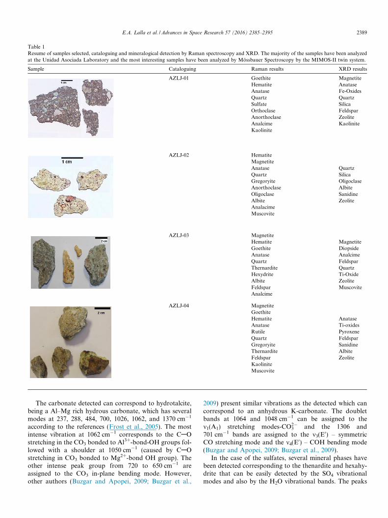
Table 1Resume of samples selected, cataloguing and mineralogical detection by Raman spectroscopy and XRD. The majority of the samples have been analyzedat the Unidad Asociada Laboratory and the most interesting samples have been analyzed by Mossbauer Spectroscopy by the MIMOS-II twin system.
Sample Cataloguing Raman results XRD results
AZLJ-01 Goethite MagnetiteHematite AnataseAnatase Fe-OxidesQuartz QuartzSulfate SilicaOrthoclase FeldsparAnorthoclase ZeoliteAnalcime KaoliniteKaolinite
AZLJ-02 HematiteMagnetiteAnatase QuartzQuartz SilicaGregoryite OligoclaseAnorthoclase AlbiteOligoclase SanidineAlbite ZeoliteAnalacimeMuscovite
AZLJ-03 MagnetiteHematite MagnetiteGoethite DiopsideAnatase AnalcimeQuartz FeldsparThernardite QuartzHexydrite Ti-OxideAlbite ZeoliteFeldspar MuscoviteAnalcime
AZLJ-04 MagnetiteGoethiteHematite AnataseAnatase Ti-oxidesRutile PyroxeneQuartz FeldsparGregoryite SanidineThernardite AlbiteFeldspar ZeoliteKaoliniteMuscovite
E.A. Lalla et al. / Advances in Space Research 57 (2016) 2385–2395 2389
The carbonate detected can correspond to hydrotalcite,being a Al–Mg rich hydrous carbonate, which has severalmodes at 237, 288, 484, 700, 1026, 1062, and 1370 cm�1
according to the references (Frost et al., 2005). The mostintense vibration at 1062 cm�1 corresponds to the CAOstretching in the CO3 bonded to Al3+-bond-OH groups fol-lowed with a shoulder at 1050 cm�1 (caused by CAOstretching in CO3 bonded to Mg2+-bond OH group). Theother intense peak group from 720 to 650 cm�1 areassigned to the CO3 in-plane bending mode. However,other authors (Buzgar and Apopei, 2009; Buzgar et al.,
2009) present similar vibrations as the detected which cancorrespond to an anhydrous K-carbonate. The doubletbands at 1064 and 1048 cm�1 can be assigned to them1(A1) stretching modes-CO3
2� and the 1306 and701 cm�1 bands are assigned to the m3(E0) – symmetricCO stretching mode and the m4(E0) – COH bending mode(Buzgar and Apopei, 2009; Buzgar et al., 2009).
In the case of the sulfates, several mineral phases havebeen detected corresponding to the thenardite and hexahy-drite that can be easily detected by the SO4 vibrationalmodes and also by the H2O vibrational bands. The peaks
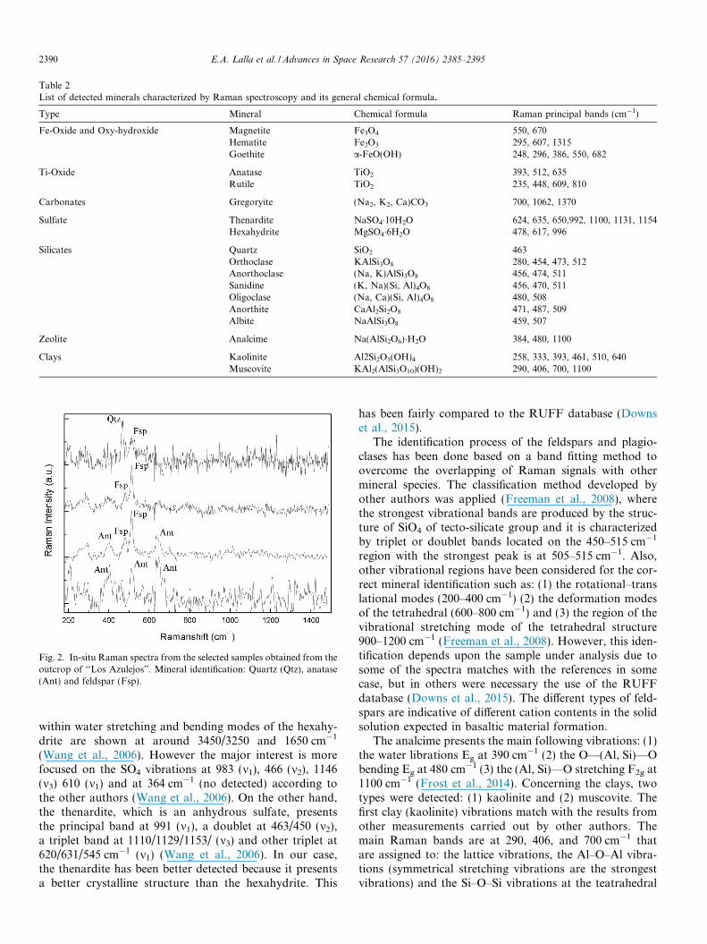
Fig. 2. In-situ Raman spectra from the selected samples obtained from theoutcrop of ‘‘Los Azulejos”. Mineral identification: Quartz (Qtz), anatase(Ant) and feldspar (Fsp).
Table 2List of detected minerals characterized by Raman spectroscopy and its general chemical formula.
Type Mineral Chemical formula Raman principal bands (cm�1)
Fe-Oxide and Oxy-hydroxide Magnetite Fe3O4 550, 670Hematite Fe2O3 295, 607, 1315Goethite a-FeO(OH) 248, 296, 386, 550, 682
Ti-Oxide Anatase TiO2 393, 512, 635Rutile TiO2 235, 448, 609, 810
Carbonates Gregoryite (Na2, K2, Ca)CO3 700, 1062, 1370
Sulfate Thenardite NaSO4�10H2O 624, 635, 650,992, 1100, 1131, 1154Hexahydrite MgSO4�6H2O 478, 617, 996
Silicates Quartz SiO2 463Orthoclase KAlSi3O8 280, 454, 473, 512Anorthoclase (Na, K)AlSi3O8 456, 474, 511Sanidine (K, Na)(Si, Al)4O8 456, 470, 511Oligoclase (Na, Ca)(Si, Al)4O8 480, 508Anorthite CaAl2Si2O8 471, 487, 509Albite NaAlSi3O8 459, 507
Zeolite Analcime Na(AlSi2O6)�H2O 384, 480, 1100
Clays Kaolinite Al2Si2O5(OH)4 258, 333, 393, 461, 510, 640Muscovite KAl2(AlSi3O10)(OH)2 290, 406, 700, 1100
2390 E.A. Lalla et al. / Advances in Space Research 57 (2016) 2385–2395
within water stretching and bending modes of the hexahy-drite are shown at around 3450/3250 and 1650 cm�1
(Wang et al., 2006). However the major interest is morefocused on the SO4 vibrations at 983 (m1), 466 (m2), 1146(m3) 610 (m1) and at 364 cm�1 (no detected) according tothe other authors (Wang et al., 2006). On the other hand,the thenardite, which is an anhydrous sulfate, presentsthe principal band at 991 (m1), a doublet at 463/450 (m2),a triplet band at 1110/1129/1153/ (m3) and other triplet at620/631/545 cm�1 (m1) (Wang et al., 2006). In our case,the thenardite has been better detected because it presentsa better crystalline structure than the hexahydrite. This
has been fairly compared to the RUFF database (Downset al., 2015).
The identification process of the feldspars and plagio-clases has been done based on a band fitting method toovercome the overlapping of Raman signals with othermineral species. The classification method developed byother authors was applied (Freeman et al., 2008), wherethe strongest vibrational bands are produced by the struc-ture of SiO4 of tecto-silicate group and it is characterizedby triplet or doublet bands located on the 450–515 cm�1
region with the strongest peak is at 505–515 cm�1. Also,other vibrational regions have been considered for the cor-rect mineral identification such as: (1) the rotational–translational modes (200–400 cm�1) (2) the deformation modesof the tetrahedral (600–800 cm�1) and (3) the region of thevibrational stretching mode of the tetrahedral structure900–1200 cm�1 (Freeman et al., 2008). However, this iden-tification depends upon the sample under analysis due tosome of the spectra matches with the references in somecase, but in others were necessary the use of the RUFFdatabase (Downs et al., 2015). The different types of feld-spars are indicative of different cation contents in the solidsolution expected in basaltic material formation.
The analcime presents the main following vibrations: (1)the water librations Eg at 390 cm�1 (2) the O—(Al, Si)—Obending Eg at 480 cm�1 (3) the (Al, Si)—O stretching F2g at1100 cm�1 (Frost et al., 2014). Concerning the clays, twotypes were detected: (1) kaolinite and (2) muscovite. Thefirst clay (kaolinite) vibrations match with the results fromother measurements carried out by other authors. Themain Raman bands are at 290, 406, and 700 cm�1 thatare assigned to: the lattice vibrations, the Al–O–Al vibra-tions (symmetrical stretching vibrations are the strongestvibrations) and the Si–O–Si vibrations at the teatrahedral
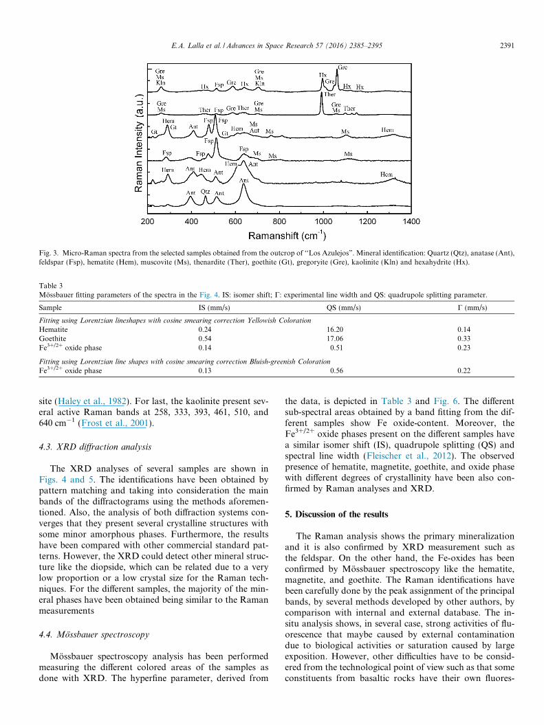
Fig. 3. Micro-Raman spectra from the selected samples obtained from the outcrop of ‘‘Los Azulejos”. Mineral identification: Quartz (Qtz), anatase (Ant),feldspar (Fsp), hematite (Hem), muscovite (Ms), thenardite (Ther), goethite (Gt), gregoryite (Gre), kaolinite (Kln) and hexahydrite (Hx).
Table 3Mossbauer fitting parameters of the spectra in the Fig. 4. IS: isomer shift; C: experimental line width and QS: quadrupole splitting parameter.
Sample IS (mm/s) QS (mm/s) C (mm/s)
Fitting using Lorentzian lineshapes with cosine smearing correction Yellowish Coloration
Hematite 0.24 16.20 0.14Goethite 0.54 17.06 0.33Fe3+/2+ oxide phase 0.14 0.51 0.23
Fitting using Lorentzian line shapes with cosine smearing correction Bluish-greenish Coloration
Fe3+/2+ oxide phase 0.13 0.56 0.22
E.A. Lalla et al. / Advances in Space Research 57 (2016) 2385–2395 2391
site (Haley et al., 1982). For last, the kaolinite present sev-eral active Raman bands at 258, 333, 393, 461, 510, and640 cm�1 (Frost et al., 2001).
4.3. XRD diffraction analysis
The XRD analyses of several samples are shown inFigs. 4 and 5. The identifications have been obtained bypattern matching and taking into consideration the mainbands of the diffractograms using the methods aforemen-tioned. Also, the analysis of both diffraction systems con-verges that they present several crystalline structures withsome minor amorphous phases. Furthermore, the resultshave been compared with other commercial standard pat-terns. However, the XRD could detect other mineral struc-ture like the diopside, which can be related due to a verylow proportion or a low crystal size for the Raman tech-niques. For the different samples, the majority of the min-eral phases have been obtained being similar to the Ramanmeasurements
4.4. Mossbauer spectroscopy
Mossbauer spectroscopy analysis has been performedmeasuring the different colored areas of the samples asdone with XRD. The hyperfine parameter, derived from
the data, is depicted in Table 3 and Fig. 6. The differentsub-spectral areas obtained by a band fitting from the dif-ferent samples show Fe oxide-content. Moreover, theFe3+/2+ oxide phases present on the different samples havea similar isomer shift (IS), quadrupole splitting (QS) andspectral line width (Fleischer et al., 2012). The observedpresence of hematite, magnetite, goethite, and oxide phasewith different degrees of crystallinity have been also con-firmed by Raman analyses and XRD.
5. Discussion of the results
The Raman analysis shows the primary mineralizationand it is also confirmed by XRD measurement such asthe feldspar. On the other hand, the Fe-oxides has beenconfirmed by Mossbauer spectroscopy like the hematite,magnetite, and goethite. The Raman identifications havebeen carefully done by the peak assignment of the principalbands, by several methods developed by other authors, bycomparison with internal and external database. The in-situ analysis shows, in several case, strong activities of flu-orescence that maybe caused by external contaminationdue to biological activities or saturation caused by largeexposition. However, other difficulties have to be consid-ered from the technological point of view such as that someconstituents from basaltic rocks have their own fluores-
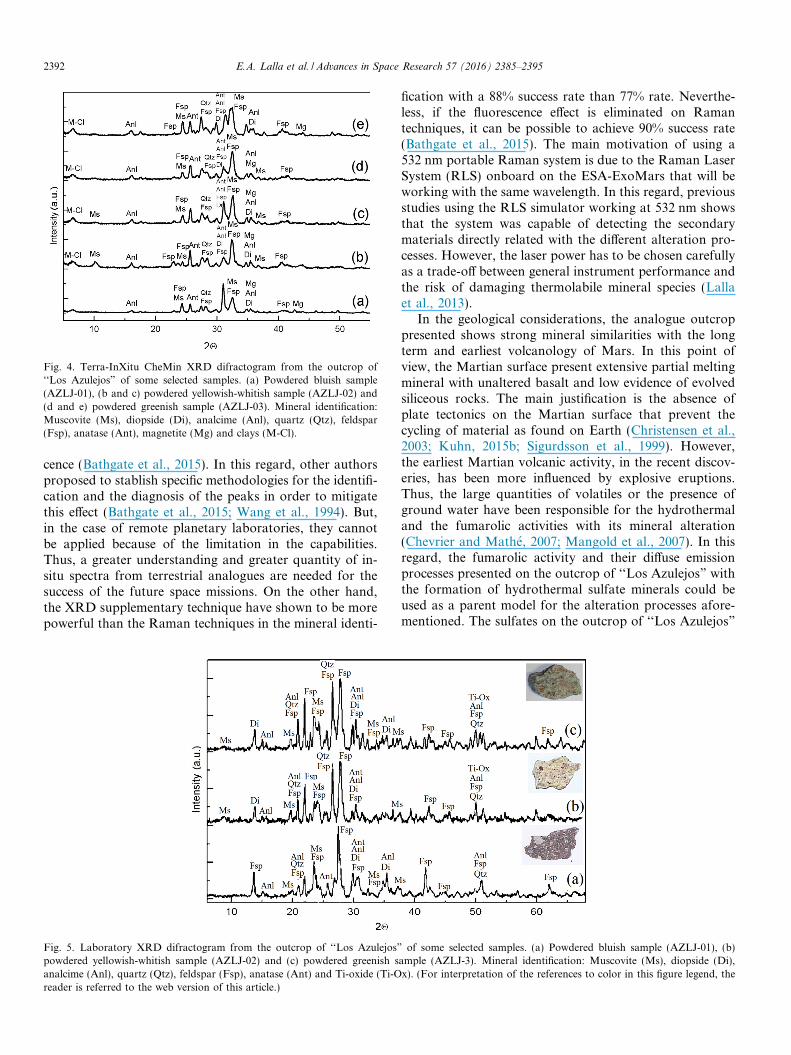
Fig. 4. Terra-InXitu CheMin XRD difractogram from the outcrop of‘‘Los Azulejos” of some selected samples. (a) Powdered bluish sample(AZLJ-01), (b and c) powdered yellowish-whitish sample (AZLJ-02) and(d and e) powdered greenish sample (AZLJ-03). Mineral identification:Muscovite (Ms), diopside (Di), analcime (Anl), quartz (Qtz), feldspar(Fsp), anatase (Ant), magnetite (Mg) and clays (M-Cl).
2392 E.A. Lalla et al. / Advances in Space Research 57 (2016) 2385–2395
cence (Bathgate et al., 2015). In this regard, other authorsproposed to stablish specific methodologies for the identifi-cation and the diagnosis of the peaks in order to mitigatethis effect (Bathgate et al., 2015; Wang et al., 1994). But,in the case of remote planetary laboratories, they cannotbe applied because of the limitation in the capabilities.Thus, a greater understanding and greater quantity of in-situ spectra from terrestrial analogues are needed for thesuccess of the future space missions. On the other hand,the XRD supplementary technique have shown to be morepowerful than the Raman techniques in the mineral identi-
Fig. 5. Laboratory XRD difractogram from the outcrop of ‘‘Los Azulejos”powdered yellowish-whitish sample (AZLJ-02) and (c) powdered greenish sanalcime (Anl), quartz (Qtz), feldspar (Fsp), anatase (Ant) and Ti-oxide (Ti-Oreader is referred to the web version of this article.)
fication with a 88% success rate than 77% rate. Neverthe-less, if the fluorescence effect is eliminated on Ramantechniques, it can be possible to achieve 90% success rate(Bathgate et al., 2015). The main motivation of using a532 nm portable Raman system is due to the Raman LaserSystem (RLS) onboard on the ESA-ExoMars that will beworking with the same wavelength. In this regard, previousstudies using the RLS simulator working at 532 nm showsthat the system was capable of detecting the secondarymaterials directly related with the different alteration pro-cesses. However, the laser power has to be chosen carefullyas a trade-off between general instrument performance andthe risk of damaging thermolabile mineral species (Lallaet al., 2013).
In the geological considerations, the analogue outcroppresented shows strong mineral similarities with the longterm and earliest volcanology of Mars. In this point ofview, the Martian surface present extensive partial meltingmineral with unaltered basalt and low evidence of evolvedsiliceous rocks. The main justification is the absence ofplate tectonics on the Martian surface that prevent thecycling of material as found on Earth (Christensen et al.,2003; Kuhn, 2015b; Sigurdsson et al., 1999). However,the earliest Martian volcanic activity, in the recent discov-eries, has been more influenced by explosive eruptions.Thus, the large quantities of volatiles or the presence ofground water have been responsible for the hydrothermaland the fumarolic activities with its mineral alteration(Chevrier and Mathe, 2007; Mangold et al., 2007). In thisregard, the fumarolic activity and their diffuse emissionprocesses presented on the outcrop of ‘‘Los Azulejos” withthe formation of hydrothermal sulfate minerals could beused as a parent model for the alteration processes afore-mentioned. The sulfates on the outcrop of ‘‘Los Azulejos”
of some selected samples. (a) Powdered bluish sample (AZLJ-01), (b)ample (AZLJ-3). Mineral identification: Muscovite (Ms), diopside (Di),x). (For interpretation of the references to color in this figure legend, the

Fig. 6. Representative Mossbauer spectra from the outcrop of ‘‘Los Azulejos” observed on the fitting process of the experimental data presenting theweathering phases such as hematite, goethite and Fe3+ phases. (a) Yellowish coloration sample and (b) bluish-greenish coloration sample. (Forinterpretation of the references to color in this figure legend, the reader is referred to the web version of this article.)
Table 4Comparison of the capabilities of the three systems employed on this investigation.
Technique Structural identification Elemental composition Geological context Penetration capability
Raman spectroscopy Yes Potentially Yes NoXRD Yes No Potentially NoMossbauer Spectroscopy Only with Fe-content materials No Yes Yes
E.A. Lalla et al. / Advances in Space Research 57 (2016) 2385–2395 2393
are products of the interaction of magmatic fluids, volcanicgases (H2S and SO2), and the surrounding rocks. They canbe of interest for future studies and to increase our knowl-edge of the processes occurred over the Martian geologicaltime. Taking into account the recent space missions likeNASA-Curiosity mission and NASA-MER-mission, wheretwin instruments have been used in this investigation, pre-sent similar mineral phases (Anderson et al., 2015;Klingelhofer et al., 2003). Moreover, the minerals detectedon Mars are in accordance with our investigations on theoutcrop of ‘‘Los Azulejos”, and this could be of impor-tance for future testing of Raman spectrometers of Martianautomatic laboratories. Also, our Raman results encourageto the continue development of Raman systems withinspace science and for the Mars exploration. A comparisonof the different techniques is available on Table 4 showingthe capabilities of the Raman spectroscopy applied to thegeological context. Its combination with the other tech-niques shows that they are capable to obtain a full detailof the mineralogy and the geological process occurred onthe selected target.
6. Conclusions
Different measurements have been performed by in-situand laboratory Raman spectroscopic techniques, X-raydiffractometers and Mossbauer spectrometer for the veryfirst time on the outcrop of ‘‘Los Azulejos”, through a com-plete analysis of the mineralogy. Regarding to this, its possi-
ble relation toMars based on its possible alteration processeshas been also studied. Crystalline primary phases such aspyroxene, feldspar, oxides as well as secondary minerals likecarbonate, Fe-oxides/oxy-hydroxides, sulfates, zeolites, andclays have been confirmed by Raman spectroscopy andXRD analyses. Moreover, theMossbauer spectroscopy alsodetected Fe-oxides and other amorphous Fe3+/2+ oxidephases as a result of hydrothermal alteration. The phasesof mineral species described along the paper are similar tothose reported on other volcanic terrestrial analoguemateri-als and Martian observations. The possibility to distinguishbetween different alteration processes by Raman spec-troscopy will help us to deduce the rock-process formationby the combined processes occurred on the outcrop of‘‘Los Azulejos” and its possible extension to Mars. Thus,the enlargement of knowledge on terrestrial analogues pro-vide an aid and a relevant knowledge support for the plane-tary research field, especially when astrogeologicalimplications are addressed. The results reveal that Ramantechnique, XRD, andMossbauer spectroscopy are powerfuland robust systems.The three techniques in combinationwillbe suitable for a complete identification of alteration pro-cesses inferred on Mars. In this regard, the measurementssupport the continued endeavors of the systemdevelopmentsfor theMars exploration on the future space mission such asthe Raman Laser Spectrometer (RLS) included in the ESAExo-Mars Mission. However, a continued improvement ofthe Raman technique is needed for the success of the futurespace missions
2394 E.A. Lalla et al. / Advances in Space Research 57 (2016) 2385–2395
Acknowledgements
The work was supported by the MICINN with the Pro-ject AYA-2008-04529 for the development of the Raman-LIBS combined spectrometer for the ESA-ExoMars Mis-sion. Emmanuel Lalla thanks MICINN for the FPI grant(BES-2009-024992). The authors also want to thank Prof.Dr. F. Garcıa Talavera for the logistic organization andcollaboration during the fieldtrip to the Museo del Hombrey la Naturaleza, (Tenerife Spain) and to Parques Nacio-nales – Gobierno de Canarias, (Tenerife, Spain).
References
Anderson, R., Bridges, J.C., Williams, A., Edgar, L., Ollila, A., Williams,J., Nachon, M., Mangold, N., Fisk, M., Schieber, J., Gupta, S.,Dromart, G., Wiens, R., Le Mouelic, S., Forni, O., Lanza, N.,Mezzacappa, A., Sautter, V., Blaney, D., Clark, B., Clegg, S.,Gasnault, O., Lasue, J., Leveille, R., Lewin, E., Lewis, K.W., Maurice,S., Newsom, H., Schwenzer, S.P., Vaniman, D., 2015. ChemCamresults from the Shaler outcrop in Gale crater, Mars. Icarus 249, 2–21.http://dx.doi.org/10.1016/j.icarus.2014.07.025.
Bathgate, E.J., Maynard-Casely, H.E., Caprarelli, G., Xiao, L., Stuart, B.,Smith, K.T., Pogson, R., 2015. Raman, FTIR and XRD study ofIcelandic tephra minerals: implications for Mars. J. Raman Spectrosc.46, 846–855. http://dx.doi.org/10.1002/jrs.4694.
Bishop, J.L., Murad, E., Lane, M.D., Mancinelli, R.L., 2004. Multipletechniques for mineral identification on Mars: a study of hydrothermalrocks as potential analogues for astrobiology sites on Mars. Icarus 169,311–323. http://dx.doi.org/10.1016/j.icarus.2003.12.025.
Bost, N., Ramboz, C., LeBreton, N., Foucher, F., Lopez-Reyes, G., DeAngelis, S., Josset, M., Venegas, G., Sanz-Arranz, A., Rull, F.,Medina, J., Josset, J.-L., Souchon, A., Ammannito, E., De Sanctis, M.C., Di Iorio, T., Carli, C., Vago, J.L., Westall, F., 2015. Testing theability of the ExoMars 2018 payload to document geological contextand potential habitability on Mars. Planet. Space Sci. 108, 87–97.http://dx.doi.org/10.1016/j.pss.2015.01.006.
Bustillo, M.A., 1989. Alteracion Hidrotermal en los Azulejos, in: Arana,V., Coello, J. (Eds.), Los Volcanes Y La Caldera Del Parque NacionalDel Teide. pp. 311–314.
Buzgar, N., Apopei, A.I., 2009. The Raman Study on Certain Carbonates.Analele Stiint. ale Univ. ‘‘Al. I. Cuza”, Iasi, pp. 97–112, vol. 55.
Buzgar, N., Buzatu, A., Sanislav, I.V., 2009. The Raman Study on CertainSulfates. Analele Stiint. ale Univ. ‘‘Al. I. Cuza”, pp. 5–23, vol. 55.
Chen, Y., Zhou, Y., Zhang, L., Wu, M., Yan, S., 2007. Discovery of CH4-rich high-pressure fluid inclusions hosted in analcime from Dongyingdepression. China J. Pet. Sci. Eng. 56, 311–314. http://dx.doi.org/10.1016/j.petrol.2006.10.005.
Chevrier, V., Mathe, P.E., 2007. Mineralogy and evolution of the surfaceof Mars: a review. Planet. Space Sci. 55, 289–314. http://dx.doi.org/10.1016/j.pss.2006.05.039.
Chio, C.H., Sharma, S.K., Muenow, D.W., 2005. Micro-Raman studies ofhydrous ferrous sulfates and jarosites. Spectrochim. Acta, Part A: Mol.Biomol. Spectrosc. 61, 2428–2433. http://dx.doi.org/10.1016/j.saa.2005.02.021.
Christensen, P.R., Bandfield, J.L., Bell III, J.F., Gorelick, N., Hamilton,V.E., Ivanov, A., Jakosky, B.M., Kieffer, H.H., Lane, M.D., Malin,M.C., McConnochie, T., McEwen, A.S., McSween, H.Y., Mehall, G.L., Moersch, J.E., Nealson, K.H., Rice, J.W., Richardson, M.I., Ruff,S.W., Smith, M.D., Titus, T.N., Wyatt, M.B., 2003. Morphology andcomposition of the surface of Mars: Mars odyssey THEMIS results.Science 300, 2056–2061. http://dx.doi.org/10.1126/science.1080885.
Courreges-Lacoste, G.B., Ahlers, B., Rull Perez, F., 2007. CombinedRaman spectrometer/laser-induced breakdown spectrometer for the
next ESA mission to Mars. Spectrochim. Acta, Part A: Mol. Biomol.Spectrosc. 68, 1023–1028. http://dx.doi.org/10.1016/j.saa.2007.03.026.
Donoghue, E., Troll, V.R., Harris, C., O’Halloran, A., Walter, T.R., PerezTorrado, F.J., 2008. Low-temperature hydrothermal alteration ofintra-caldera tuffs, Miocene Tejeda caldera, Gran Canaria, CanaryIslands. J. Volcanol. Geotherm. Res. 176, 551–564. http://dx.doi.org/10.1016/j.jvolgeores.2008.05.002.
Downs, B., Robinson, S., Yang, H., Mooney, P., 2015. RRUFF Project.Dep. Geosci. Univ, Arizona.
Fleischer, I., Klingelhofer, G., Morris, R.V., Schroder, C., Rodionov, D.,de Souza, P.A., 2012. In-situ Mossbauer spectroscopy with MIMOS II.Hyperfine Interact. 207, 97–105.
Freeman, J.J., Wang, A., Kuebler, K.E., Jolliff, B.L., Haskin, L.A., 2008.Characterization of natural feldspars by Raman spectroscopy forfuture planetary exploration. Can. Mineral. 46, 1477–1500. http://dx.doi.org/10.3749/canmin.46.6.1477.
Frost, R.L., Fredericks, P.M., Kloprogge, J.T., Hope, G.A., 2001. Ramanspectroscopy of kaolinites using different excitation wavelengths. J.Raman Spectrosc. 32, 657–663. http://dx.doi.org/10.1002/jrs.722.
Frost, R.L., Adebajo, M.O., Erickson, K.L., 2005. Raman spectroscopyof synthetic and natural iowaite. Spectrochim. Acta, Part A: Mol.Biomol. Spectrosc. 61, 613–620. http://dx.doi.org/10.1016/j.saa.2004.05.015.
Frost, R.L., Lopez, A., Theiss, F.L., Romano, A.W., Scholz, R., 2014. Avibrational spectroscopic study of the silicate mineral analcime –Na2(Al4SiO4O12)�2H2O – A natural zeolite. Spectrochim Acta, Part A:Mol. Biomol. Spectrosc. 133, 521–525. http://dx.doi.org/10.1016/j.saa.2014.06.034.
Galindo, I., Soriano, C., Martı, J., Perez, N., 2005. Graben structure in theLas Canadas edifice (Tenerife, Canary Islands): Implications for activedegassing and insights on the caldera formation. J. Volcanol.Geotherm. Res. 144, 73–87. http://dx.doi.org/10.1016/j.jvolgeores.2004.11.017.
Graham, L., Graff, T.G., Aileen Yingst, R., ten Kate, I.L., Russell, P.,2015. 2012 Moon Mars analog mission activities on Mauna Kea,Hawaii. Adv. Space Res. 55, 2405–2413. http://dx.doi.org/10.1016/j.asr.2015.01.024.
Grossman, L., 2013. NASA urged to seek live Martians with 2020 rover.New Sci. 219, 9. http://dx.doi.org/10.1016/S0262-4079(13)61775-3.
Haley, L.V., Wylie, I.W., Koningstein, J.A., 1982. An investigation of thelattice and interlayer water vibrational spectral regions of muscoviteand vermiculite using Raman microscopy. A Raman microscopicstudy of layer silicates. J. Raman Spectrosc. 13, 203–205. http://dx.doi.org/10.1002/jrs.1250130217.
Hanesch, M., 2009. Raman spectroscopy of iron oxides and (oxy)hydroxides at low laser power and possible applications in environ-mental magnetic studies. Geophys. J. Int. 177, 941–948. http://dx.doi.org/10.1111/j.1365-246X.2009.04122.x.
Jubb, A.M., Allen, H.C., 2010. Vibrational spectroscopic characterizationof hematite, maghemite, and magnetite thin films produced by vapordeposition. ACS Appl. Mater. Interfaces 2, 2804–2812. http://dx.doi.org/10.1021/am1004943.
Karwowski, Ł., Helios, K., Kryza, R., Muszynski, A., Dro_zd_zewski, P.,2013. Raman spectra of selected mineral phases of the Morasko ironmeteorite. J. Raman Spectrosc. 44, 1181–1186. http://dx.doi.org/10.1002/jrs.4340.
Klingelhofer, G., Morris, R.V., Bernhardt, B., Rodionov, D., de Souza, P.A., Squyres, S.W., Foh, J., Kankeleit, E., Bonnes, U., Gellert, R.,Schroder, C., Linkin, S., Evlanov, E., Zubkov, B., Prilutski, O., 2003.Athena MIMOS II Mossbauer spectrometer investigation. J. Geophys.Res. Planets 108. http://dx.doi.org/10.1029/2003JE002138.
Koura, N., Kohara, S., Takeuchi, K., Takahashi, S., Curtiss, L.A.,Grimsditch, M., Saboungi, M.-L., 1996. Alkali carbonates: Ramanspectroscopy, ab initio calculations, and structure. J. Mol. Struct. 382,163–169. http://dx.doi.org/10.1016/0022-2860(96)09314-3.
Kuhn, N., 2015. Experiments in Reduced Gravity. Elsevier, Experimentsin Reduced Gravity, doi: 10.1016/B978-0-12-799965-4.00002-9.
E.A. Lalla et al. / Advances in Space Research 57 (2016) 2385–2395 2395
Kuhn, N., 2015b. Chapter 2 – Overview of Mars, in: Kuhn, N.B.T.-E. inR.G. (Ed.), Experiments in Reduced Gravity. Elsevier, Oxford,pp. 17–26. doi: http://dx.doi.org/10.1016/B978-0-12-799965-4.00002-9.
Lalla, E., 2014. Tenerife como analogo de Marte: Caracterizacionmultianalıtica (Raman, DRX, ATR-FTIR, SEM y MossBaeur) demuestras de interes planetario y astrobiologico. Dep. Fısica la Mater.Condens. Cristalogr. y Minerealogıa – Univ. Valladolid, University ofValladolid.
Lalla, E., Caramazana Sansano, A., Sanz-Arranz, A., Alonso Alonso, P.,Medina Garcıa, J., Martinez-frıas, J., Rull-Perez, F., 2010. Espectro-scopıa Raman de Basaltos Correspondientes al Volcan de Las Arenas.Tenerife. MACLA – Soc. Espanola Mineral. 13, 129–130.
Lalla, E., Lopez-Reyes, G., Rull, F., Medina, J., Martinez-Frias, J.,Sansano, A., Navarro, R., 2013. Raman analysis of basaltic samplesfrom Tenerife Island (Canadas, Azulejos, and Historical eruptions)with the ExoMars RLS instrument, in: 44th Lunar and PlanetaryScience Conference. Houston, Texas, p. 2403.
Lalla, E.A., Lopez-Reyes, G., Sansano, A., Sanz-Arranz, A., Martinez-Frıas, J., Medina, J., Rull-Perez, F., 2015. Raman-IR vibrational andXRD characterization of ancient and modern mineralogy fromvolcanic eruption in Tenerife Island: Implication for Mars. Geosci.Front. http://dx.doi.org/10.1016/j.gsf.2015.07.009.
Lalla, E.A., Lopez-Reyes, G., Sansano, A., Sanz-Arranz, A., Schmanke,D., Klingelhofer, G., Medina-Garcıa, J., Martınez-Frıas, J., Rull-Perez, F., 2015. Estudio espectroscopico y DRX de afloramientosterrestres volcanicos en la isla de Tenerife como posibles analogos de lageologıa marciana. Estud. Geologicos 71, 1–19. http://dx.doi.org/10.3989/egeol.41927.354.
Lukacevic, I., Gupta, S.K., Jha, P.K., Kirin, D., 2012. Lattice dynamicsand Raman spectrum of rutile TiO2: The role of soft phonon modes inpressure induced phase transition. Mater. Chem. Phys. 137, 282–289.http://dx.doi.org/10.1016/j.matchemphys.2012.09.022.
Mahaney, W.C., Fairen, A.G., Dohm, J.M., Krinsley, D.H., 2012.Weathering rinds on clasts: examples from Earth and Mars as shortand long term recorders of paleoenvironment. Planet. Space Sci. 73,243–253. http://dx.doi.org/10.1016/j.pss.2012.08.025.
Mangold, N., Poulet, F., Mustard, J.F., Bibring, J.P., Gondet, B.,Langevin, Y., Ansan, V., Masson, P., Fassett, C., Head, J.W.,Hoffmann, H., Neukum, G., 2007. Mineralogy of the Nili Fossaeregion with OMEGA/Mars express data: 2. Aqueous alteration of thecrust. J. Geophys. Res. E Planets 112.
Martens, W.N., Ding, Z., Frost, R.L., Kristof, J., Kloprogge, J.T., 2002.Raman spectroscopy of hydrazine-intercalated kaolinite at 77, 298,323, 343 and 358 K. J. Raman Spectrosc. 33, 31–36. http://dx.doi.org/10.1002/jrs.812.
Marti, J., Gudmundsson, A., 2000. The Las Canadas Caldera (Tenerife,Canary Islands): an overlapping collapse caldera generated by magma-chamber migration. J. Volcanol. Geotherm. Res. 103, 161–173. http://dx.doi.org/10.1016/S0377-0273(00)00221-3.
Martin-Ramos, J.D., 2004. XPowder: A Software Package for Powder X-Ray Diffraction Analysis Powder Methods.
View publication statsView publication stats
Merle, O., Barde-Cabusson, S., van Wyk de Vries, B., 2010. Hydrothermalcalderas. Bull. Volcanol. 72, 131–147. http://dx.doi.org/10.1007/s00445-009-0314-6.
Minitti, M.E., Hamilton, V.E., 2010. A search for basaltic-to-intermediateglasses on Mars: assessing Martian crustal mineralogy. Icarus 210,135–149. http://dx.doi.org/10.1016/j.icarus.2010.06.028.
Mouginis-Mark, P., Robinson, M., 1992. Evolution of the Olympus monscaldera. Mars Bull. Volcanol. 54, 347–360. http://dx.doi.org/10.1007/BF00312318.
Prinsloo, L.C., Colomban, P., Brink, J.D., Meiklejohn, I., 2011. A Ramanspectroscopic study of the igneous rocks on Marion Island: a possibleterrestrial analogue for the geology on Mars. J. Raman Spectrosc. 42,626–632. http://dx.doi.org/10.1002/jrs.2756.
Rayl, A.J.S., 2014. Mars Exploration Rovers Update: Mission Nears 10-Year Milestone, Oppy Roves On, We Look Back on 2013 [WWWDocument]. Planet. Soc. URL http://www.planetary.org/explore/space-topics/space-missions/mer-updates/2013/12-mer-update-mis-sion-nears-10-year-milestone.html.
Ruff, S.W., 2004. Spectral evidence for zeolite in the dust on Mars. Icarus168, 131–143. http://dx.doi.org/10.1016/j.icarus.2003.11.003.
Rull, F., Martinez-Frias, J., Rodrıguez-Losada, J.A., 2007. Micro-Ramanspectroscopic study of El Gasco pumice, western Spain. J. RamanSpectrosc. 38, 239–244. http://dx.doi.org/10.1002/jrs.1628.
Rull-Perez, F., Martinez-Frias, J., 2006. Raman spectroscopy goes toMars. Spectrosc. Eur. 18, 18–21.
Sekiya, T., Ohta, S., Kamei, S., Hanakawa, M., Kurita, S., 2001. Ramanspectroscopy and phase transition of anatase TiO2 under highpressure. J. Phys. Chem. Solids 62, 717–721. http://dx.doi.org/10.1016/S0022-3697(00)00229-8.
Sigurdsson, H., Bruce, H., Rymer, H., John, S., McNutt, S., 1999.Encyclopedia of Volcanoes. Academic Press.
Villasante-Marcos, V., Finizola, A., Abella, R., Barde-Cabusson, S.,Blanco, M.J., Brenes, B., Cabrera, V., Casas, B., De Agustın, P., DiGangi, F., Domınguez, I., Garcıa, O., Gomis, A., Guzman, J.,Iribarren, I., Levieux, G., Lopez, C., Luengo-Oroz, N., Martın, I.,Moreno, M., Meletlidis, S., Morin, J., Moure, D., Pereda, J., Ricci, T.,Romero, E., Schutze, C., Suski-Ricci, B., Torres, P., Trigo, P., 2014.Hydrothermal system of Central Tenerife volcanic complex, CanaryIslands (Spain), inferred from self-potential measurements. J. Vol-canol. Geotherm. Res. 272, 59–77. http://dx.doi.org/10.1016/j.jvolgeores.2013.12.007.
Wang, A., Han, J., Guo, L., Yu, J., Zeng, P., 1994. Database of standardRaman spectra of minerals and related inorganic crystals. Appl.Spectrosc. 48, 959–968.
Wang, A., Freeman, J.J., Jolliff, B.L., Chou, I.-M., 2006. Sulfates onMars: a systematic Raman spectroscopic study of hydration states ofmagnesium sulfates. Geochim. Cosmochim. Acta 70, 6118–6135.http://dx.doi.org/10.1016/j.gca.2006.05.022.
Zotov, N., Ebbsjo, I., Timpel, D., Keppler, H., 1999. Calculation ofRaman spectra and vibrational properties of silicate glasses: compar-ison between Na2Si4O9 and SiO2 glasses. Phys. Rev. B. http://dx.doi.org/10.1103/PhysRevB.60.6383.


![MultiSpec® Raman: Raman Spectrometer for Process and ... · Product Information Systems [ MultiSpec® Raman] Spectrometer Module The Raman system uses a high throughput, high-resolution](https://img.pdfslide.net/doc/110x75/5cf715f188c99346318c70a0/multispec-raman-raman-spectrometer-for-process-and-product-information.jpg)