Embed Size (px)
Citation preview
VETERINARY RESEARCHRamis et al. Veterinary Research 2014, 45:84http://www.veterinaryresearch.org/content/45/1/84
RESEARCH Open Access
Experimental infection of highly pathogenic avianinfluenza virus H5N1 in black-headed gulls(Chroicocephalus ridibundus)Antonio Ramis1*, Geert van Amerongen2, Marco van de Bildt2, Loneke Leijten2, Raphael Vanderstichel3,Albert Osterhaus2 and Thijs Kuiken2
Abstract
Historically, highly pathogenic avian influenza viruses (HPAIV) rarely resulted in infection or clinical disease in wildbirds. However, since 2002, disease and mortality from natural HPAIV H5N1 infection have been observed in wildbirds including gulls. We performed an experimental HPAIV H5N1 infection of black-headed gulls (Chroicocephalusridibundus) to determine their susceptibility to infection and disease from this virus, pattern of viral shedding, clinicalsigns, pathological changes and viral tissue distribution. We inoculated sixteen black-headed gulls with 1 × 10⁴
median tissue culture infectious dose HPAIV H5N1 (A/turkey/Turkey/1/2005) intratracheally and intraesophageally.Birds were monitored daily until 12 days post inoculation (dpi). Oropharyngeal and cloacal swabs were collecteddaily to detect viral shedding. Necropsies from birds were performed at 2, 4, 5, 6, 7, and 12 dpi. Sampling fromselected tissues was done for histopathology, immunohistochemical detection of viral antigen, PCR, and viral isolation.Our study shows that all inoculated birds were productively infected, developed systemic disease, and had a highmorbidity and mortality rate. Virus was detected mainly in the respiratory tract on the first days after inoculation, andthen concentrated more in pancreas and central nervous system from 4 dpi onwards. Birds shed infectious virus until 7dpi from the pharynx and 6 dpi from the cloaca. We conclude that black-headed gulls are highly susceptible to diseasewith a high mortality rate and are thus more likely to act as sentinel species for the presence of the virus than aslong-distance carriers of the virus to new geographical areas.
IntroductionHistorically, highly pathogenic avian influenza viruses(HPAIV) were rarely found in wild birds, and, if theywere, they typically did not cause clinical disease [1].However, in late 2002, HPAIV H5N1 outbreaks were de-scribed in Hong Kong parks causing disease and deathin wild local and migratory birds; concurrently withthese outbreaks, HPAIV H5N1 were isolated from deadchickens and human beings [2]. It was shown throughantigenic analysis that these new isolates of HPAIVH5N1 presented a reactivity pattern different from thatof HPAIV H5N1 isolated in 1997 and 2001 and causedsystemic infection in experimentally infected domesticducks, with high virus titres and lesions in multipleorgans, particularly in the brain [3].
* Correspondence: [email protected] and Departament de Sanitat i Anatomia Animals, UniversitatAutonoma de Barcelona, Barcelona, SpainFull list of author information is available at the end of the article
© 2014 Ramis et al.; licensee BioMed Central LCommons Attribution License (http://creativecreproduction in any medium, provided the orDedication waiver (http://creativecommons.orunless otherwise stated.
Since 2002, disease and mortality from natural HPAIVH5N1 infection have been described for several gull spe-cies: black-headed gull (Chroicocephalus ridibundus) [2],great black-headed gull (Larus ichthyaetus) and brown-headed gull (Larus brunnicephalus) [4]. Also, experimen-tal infections have been performed in laughing gulls(Larus atricilla) [5] and herring gulls (Larus argentatus)[6] showing that these species were highly susceptible(100%) to different HPAIV H5N1 isolates (A/WhooperSwan/Mongolia/244/05 and A/Duck Meat/Anyang/01),presenting a severe and short (4 to 10 dpi) clinicalcourse, consisting mainly of neurologic signs with a highmortality rate (up to 100% in some cases). The main tar-get organs for viral replication were central nervous sys-tem (CNS), pancreas, and adrenal gland, and theduration of the viral shedding after inoculation was in arange of 1 to 10 dpi depending on the isolate, the gullspecies, and inoculation route. In these experiments,
td. This is an Open Access article distributed under the terms of the Creativeommons.org/licenses/by/4.0), which permits unrestricted use, distribution, andiginal work is properly credited. The Creative Commons Public Domaing/publicdomain/zero/1.0/) applies to the data made available in this article,
Ramis et al. Veterinary Research 2014, 45:84 Page 2 of 10http://www.veterinaryresearch.org/content/45/1/84
virus dynamics and in-depth pathogenesis were not in-vestigated. Lastly, in an experimental infection in com-mon gulls (Larus canus) with a HPAIV H5N1 isolate (A/Gull/Chany/P/06) isolated from healthy birds of this spe-cies, the virus was shown to replicate in respiratory anddigestive tracts and to be excreted from pharynx andcloaca for 2 weeks after the infection, with no clinicalsigns in infected birds [7].The black-headed gull is the most ubiquitous gull in
Europe and Asia, is a peridomestic species that often oc-curs in urban and agricultural environments [8] and isknown to be susceptible to HPAIV H5N1 infection [2].Also, black-headed gulls are listed by the European FoodSafety Authority [9] as having higher probability to beexposed to Asian linage HPAIV H5N1 during migrationoutside the European Union, because of their gregariousbehaviour and their tendency to use both wild areas andbuilt-up areas. However, it is known neither how longblack-headed gulls can shed HPAIV H5N1 nor how sus-ceptible they are to disease from HPAIV H5N1 infection.These are important considerations to decide whetherblack-headed gulls are more likely to act as sentinels forthe presence of the virus in the environment, or to actas long-distance vectors of the virus to other geograph-ical areas [10]. Therefore, we performed an experimentalinfection of black-headed gulls using HPAIV A/turkey/Turkey/1/2005 (H5N1) to determine the susceptibility ofthis host species to infection and disease from this virus;the pattern of pharyngeal and cloacal viral shedding; theclinical signs and pathological changes; and lastly thevirus distribution in different tissues. We chose this virusisolate to allow comparison of the results between black-headed gulls and 6 different wild species of Anseriformes[10], which jointly with Charadriiformes—to which theblack-headed gull belongs—have traditionally been con-sidered the natural reservoir for avian influenza viruses;and also because this isolate had spread quickly fromAsia to Europe in 2005–2006 [11].
Material and methodsVirus preparationA virus stock of HPAIV A/turkey/Turkey 1/2005 (H5N1)was prepared by propagation in Madin-Darby canine kid-ney (MDCK) cells and titrated according to standardmethods [12]. The fluid had a titre of 1 × 108.1 median tis-sue culture infectious dose (TCID50)/mL and was dilutedwith phosphate-buffered saline (PBS) to obtain a final titreof 104 TCID50/mL. All the experiments with HPAIV H5N1were performed under Biosafety level 3 + conditions.
Experimental designWe used 22 black-headed gulls (2–3 weeks old) obtainedfrom a breeding colony site at Blauwestad, TheNetherlands. The gulls were housed in negative isolator
units for 15 days to acclimatize them to the new envir-onment. Two days before the experimental inoculation,they were tested for antibodies against influenza virusesby use of a commercially available nucleoprotein-basedELISA test (European Veterinary Laboratory, Woerden,The Netherlands) to assure they were not previously in-fected. Sixteen birds were inoculated with 1 × 10⁴ TCID50
HPAIV H5N1, 1.5 mL intratracheally and 1.5 mL intraoe-sophageally, which was the same virus, inoculum dose,and inoculation route as in a previous study [10]. We usedthis dose to increase the chance of inducing subclinical in-fection and to simulate field circumstances. In addition, 6birds that served as negative controls were sham-inoculated in the same manner with PBS-diluted sterile al-lantoic fluid. Daily, a qualified veterinarian scored clinicalsigns of disease in all birds according to a standardizedlist. Also daily, the body weight of each bird was mea-sured, and cloacal and pharyngeal swabs were collected in1 mL transport medium. Birds were euthanatized by ex-sanguination under isofluorane anesthesia. After euthan-asia, blood samples were taken and necropsies and tissuesampling were performed. The original plan was to eu-thanatize four HPAIV H5N1-inoculated birds on each ofdays 2, 4, 6 and 12 post inoculation, two sham-inoculatedbirds at 3 days post inoculation (dpi) and four sham-inoculated birds at 12 dpi. Because some H5N1-inoculated birds died spontaneously before the plannedday of euthanasia, we were unable to completely followthe original plan, and the timing of necropsies and sam-pling for H5N1-inoculated birds was as follows: four birdsat 2 dpi; five birds at 4 dpi; two birds at 5 dpi; two birds at6 dpi; one bird at 7 dpi; and two birds at day 12 dpi(Table 1).
Pathologic and immunohistologic examinationNecropsies and tissue sampling were performed accordingto a standard protocol. After fixation in 10% neutral-buffered formalin and embedding in paraffin, 3-μm-thicktissue sections were made and stained by two methods:hematoxylin and eosin staining for histologic evaluationand an immunohistologic method that used a monoclonalantibody against nucleoprotein for influenza A virus as aprimary antibody for detection of influenza viral antigen[12]. The positive control was a lung of a HPAIV (H5N1)infected ferret; negative controls were substitution of theprimary antibody by an irrelevant monoclonal antibody ofthe same isotype, and testing of tissues from sham-inoculated black-headed gulls. The following tissues wereexamined: brain (cerebrum, cerebellum and brain stem),trachea, bronchus, lung, air sac, oesophagus, proventricu-lus, duodenum, pancreas, jejunum, ileum, cecum, colon,liver, gallbladder, cloaca, cloacal bursa, spleen, thymus,kidney (cranial and caudal lobes), gonad, adrenal gland,feathered skin, myocardium and skeletal muscle.
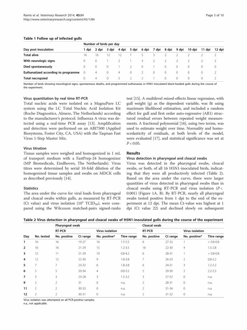
Table 1 Follow up of infected gulls
Number of birds per day
Day post inoculation 1 dpi 2 dpi 3 dpi 4 dpi 5 dpi 6 dpi 7 dpi 8 dpi 9 dpi 10 dpi 11 dpi 12 dpi
Total alive 16 16 12 12 7 5 3 2 2 2 2 2
With neurologic signs 0 0 1 2 1 3 2 2 2 2 2 2
Died spontaneously 0 0 0 1 2 0 1 0 0 0 0 0
Euthanatized according to programme 0 4 0 4 0 2 0 0 0 0 0 2
Total necropsied 0 4 0 5 2 2 1 0 0 0 0 2
Number of birds showing neurological signs, spontaneous deaths, and programmed euthanasias in H5N1-inoculated black-headed gulls during the course ofthe experiment.
Ramis et al. Veterinary Research 2014, 45:84 Page 3 of 10http://www.veterinaryresearch.org/content/45/1/84
Virus quantitation by real time RT-PCRTotal nucleic acids were isolated on a MagnaPure LCsystem using the LC Total Nucleic Acid Isolation Kit(Roche Diagnostics, Almere, The Netherlands) accordingto the manufacturer’s protocol. Influenza A virus was de-tected using a real-time PCR assay [13]. Amplificationand detection were performed on an ABI7500 (AppliedBiosystems, Foster City, CA, USA) with the Taqman FastVirus 1-Step Master Mix.
Virus titrationTissue samples were weighed and homogenized in 1 mLof transport medium with a FastPrep-24 homogenizer(MP Biomedicals, Eindhoven, The Netherlands). Virustitres were determined by serial 10-fold dilution of thehomogenized tissue samples and swabs on MDCK cellsas described previously [14].
StatisticsThe area under the curve for viral loads from pharyngealand cloacal swabs within gulls, as measured by RT-PCR(Ct value) and virus isolation (10x TCID50), were com-pared using the Wilcoxon matched-pairs signed-ranks
Table 2 Virus detection in pharyngeal and cloacal swabs of H
Pharyngeal swab
RT-PCR Virus isolation
Day No. tested No. positive Ct range No. positive* Titr
1 16 16 19-27 16 1.5-
2 16 16 21-29 15 1.2-
3 12 11 21-29 10 0.8-
4 12 12 22-30 9 1.8-
5 7 7 23-32 6 1.8-
6 5 5 20-34 4 0.8-
7 3 3 23-28 3 1.5-
9 2 2 31 0 n.a.
11 2 2 30-33 0 n.a.
12 2 2 30-31 0 n.a.
Virus isolation was attempted on all PCR-positive samples.n.a., not applicable.
test [15]. A multilevel mixed-effects linear regression, withgull weight (g) as the dependent variable, was fit usingmaximum likelihood estimation, and included a randomeffect for gull and first order auto-regressive (AR1) struc-tured residual errors between repeated weight measure-ments. A fractional polynomial [16], using two terms, wasused to estimate weight over time. Normality and homo-scedasticity of residuals, at both levels of the model,were evaluated [17], and statistical significance was set atP < 0.05.
ResultsVirus detection in pharyngeal and cloacal swabsVirus was detected in the pharyngeal swabs, cloacalswabs, or both, of all 16 H5N1-inoculated birds, indicat-ing that they were all productively infected (Table 2).Based on the area under the curve, there were largerquantities of virus detected in pharyngeal swabs than incloacal swabs using RT-PCR and virus isolation (P <0.001) (Figure 1A, B). By RT-PCR, nearly all pharyngealswabs tested positive from 1 dpi to the end of the ex-periment at 12 dpi. The mean Ct-value was highest at 1dpi (Ct value 22) and declined slowly on subsequent
5N1-inoculated gulls during the course of the experiment
Cloacal swab
RT-PCR Virus isolation
e range No. positive Ct range No. positive* Titre range
5.5 6 27-32 1 < 0.8-0.8
3.5 16 22-30 4 1.5-2.8
4.2 6 28-31 1 < 0.8-0.8
3.8 7 26-33 2 0.8-2.2
3.8 6 24-31 3 1.2-2.2
3.5 5 20-30 2 2.2-2.5
3.2 3 27-32 0 n.a.
2 28-31 0 n.a.
2 31-34 0 n.a.
2 31-32 0 n.a.
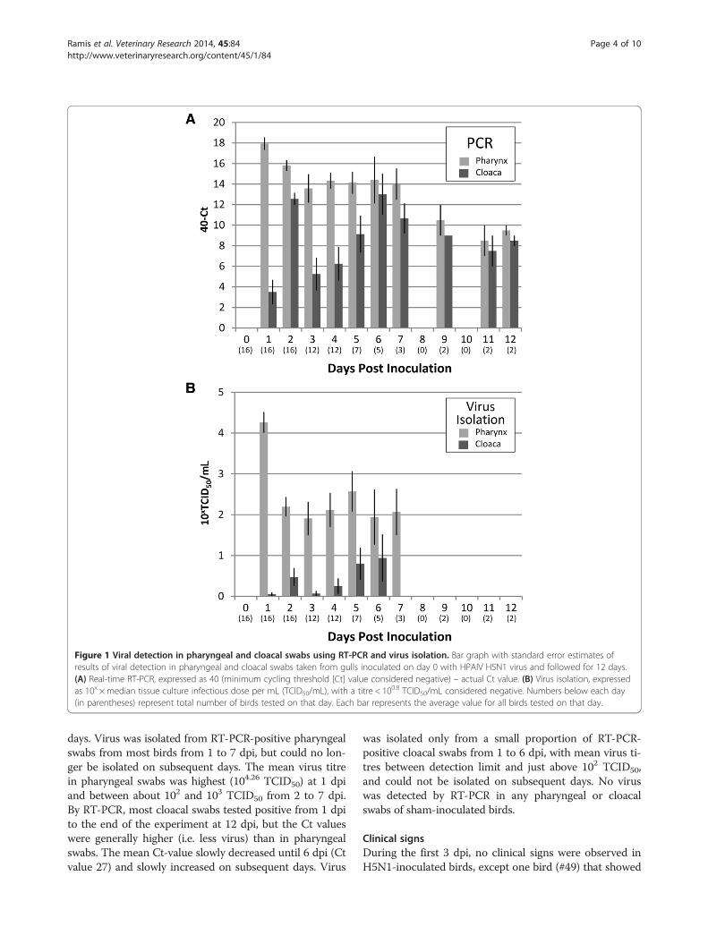
Figure 1 Viral detection in pharyngeal and cloacal swabs using RT-PCR and virus isolation. Bar graph with standard error estimates ofresults of viral detection in pharyngeal and cloacal swabs taken from gulls inoculated on day 0 with HPAIV H5N1 virus and followed for 12 days.(A) Real-time RT-PCR, expressed as 40 (minimum cycling threshold [Ct] value considered negative) – actual Ct value. (B) Virus isolation, expressedas 10x × median tissue culture infectious dose per mL (TCID50/mL), with a titre < 100.8 TCID50/mL considered negative. Numbers below each day(in parentheses) represent total number of birds tested on that day. Each bar represents the average value for all birds tested on that day.
Ramis et al. Veterinary Research 2014, 45:84 Page 4 of 10http://www.veterinaryresearch.org/content/45/1/84
days. Virus was isolated from RT-PCR-positive pharyngealswabs from most birds from 1 to 7 dpi, but could no lon-ger be isolated on subsequent days. The mean virus titrein pharyngeal swabs was highest (104.26 TCID50) at 1 dpiand between about 102 and 103 TCID50 from 2 to 7 dpi.By RT-PCR, most cloacal swabs tested positive from 1 dpito the end of the experiment at 12 dpi, but the Ct valueswere generally higher (i.e. less virus) than in pharyngealswabs. The mean Ct-value slowly decreased until 6 dpi (Ctvalue 27) and slowly increased on subsequent days. Virus
was isolated only from a small proportion of RT-PCR-positive cloacal swabs from 1 to 6 dpi, with mean virus ti-tres between detection limit and just above 102 TCID50,and could not be isolated on subsequent days. No viruswas detected by RT-PCR in any pharyngeal or cloacalswabs of sham-inoculated birds.
Clinical signsDuring the first 3 dpi, no clinical signs were observed inH5N1-inoculated birds, except one bird (#49) that showed
Ramis et al. Veterinary Research 2014, 45:84 Page 5 of 10http://www.veterinaryresearch.org/content/45/1/84
less activity than normal and slight nonspecific neuro-logical signs at 3 dpi: regularly fluffing of feathers andwalking slightly to one side. At 4, 5 and 6 dpi, all H5N1-inoculated birds were less active than sham-inoculatedbirds, showing increased recumbency and regularly fluff-ing of feathers. Four of them (#46, 47, 49, 51) showedobvious neurological signs, consisting mainly of tortico-lis, circling, loss of balance and head tremors. Respira-tory or digestive signs, such as diarrhoea, were notobserved in any H5N1-inoculated birds. From 7 dpi on-wards, the two remaining H5N1-inoculated birds (#47,51) partially recovered, alternating between normalityand neurological signs (Table 1). At 4 dpi, the H5N1-inoculated bird that showed clinical signs at 3 dpi (#49)died spontaneously. Two birds (#46, 50) at 5 dpi andone bird (#48) at 7 dpi, all three H5N1-inoculated, alsodied spontaneously. No clinical signs or spontaneousmortality were observed in any of the sham-inoculatedbirds. Based on the statistical model: the body weight ofH5N1-inoculated birds was 26 g lower than sham-inoculated birds (P = 0.029) after inoculations; within theH5N1-inoculated group, the body weight of birds thatdied was 26 g lower after inoculations than that of birdsthat survived (P = 0.047). Regardless of their inoculationstatus, gulls were losing weight during the first weekpost inoculation, and this trend was reversed after 10dpi (P < 0.001, Figure 2).
Gross lesionsNo relevant gross lesions were observed in any H5N1-inoculated or sham-inoculated birds. In general, all birds
Figure 2 Estimated gull body weights (g) throughout the study periomixed-effects linear regression, for control gulls that remained alive for theintervals. The same statistical model predicted an average constant loss of 26of 26 grams for gulls that died during the study (P = 0.047)—these estimates
showed good body condition and had abundant subcuta-neous and perivisceral fat deposits.
Histopathology and immunohistochemistryThe evaluation of histological lesions and the presenceof virus antigen in the different cells and tissues of eval-uated organs of H5N1-inoculated birds are summarizedin Table 3. On day 2, air sacs in all birds (#36, 37, 38,39) had marked diffuse infiltration of heterophils andmultiple variably-sized well-demarcated aggregates ofheterophils and macrophages. The adrenal gland in onebird (#37) had multiple foci of necrotic adrenocorticaland chromaffin cells. Virus antigen expression was ob-served in epithelial cells (#36, 37, 38, 39) and unspecifiedinflammatory cells (#36, 37, 38, 39) of the air sacs, andin adrenocortical cells (#37) of the adrenal gland, local-ized to lesions in these tissues. Virus antigen expressionalso was observed in individual tracheal and bronchialciliated epithelial cells (#38) (Figure 3A, B), air capillarycells (#36, 37), and thymic epithelial cells in absence ofhistological lesions.On day 4, all birds (#40, 41, 42, 43, 49) had airsacculi-
tis and air sac granulomas similar to those on day 2. Thetrachea in one bird (#42) had many heterophils in thelumen. The pancreas in two birds (#40, 49) had multiplefoci of necrotic acinar cells with scarce associated in-flammatory cells (Figure 3C). The cerebrum in all birds(#40, 41, 42, 43, 49) had multiple randomly distributedfoci of necrotic neurons, characterized by chromatolysisand satellitosis (Figure 3E). These foci of necrosis wereassociated with predominantly perivascular infiltration
d. It was determined by the fractional polynomial terms in theduration of the study; shaded area represents the 95% confidencegrams for H5N1-inoculated gulls (P = 0.029), and an average constant lossare not shown in graph.

Table 3 Histological lesions (HE) and presence of antigen (IHC) in tissues of H5N1-inoculated gulls during the course ofthe experiment
Number of birds positive/number of birds examined per day
2 dpi 4 dpi 5 dpi 6 dpi 7 dpi 12 dpi
Tissue HE1 IHC2 HE IHC HE IHC HE IHC HE IHC HE IHC
Thymus 0/4 2/4 0/5 0/5 0/2 0/2 0/2 0/2 0/1 0/1 0/2 0/2
Adrenal 1/4 1/4 0/5 0/5 0/2 0/2 1/2 1/2 0/1 0/1 0/2 0/2
Lung/Trachea 0/4 3/4 1/5 0/5 1/2 0/2 1/2 0/2 0/1 0/1 0/2 0/2
Air sacs 4/4 4/4 5/5 5/5 2/2 1/2 1/2 1/2 0/1 0/1 1/2 1/2
Pancreas 0/4 0/4 2/5 2/5 1/2 1/2 2/2 2/2 1/1 1/1 1/2 0/2
CNS 0/4 0/4 5/5 5/5 2/2 2/2 2/2 2/2 1/1 1/1 1/2 0/21HE: presence of histological lesions by examination of hematoxylin-and-eosin-stained tissue sections.2IHC: presence of viral antigen by examination of tissue sections stained by immunohistochemical technique.
Ramis et al. Veterinary Research 2014, 45:84 Page 6 of 10http://www.veterinaryresearch.org/content/45/1/84
of inflammatory cells, consisting of variable proportionsof lymphocytes, plasma cells, macrophages, and hetero-phils. The cerebellum of one bird (#49) and brainstem ofanother one (#43) had similar lesions as the cerebrum.Virus antigen expression was observed in epithelial cells
Figure 3 Histopathology and antigen expression in selected tissues frexpression is visible as a red staining. (A, B): Bronchus of 2 dpi infected gushowing positivity on the immuno-stained consecutive section (arrows) (B)necrosis (C), and intense positivity associate to necrotic foci in the immuneneurons, and satellitosis (E), and intense positivity in neurons and associate(A, C, E) and immunoperoxidase counterstained with hematoxylin (B, D, F
(#42, 49) and unspecified inflammatory cells (#40, 41,42, 43, 49) of the air sacs, in acinar cells (#40, 49) of thepancreas (Figure 3D), and in neurons, glial cells, andependymal cells of the brain, localized to lesions in thesetissues (Figure 3F). There also was virus antigen
om infected gulls. By immunohistochemistry, influenza virus antigenll without histologic lesions (A), and scarce bronchial epithelial cells. (C, D): Pancreas of 4 dpi infected gull showing multiple foci of lytic-stained consecutive section. (E, F): Cerebral cortex showing necroticd glial cells in the immune-stained consecutive section. HE stain). All the pictures are at 10x power fields.
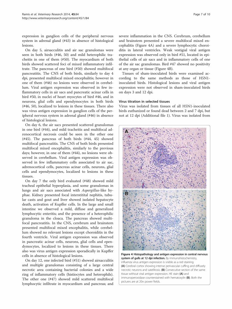
Figure 4 Histopathology and antigen expression in central nervoussystem of gulls at 12 dpi infection. By immunohistochemistry,influenza virus antigen expression is visible as a red staining.(A) Cerebral cortex showing intense perivascular cuffing and diffuselynecrotic neurons and satellitosis. (B) Consecutive section of the sametissue without viral antigen expression. HE stain (A) andimmunoperoxidase counterstained with hematoxylin (B). Both thepictures are at 20× power fields.
Ramis et al. Veterinary Research 2014, 45:84 Page 7 of 10http://www.veterinaryresearch.org/content/45/1/84
expression in ganglion cells of the peripheral nervoussystem in adrenal gland (#43) in absence of histologicallesions.On day 5, airsacculitis and air sac granulomas were
seen in both birds (#46, 50) and mild heterophilic tra-cheitis in one of them (#50). The myocardium of bothbirds showed scattered foci of mixed inflammatory infil-trate. The pancreas of one bird (#50) showed multifocalpancreatitis. The CNS of both birds, similarly to day 4dpi, presented multifocal mixed encephalitis; however inone of them (#46) no lesions were observed in cerebel-lum. Viral antigen expression was observed in few in-flammatory cells in air sacs and pancreatic acinar cells inbird #50, in nuclei of heart myocytes of bird #46, and inneurons, glial cells and ependymocytes in both birds(#46, 50), localized to lesions in these tissues. There alsowas virus antigen expression in ganglion cells of the per-ipheral nervous system in adrenal gland (#46) in absenceof histological lesions.On day 6, the air sacs presented scattered granulomas
in one bird (#44), and mild tracheitis and multifocal ad-renocortical necrosis could be seen in the other one(#45). The pancreas of both birds (#44, 45) showedmultifocal pancreatitis. The CNS of both birds presentedmultifocal mixed encephalitis, similarly to the previousdays; however, in one of them (#44), no lesions were ob-served in cerebellum. Viral antigen expression was ob-served in few inflammatory cells associated to air sac,adrenocortical cells, pancreas acinar cells, neurons, glialcells and ependymocytes, localized to lesions in thesetissues.On day 7 the only bird evaluated (#48) showed mild
tracheal epithelial hyperplasia, and some granulomas inlungs and air sacs associated with Aspergillus-like hy-phae. Kidney presented focal interstitital nephitis, tubu-lar casts and gout and liver showed isolated hepatocytedeath, activation of Kupffer cells. In the large and smallintestine we observed a mild, diffuse and generalizedlymphocytic enteritis; and the presence of a heterophilicgranuloma in the cloaca. The pancreas showed multi-focal pancreatitis. In the CNS, cerebrum and brainstempresented multifocal mixed encephalitis, while cerebel-lum showed no relevant lesions except choroiditis in thefourth ventricle. Viral antigen expression was observedin pancreatic acinar cells, neurons, glial cells and epen-dymocytes, localized to lesions in these tissues. Therealso was virus antigen expression sporadically in Kupffercells in absence of histological lesions.On day 12, one infected bird (#51) showed airsacculitis
and multiple granulomas consisting of a large centralnecrotic area containing bacterial colonies and a widering of inflammatory cells (histiocytes and heterophils).The other one (#47) showed mild scattered multifocallymphocytic infiltrate in myocardium and pancreas; and
severe inflammation in the CNS. Cerebrum, cerebellumand brainstem presented a severe multifocal mixed en-cephalitis (Figure 4A) and a severe lymphocytic choroi-ditis in lateral ventricles. Weak vestigial viral antigenexpression was observed only in bird #51, located in epi-thelial cells of air sacs and in inflammatory cells of oneof the air sac granulomas. Bird #47 showed no positivityat any organ or tissue (Figure 4B).Tissues of sham-inoculated birds were examined ac-
cording to the same methods as those of H5N1-inoculated birds. Histological lesions and viral antigenexpression were not observed in sham-inoculated birdson days 3 and 12 dpi.
Virus titration in selected tissuesVirus was isolated from tissues of all H5N1-inoculatedbirds euthanized or found dead between 2 and 7 dpi, butnot at 12 dpi (Additional file 1). Virus was isolated from
Ramis et al. Veterinary Research 2014, 45:84 Page 8 of 10http://www.veterinaryresearch.org/content/45/1/84
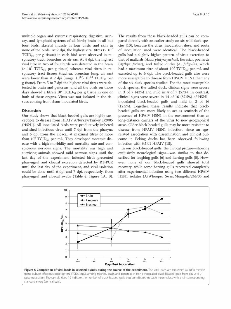
multiple organ and systems: respiratory, digestive, urin-ary, and lymphoid systems of all birds; brain in all butfour birds; skeletal muscle in four birds; and skin innone of the birds. At 2 dpi, the highest viral titres (> 107
TCID50 per g tissue) in each bird were observed in re-spiratory tract: bronchus or air sac. At 4 dpi, the highestviral titre in two of four birds was detected in the brain(≥ 107 TCID50 per g tissue) whereas viral titres in re-spiratory tract tissues (trachea, bronchus lung, air sac)were lower than at 2 dpi (range: 102.7- 106.4 TCID50 perg tissue). From 5 to 7 dpi the highest viral titres were de-tected in brain and pancreas, and all the birds on thosedays showed a titre ≥ 107 TCID50 per g tissue in one orboth of these organs. Virus was not isolated in the tis-sues coming from sham-inoculated birds.
DiscussionOur study shows that black-headed gulls are highly sus-ceptible to disease from HPAIV A/turkey/Turkey 1/2005(H5N1). All inoculated birds were productively infectedand shed infectious virus until 7 dpi from the pharynxand 6 dpi from the cloaca, at maximal titres of morethan 104 TCID50 per mL. They developed systemic dis-ease with a high morbidity and mortality rate and con-spicuous nervous signs. The mortality was high andsurviving animals showed mild nervous signs until thelast day of the experiment. Infected birds presentedpharyngeal and cloacal excretion detected by RT-PCRuntil the last day of the experiment, and viral isolationcould be done until 6 dpi and 7 dpi, respectively, frompharyngeal and cloacal swabs (Table 2; Figure 1A, B).
Figure 5 Comparison of viral loads in selected tissues during the coutissue culture infectious dose per mL (TCID50/mL), among trachea, brain, anpost inoculation. The sample sizes (n) indicate the number of black-headedstandard errors (vertical bars).
The results from these black-headed gulls can be com-pared directly with an earlier study on six wild duck spe-cies [10], because the virus, inoculation dose, and routeof inoculation used were identical. The black-headedgulls had a slightly higher pattern of virus excretion tothat of mallards (Anas platyrhynchos), Eurasian pochards(Aythya ferina), and tufted ducks (A. fuligula), whichhad a maximum titre of about 103 TCID50 per mL andexcreted up to 6 dpi. The black-headed gulls also weremore susceptible to disease from HPAIV H5N1 than anyof the six duck species studied. For the most susceptibleduck species, the tufted duck, clinical signs were severein 3 of 7 (43%) and mild in 4 of 7 (57%). In contrast,clinical signs were severe in 14 of 16 (87.5%) of H5N1-inoculated black-headed gulls and mild in 2 of 16(12.5%). Together, these results indicate that black-headed gulls are more likely to act as sentinels of thepresence of HPAIV H5N1 in the environment than aslong-distance carriers of the virus to new geographicalareas. Older black-headed gulls may be more resistant todisease from HPAIV H5N1 infection, since an age-related association with dissemination and clinical out-come in Peking ducks has been observed followinginfection with H5N1 HPAIV [18].In our black-headed gulls, the clinical picture—showing
exclusively neurological signs—was similar to that de-scribed for laughing gulls [6] and herring gulls [5]. How-ever, none of our black-headed gulls showed totalrecovery, while some herring gulls recovered completelyafter experimental infection using two different HPAIVH5N1 isolates (A/Whooper Swan/Mongolia/244/05 and
rse of the experiment. The viral loads are expressed as 10x ×mediand pancreas in H5N1-inoculated black-headed gulls from day 2 to 7gulls that contributed to each mean value, with their corresponding
Ramis et al. Veterinary Research 2014, 45:84 Page 9 of 10http://www.veterinaryresearch.org/content/45/1/84
A/Duck Meat/Anyang/01), and one laughing gull, infectedwith HPAIV H5N1 (A/Whooper Swan/Mongolia/244/05H5N1) recovered without clinical signs. In these species,clinically healthy birds showed pharyngeal viral excretiondetected by viral isolation, but no longer than 10 dpi forlaughing gulls [6] and 5 dpi for herring gulls [5]. Althoughcomparison between our study and these studies is diffi-cult because of differences in virus strains, inoculationdose, and route of inoculation, it suggests that black-headed gulls are more susceptible to disease from HPAIVH5N1 than either herring gulls or laughing gulls and showa similar level of virus excretion as laughing gulls. In thecase of common gulls, it is not clear whether they are re-sistant to the infection with (A/Gull/Chany/P/06) H5N1or this isolate is not pathogenic for gulls [7].The absence of gross lesions in our black-headed gulls,
despite abundant virus replication and associated histo-logical lesions in multiple organs, is remarkable, but cor-responds to results of other experimental HPAIV H5N1infections in wild birds, where only a small proportionof infected individuals shows gross lesions [10]. Specific-ally, we did not observe multifocal pancreatic necrosis,the most common gross lesion in most wild birds, ormulti-organ haemorrhage, as is seen specifically in swans[19]. We did observe a clear decrease of the weight inthe H5N1-inoculated birds in comparison with sham-inoculated ones, and also between the birds that diedand birds that survived within the inoculated group.These findings correspond with severe anorexia, leadingto rapid loss in body weight.The location and severity of microscopic lesions gener-
ally corresponded to the distribution and intensity of theimmunostaining and to the viral load in each evaluatedorgan or tissue. At 2 dpi, viral antigen expression andviral titres were higher in organs of the respiratory tractthan in other organ systems, although viral antigen wasexpressed in some other organs (thymus, adrenal gland).This suggests that the viremic period is reached verysoon after virus inoculation. At 4 dpi, the respiratorytract presented a similar degree of microscopic lesions,viral antigen expression and viral titres as at 2 dpi.Moreover, necrotizing pancreatitis and neuronal necrosisin the brain were present, as described in other gull spe-cies (6, 5). Viral titres in pancreas and brain were similarto those respiratory tract tissues. From 4 dpi onwards,the severity of microscopic lesions, viral antigen expres-sion and viral titres were increased in brain and pancreasand decreased in respiratory tract tissues. Interestingly,while the two birds euthanatized on day 12 dpi bothshowed neurological signs, and one of them showedmicroscopic brain lesions, no virus could be detected inthe brain or any other tissue by virus isolation or immu-nohistochemistry in either bird (Table 3 and Additionalfile 1). As a general trend in this HPAIV H5N1 infection
of black-headed gulls, we can assert that the virus ismainly in the respiratory tract on the first days after in-oculation and then concentrates more in pancreas andCNS from 4 dpi onwards (Figure 5).
Additional file
Additional file 1: Distribution of infectious virus in tissues ofH5N1-inoculated gulls during the course of the experiment.
Competing interestsTK is part-time advisor and AO is part-time chief scientific officer of ViroclinicsBiosciences B.V. The other authors declare that they have no competinginterests.
Authors’ contributionsConceived and designed the study: AR, AO, TK. Performed the study: AR,GvA, MvdB, LL, TK. Analysed the data: AR, MvdB, RV, TK. Wrote themanuscript: AR, MvdB, RV, TK. All authors read and approved the finalmanuscript.
AcknowledgementsThis work was funded by the Dutch Ministry of Economic Affairs (ImpulseVeterinary Avian Influenza Research in the Netherlands program). ARsabbatical stay at the Department of Virology in the Erasmus Medical Centerwas supported by a grant of the Spanish Government (PR-2009-0145). Wethank P. van Run for technical assistance and F. van der Panne for figurepreparation.
Author details1CReSA and Departament de Sanitat i Anatomia Animals, UniversitatAutonoma de Barcelona, Barcelona, Spain. 2Department of Viroscience,Erasmus Medical Center, Rotterdam, The Netherlands. 3Department of HealthManagement, Atlantic Veterinary College, University of Prince Edward Island,Charlottetown, Canada.
Received: 4 March 2014 Accepted: 30 July 2014
References1. Webster RG, Bean WJ, Gorman OT, Chambers TM, Kawaoka Y: Evolution
and ecology of influenza A viruses. Microbiol Revs 1992, 56:152–179.2. Ellis TM, Bousfield RB, Bissett LA, Dyrting KC, Luk GSM, Tsim ST, Sturm-Ramirez K,
Webster RG, Guan Y, Peiris JSM: Investigation of outbreaks of highly pathogenicH5N1 avian influenza in waterfowl and wild birds in Hong Kong in late 2002.Avian Pathol 2004, 33:492–505.
3. Sturm-Ramirez KM, Ellis T, Bousfield B, Bissett L, Dyrting K, Rehg JE, Poon L,Guan Y, Peiris M, Webster RG: Reemerging H5N1 influenza viruses inHong Kong in 2002 are highly pathogenic to ducks. J Virol 2004,78:4892–4901.
4. Liu J, Xiao H, Lei F, Zhu Q, Qin K, Zhang XW, Zhang XL, Zhao D, Wang G,Feng Y, Ma J, Liu W, Wang J, Gao GF: Highly pathogenic H5N1 influenzavirus infection in migratory birds. Science 2005, 309:1206.
5. Brown JD, Stallknecht DE, Swayne DE: Experimental infections of herringgulls (Larus argentatus) with H5N1 highly pathogenic avian influenzaviruses by intranasal inoculation of virus and ingestion of virus-infectedchicken meat. Avian Pathol 2008, 37:393–397.
6. Brown JD, Stallknecht DE, Beck JR, Suarez DL, Swayne DE: Susceptibility ofNorth American ducks and gulls to H5N1 highly pathogenic avianinfluenza viruses. Emerg Infect Dis 2006, 12:1663–1670.
7. Zaykovskaya AV, Sharshow KA, Sherstkov EA, Yurlov AK, Shestopalov AM:Experimental infection caused by influenza A (H5N1) virus in commongull (Larus canus). Vopr Virusol 2012, 57:43–45 (in Russian).
8. Piersma T, Wiersma P: Familia Charadriidae. In Handbook of the Birds of theWorld, Volume 3. Edited by del Hoyo J, Elliott A, Sargatal J. Barcelona, Spain:Lynx Edicions; 1996:615–616.
9. European Food Safety Authority (EFSA): Scientific Statement on MigratoryBirds and their Role in the Spread of Highly Pathogenic Avian Influenza. 2006.

Ramis et al. Veterinary Research 2014, 45:84 Page 10 of 10http://www.veterinaryresearch.org/content/45/1/84
10. Keawcharoen J, van Riel D, van Amerongen G, Bestebroer T, Beyer WE,van Lavieren R, Osterhaus ADME, Fouchier RA, Kuiken T: Wild ducks aslong-distance vectors of highly pathogenic avian influenza virus (H5N1).Emerg Infect Dis 2008, 14:600–607.
11. Löndt BZ, Nunez A, Banks J, Nili H, Johnson LK, Alexander DJ: Pathogenesisof highly pathogenic avian influenza A/turkey/Turkey/1/2005 H5N1 inPekin ducks (Anas platyrhynchos) infected experimentally. Avian Pathol2008, 37:619–627.
12. Rimmelzwaan GF, Kuiken T, van Amerongen G, Bestebroer TM, Fouchier RA,Osterhaus AD: Pathogenesis of influenza A (H5N1) virus infection in aprimate model. J Virol 2001, 75:6687–6691.
13. Ward C: Design and performance testing of quantitative real time PCRassays for influenza A and B viral load measurement. J Clin Virol 2004,29:179–188.
14. Rimmelzwaan GF, Baars M, Claas EC, Osterhaus AD: Comparison of RNAhybridization, hemagglutination assay, titration of infectious virus andimmunofluorescence as methods for monitoring influenza virusreplication in vitro. J Virol Methods 1998, 74:57–66.
15. Wilcoxon F: Individual comparisons by ranking methods. Biometrics 1945,1:80–83.
16. Royston P, Sauerbrei W: Multivariable Model-building, a Pragmatic Approachto Regression Analysis Based on Fractional Polynomials for Modelling ContinuousVariables. New Jersey, USA: John Wiley & Sons Ltd; 2008.
17. Dohoo I, Martin W, Stryhn H: Veterinary Epidemiologic Research. Canada:AVC Incorporated Charlottetown; 2009.
18. Löndt BZ, Núñez A, Banks J, Alexander DJ, Russell C, Richard- Löndt AC,Brown IH: The effect of age on the pathogenesis of a highly pathogenicavian influenza (HPAI) H5N1 virus in Pekin ducks (Anas platyrhynchos)infected experimentally. Influenza Other Respir Viruses 2010, 4:17–25.
19. Reperant LA, Osterhaus ADME, Kuiken T: Influenza virus infections. InInfectious Diseases of Wild Mammals and Birds in Europe. Edited by Gavier-Widen D, Duff JP, Meredith A. Chichester, U.K: Wiley-Blackwell; 2012:37–58.
doi:10.1186/s13567-014-0084-9Cite this article as: Ramis et al.: Experimental infection of highlypathogenic avian influenza virus H5N1 in black-headed gulls(Chroicocephalus ridibundus). Veterinary Research 2014 45:84.
Submit your next manuscript to BioMed Centraland take full advantage of:
• Convenient online submission
• Thorough peer review
• No space constraints or color figure charges
• Immediate publication on acceptance
• Inclusion in PubMed, CAS, Scopus and Google Scholar
• Research which is freely available for redistribution
Submit your manuscript at www.biomedcentral.com/submit