Embed Size (px)
Citation preview

155
Size 7.25 x 10 inches
Ranaviruses of Fish, Amphibians and Reptiles: Diversity and theRequirement for Revised Taxonomy
ALEX D. HYATTAustralian Animal Health Laboratory (AAHL), CSIRO, P.O. Bag 24, Geelong,
Victoria, 3220 Australia
RICHARD J. WHITTINGTONFaculty of Veterinary Science, University of Sydney, Private Bag 3, Camden,
NSW 2570, Australia
ABSTRACT
The genus Ranavirus from the family Iridoviridae includes a limited number of ICTV(International committee on Taxonomy of Viruses) recognised viruses and a larger numberof tentative viral assignments. Within the scientific literature an even larger number ofranaviruses, as yet not recognised by the ICTV, have been reported. In this paper we reviewthe major viruses identified within finfish of Australia and discuss the relevance of this interms of new and emerging viruses, natural viral assemblages and the importance oftaxonomy of emerging ranaviruses. We suggest future reviews of the taxonomy ofranaviruses should include universal and polythetic class criteria covering a range of viraland host biotic characteristics. The suggested approach to taxonomy questions the currenttrend to classify viruses solely by genomic analyses. A suggested approach to classifyranaviruses is outlined whereby iridoviruses from poikilothermic vertebrates can becategorised into genera, species and genotypes.
INTRODUCTION
Over the past 10 to 15 years many new viruses have been identified within Australianpoikilothermic vertebrates (Table 1). The identification of these viruses raises many topicalquestions. For example, where did the viruses come from, what were the circumstanceswhereby they appeared, are they expanding in their geographic range, are they associatedwith free-ranging population declines (i.e. amphibian population declines) and are they athreat to commercial aquaculture and trade activities? These questions are complex andwill require significant research effort to generate data that may offer some insights intothis area of host-virus ecology. To facilitate such research, a prerequisite will be the capacityto accurately identify, characterise and differentiate the pertinent viruses.
To assist in exploring the above questions in the context of the Australian environment it isimportant to explore the diversity of viruses isolated from Australian poikliotherms. In thispaper we shall discuss (A) ranaviruses isolated from Australian poikliotherms (fish and
Diseases in Asian Aquaculture V
Hyatt, A.D. and R.J. Whittington. 2005. Ranaviruses of fish, amphibians and reptiles: diversity and the requirement forrevised taxonomy. In P. Walker, R. Lester and M.G. Bondad-Reantaso (eds). Diseases in Asian Aquaculture V, pp. 155-170.Fish Health Section, Asian Fisheries Society, Manila.

Alex D. Hyatt and Richard J. Whittington
156
Size 7.25 x 10 inches
reptiles) and discuss these in (B) the context of overseas ranaviruses and the importance ofusing basic biological data for identifying and differentiating ranaviruses whereby they canbe classified into discrete families, sub-families, genera, species and genotypes.
NEW AND EMERGING VIRUSES OF AUSTRALIAN LOWER VERTEBRATES
Within the context of this paper, new viruses are defined as those that have not previouslybeen described; emerging viruses are those that are currently expanding their geographicalrange (Daszak et al., 2000).
Table 1. Viruses identified from Australian fish, amphibians and reptiles.
Host Virus Origin Associat Endemic/ Referenceed Exotic
Disease*
FISHRed-fin perch Epizootic Victoria, 1985 Y Endemic Langdon et al. (1986)(Perca fluviatilis) haematopoietic YRainbow trout necrosis virus, EHNV(Oncorhynchus (ranavirus)mykiss)Barramundi (Lates Lymphocystis Queensland, 1989 Y Endemic Pearce et al. (1990)calcarifer) virusBarramundi Barramundi Queensland, 1987 Y Endemic Munday et al. (1992)(Lates calcarifer) nodavirusAtlantic salmon Tasmanian Tasmania, 1990 N Endemic Hyatt (unpub.)(Salmo salar) aquareovirusAtlantic salmon Tasmanian Tasmania, 1997 N Endemic Crane et al. (2000)(Salmo salar) aquabirnavirusFlounder Herpesvirus Tasmania, 1996 Y Endemic (?) Hyatt (unpub.)(Unknown species)Pilchards Herpesvirus Australian coastline, Y Exotic (?) Hyatt et al. (1997)(Sardinops sagax) 1995, 1998 & Whittington et al.
(1997)Pilchards Orthomyxo-like South Australia, N Unknown Hyatt (unpub.)(Sardinops sagax) virus 1998Imported dwarf ‘Ranavirus’ Tasmania, 1992 Y Exotic Hyatt (unpub.)gouramiUnknown imported Birnavirus Victoria, 1987 Y Exotic Hyatt (unpub.)ornamental fish
AMPHIBIANSOrnate burrowing frog Bohle iridovirus, Australia N Endemic Spear et al., 1991;Limnodynastes ornatus BIV (ranavirus) Hengstberger et al.
(1993).
REPTILESPython (Aspidites Erthrocytic virus Victoria 1993 Y Endemic (Hyatt unpub.)mmelanocephalus)Green tree python Wamena virus Irian Jaya (illegal Y Exotic Hyatt et al. (2002)(Chondropython viridis) (ranavirus) import) Queensland*
*Disease, defined as animals displaying either abnormal clinical signs, morbidity and/or mortalities.; (?), origin of virusnot proved.

Ranaviruses of Fish, Amphibians and Reptiles:Diversity and the Requirement for Revised Taxonomy
157
Size 7.25 x 10 inches
Table (1) lists the viruses, which we have identified within a variety of Australian fish,amphibians and reptiles. Most of these viruses are assumed to be endemic as they are yet tobe identified outside of Australia. Of these viruses, some are associated with disease whilstothers are not (benign). Hyatt et al. (2004) discusses the possible significance of endemic,benign viruses in terms of host viral assemblages. From an evolutionary viewpoint it isgenerally accepted that such viruses have evolved with their hosts and form part of theirnatural ecology (Hurst, 2000). Based on this hypothesis it is not surprising that we continueto identify ‘new’ viruses such as Bohle iridovirus (BIV), Australian aquareovirus andAustralian aquabirnavirus (Table 1). We should, however, be cautious about the usage ofthe term ‘benign’ because such viruses are most likely associated with functions essentialto the long-term ‘health’ of the host ecology (e.g. population dynamics). It should be notedthat if selection pressures are present whereby a ‘benign’ virus can spill-over from theirnatural to new hosts then the virus can become of overt significance (Daszak et al., 2000).Such an example may be EHNV. Experimental trials with EHNV and redfin perch havedemonstrated that these fish are exquisitely sensitive to the virus (Langdon et al., 1986;Reddacliff and Whittington, 1996) and are therefore unlikely to be the natural host.Consequently EHNV is most likely a member of the natural viral assemblage of an as yetunidentified animal within the Australian environment. The spill-over of such a virus wouldoccur upon its exposure to naïve hosts (e.g. redfin perch) in conditions of optimum hostsusceptibility, viral replication and propagation (e.g. status of host immune system, age,population density and temperatures).
Within Table 1 there are also a number of viruses that are associated with clinical disease infree-ranging and/or farmed animals. Of these two viruses, EHNV and Barramundi Nodavirus(Lates calcarifer encephalitis virus) appear to be increasing their geographical range andcan be referred to as emerging viruses (e.g. Whittington et al., 1996; and unpublished data).
Ranaviruses are emerging pathogens of poikliothermic vertebrates
This paper will restrict further discussion to the emerging ranaviruses which encompass abroad collection of viruses from Australia and elsewhere in the world and which collectivelyhave the potential to cause significant mortalities within a broad range of fish, reptiles andamphibians (e.g. Langdon et al., 1986; Langdon et al., 1988; Moody and Owens, 1994;Jancovich et al., 1998; Hyatt et al., 1997; Dury et al., 1995). Of these viruses, the Office ofthe International des Epizooties (2002) (OIE) recognises EHN as a notifiable list B diseasei.e. a ‘transmissible disease that is considered to be of socio-economic and/or public healthimportance within countries and that is significant in the international trade of animals andanimal products’. The identification of other ‘iridoviruses’ (e.g. WSIV and RSIV) fromdiseased farmed animals may lead to the similar listing of the associated diseases by theOIE. Infectious diseases of wildlife are also identified by the OIE (Rev. sci. tech. Off. int.Epiz., 2002, 21 (2), 217) as an emerging animal health issue of world-wide importance; assuch, ranaviruses associated with disease of free-ranging poikliotheric vertebrates will most-likely be formally recognised by the organisation within the near future. With the increasingrecognition of the importance of ranaviruses within wildlife and commercial species, it isobvious that the future identification of ‘ranaviruses’ will have to be more precise.
Alex D. Hyatt and Richard J. Whittington
158
Size 7.25 x 10 inches
Examination of Table 2 shows the number of ranaviruses reported outside Australia. Thelist is not exhaustive but indicates the large number of viruses and confusion (refer below)that is present in the identification of putative ranaviruses. ‘Ranaviruses’ have been identifiedfrom most continents and the United Kingdom and extend from temperate to tropical waters.Animals include fish (freshwater and marine), reptiles (snakes and turtles) and amphibians(frogs, toads and salamanders). In most descriptions the identifications are associated withdisease and death but to date no long-term animal population declines have been documented;the only reported infectious agent that is consistently associated with amphibian populationdeclines is Batrachochytrium dendrobatitis, which causes the fatal epidermal diseasechytridiomycosis (Berger et al., 1998).
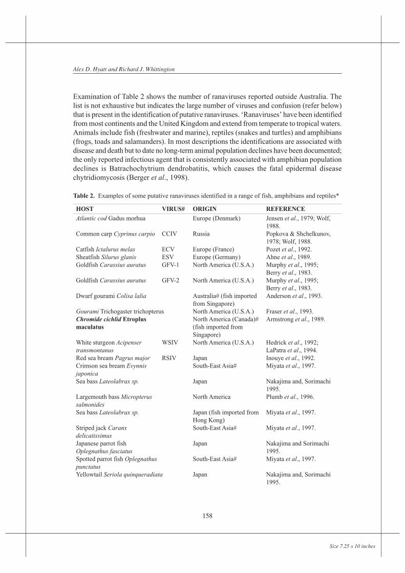
Table 2. Examples of some putative ranaviruses identified in a range of fish, amphibians and reptiles*
HOST VIRUS# ORIGIN REFERENCE
Atlantic cod Gadus morhua Europe (Denmark) Jensen et al., 1979; Wolf,1988.
Common carp Cyprinus carpio CCIV Russia Popkova & Shchelkunov,1978; Wolf, 1988.
Catfish Ictalurus melas ECV Europe (France) Pozet et al., 1992.Sheatfish Silurus glanis ESV Europe (Germany) Ahne et al., 1989.Goldfish Carassius auratus GFV-1 North America (U.S.A.) Murphy et al., 1995;
Berry et al., 1983.Goldfish Carassius auratus GFV-2 North America (U.S.A.) Murphy et al., 1995;
Berry et al., 1983.Dwarf gourami Colisa lalia Australia# (fish imported Anderson et al., 1993.
from Singapore)Gourami Trichogaster trichopterus North America (U.S.A.) Fraser et al., 1993.Chromide cichlid Etroplus North America (Canada)# Armstrong et al., 1989.maculatus (fish imported from
Singapore)White sturgeon Acipenser WSIV North America (U.S.A.) Hedrick et al., 1992;transmontanus LaPatra et al., 1994.Red sea bream Pagrus major RSIV Japan Inouye et al., 1992.Crimson sea bream Evynnis South-East Asia# Miyata et al., 1997.japonicaSea bass Lateolabrax sp. Japan Nakajima and, Sorimachi
1995.Largemouth bass Micropterus North America Plumb et al., 1996.salmonidesSea bass Lateolabrax sp. Japan (fish imported from Miyata et al., 1997.
Hong Kong)Striped jack Caranx South-East Asia# Miyata et al., 1997.delicatissimusJapanese parrot fish Japan Nakajima and SorimachiOplegnathus fasciatus 1995.Spotted parrot fish Oplegnathus South-East Asia# Miyata et al., 1997.punctatusYellowtail Seriola quinqueradiata Japan Nakajima and, Sorimachi
1995.

Ranaviruses of Fish, Amphibians and Reptiles:Diversity and the Requirement for Revised Taxonomy
159
Size 7.25 x 10 inches
HOST VIRUS# ORIGIN REFERENCE
Amberjack Seriola dumerili South-East Asia# Miyata et al., 1997.Goldstriped amberjack Seriola South-East Asia# Miyata et al., 1997.aureovittataBrown-spotted grouper SGD South-East Asia Chua et al., 1994.Epinephelus tauvina (Singapore)#Brown-spotted grouper South-East Asia (Thailand) Miyata et al., 1997.Epinephelus malabaricusRed spotted grouper South-East Asia# Miyata et al., 1997.Epinephelus akaaraTiger puffer Takifugu rubripes South-East Asia# Miyata et al., 1997.Guppy fish Poecilia reticlata North America (U.S.A.) Hedrick and McDowell, 1995
(fish imported fromSouth-East Asia)
Doctor fish Labroides dimidatus North America (U.S.A.) Hedrickand McDowell, 1995.(fish imported fromSouth-East Asia)
Turbot Scophthalmus maximus Europe (Denmark)# Bloch et al., 1993.Angelfish Pterophyllum scalare North America (Canada)# Schuh and Shirley 1990.
(fish imported fromunknown source)
Pike perch Stizostedion lucioperca Finland Tapiovaara et al., 1998.Mudskipper Parapocryptes serperaster Europe (Spain)# Martinez-Picado et al., 1993.
(fish imported from Malaysia)Infectious spleen and Kidney ISKV China He et al., 2002necrosis virus
AMPHIBIANSLeopard frog Rana pipiens frog North America (U.S.A.) Granoff et al., 1965.
virus 3 (FV3)(type exampleof sympatricisolates FV1, 2, 9-23)
Leopard frog Rana pipiens LT1-LT4 North America (U.S.A.) Clark et al., 1968.Red eft Diemictylus viridescens T6-20 North America (U.S.A.) Clark et al., 1969.North American bullfrog tadpole North America (U.S.A.) Wolf et al., 1968.Rana catesbeiana edema
virus (TEV)Edible frog Rana esculenta REIR Europe (Croatia) Fijan et al., 1991.Cane toad Bufo marinus GV South America Zupanovic et al., 1998.
(Venezuela)Common frog Rana temporaria Europe (U.K.) Drury et al., 1995.Red-legged frog Rana aurora 276 North America (U.S.A.) Mao et al., 1997.Tiger salamander Ambystoma ATV North America (U.S.A.) Janovich et al., 1998.tigrinum stebbensi
REPTILESBox turtle Terrapene c. carolina TV3 North America (U.S.A.) Mao et al., 1997.Central Asian tortoise TV5 North America (U.S.A.) Mao et al., 1997.Testudo horsefieldiGopher tortoise Gopherus polyphemus North America (U.S.A.) Westhouse et al., 1996.Testudo hermanni ranavirus ThRV Marschang et al., 1999
* Australian ranaviruses not included; #, nomenclature as per cited reference.

Alex D. Hyatt and Richard J. Whittington
160
Size 7.25 x 10 inches
Fig
ure
1. S
chem
atic
of
sugg
este
d st
rate
gy f
or th
e cl
assi
fica
tion
of r
anav
irus
es (
base
d on
Tab
le 3
). S
chem
atic
is s
how
n to
the
leve
l of
genu
s.

Ranaviruses of Fish, Amphibians and Reptiles:Diversity and the Requirement for Revised Taxonomy
161
Size 7.25 x 10 inches
The identification of many putative ranaviruses in one region of Northern America (e.g.Green et al., 2002) raises questions such as why are we now identifying these viruses andwhat has happened (eg anthropogenic changes) to cause the emergence of these viruses? Toanswer these questions it is critical that we can accurately identify these new viruses interms of genera, species and genotypes. The ability to identify and differentiate ranaviruseswill provide the background knowledge so that it may be possible to state whether a specificpopulation of viruses is present/absent from any one region or country and whether a specificvirus is increasing its range (issues in trade and conservation). It will also provide the basicknowledge to initiate research activities into attempting to answer the topical questionsreferred to in the Introduction.
Biology and Taxonomy of Ranaviruses
Identification of a ranavirus infection is based upon pathology and a battery of diagnosticassays including cell culture, ultrastructure/morphogenesis, (electron microscopy), antigenicanalyses (ELISA, histochemistry, immunoelectron microscopy), SDS-PAGE, restrictionendonuclease digestion, hybridisation, PCR analyses and sequencing (e.g. Hyatt et al., 2000).The data from all of these assays should be used in the overall classification and identificationof any putative ranavirus. To explain the significance of this statement each of the aboveareas will be discussed in reference to categorising a virus to a specific level of classification.In addition a classification strategy for the identification of ranaviruses will be suggested.
Requirement for redefining iridovirus classification
The genus Ranavirus (refer below) contains a large group of viruses identified from fish,amphibians and reptiles. The many viruses described have differences in pathology, proteinprofiles, restriction fragment polymorphisms, antigenicity and sequence (refer to referencesin Table 2). That is, this group of viruses has become a large ‘holding bag’ for all ‘iridoviruses’isolated from poikilothermic animals (excluding invertebrates), are not ‘erythrocytic’ orbelong to the genus Lymphocystivirus. If we are to suggest a reclassification scheme for thevertebrate iridoviruses, specifically the ranaviruses, we should begin at the level of ‘Family’.
Currently the ICTV classifies the family Iridoviridae into the genera, Iridovirus,Chloridovirus, Lymphocystivirus and Ranavirus (Williams et al., 2000). An outline andschematic of a proposed strategy for the classification of ranaviruses is shown in Fig. 1 andTable 3. Table 3 highlights a major change in the classification of iridoviruses with theintroduction of ‘sub-families’. The suggested classification strategy includes criteria basedon either universal or polythetic classes and takes into consideration replicative lineage andecological niches for species definition. An example of using an universal selection criterionis the presence/absence of methylated genomes. That is, this characteristic may be used todivide the family Iridoviridae into two sub-families namely ‘Methylated Iridoviruses’(including the current genera Ranavirus and Lymphocystivirus) and ‘Non-methylatedIridoviruses’ (including the current genera Chloridovirus and Iridovirus.). Alternatively,the families could be divided into sub-families Entomovirinae and Chordovirinae as forpoxviruses (Moyer et al., 2000). If consistency in the taxonomy of viruses is to be achievedthen the taxonomy of iridoviruses should follow that existing for the more closely relatedfamilies. As such, we suggest (based on the viruses listed by the ICTV) that the names of
Alex D. Hyatt and Richard J. Whittington
162
Size 7.25 x 10 inches
the sub-families be Entomovirinae (non-methylated iridoviruses) and Chordovirinae(methylated iridoviruses); this scheme would have the advantage of accommodating otherfuture sub-families encompassing other major groups of invertebrates.
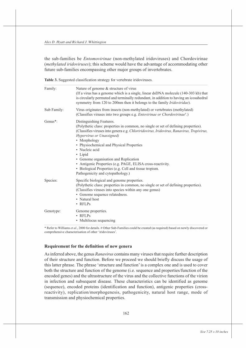
Table 3. Suggested classification strategy for vertebrate iridoviruses.
Family: Nature of genome & structure of virus(If a virus has a genome which is a single, linear dsDNA molecule (140-303 kb) thatis circularly permuted and terminally redundant, in addition to having an icosahedralsymmetry from 120 to 200nm then it belongs to the family Iridoviridae).
Sub Family: Virus originates from insects (non-methylated) or vertebrates (methylated)(Classifies viruses into two groups e.g. Entovirinae or Chordovirinae#.)
Genus*: Distinguishing Features.(Polythetic class: properties in common, no single or set of defining properties).(Classifies viruses into genera e.g. Chloriridovirus, Iridovirus, Ranavirus, Tropivirus,Hypervirus or Unassigned)• Morphology• Physiochemical and Physical Properties• Nucleic acid• Lipid• Genome organisation and Replication• Antigenic Properties (e.g. PAGE, ELISA cross-reactivity.• Biological Properties (e.g. Cell and tissue tropism.Pathogenicity and cytopathology.)
Species: Specific biological and genome properties.(Polythetic class: properties in common, no single or set of defining properties).(Classifies viruses into species within any one genus)• Genome sequence relatedness.• Natural host• RFLPs
Genotype: Genome properties.• RFLPs• Multilocus sequencing
* Refer to Williams et al., 2000 for details. # Other Sub-Families could be created (as required) based on newly discovered orcomprehensive characterisation of other ‘iridoviruses’.
Requirement for the definition of new genera
As inferred above, the genus Ranavirus contains many viruses that require further descriptionof their structure and function. Before we proceed we should briefly discuss the usage ofthis latter phrase. The phrase ‘structure and function’ is a complex one and is used to coverboth the structure and function of the genome (i.e. sequence and properties/function of theencoded genes) and the ultrastructure of the virus and the collective functions of the virionin infection and subsequent disease. These characteristics can be identified as genome(sequence), encoded proteins (identification and function), antigenic properties (cross-reactivity), replication/morphogenesis, pathogenicity, natural host range, mode oftransmission and physiochemical properties.

Ranaviruses of Fish, Amphibians and Reptiles:Diversity and the Requirement for Revised Taxonomy
163
Size 7.25 x 10 inches
Selection of demarcation criteria for new genera
To divide the current ranaviruses into more appropriate groups (e.g. genera and species) wemust define a list of ‘Distinguishing Features’ (demarcation criteria) for each level ofclassification. For a virus to be classified into any one group then it must satisfy most (butnot necessarily all) of the defined properties for that group. To illustrate how this classificationscheme would work we will take a closer look at the current group of ranaviruses. Forexample, there are two obvious groups of ranaviruses. One group is restricted to tropicalfish, is associated with splenomegaly, anaemia, swollen kidneys, “highly ballooned cells”and little to no cross-reactivity to EHNV and FV3 (Sudthongkong et al., 2002). The secondgroup includes the FV3-like viruses that are found in a range of fish, reptiles and amphibians,is not associated with the development of hypertrophied cells and cross-reacts in the EHNVantigen capture ELISA (Hyatt et al., 2000). Table 4 illustrates how these two groups can beobjectively grouped into two different genera using seven demarcation criteria and a selectionof viruses. We suggest that the name Ranavirus be retained as it is entrenched within thescientific literature. We also suggest that the second genus be called either Hyperiridovirusor Tropivirus. Sudthongkong et al. (2002) suggested that the name Tropivirus be used as itis representative of the geographical area from which all viruses that can be phylogenticallygrouped originated. An alternative name could be Hypervirus or indeed any another namethat would represent the common property of this group namely the excessively large natureof the inclusion bodies (virus assembly sites) of the infected cells that contribute to theirhypertrophic appearance. At this point in time, we shall refer to this second genus asHyperiridovirus as it is a descriptive term that represents a common biological characteristicof this group.
From this analysis EHNV, BIV, European sheatfish virus (ESV) and doctorfish virus (DFV)are grouped into the genus Ranavirus whereas Red sea bream iridovirus (RSIV) satisfiedonly a few of the listed demarcation criteria for the same genus. For example, examinationof the scientific literature (e.g. Inouye et al., 1992; Sudthongkong et al., 2002) reveals thatRSIV is associated with marine fish, does not cross-react with antibodies against EHNV,requires a different set of PCR primers, has a 44% identity with FV3 and generates‘hypertrophied’ cells. An analogous set of demarcation criteria could be generated to groupthe viruses similar to RSIV. Here differentiating criteria within “Distinguishing Features”(refer to Fig. 4) could include (i) ‘viruses do not cross-react with the EHNV antigen captureELISA but cross-react with polyclonal antibodies against RSIV’ (‘Antigenic Properties’);(ii) ‘infected cells are enlarged due to the presence of large inclusion bodies/assembly bodies’(‘Biological Properties’) and (iii) viruses do not replicate in a range of continuous amphibianpiscine and mammalian cell lines’ (‘Biological Properties’). If these criteria were used thenRSIV would be included into the genus Hyperiridovirus.
An important point in the taxonomy of ranaviruses is that classification at the level of genuscannot be done on molecular biology alone. The reason for this is that unless the entiregenome is sequenced and compared for numerous isolates then such taxonomy is of limitedvalue. For example, the function of the genome comes from its structure; the varyingbiological properties that influence replication, virulence, host animals and transmission isa consequence of the structure of the genome; what may appear as inconsequential sequencedifferences may result in a significant biological phenotypic property (biological

Alex D. Hyatt and Richard J. Whittington
164
Size 7.25 x 10 inches
characteristic). This is not to underestimate the importance of molecular biology. Sequencedata is extremely important but it must be placed into context. It is important in determiningthe replicative lineage of viruses and increases in significance as the resolution of taxonomy(i.e. from species to genotype) increases.
Table 4. Suggested demarcation criteria for identifying viruses belonging to the genus Ranavirus.
Genus: Ranavirus(Type species FV3)
EHNV ESV DFV RSIV BIV
1. >60 aa identity of MCP of type Y Y Y N Yspecies.
2. Systemic and necrotising infection# Y Y (?) N Y
3. Acquire plasma membrane and bud. Y Y Y N Y
4. Cytoplamic assembly bodies, Y Y Y N Yno hypertrophy/cytomegally.
5. Host range: Fish/amphibians/reptiles Y Y Y Y Y(one or more).
6. Replicate in a range of continuous Y Y Y N Ycell lines (including amphibian,piscine and mammalian)
7. Reactivity in EHNV antigen-capture Y Y N N YELISA*.
Total 7/7 7/7 5(?)/7 1/7 7/7
Conclusion: EHNV, BIV, ESV & DFV are members of the genus: Ranavirus.
aa: amino acid; (?), data not available; # pathology may also include haemorrhage and/or ulcers; *refer to Hyatt et al., 2000.Note, information relating to each of the criteria originates from the scientific literature cited in Table 2.
Selection of demarcation criteria for species, genotypes and importance thereof
As stated above it is important from trade, animal health and biological viewpoints to havethe capacity to identify and differentiate species and genotypes within the current genusRanavirus. For example, EHNV is listed by the OIE as a list B pathogen for the diseaseepizootic haematopoietic necrosis (EHN). Other viruses recognised by the OIE in associationwith EHN are ESV and ECV. This raises several questions namely, is EHNV a member ofthe species FV3, are redfin perch virus and rainbow trout virus (isolates of EHNV) differentto each other and are ESV and ECV European isolates of EHNV? To answer these questionsdemarcation criteria must be listed for proposed species within the genus.

Ranaviruses of Fish, Amphibians and Reptiles:Diversity and the Requirement for Revised Taxonomy
165
Size 7.25 x 10 inches
Demarcation criteria for ‘species’. To date the ICTV recognises one species (FV3) withinthe existing genus Ranavirus. Within this species are listed the following isolates, box turtle3 (TV3), Lucke titurus virus 1, tadpole edema virus and tortoise virus 5. Tentative specieswithin the genus include BIV, EHNV (rainbow trout and redfin perch virus), RedwoodPark virus (tadpole virus2, stickleback virus), Regina ranavirus (tiger salamander virus,Ambystoma tigrinum stebbinsi virus) and Santee-Cooper ranavirus (Largemouth bass virus,doctor fish virus, guppy virus 6).
Table 5 lists 4 demarcation criteria for the species FV3. In this table the viruses EHNV,BIV, ESV and DFV have been included for comparison. The analyses illustrates that EHNVis an individual species. If these criteria are used to analyse the data from Hyatt et al. (2000)then it can be proposed that there are currently five species within the current genus Ranavirusnamely (i) FV3, (ii) ESV, (iii) DFV, (iv) BIV and (v) EHNV.
Are all members of a single species isolates of the one ‘population’? It is generallyaccepted that members within a group such as ‘species’ are ‘plastic’. That is, due toevolutionary pressures there is some variation in the structure and function of the genome.Therefore, without the use of differentiating neutralising antibodies and access to sequencedata of complete genomes or validated portions thereof, how can we identify distinctranavirus populations within a species (i.e. identify different genotypes)? Most diagnosticassays provide data pertinent to identifying viruses to the levels of genus and on occasion,species. For example, SDS-PAGE identifies polypeptide profiles indicative of a specificgenus and the presence of a major 48 to 52 kDa MCP (Hyatt et al., 2000). Antigen captureELISAs (and other antigen - based assays) illustrate cross-reactivity with most viruses atthe genus level, and ultrastructure, which as a ‘rule of thumb’ provides general information
Table 5. Suggested demarcation criteria for differentiating species within the genus ‘Ranavirus’.
Species demarcation criteria
EHNV BIV ESV DFV
1. MCP gene and one other gene Y Y Y Y
(or part of) are different (e.g. 2%*)with other viruses within the genus.
2. Specific natural host. Y Y Y (?)
3. RFLP bands differ significantly to Y/Y Y/(?) Y/(?) Y/(?)other viruses (species) within genus(e.g. 20-30% bands in common).Viruses within same genus shouldshare approximately 70-80% bands).
4/4 3/4 3/4 1/4
Conclusion: EHNV, BIV & ESV are distinct species. There is insufficient data to categorise DFV as aseparate species.
*Refer to Hyatt et al. (2000); (?) insufficient data. Note, information relating to each of the criteria originates from thescientific literature cited in Table 2.

Alex D. Hyatt and Richard J. Whittington
166
Size 7.25 x 10 inches
on replication and cytopathology useful at the genus level. It should, however, be noted thatsubtle ultrastructural differences can be observed in ultrastructural pathology (manifestationsof changes in genomic structure) which are indicative of specific species (e.g. Wamenavirus, Hyatt et al., 2002). Collectively, these diagnostic assays cannot differentiate betweendistinct species or between distinct populations within anyone species.
Within this paper, genotypes are defined as sub-populations of a species that generate progenyvirions of high fidelity (maintenance of the specific genomic structure and function). Toidentify such genotypes high-resolution diagnostic assays are required. These analyticaltools should examine the fidelity of the entire genome. To test fidelity many isolates shouldbe analysed as a function of time, host(s) and geographical range. If fidelity is conservedthen assumptions in relation to the taxonomic status of future new isolates can be made (i.e.the use of a set number of genes or part there of). At present only a few methylated iridovirusgenomes have been sequenced in their entirety; key genes to define specific species andgenotypes have therefore yet to be identified. Alternatively, restriction endonuclease digestionof complete genomes can be performed. To increase the sensitivity of this technique aminimum of three enzymes should be used.
As one of the suggested demarcation criteria for identification of a species is that all membersshould have a minimum of 60% to 80% RFLP bands between similar isolates we suggestthat genotypes should display greater than 80% homology. Using this approach we canchallenge inferences that there are two currently identified genotypes of EHNV namelyrainbow trout virus (RTV) and redfin perch virus (RFPV) (Williams et al., 2000). TheRFLPs of various EHNV isolates collected over different time periods, geographical rangesand the two different hosts (Hyatt et al., 2000) indicate that the isolates are very similar, i.e.whilst there are differences between the isolates, these differences appear random and cannotbe explained on the basis of host animal.
The use of an objective, logical taxonomic strategy can therefore be used to demonstratethat EHNV is a distinct species. Furthermore the data from Hyatt et al. (2000) suggests thatthere are currently five species within the current genus Ranavirus namely (i) FV3, (ii)ESV, (iii) DFV, (iv) BIV and (v) EHNV. The data also suggests that of the many isolates ofEHNV so far characterised there are no distinct genotypes (correlation of RFLPs with diseasecharacteristics).
Summary
Over the past ten to fifteen years many viruses have been identified from Australianpoikilotherms. Of these, ranaviruses are the only viruses that increased their geographicalrange. This genus of viruses is distinct from the group of viruses isolated from tropical fishthat are associated with hypertrophied cells. Ranaviruses have been identified from manyother countries including North and South America, Europe and Asia. Whilst pathogenic tomany animals, and probably representative of many of their natural viral assemblage, thisgroup of viruses has not been associated with long-term population declines. They arehowever, identified by the OIE and are recognised as potential threats to aquaculture andfree-ranging animals.

Ranaviruses of Fish, Amphibians and Reptiles:Diversity and the Requirement for Revised Taxonomy
167
Size 7.25 x 10 inches
The current taxonomy of ‘ranaviruses’ is in need of revision. We have discussed what webelieve to be the shortcomings and have suggested a more complex and rigorous classificationscheme. From such an approach we suggest that the family Iridoviridae be divided into twosub-families Entomovirinae and Chordovirinae. Within the sub-family Chordovirinae wesuggest that (i) the genus Ranavirus be retained (i.e. not renamed) and (ii) at least one othergenus be created (eg. Hyperiridovirus or Tropivirus) to include the tropical viruses that areassociated with the development of hypertrophied cells. We further suggest that a list ofdemarcation criteria be established for the identification of specific genotypes which shoulddecrease confusion about identity of specific viruses, i.e. is a newly identified virus anisolate of an existing species and genotype or is it a genuinely new virus constituting a newspecies or new genotype of an existing species? Finally, with the implementation of a newmethodical approach to the taxonomy of ranaviruses meaningful research into topicalquestions referred to in the ‘Introduction’ can be initiated.
Note: Since the writing of this manuscript the International Committee on the Taxonomyof Viruses (ICTV) have accepted the naming of the genus referred to within this paper as“Hyperiridovirus” or “Tropivirus” as “Meglaocytivirus” from the Greek meaning “enlargedcell”.
REFERENCES
Ahne, W., Schlotfeldt, H.J., and Thomsen, I. 1989. Fish viruses: isolation of an icosahedral cytoplasmicdeoxyribovirus from sheatfish (Silurus glanis). Journal of Veterinary Medicine B 36, 333-336.
Anderson, I.G, Prior H.C, Rodwell, B.J., and Harris, G.O. 1993. Iridovirus-like virions in importeddwarf gourami (Colisa Ialia) with systemic amoebiasis Australian Veterinary Journal 70, 66-67.
Armstrong, R.D. and Ferguson, H.W. 1989. A systemic viral disease of chromide cichlids, Etropusmaculatus Bloch. Diseases of Aquatic Organisms 7, 155-157.
Berger, L., Speare, R., Daszak, P., Green, D.E., Cunningham, A.A., Goggin, C.L., Slocombe, R.,Ragan, M.A., Hyatt, A.D., McDonald, K.R., Hines, H.B., Lips, K.R., Marantelli, G. andParkes, H. 1998. Chytridiomycosis causes amphibian mortality associated with populationdeclines in the rain forests of Australia and Central America. Proceedings of the NationalAcademy of Science USA 95, 9031-9036.
Berry, E.S., Shea, T.B. and Galiks, J. 1983. Two iridovirus isolates from Carassius auratus (L).Journal of Fish Diseases 6, 501-510.
Bloch, B. and Larsen, J.L. 1993. An iridovirus-like agent associated with systemic infection in culturedturbot Scophthalmus maximus fry in Denmark. Diseases of Aquatic Organisms 15, 235-240.
Chua, F.H.C., Ng, M.L., Ng, K.L., Loo, J.J. and Wee, J.Y. 1994. Investigation of outbreaks of a noveldisease, “Sleepy Grouper Disease”, affecting the brown spotted grouper, Epinephelus tauvinaForskal. Journal of Fish Diseases 17, 417-427.
Clark, H.F., Brennan, J.C., Zeigel, R.F., Karzon, D.T. 1968. Isolation and characterization of virusesfrom the kidneys of Rana pipiens with renal adenocarcinoma before and after passage in redeft (Triturus viridescens). Journal of Virology 2, 629-640.
Clark, H.F., Gray, C., Fabian, F., Zeigal, R. and Karzon, D.T. 1969. Comparative studies of amphibiancytoplasmic virus strains isolated from the leopard frog, bullfrog and newt. Mizell, M. (ed.).Biology of Amphibian Tumors. Springer-Verlag New York Heidelberg Berlin. pp 310-326.
Alex D. Hyatt and Richard J. Whittington
168
Size 7.25 x 10 inches
Crane, M.S., Hardy-Smith, P., Williams, L.M., Hyatt, A.D., Eaton, L.M., Gould, A., Handlinger, J.,Kattenbelt, J. and Gudkovs, N. 2000. First isolation of an aquatic birnavirus from farmed andwild fish species in Australia. Disease of Aquatic Organisms 43, 1-14.
Daszak, P, Cunningham, A.A, and Hyatt, A.D. 2000. Emerging Infectious Diseases of Wildlife -Threats to Biodiversity and Human Health. Science 287, 443-449.
Drury, S.E.N., Gough, R.E. and Cunningham, A.A. 1995. Isolation an iridovirus-like agent fromcommon frogs (Rana temporaria). Veterinary Record 137, 72-73.
Fijan, N., Matasin, Z., Petrinec, Z., Valpotic, I. and Zwillenberg, L.O. 1991. Isolation of an iridovirus-like agent from the green frog (Rana esculenta L). Veterinary Archives Zagreb 3, 151-158.
Fraser, W.A., Keefe, T.J. and Bolon, B. 1993. Isolation of an iridovirus from farm raised gouramis(Trichogaster trichopterus) with fatal disease. Journal of Veterinary Diagnostic Investigation5, 250-253.
Granoff, A., Cam,e P.E. and Rafferty, K. 1965. The isolation and properties of viruses from Ranapipiens: their possible relationship to the renal adenocarcinoma of the leopard frog. Annals ofthe New York Academy of Science 126, 237-255.
Green, D.E., Converse, K.A. and Schrader, A.K. 2002. Epizootiology of sixty-four amphibianmorbidity and mortality events in the USA, 1996-2001. Annals of the New York Academy ofScience 969, 323-39.
He, J.G., Wang, S.P., Zeng, K., Huang, Z.J. and Chan, S.M. 2000. Systemic disease caused by aniridovirus-like ageny in cultured mandarinfish, Siniperca chuatsi (Basilewsky), in China.Journal of Fish Diseases 23, 219-222.
Hedrick, R.P., McDowell, T.S., Ahne, W., Torhy, C. and de Kinkelin, P. 1992. Properties of threeiridovirus-like agents associated with systemic infections of fish. Diseases of AquaticOrganisms 13, 203-209.
Hedrick, R.P. and McDowell, T.S. 1995. Properties of Iridoviruses from ornamental fish. VeterinaryResearch 26, 423-427.
Hengstberger, S.G., Hyatt A.D., Speare R. and Coupar B.E.H. (1993). Comparison of epizootichaematopoietic necrosis and Bohle iridoviruses, recently isolated Australian iridoviruses.Diseases of Aquatic Organisms 15, 93-107.
Hurst, C.J. 2000. Viral Ecology. San Diego, California; Academic Press, London.
Hyatt, A.D. 1998. Identification, Characterisation and Assessment of Venezuelan viruses for potentialuse as biological control agents against the cane toad (Bufo marinus) in Australia. A report tothe Federal Government. (Environment Australia and Division of Wildlife & Ecology); twovolumes.
Hyatt, A.D., Gould, A.R., Zupanovic, Z., Cunningham, A.A., Hengstberger, S., Whittington, R.J.,Kattenbelt, J. and Coupar, B.E. 2002. Comparative studies of piscine and amphibianiridoviruses. Archives of Virology 145, 301-31.
Hyatt, A.D., Hine, P.M., Jones, B., Whittington, R., Wise, T. and Crane, M. 1997. Epizootic mortalityin the pilchard (Sardinops sagax neopilchardus) in Australia and New Zealand in 1995 II.Identification of a herpesvirus within the gill epithelium. Diseases of Aquatic Organisms 28,17-29.
Hyatt, A.D., Gould, A.R., Zupanovic, Z., Cunningham, A.A., Hengstberger, S., Whittington, R.J.,Kattenbelt, J. and Coupar, B.E. 2000. Comparative studies of piscine and amphibianiridoviruses. Archives of Virology 145:301-31.

Ranaviruses of Fish, Amphibians and Reptiles:Diversity and the Requirement for Revised Taxonomy
169
Size 7.25 x 10 inches
Hyatt, A.D., Williamson, M., Coupar, B.E., Middleton, D., Hengstberger, S.G., Gould, A.R., Selleck,P., Wise, T.G., Kattenbelt, J., Cunningham, A.A., Lee, J. 2002. First identification of a ranavirusfrom green pythons (Chondropython viridis). Journal of Wildlife Diseases 38, 239-252.
Hyatt, A.D., Daszak, P., Cunningham, A.A., Field, H. and Gould, A.R. 2004. Henipaviruses: Gaps inthe knowledge of emergenge. EcoHealth. (in press).
Inouye, K., Yamano, K., Maeno, Y., Nakajima, K., Matsuoka, M., Wada, Y. and Sorimachi, M. 1992.Iridovirus infection of cultured red sea bream, Pagrus major. Fish Pathology 27, 19-27.
Jancovich, J.K., Davidson, E.W., Morado, J.F., Jacobs, B.L. and Collins, J.P. 1998. Isolation oflethal virus from the endangered salamander Ambystoma tigrinum stebbinsi. Diseases ofAquatic Organisms 31, 161-167.
Jensen, N.J. and Larsen, J.L. 1979. The ulcus-syndrome in cod (Gadus morhua). I. A pathologicaland histopathological study. Nordic Veterinary Medicine 31, 436-442.
Langdon, J.S., Humphrey, J.D, Williams, L.M, Hyatt, A.D. and Westbury, H.A. 1986. First virusisolation from Australian fish: An iridovirus-like pathogen from redfin perch, Perca fluviatilisL. Journal of Fish Diseases 9, 263-268.
Langdon, J.S. and Humphrey, J.D. and Williams, L.D. 1998. Outbreaks of an EHNV-like iridovirusin cultured rainbow trout, Salmo gairdneri Richardson, in Australia. Journal of Fish Diseases11, 93-96.
LaPatra, S.E., Groff, J.M., Jones, G.R., Munn, B., Patterson, T.L., Holt, R.A., Hauck, A.K. andHedrick, R.P. 1994. Occurrence of white sturgeon iridovirus infections among cultured whitesturgeon in the Pacific Northwest. Aquaculture 126, 201-210.
Mao, J., Hedrick, R.P. and Chinchar, V.G. 1997. Molecular characterization, sequence analysis, andtaxonomic position of newly isolated fish iridoviruses. Virology 229, 212-220.
Marschang, R.E., Becher, P., Posthaus, H., Wild, P., Thiel H-J, Muller-Doblies, U., Kaleta, E.F. andBacciarini, L.N. 1999. Isolation and characterisation of an iridovirus from Hermann’s tortoises(Testudo hermanni). Archives of Virology. 144, 1909-1922.
Martinez-Picado, J., Blanch, A.R. and Jofre, J. 1993. Iridovirus-like particles associated with nodularskin lesions and vesicles in Parapocryptes serperaster. Journal of Aquatic Animal Health 5,148-151.
Miyata, M., Matsuno, K., Jung, S.J., Danayadol, Y. and Miyazaki, T. 1997. Genetic similarity ofiridoviruses from Japan and Thailand. Journal of Fish Diseases 20, 127-134.
Moody, N.J.G. and Owens, L. 1994. Experimental demonstration of the pathogenicity of a frogvirus, Bohle iridovirus, for a fish species, barramundi Lates calcarifer. Diseases of AquaticOrganisms 18, 265-102.
Moyer, R.W., Arif, B.M., Black, D.N., Boyle, D.B., Buller, R.M., Dumbell, K.R., Esposito, J.J.,McFadden, G., Moss, B., Mercer, A.A., Ropp, S., Tripathy, D.N. and Upton, C. 2000. FamilyPoxviridae. In van Regen Mortel, M.H.V. et al. (eds.). Virus Taxonomy, Seventh Report ofthe International Committee of Viruses. Academic Press, San Diego. pp. 167-182.
Munday, B.L., Langdon, J.S., Hyatt, A.D. and Humphrey, J.D. 1992. Mass mortality associated witha viral-induced vacuolating encephalopathy and retinopathy of larval and juvenile barramundi,Lates calcarifer Bloch. Aquaculture 103, 197-211.
Murphy, F.A., Fauquet, C.M., Bishop, D.H.L., Ghabrial, S.A., Jarvis, A.W., Martelli, P., Mayo, M.A.and Summers, M.D. 1995. Virus Taxonomy: Classification and Nomenclature of Viruses.Sixth Report of the International Committee on Taxonomy of Viruses. Springer-Verlag. Wien,New York (Archives of Virology Supplement 10).
Nakajima, K. and Sorimachi, M. 1995. Production of monoclonal antibodies against red sea breamiridovirus. Fish Pathology 30, 47-52.

170
Size 7.25 x 10 inches
Office International des Epizooties: Annual Reports of OIE Reference Laboratories and CollaboratingCentres 2002, p351.
Pearce, M., Humphrey, J.D., Hyatt, A.D. and Williams, L.M. 1990. Lymphocystis disease in captivebarramundi Lates calcarifer. Australian Veterinary Journal 67, 144-145.
Plumb J.A., Grizzle J.M., Young H.E. and Noyes A.D. (1996). An iridovirus isolated from wildlargemouth bass. Journal of Aquatic Animal Health 8, 265-270.
Popkova, T.I., Shchelkunov, I.S. 1978. Isolation of virus from carp afflicted with gill necrosis(Vedelenie virusa of karpov, bol’nykh zhabernym nekrozom.) VNIIPRKh. Rybn. Khoz. 4,34-38.
Pozet, F., Morand, M., Moussa, A., Torhy, C. and de Kinkelin, P. 1992. Isolation and preliminarycharacterisation of a pathogenic icosahedral deoxyribovirus from the catfish Ictalurus melas.Diseases of Aquatic Organisms 14, 35-42.
Reddacliff, L.A. and Whittington, R.J. 1996. Pathology of epizootic haematopoietic necrosis virus(EHNV) infection in rainbow trout (Oncorhynchus mykiss Walbaum) and redfin perch (Percafluviatilis L). Journal of Comparative Pathology 115, 103-115.
Schuh, J.C.L. and Shirley, I. 1990. Viral hematopoietic necrosis in an angelfish (Pterrophyllumscalare). J. Zoo. Wildlife Medicine 21, 95-98.
Speare, R., Freeland, W.J. and Bolton, S.J. 1991. A possible iridovirus in erythrocytes of Bufo marinusin Costa Rica. Journal of Wildlife Diseases 14, 35-42.
Sudthongkong, M., Miyata, M. and Miyazaki, T. 2002. Viral DNA sequences of genes encoding theATPase and the outer capsid protein of tropical isolates which are pathogenic to fishes inJapan, South China Sea and South China Sea and Southeast Asian countries. Archives ofVirology 147, 2089-2109.
Tapiovaara, H., Olesen, N.J., Linden, J., Rimaila-Parnanen, E. and von Bonsdorff, C.H. 1998. Isolationof an iridovirus from pike-perch Stizostedion lucioperca. Diseases of Aquatic Organisms 32,185-193.
Westhouse, R.A., Jacobson, E.R., Harris, R.K., Winter, K.R. and Homer, B.L. 1996. Respiratory andpharyngo-esophageal iridovirus infection in a gopher tortoise (Gopherus polyphemus). Journalof Wildlife Diseases 32, 682-686.
Whittington, R.J., Kearns, C., Hyatt, A.D., Hengstberger, S. and Rutzou, T. 1996. Spread of epizootichaematopoietic necrosis virus (EHNV) in redfin perch (Perca fluviatilis) in southern Australia.Australian Veterinary Journal 73, 112-114.
Whittington, R.J., Jones, J.B., Hine, P.M. and Hyatt, A.D. 1997. Epizootic mortality in the pilchard(Sardinops sagax neopilchardus) in Australia and New Zealand in 1995. I Pathology andepizootiology. Diseases of Aquatic Organisms 28, 1-16.
Williams, T., Chinchar, V.G., Darai, G., Hyatt, A.D., Kalmakoff, J. and Seligy, V. 2000. FamilyIridoviridae, In van Regenmortel, M.H.V. et al. (eds.). Virus Taxonomy, Seventh Report ofthe International Committee of Viruses. Academic Press, San Diego. pp. 167-182.
Wolf, K., Bullock, G.L., Dumbar, D.E. and Quimby, M.C. 1968. Tadpole edema virus: a viscerotropicpathogen for anuran amphibians. Journal of Infectious Diseases 118, 253-262
Wolf, K. 1988. Viral infections of indeterminate pathogenicity, In Fish Viruses and Fish Viral Diseases.Comstock Publishing Associates, Cornell University Press. p352.
Zupanovic, Z., Musso, C., Lopez, G., Louriero, C.L., Hyatt, A.D., Hengstberger, S. and Robinson,A.J. 1998. Isolation and characterization of iridoviruses from the giant toad Bufo marinus inVenezuela. Diseases of Aquatic Organisms 33, 1-9
Alex D. Hyatt and Richard J. Whittington