Embed Size (px)
Citation preview
British Journal ofHaernatoZogy. 1995, 89. 417-420
SHORT REPORT
Rapid and sensitive non-radioactive assay for the detection of clonal gene rearrangements in B-lineage acute leukaemia
GERALD MARTIN, ROBERT MAIRS, JEREMY PLATT* A N D BRENDA GIBSON* Department of Radiation Oncology, Glasgow University, CRC Beatson Laboratories, Bearsden, Glasgow, and *Department of Haematology, Royal Hospital for Sick Children, Yorkhill, Glasgow
Received 2 June 1994; accepted for publication 10 November 1994
Summary. We have applied a simple and non-radioactive assay system used in conjunction with polymerase chain reaction amplification and high-resolution polyacrylamide gel electrophoresis to detect clonal immunoglobulin gene Keywords: B-lineage acute leukaemia, immunoglobulin rearrangements in leukaemia patient DNA. This technique is
as sensitive as isotopic methods, allowing the detection of clonal DNA diluted 1 in 10 000 in non-clonal DNA.
gene rearrangements, PCR, silver staining, non-isotopic.
Rearrangements of the immunoglobulin (Ig) and T cell receptor (TCR) genes can be used to identify clonal leukaemic cell populations in the bone marrow of patients with acute lymphoblastic leukaemia (ALL) (Deane & Hoarand, 1993). The ability to detect these rearrange- ments has been an integral part of the evaluation of such patients (British Society of Haematology, 1991).
Current protocols for high sensitivity detection of Ig gene rearrangements rely on the use of the polymerase chain reaction (PCR) with radiolabelled probes or primers (Deane & Norton, 1990, 1991; Deane et al, 1991b), or labour- intensive and time-consuming amplification, cloning and sequencing strategies (Yamada et al, 1989, 1990; Brisco et al, 1993, 1994; Jonsson et al, 1990; Billadeau et al, 1991, 1992: Ito et al, 1993). Due to the potential hazards associated with radioactive material and the elaborate nature of current strategies, we have applied a simple and non-isotopic detection system coupled to the PCR and high- resolution polyacrylamide gel electrophoresis to detect clonal leukaemic DNA in a rapid and highly sensitive assay.
MATERIALS AND METHODS
DNA was isolated as previously described (Martin & Lawlor, 1991) from blood or bone marrow mononuclear cells of 2 1 B-lineage ALL patients and control volunteers.
Nun-radioactive PCR. PCR was performed on a Hybaid
Correspondence: Dr Robert Mairs, Department of Radiation Oncology, Glasgow University, CRC Beatson Laboratories, Garscube Estate, Bearsden, Glasgow G 6 1 1BD.
Omnigene programmable thermocycler. The Ig primers were those described by Deane & Norton (1991) and by Trainor et a1 (1990) and Brisco et ai (1990). DNA derived from patient bone marrow was diluted in 10-fold increments with normal polyclonal mononuclear cell DNA. A total of 0.5 pg of DNA was amplified under the following conditions. Initial denaturation was at 92°C for Smin, followed by 35 cycles of 1 min denaturation at 90T , 1 min annealing at various temperatures, and 2 min extension at 72°C. Finally, there was another cycle with an extension time of 7min. An annealing temperature of 56°C was used for the FR3 5’ primer, 61°C for vH6, and 63°C for vH1 and vH2 (Table I). Non-isotopic PCR was performed in a 30 pl reaction volume, consisting of 1 x reaction buffer (Promega), 2.5 m~ MgClz, 10% (w/v) DMSO. 500 p~ each dNTP (Promega), 1 ~ L M each primer (Oswel DNA, Edinburgh), with 0.75units of Taq polymerase (Promega). Amplification was confirmed by 2% (w/v) agarose gel electrophoresis of 8p1 of PCR product. Electrophoresis through 6% (w/v) denaturing polyacryla- mide gels (National Diagnostic ‘Sequagel 6’) of 8 p1 of each PCR product was followed by silver staining using a commercially available kit (Biorad) based on the method of Merril et a1 (1981). The gels were fixed for 45 min in three changes of 40% (v/v) methanol. Oxidation of the gel was performed for 5min with a 10% (v/v) solution of the provided oxidizer (chromic acid). Following a single 30 s ddHzO wash, the gel was incubated for 20 min in a 10% (v/ v) solution of the provided silver nitrate. Excess silver nitrate was removed by rinsing for 10 s in ddHzO. followed by the addition of developing reagent (32g/l). The first two incubations with developer were replaced rapidly (within
41 7
418 Gerald Martin et al Table I. PCR primers for non-isotopic Ig gene rearrangement detection.
Detectable dilution by:
Silver [3SS]dATP Primer
Name Sequence staining autoradiogram Primer source
JH j’ACCTGAGGAGACGGTGACCAGGGT3‘ Dean & Norton (1990) VH 1 j’CCTCAGTGAAGGTCTCCTGCAAGG3’ 1 0-3 10-3 Dean &Norton (1990) vH2 5’TCCTGCGCTGGTGAAAGCCACACA3’ Dean & Norton (1990) VH6 j’CCTGTGCCATCTCCGGGGACAGTG3’ 10-4 Dean & Norton (1990) FR3 S’ACACGGC(C/T)(G/C)TGTATTACTGT3’ 10-~ Brisco et a1 (1990)
PCR primers used and their relative sensitivity by isotopic and non-isotopic detection methods.
10-1 5 s) to reduce background staining. Subsequent incubations were of longer duration, and continued until satisfactory staining with minimal background was achieved. Gels were fixed for 30min with 7.5% (v/v) acetic acid. All incubations, particularly during developing, were accompanied by gentle shaking, ensuring minimal disrup- tion to the gel surface. Buffers were removed by suction. The duration of the ddH20 wash and rinse stages were critical as prolonged washing increased the level of the non-specific background stain. The gel was then transferred to chromatography paper covered with Saran Wrap and dried at 80°C under vacuum for 30 min.
Radioactive PCR. The radioactive PCR was performed in a 20 pl reaction volume of the above buffer, but with 500 ,UM dGTP, dCTP, dTTP, and 5 0 p ~ dATP, 0.5units Tuq polymerase and 1 pl 37x TBq/mmole [35S]dATP (Amer- sham). As with Deane & Norton (1990, 1991), only 26 cycles were performed for the radioactive PCR. Polyacryl- amide gels which had been loaded with 5pl of each radioactive PCR product were fixed in 10% (v/v) methanol and 10% (v/v) acetic acid for 30min and dried on
chromatography paper at 80°C under vacuum for 2 h . Autoradiography was performed using Fuji RX film. The exposure time was 10-14d.
RESULTS
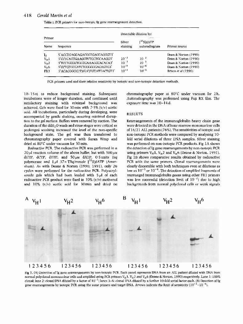
Rearrangements of the immunoglobulin heavy chain gene were detected in the DNA of bone marrow mononuclear cells of 16/21 ALL patients (76%). The sensitivities of isotopic and non-isotopic PCR methods were compared by analysing 10- fold serial dilutions of three DNA samples. Silver staining was performed on non-isotopic PCR products. Fig 1A shows the detection of Ig gene rearrangements by non-isotopic PCR using primers VH1, VH2 and VH6 (Deane & Norton, 1991). Fig 1 B shows comparative results obtained by radioactive PCR with the same primers. Clonal rearrangements were clearly discernible with both techniques even at dilutions as low as or The detection of amplified fragments of rearranged immunoglobulin genes using other FR1 primers was less successful (detection limit of lop2) due to high backgrounds from normal polyclonal cells or weak signals
Fig 1. (A) Detection of Ig gene rearrangements by non-isotopic PCR. Each panel represents DNA from an ALL patient diluted with DNA from normal polyclonal mononuclear cells and amplified using PCR primers v H 1 , vH2 and VH6 (Deane & Norton, 1990) respectively. Lane 1: 100% clonal: lane 2: clonal DNA diluted by a factor of lanes 3-6: clonal DNA diluted by a further 10-fold serial factor each. (B) Detection of Ig gene rearrangements by isotopic PCR using the same primers and target DNA. Arrows indicate the limit of sensitivity ( w - ~ O - ~ ) .

Short Report 4 19 achieve a 10-fold increase in sensitivity using FR3 primers in a single PCR (Trainor et al, 1990) without the inclusion of radionuclide. This compares favourably with the sensitivity achieved using the same methodology with the incorpora- tion of radiolabelled nucleotides.
The non-isotopic system, however, has several advantages in terms of routine application. The entire staining reaction is rapid, requiring approximately 1.5 h to achieve adequate strength of signal. The staining kit is relatively inexpensive (roughly t3/gel compared to €7S/gel for radioactive PCR (24 reactions)) and is stable at 4°C for at least 6 months. In addition, whereas the detection of radiolabelled PCR products becomes more difficult with time due to the decay of the incorporated radionuclide, repetitive analysis of unlabelled PCR products can be performed without loss of signal intensity. Furthermore, the special storage, handling and disposal precautions associated with radioactive material can be avoided.
Recent reports by Brisco et a1 (1993, 1994) imply that quantitative detection of clonality at the end of induction therapy may be of prognostic significance. We are currently investigating whether silver-stained gels can be used for highly sensitive non-isotopic quantitative analysis following the use of nested and/or patient-specific primers during the PCR amplification.
In conclusion, this rapid and sensitive system can be used to routinely detect clonal gene rearrangements from small leukaemic cell numbers in a safe non-isotopic clinical laboratory setting.
Fig 2. (A) and (B) Detection of Ig gene rearrangements using FR3 primer (Trainor et al, 1990) by non-isotopic and isotopic PCR respectively. In each panel, lane 1: 100% clonal; lane 2: clonal DNA diluted by a factor of lanes 3-6: clonal DNA diluted by a further 10-fold serial dilution factor each. Arrows indicate the limit of sensitivity
from the clonal DNA. Silver staining did, however, still match the sensitivity of radioactive PCR with these primers (data not shown). Figs 2A and 2B show a clonal gene rearrangement detected in DNA from an ALL patient by the non-isotopic and isotopic methods respectively using FR3 primer (Trainor et al, 1990), even when the clonal DNA was diluted by a factor of This represents a 10-fold increase in sensitivity over the original published methodology which employed ethidium bromide staining of DNA fragments in agarose gels (Trainor et al. 1990).
DISCUSSION
Non-isotopic systems for revealing clonal rearrangements of the immunoglobulin heavy chain gene in ALL patients have been of limited use for detecting residual disease due to their lack of sensitivity (detection limits of 31%) (Martin & Lawlor, 1991; Deane et aI, 1991a; Trainor et al, 1990), or due to the considerable amount of technical imput required (Brisco et al, 1990, 1993, 1994). We have applied a straightforward nucleic acid staining technique capable of detecting a clonal population consisting of 0.1-0.0 1 % of the mononuclear cell content of the marrow using PCR primers specific for the Ig VH1. vH2 and VH6 gene families (Deane & Norton, 1990). We have also applied this technique to
ACKNOWLEDGMENTS
We thank Frank Rinaldi for technical assistance and Ann Barrett and Tom Wheldon for useful discussion. This work was supported by grants from the SHERT and LRFC.
REFERENCES
Billadeau, D., Blackstadt, M., Greipp, P., Kyle, R.A., Oken, M.M., Kay, N. & van Ness, €3. (1991) Analysis of B-lymphoid malignancies using allele-specific polymerase chain reaction: a technique for sequentialquantitationofresidualdisease. Blood, 78,3021-3029.
Billadeau, D., Quam, L.. Thomas, W., Kay, N., Greipp, P., Kyle R., Oken. M.M. & van Ness, B. (1992) Detection and quantitation of malignant cells in the peripheral blood of multiple myeloma patients. Blood. 80, 1818-1824.
Brisco, M.J., Condon, J., Hughes, E., Noeh, S-H., Nicholson, I., Sykes, P.J., Tauro, G.. Ekert. H., Waters, K., Toogood. I., Seshandri. R.. Morley, A.A.. and the Australian and New Zealand Children's Cancer Study Group (1993) Prognostic significance of detection of rnonoclonality in remission marrow in acute lymphoblastic leukemia in children. Leukemia. 7, 1514-1520.
Brisco, M.J., Condon. J., Hughes, E., Noeh S-H., Sykes, P.J., Seshandri. R., Toogood, I.. Waters, K., Tauro, G., Ekert, H. & Morley, A.A. (1994) Outcome prediction in childhood acute lymphoblastic leukaemia by molecular quantitation of residual disease at the end of induction. Lancet, 343, 196-200.
Brisco, M.J., Tan. L.W.. Orsborn, A.M. & Morley, A.A. (1990) Development of a highly sensitive assay, based on the polymerase chain reaction, for rare B-lymphocyte clones in a polyclonal population. British Journal of Haernatology, 75, 163-167.
420 Gerald Martin et al British Society of Haematology (1991) Recommendations for the
Application of Molecular Genetic and Cytogenetic Techniques in Haematology Departments in the U.K. McMillan Press, U.K.
Deane, M. & Hoflbrand. A.V. (1993) Detection of minimal residual disease in W . Cancer Treatment Research, 64, 135-170.
Deane, M., McCarthy. K.P., Wiedemann. L.M. & Norton, J.D. (1991a) An improved method for detection of B-lymphoid clonality by polymerase chain reaction. Leukemia, 5, 726-730.
Deane, M. &Norton, J.D. (1990) Detection of immunoglobulin gene rearrangement in B lymphoid malignancies by polymerase chain reaction gene amplification. British Journal of Haematology. 74, 2 5 1-2 56.
Deane, M. & Norton, J.D. (1991) Immunoglobulin gene 'fingerprint- ing': an approach to analysis of B lymphoid clonality in lymphoproliferative disorders. British Iournal of Haematology, 77,
Deane, M.. Pappas, H. & Norton, J.D. (1991b) Immunoglobulin heavy chain gene fingerprinting reveals widespread oligoclonality in B-lineage acute lymphoblastic leukemia. Leukemia, 5, 832- 838.
Ito, Y., Wasserman, R., Galili, N.. Reichard, B.A.. Shane, S., Lange, B. & Rovera, G. (1993) Molecular residual disease status at the end of chemotherapy fails to predict subsequent relapse in children with B-lineage acute lymphoblastic leukaemia. Journal of Clinical Oncology. 11, 546-553.
Jonsson O.G., Kitchens. R.L., Scott, F.C. & Smith, R.G. (1990)
274-28 1.
Detection of minimal residual disease in acute lymphoblastic leukemia using immunoglobulin hypervariable region specific oligonucleotide probes. Blood, 76, 2072-2079.
Martin, G. & Lawlor, E. (1991) Non-radioactive detection of immunoglobulin and T cell receptor gene rearrangements in acute lymphoblastic leukaemia. British Journal of Haematology. 79,
Merril, C.R.. Goldman. D., Sedman, S.A. & Ebert, M.H. (1981) Ultrasensitive stain for proteins in polyacrylamide gels shows regional variation in cerebrospinal fluid proteins. Science, 2 11,
Trainor. K.J., Brisco, M.J., Story, C.J. & Morley, A.A. (1990) Monoclonality in B-lymphoproliferative disorders detected at the DNA level. Blood, 75, 2220-2222.
Yamada M., Hudson, S., Tournay, O., Bittenbender, S., Shane, S.S.. Lange, B., Tsujimoto, Y., Caton, A.J. & Rovera, G. (1989) Detection of minimal residual disease in hematopoietic malig- nancies of the B-cell lineage by using third-complementarity- determining region (CDR-111)-specific probes. Proceedings of the National Academy of Sciences of the United States of America, 86,
Yamada, M., Wasserman, R., Lange, B., Reichard. B.A.. Womer, R.B. & Rovera, G. (1990) Minimal residual disease in childhood B- lineage lymphoblastic leukemia: persistance of leukemic cells during the first 18 months of treatment. New England journal of Medicine. 323, 448-455.
516-519.
1437-1438.
5123-5127.


![[3,3]-Sigmatropic rearrangements - Massey Universitygjrowlan/stereo2/lecture11.pdf · 123.702 Organic Chemistry Claisen rearrangements • One of the most useful sigmatropic rearrangements](https://img.pdfslide.net/doc/110x75/5adcada77f8b9a213e8bd8b0/33-sigmatropic-rearrangements-massey-gjrowlanstereo2lecture11pdf123702.jpg)