Embed Size (px)
Citation preview
Journal of Neuroendocrinology. 2018;30:e12561. wileyonlinelibrary.com/journal/jne | 1 of 13https://doi.org/10.1111/jne.12561
© 2017 British Society for Neuroendocrinology
Received:1April2017 | Revised:24November2017 | Accepted:27November2017DOI: 10.1111/jne.12561
R E V I E W A R T I C L E
Rapid nongenomic modulation by neurosteroids of dendritic spines in the hippocampus: Androgen, oestrogen and corticosteroid
G. Murakami1 | Y. Hojo2 | A. Kato3 | Y. Komatsuzaki4 | S. Horie5 | M. Soma6 | J. Kim6 | S. Kawato3,5,6
1Department of Liberal Arts, Faculty of Medicine, Saitama Medical University, Iruma, Saitama, Japan2Department of Biochemistry, Faculty of Medicine, Saitama Medical University, Iruma, Saitama, Japan3Department of Biophysics and Life Sciences, Graduate School of Arts and Sciences, University of Tokyo, Meguro, Tokyo, Japan4Department of Physics, College of Science andTechnology,NihonUniversity,Chiyoda,Tokyo, Japan5Department of Urology, Graduate School of Medicine, Juntendo University, Hongo, Tokyo, Japan6Department of Cognitive Neuroscience,FacultyofPharma-Science, Teikyo University, Itabashi, Tokyo, Japan
CorrespondenceSuguru Kawato, Department of Biophysics and Life Sciences, Graduate School of Arts and Sciences, University of Tokyo, Meguro, Tokyo, Japan.Email:[email protected]
AbstractMemories are stored in synapses that consist of axon terminals and dendritic spines. Dendritic spines are postsynaptic structures of synapses and are essential for synaptic plasticity and cognition. Therefore, extensive investigations concerning the functions and structures of spines have been performed. Sex steroids and stress steroids have been shown to modulate hippocampal synapses. Although the rapid modulatory ac-tion of sex steroids on synapses has been studied in hippocampal neurones over sev-eral decades, the essential molecular mechanisms have not been fully understood. Here,adescriptionofkinase-dependentsignallingmechanisms isprovidedthatcanexplain the rapid nongenomic modulation of dendritic spinogenesis in rat and mouse hippocampal slices by the application of sex steroids, including dihydrotestosterone, testosterone, oestradiol and progesterone. We also indicate the role of synaptic (clas-sic) sex steroid receptors that trigger these rapid synaptic modulations. Moreover, we describe rapid nongenomic spine modulation by applying corticosterone, which is an acute stress model of the hippocampus. The explanations for the results obtained are mainly based on the optical imaging of dendritic spines. Comparisons are also per-formed with results obtained from other types of imaging, including electron micro-scopic imaging. Relationships between spine modulation and modulation of cognition are discussed. We recognise that most of rapid effects of exogenously applied oestro-genandandrogenwereobservedinsteroid-depletedconditions,includingacuteslicesof the hippocampus, castrated male animals and ovariectomised female animals. Therefore, the previously observed effects can be considered as a type of recovery event, which may be essentially similar to hormone replacement therapy under hormone-decreasedconditions.Ontheotherhand,ingonadallyintactyounganimalswith high levels of endogenous sex hormones, further supplementation of sex hor-mones might not be effective, whereas the infusion of blockers for steroid receptors or kinases may be effective, with respect to suppressing sex hormone functions, thus providing useful information regarding molecular mechanisms.
K E Y W O R D S
androgens, cortisol/corticosterone, glucocorticoids, membrane/nuclear receptors, neuroactive steroids, oestrogens, steroids
2 of 13 | MURAKAMI et Al.
1 | INTRODUCTION
The hippocampus is a centre for learning and memory, as well as a targetofage-dependentcognitive impairment, includingAlzheimer’sdisease. Synaptic modulations via treatment with oestrogen and an-drogen are essential for understanding the molecular mechanisms of hormone replacement therapy. The hippocampus is also a target of stress because this area isvulnerable to stress-inducedneuroendo-crine responses. These events are associated in part with changes in hippocampal synaptic structures such as dendritic spines.
In the rat and mouse hippocampus, in addition to the slow/ge-nomic actions of sex steroids, the rapid action (eg, occurring between 30 and 120 minutes) of sex steroids has been a target of a number of electrophysiological investigations, including long-term potentiation(LTP)1-3 and dendritic spine analyses.4,5
The possibility of rapid signalling mechanisms of sex steroids via multiplekinases, includingmitogen-activatedproteinkinase (MAPK)andphosphoinositide3-kinase(PI3K),wassuggestedonthebasisofseveral investigations.5-8 However, the contributions of many other essential kinases that participate in the modulation of synaptic plas-ticity have not been extensively studied. These important kinases are protein kinase A (PKA), protein kinase C (PKC) and LIM domain ki-nase (LIMK), which all are essential for the regulation of the synaptic plasticity.9,10
The candidates of membrane receptors, triggering the rapid ef-fects of sex steroids, may be classic nuclear receptors, localised on the synaptic membrane, as indicated by the results of many recent investigations.11-15 These synaptic classic receptors comprise: andro-gen receptor (AR), oestrogen receptor (ER) and progesterone receptor (PR). Synaptic membrane localisation of these receptors might be per-formed via the palmitoylation of receptors.
Rapid synaptic modulation by sex steroids in the hippocampus may be involved in physiological events because androgen and oestrogen are locally and rapidly synthesised.3,14,16–22 As a result of local synthesis, the in vivo levels of dihydrotestosterone (DHT), testosterone, oestradiol (E2) and progesterone (PROG) in the adult hippocampus are higher than those in the plasma, as determined by mass spectrometric analysis.23,24 Therefore, hippocampal sex steroids(ie,hippocampus-synthesisedsteroidsplusplasmasteroids)play a much more important role in synaptic modulation than only plasma sex steroids.
Rapid nongenomic steroid signalling is also observed for corticos-terone (CORT) in hippocampal acute stress reactions, in addition to slow/genomic processes.25,26 Therefore, a comparison between the actions of sex steroids and CORT would be interesting in hippocampal spinogenesis.
Inthismini review,wefocusonthekinase-dependentsignallingmechanisms involved in the rapid modulation of dendritic spinogene-sis by sex steroids (DHT, testosterone, E2, PROG) and stress steroids (CORT) in adult male rat hippocampal slices. Because investigations of spine modulations were also performed on cultured neurones, in vivo hippocampus and the female hippocampus, we compare all these re-sults and provide explanations about the similarities and discrepancies
between them. We then consider the role of classic steroid receptors as synaptic (membrane) steroid receptors in these rapid effects.
Finally,possiblerelationshipsbetweenneurosteroid-inducedmod-ulations of cognitive function and spines are discussed.
2 | RAPID MODULATION OF DENDRITIC SPINES BY ANDROGEN, OESTROGEN, PROG AND CORT IN THE HIPPOCAMPUS
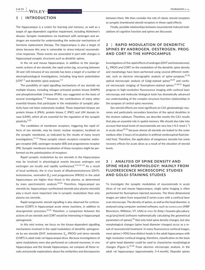
Investigations of the rapid effects of androgen (DHT and testosterone), E2, PROG and CORT on the modulation of the dendritic spine density and morphology have been performed using several different meth-ods, such as electron micrographic analysis of spine-synapses,27,28 optical microscopic analysis of Golgi-stained spines10,29 and confo-cal microscopic imaging of fluorophore-stained spines.15,30,31 Rapid progress inhigh-resolutionfluorescenceimagingwithconfocal lasermicroscopy and molecular biological tools has dramatically advanced ourunderstandingofthecomplexstructure-functionrelationshipsinthe synapses of central spiny neurones.
Sex steroid effects are most significant on CA1 glutamatergic neu-rones and particularly secondary branches of the apical dendrites in the stratum radiatum. Therefore, we describe mostly the CA1 results that play an essential role in spatial memory. We should also take into account that basal levels of neurosteroids are very low (<0.5 nmol L-1) in acute slices32,33 because almost all steroids are leaked to the outer medium after 2 hours of incubation in artificial cerebrospinal fluid (ste-roid free). Therefore, the application of exogenous steroids has some recovery effects for acute slices as a result of the elevation of steroid levels.
3 | ANALYSIS OF SPINE DENSITY AND SPINE HEAD MORPHOLOGY: MAINLY FROM FLUORESCENCE MICROSCOPIC STUDIES AND GOLGI- STAINING STUDIES
To investigate the synaptic modulation of neurosteroids in acute slices of rat and mouse hippocampus, single spine imaging is often performedforfluorophore-injectedneurones.Thethree-dimensionalimagesaretakenfromsequentialZ-seriesscanswithaconfocallaserscan microscope. The density of spines, as well as the head diameter, is analysedusingcomputer-assistedsoftware,suchasnuerolucida (MBF Bioscience, Williston, VT, USA) or spiso-3d(http://kawato-glia.sakura.ne.jp/proj.html) (software mathematically calculating the geometrical parameters of spines).34Notonlytotalspinedensitychanges,butalsomorphological changes (spine head diameter changes) occur as a re-sult of neurosteroid treatment. In many fluorescence confocal images, most spines (>90%) have distinct heads in the adult hippocampus with high-resolutionconfocalimaging;therefore,classificationonthebasisof spine head diameter could be used to characterise morphological changes (Figure 1).15,30 From electron microscopic analysis, in the adult rat hippocampus (approximately 3-4month), filopodia spines
| 3 of 13MURAKAMI et Al.
represent a very minor population (<1%)35 and the stubby spine popu-lation comprises no more than 10%. Although, historically, spines with distinct heads have often been classified into two categories, includ-ing thin spines (with head diameter <0.6 μm) and mushroom spines (with head diameter >0.6 μm), these two classes are not sufficient to distinguishcomplicatedneurosteroid-inducedchanges.Forexample,the effects of some kinase inhibitors cannot be clearly observed when only intermediate sized spines (intermediate size between thin andmushroom) are changed.
For statistical analysis of fluorescence confocal images, the clas-sification of the spines (which have clear heads) into three categories is usefulwith respect to their headdiameters: 0.2-0.4μmas small-head spines, 0.4-0.5μm as middle-head spines and 0.5-1.2μm as large-head spines.34 Small-, middle- and large-head spines may besignificantly different regarding the number of AMPA receptors and therefore these three types of spines may have different efficiencies with respect to signal transduction. The number of AMPA receptors (including GluR1 subunit) in the spine increases as the surface area of postsynapse increases,whereas the number ofNMDA receptors(includingNR2Bsubunit)mayberelativelyconstant.36
Here,notonlyfluorophore-stainedspines,butalsoGolgi-stainedspines are compared together in terms of the essential effects of neu-rosteorids. From Golgi-staining experiments of hippocampal slices,many stubby and filopodia spines (which do not have clear heads) were observed in addition to mushroom and thin spines (which have clear heads and necks) and research has mainly focused on changes in the density of mushroom and thin spines upon the application of neuros-teroids.37InsomecasesofGolgi-staining,stubbyandfilopodiaspinesoccupiedalmost40%ofthetotalspines,respectively.37 Currently, we cannot explain well why there are so many stubby and filopodia spines inGolgi-stainedspines,norwhythesespinescompriselessthan10%ofthepopulationinfluorophore-stainedspines.Onepossiblereason
mightbethelowresolutionoftheGolgi-stainedspines;therefore,thenecks of spines might not be visible, leading to more stubby spines being observed.
3.1 | Androgen (DHT and testosterone) effects
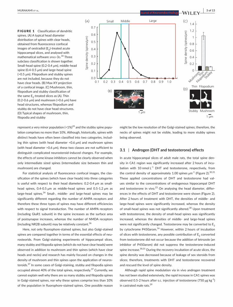
In acute hippocampal slices of adult male rats, the total spine den-sity in CA1 region was significantly increased after 2 hours of incu-bation with 10 nmol L-1 DHT and testosterone, respectively, from the control density of approximately 1.00 spines μm-1 (Figure 2).30,31 These applied concentrations of DHT and testosterone had val-ues similar to the concentrations of endogenous hippocampal DHT and testosterone in vivo.23 On analysing the head diameter, differ-ences in the effects of DHT and testosterone were shown (Figure 2). After 2hours of treatmentwithDHT, the densities ofmiddle- andlarge-head spineswere significantly increased,whereas the densityofsmall-headspineswasnotsignificantlyaltered.30 Upon treatment withtestosterone,thedensityofsmall-headspineswassignificantlyincreased, whereas the densities of middle- and large-head spineswere not significantly changed. Testosterone may be converted to E2 bycytochromeP450arom.16 However, within 2 hours of incubation of slices with testosterone, any possible contribution of E2 converted fromtestosteronedidnotoccurbecausetheadditionofletrozole(aninhibitor of P450arom) did not suppress the testosterone-inducedspine increase.30,31 During the recovery incubation of acute slices, the spine density was decreased because of leakage of sex steroids from slices; therefore, treatments with DHT and testosterone recovered and rescued the level of spine density.
Although rapid spine modulation via in vivo androgen treatment has not been studied extensively, the rapid increase in CA1 spines was observed0.5-2hoursafters.c.injectionoftestosterone(750μg kg-1) in castrated male rats.29
F IGURE 1 Classification of dendritic spines. (A) A typical head diameter distribution of spines with clear heads, obtained from fluorescence confocal images of oestradiol (E2)-treatedacutehippocampal slices, and analysed with mathematical software spiso-3d.34 Three subclass classification is shown together. Small-headspine(0.2-0.4μm),middle-headspine(0.4-0.5μm)andlarge-headspine(>0.5 μm). Filopodium and stubby spines are not included, because they do not haveclearheads.(B)Max-XYprojectionof a confocal image. (C) Mushroom, thin, filopodium and stubby classification of the same E2-treatedslicesas(A).Thin(0.2-0.6μm) and mushroom (>0.6 μm) have head structures, whereas filopodium and stubby do not have clear head structures. (D) Typical shapes of mushroom, thin, filopodia and stubby
(B)FilopodiumThin
MushroomStubby
1 µm
(D)
Small Middle Large
Spin
e de
nsity
(spi
nes/
µm)
Spin
e de
nsity
(spi
nes/
µm)
(C)(A)
Large Middle
Small
0.7
0.6
0.5
0.4
0.3
0.2
0.1
0.00.1 0.2 0.3 0.4 0.5 0.6 0.7 0.8 0.9 1.0
1 µm
0
Mus
hroo
m
Filop
odium
Stub
byTh
in
0.2
0.4
0.6
0.8
1
1.2
1.4
4 of 13 | MURAKAMI et Al.
Apart from rapid androgen effects, slow/genomic androgen effects invivo(withinseveraldays)onspine-synapsesobtainedfromelectronmicroscopic analyses have been studied extensively.38 Castration de-creasesspine-synapsesinmalerathippocampusasaresultofasignifi-cant decrease of DHT and testosterone in the hippocampus;23 therefore, thereplacementofDHTandtestosteronerecoveredthelevelofspine-synapse density of CA1 neurones after several days of treatment.27,39
3.2 | E2 effects
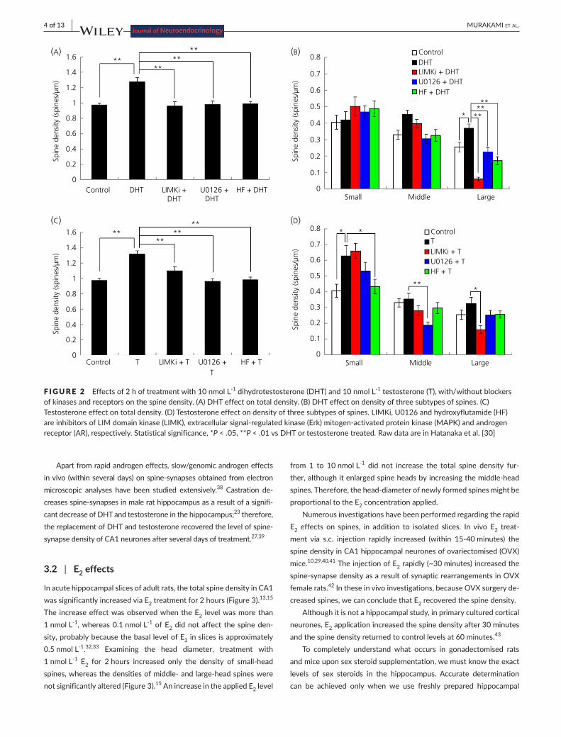
In acute hippocampal slices of adult rats, the total spine density in CA1 was significantly increased via E2 treatment for 2 hours (Figure 3).13,15 The increase effect was observed when the E2 level was more than 1 nmol L-1, whereas 0.1 nmol L-1 of E2 did not affect the spine den-sity, probably because the basal level of E2 in slices is approximately 0.5 nmol L-1.32,33 Examining the head diameter, treatment with 1 nmol L-1 E2 for 2hours increased only the density of small-headspines,whereasthedensitiesofmiddle-andlarge-headspineswerenot significantly altered (Figure 3).15 An increase in the applied E2 level
from 1 to 10 nmol L-1 did not increase the total spine density fur-ther,althoughitenlargedspineheadsbyincreasingthemiddle-headspines.Therefore,thehead-diameterofnewlyformedspinesmightbe proportional to the E2 concentration applied.
NumerousinvestigationshavebeenperformedregardingtherapidE2 effects on spines, in addition to isolated slices. In vivo E2 treat-ment via s.c. injection rapidly increased (within 15-40minutes) thespinedensityinCA1hippocampalneuronesofovariectomised(OVX)mice.10,29,40,41 The injection of E2 rapidly (~30 minutes) increased the spine-synapsedensityasaresultofsynapticrearrangementsinOVXfemale rats.42Intheseinvivoinvestigations,becauseOVXsurgeryde-creased spines, we can conclude that E2 recovered the spine density.
Although it is not a hippocampal study, in primary cultured cortical neurones, E2 application increased the spine density after 30 minutes and the spine density returned to control levels at 60 minutes.43
To completely understand what occurs in gonadectomised rats and mice upon sex steroid supplementation, we must know the exact levels of sex steroids in the hippocampus. Accurate determination can be achieved only when we use freshly prepared hippocampal
F IGURE 2 Effects of 2 h of treatment with 10 nmol L-1 dihydrotestosterone (DHT) and 10 nmol L-1 testosterone (T), with/without blockers of kinases and receptors on the spine density. (A) DHT effect on total density. (B) DHT effect on density of three subtypes of spines. (C) Testosterone effect on total density. (D) Testosterone effect on density of three subtypes of spines. LIMKi, U0126 and hydroxyflutamide (HF) areinhibitorsofLIMdomainkinase(LIMK),extracellularsignal-regulatedkinase(Erk)mitogen-activatedproteinkinase(MAPK)andandrogenreceptor (AR), respectively. Statistical significance, *P < .05, **P < .01 vs DHT or testosterone treated. Raw data are in Hatanaka et al. [30]
0
0.1
0.2
0.3
0.4
0.5
0.6
0.7
0.8
Small Middle Large
ControlTLIMKi + TU0126 + THF + T
0
0.2
0.4
0.6
0.8
1
1.2
1.4
1.6
Control T LIMKi + T U0126 + T
HF + T
0
0.1
0.2
0.3
0.4
0.5
0.6
0.7
0.8
Small Middle Large
ControlDHTLIMKi + DHTU0126 + DHTHF + DHT
0
0.2
0.4
0.6
0.8
1
1.2
1.4
1.6
Control DHT LIMKi +DHT
U0126 + DHT
HF + DHT
Spin
e de
nsity
(spi
nes/
µm)
Spin
e de
nsity
(spi
nes/
µm)
Spin
e de
nsity
(spi
nes/
µm)
Spin
e de
nsity
(spi
nes/
µm)
(A) (B)
(C) (D)
****
****
*
**
****
*
**
***
****
**
*
| 5 of 13MURAKAMI et Al.
homogenateswithanti-oxidationtreatmentsbecauseE2 is particularly unstableasaresultofoxidationofthe3-OHgroupofthephenolringduringextraction,high-performance liquidchromatographypurifica-tion and quantitative mass-spectrometric analysis.23 However, this still appears to present technical difficulties for many laboratories. Ovariectomy of female rats decreased the hippocampal E2 and tes-tosterone levels down to the level of oestrus stage (~0.5 nmol L-1);24 however, surprisingly, castration of male rats did not decrease the hippocampal E2 levels at all but, instead, decreased the hippocampal testosteronesignificantly (down to20-30%)anddecreased thehip-pocampalDHTleveltoalmostzero.23 Therefore, E2 supplementation inOVXfemaleanimalsshowedrecoveryeffectswithagoodrepro-ducibility; however, low-dose E2 supplementation in castrated male animals may not always show recovery effects. Indeed, E2 injection (approximately 30 μg kg-1 for 2 days) did not show any effect in cas-trated male rats, although DHT and testosterone injections (approxi-mately 1.5 mg kg-1) induced significant recovery effects.27,44 From our consideration,noeffectsasa resultof low-doseE2 injection should occur because of the barrier of such a high endogenous level of E2 inthemalehippocampus(2-10nmolL-1 with individual variation and
approximately 7 nmol L-1 on average) even at 2 weeks after castra-tion.23Therefore,theinjectionoflow-doseE2 could not always over-come the endogenous E2 level in the castrated male hippocampus. This high endogenous level of E2 may be a result of local E2 synthesis in the castrated rat hippocampus. Interestingly, however, a single s.c. injection of low dose E2 (approximately 20 μg kg-1) did demonstrate aspineincreaseincastratedmaleratsthatwerefedwithlowphyto-oestrogen chow, probably because of decrease in the endogenous E2 level.29Nevertheless,toobtainasignificantincreaseinspines,wemust inject a sufficiently high dose of E2 to castrated male animals (eg, 200 μg kg-1 E2).
3.3 | PROG effects
PROG is an essential female sex hormone regulating the oestrus cycle. Therefore, a possible role of PROG in rapid spine modulation was ex-amined. In acute hippocampal slices of adult male rats, treatment with 100 nmol L-1 PROG for 2 hours significantly increased the total spine density in CA1 (Figure 3).24 The applied high concentration of PROG (100 nmol L-1) was chosen based on the endogenous concentration.
F IGURE 3 Effect of 2 h of treatment with 1 nmol L-1 oestradiol (E2), 100 nmol L-1 progesterone (PROG) with/without blockers of kinases and receptors on the spine density. (A) E2 effect on total density. (B) E2 effect on density of three subtypes of spines. (C) PROG effect on total density.(D)PROGeffectondensityofthreesubtypesofspines.ICI182,780(ICI)andRU486areinhibitorsofoestrogenreceptor(ER)andprogesterone receptor (PR), respectively. Statistical significance, *P < .05, **P < .01 vs E2 treated. Some of the raw data are in Hasegawa et al. [15]andKatoetal.[45]
0
0.2
0.4
0.6
0.8
1
1.2
1.4
1.6
Control PROG LIMKi + PROG
U0126 + PROG
RU486 +PROG
Spin
e de
nsity
(spi
nes/
µm)
0
0.1
0.2
0.3
0.4
0.5
0.6
0.7
0.8
0.9
Small Middle Large
ControlE2LIMKi + E2U0126 + E2ICI + E2
Spin
e de
nsity
(spi
nes/
µm)
0
0.2
0.4
0.6
0.8
1
1.2
1.4
1.6
Control E2 LIMKi +
E2
U0126 +
E2
ICI +
E2
Spin
e de
nsity
(spi
nes/
µm)
(A)(B)
(C)
****
****
***
*
*
**
****
****
Spin
e de
nsity
(spi
nes/
µm)
(D)
0
0.1
0.2
0.3
0.4
0.5
0.6
0.7
0.8
0.9
Small Medium Large
ControlPROGLIMKi + PROGU0126 + PROGRU486 + PROG
****
*
* **
6 of 13 | MURAKAMI et Al.
From head diameter analysis, treatment with 100 nmol L-1 PROG increased the density of small-head andmiddle-head spines.45 It is known that applied PROG might be metabolised to allopregnanolone, which could modulate GABAA receptor, leading to a spine increase.46 Therefore, RU486, a PR antagonist,was applied to investigate thispossibility.BecauseRU486completelysuppressedthespineincrease,the possibility of allopregnanolone action is excluded in acute slices undergoing 2 hours of incubation of PROG (Figure 3B).
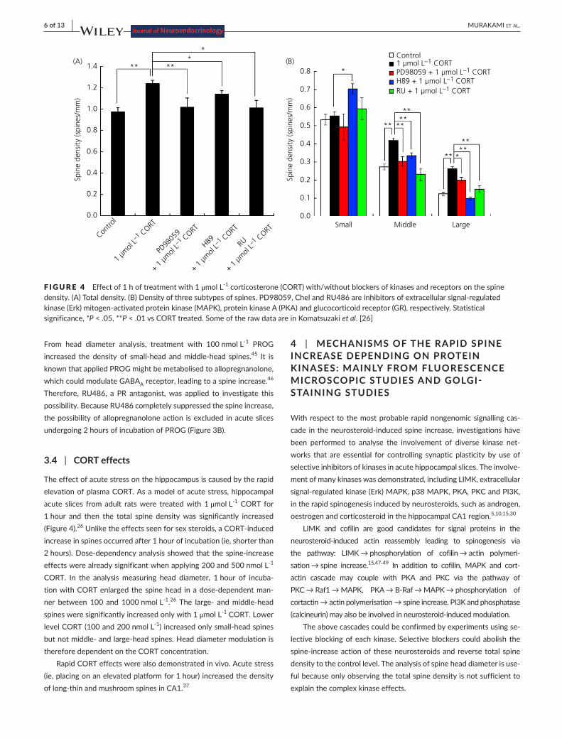
3.4 | CORT effects
The effect of acute stress on the hippocampus is caused by the rapid elevation of plasma CORT. As a model of acute stress, hippocampal acute slices from adult rats were treated with 1 μmol L-1 CORT for 1 hour and then the total spine density was significantly increased (Figure4).26Unliketheeffectsseenforsexsteroids,aCORT-inducedincrease in spines occurred after 1 hour of incubation (ie, shorter than 2hours).Dose-dependencyanalysisshowedthatthespine-increaseeffects were already significant when applying 200 and 500 nmol L-1 CORT. In the analysis measuring head diameter, 1 hour of incuba-tionwithCORTenlargedthespinehead inadose-dependentman-ner between 100 and 1000 nmol L-1.26 The large- andmiddle-headspines were significantly increased only with 1 μmol L-1 CORT. Lower level CORT (100 and 200 nmol L-1)increasedonlysmall-headspinesbutnotmiddle-andlarge-headspines.Headdiametermodulationistherefore dependent on the CORT concentration.
Rapid CORT effects were also demonstrated in vivo. Acute stress (ie, placing on an elevated platform for 1 hour) increased the density oflong-thinandmushroomspinesinCA1.37
4 | MECHANISMS OF THE RAPID SPINE INCREASE DEPENDING ON PROTEIN KINASES: MAINLY FROM FLUORESCENCE MICROSCOPIC STUDIES AND GOLGI- STAINING STUDIES
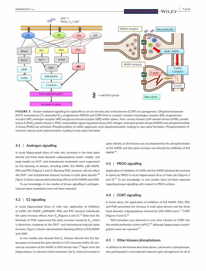
With respect to the most probable rapid nongenomic signalling cas-cade intheneurosteroid-inducedspine increase, investigationshavebeen performed to analyse the involvement of diverse kinase net-works that are essential for controlling synaptic plasticity by use of selective inhibitors of kinases in acute hippocampal slices. The involve-ment of many kinases was demonstrated, including LIMK, extracellular signal-regulatedkinase(Erk)MAPK,p38MAPK,PKA,PKCandPI3K,in the rapid spinogenesis induced by neurosteroids, such as androgen, oestrogen and corticosteroid in the hippocampal CA1 region.5,10,15,30
LIMK and cofilin are good candidates for signal proteins in the neurosteroid-induced actin reassembly leading to spinogenesis viathe pathway: LIMK→phosphorylation of cofilin→actin polymeri-sation→spine increase.15,47-49 In addition to cofilin, MAPK and cort-actin cascade may couple with PKA and PKC via the pathway of PKC→Raf1→MAPK, PKA→B-Raf→MAPK→phosphorylation ofcortactin→actinpolymerisation→spineincrease.PI3Kandphosphatase(calcineurin)mayalsobeinvolvedinneurosteroid-inducedmodulation.
The above cascades could be confirmed by experiments using se-lective blocking of each kinase. Selective blockers could abolish the spine-increaseactionof theseneurosteroidsandreversetotalspinedensity to the control level. The analysis of spine head diameter is use-ful because only observing the total spine density is not sufficient to explain the complex kinase effects.
F IGURE 4 Effect of 1 h of treatment with 1 μmol L-1 corticosterone (CORT) with/without blockers of kinases and receptors on the spine density.(A)Totaldensity.(B)Densityofthreesubtypesofspines.PD98059,ChelandRU486areinhibitorsofextracellularsignal-regulatedkinase(Erk)mitogen-activatedproteinkinase(MAPK),proteinkinaseA(PKA)andglucocorticoidreceptor(GR),respectively.Statisticalsignificance, *P < .05, **P<.01vsCORTtreated.SomeoftherawdataareinKomatsuzakiet al. [26]
0.0
0.1
0.2
0.3
0.4
0.5
0.6
0.7
0.8
Small Middle Large
Control1 µmol L–1 CORTPD98059 + 1 µmol L–1 CORTH89 + 1 µmol L–1 CORTRU + 1 µmol L–1 CORT
*
**
****
**
**
** *
**
Spin
e de
nsity
(spi
nes/
mm
)
0.0
0.2
0.4
0.6
0.8
1.0
1.2
1.4
Contro
l
1 µm
ol L–1 C
ORT
PD98
059
+ 1 µ
mol
L–1 C
ORT
H89
+ 1 µ
mol
L–1 C
ORT
RU
+ 1 µ
mol
L–1 C
ORT
Spin
e de
nsity
(spi
nes/
mm
)**
*
***(A) (B)
| 7 of 13MURAKAMI et Al.
4.1 | Androgen signalling
In acute hippocampal slices of male rats, increases in the total spine densityandthreeheaddiametersubpopulations (small-,middle-andlarge-heads)viaDHT-andtestosterone-treatmentsweresuppressedby the blocking of kinases, including LIMK, Erk MAPK, p38 MAPK, PKA and PKC (Figures 2 and 5). Blocking PI3K, however, did not affect theDHT-andtestosterone-inducedincreaseintotalspinedensity.30 Figure 2 shows representative blocking effects of Erk MAPK and LIMK.
Toourknowledge,invivostudiesofkinasesignallinginandrogen-induced spine modulation have not been reported.
4.2 | E2 signalling
In acute hippocampal slices of male rats, application of inhibitors of LIMK, Erk MAPK, p38MAPK, PKA and PKC blocked individually the spine increase effects from E2 (Figures 3 and 5).15Notethattheblockade of PI3K suppressed the spine increase caused by E2, which isdistinctivecomparedtotheDHT-andtestosterone-inducedspineincrease. Figure 3 shows representative blocking effects of Erk MAPK and LIMK.
In vivo studies also showed that E2 infusion directly into the hip-pocampus increased the spine density in CA1 neurones within 30 min-utesbyactivationofErkMAPKinOVXfemalemice.10 Apart from the hippocampus, in cultured cortical neurones, the E2-inducedincreasein
spine density at 30 minutes was accompanied by the phosphorylation of Erk MAPK and this spine increase was blocked by inhibition of Erk MAPK.43
4.3 | PROG signalling
Application of inhibitors of LIMK and Erk MAPK blocked the increase in spines by PROG in acute hippocampal slices of male rats (Figures 3 and 5).45 To our knowledge, in vivo studies have not been reported regarding kinase signalling with respect to PROG actions.
4.4 | CORT signalling
In acute slices, the application of inhibitors of Erk MAPK, PKA, PKC and PI3K prevented the increase in total spine density and the three headdiametersubpopulations inducedby200-1000nmolL-1 CORT (Figures4and5).26
PKA activation was observed in vivo after infusion of CORT into the medial prefrontal cortex (mPFC),50 although hippocampus investi-gations were not reported.25
4.5 | Other kinases/phosphatases
In addition to the kinases described above, calcineurin, a phosphatase, alsoparticipatedinneurosteroid-inducedrapidspinogenesisforallof
F IGURE 5 Kinase-mediatedsignallingforrapideffectsofsexsteroidsandcorticosterone(CORT)onspinogenesis.Dihydrotestosterone(DHT), testosterone (T), oestradiol (E2), progesterone (PROG) and CORT bind to synaptic receptors [oestrogen receptor (ER), progesterone receptor (PR), androgen receptor (AR) and glucocorticoid receptor (GR)] within spines. Then, various kinases [LIM domain kinase (LIMK), protein kinaseA(PKA),proteinkinaseC(PKC),extracellularsignal-regulatedkinase(Erk)mitogen-activatedproteinkinase(MAPK)andphosphoinositide3-kinase(PI3K)]areactivated.Phosphorylationofcofilinsuppressesactindepolymerisation,leadingtonewspineformation.Phosphorylationofcortactin induces actin polymerisation, leading to new spine formation
DHT, TPROG, E2 CORT
NMDA receptor Neurosteroids
AR
PR ER
GR
AR
RhoA
ROCK
LIMK
Cofilin ERK/MAPK
LIMK, MAPK
AMPA receptor
F-actin
CofilinCortactin
G-actin
New spine
Cortactin
Actin polymerisation
Increase in spine
MEK
Raf-1 B-Raf
PKC PKA
Akt
= palmitoyl group
PI3K
PR GRERα
8 of 13 | MURAKAMI et Al.
the sex steroids and CORT examined. In all the cases examined above, however, blocking Jun kinase (JNK) showed no suppression of theneurosteroid-inducedincreaseinspinedensity.15,26,30
It should be noted that these kinase inhibitors alone did not sig-nificantly affect total spine density, indicating that the observed inhib-itory effects are not the result of simple nonspecific blockade.
5 | SYNAPTIC (MEMBRANE) RECEPTORS FOR NEUROSTEROID ACTION
The synaptic receptor of neurosteroids must evidently be identified to explain the mechanism of rapid (1-2hours) spinogenesis that is induced by neurosteroids in the hippocampus.
5.1 | ARs
The involvement of classic AR in rapid action was observed by sup-pressingandrogen-inducedspinogenesisusinghydroxyflutamide(HF),a specific inhibitor of AR in acute slices of rat hippocampus (Figures 2 and 5).30 AR immunoreactivity was localised in CA1 neurones with op-tical microscopic analysis and the extranuclear localisation of AR (in dendrites and spines) was also observed with electron microscopic anal-ysis.51 The subcellular distribution of AR was also examined by western immunoblotanalysisusingPG-21,ananti-ARantibody.TheARproteinband was observed in the postsynaptic density (PSD) fraction, as well as in cytoplasmic and nuclear fractions, which indicates that AR localised in thePSDcouldparticipateinandrogen-inducedspinogenesis.30
InhibitionofNMDAreceptorsbyMK-801significantlysuppressedtheDHT-induced increase in the spine density, suggesting thatARpossiblylocalisesclosetoNMDAreceptors(ie,withinspines).30
In vivo studies have shown that intrahippocampal administration of anAR antagonist, flutamide, can increase anxiety-like behaviourinintactmaleratsandDHT-replacedcastratedmalerats.52 These re-sults suggest AR involvement in anxiety, which may influence learn-ingbehaviour.Notethatcastrationincreasedanxiety-likebehaviour,whereasDHT-replacementreversedit.
On theotherhand, fromspineanalyses, apossibilityofnon-ARmembrane receptors has been proposed and investigated because AR antagonistHFdidnot suppress theDHT-induced increase in spine-synapses with electron microscopic analysis.38,53 However, the puta-tivenon-ARmembranereceptorhasnotyetbeenpurifiedorcloned,preventing further definitive characterisation.54
DHT may be converted to androstanediol by 3α-hydroxysteroid-dehydrogenase and might modulate GABAA receptors, leading to synaptic changes.55 This possibility in acute hippocampal slices within 2 hours of incubation was excluded because HF, an AR antagonist, completelysuppressedtheDHT-inducedspineincrease(Figure2).
5.2 | E2 receptors
Judging from many investigations, the most probable candidates for synaptic (membrane) oestrogen receptors are classic nuclear type
receptors (ERα, ERβ). The blocking of classic ER by ICI182,780 (ICI), a specific antagonist of ERα and ERβ, completely abrogated the en-hancing effect of E2 on the spine density in acute hippocampal slices, suggesting that rapid effect of E2 on spinogenesis is mediated by ER (Figures 3 and 5).15 The involvement of ERα and ERβ in rapid spino-genesis was further demonstrated by introducing oestrogen recep-tor agonists. ERα agonist, (propyl-pyrazole-trinyl) tris-phenol (PPT),unlike ERβ agonists such as (4-hydroxyphenyl)-propionitrile (DPN),rapidly increased the spine density in hippocampal neurones in CA1 in male rats 15andOVXfemalemice.40 These results support the ex-clusive involvement of ERα in rapid signalling. An ER knockout (KO) mice study further confirmed the involvement of ERα in rapid signal-ling.56 Treament for 2 hours with E2 preferably increased the density ofmiddle-head spines inhippocampal slicesofwild-typemice.Theoestradiol-induced increase inmiddle-head spineswas observed inERβKO mice (which express ERα) but not in ERαKO mice. These results indicate that ERα is necessary for oestrogen-induced spinogenesis,whereas ERβisnotessentialforthisoestrogen-inducedspinogenesis.
On the other hand, there are contrasting reports that indicate ERβ participation.TherapidadministrationofDPNbutnotPPTenhancedobjectandplacerecognitionmemoryinOVXfemalerats.4 Treatment withWAY-200070,anERβ agonist, increased spine density and spine size, as well as PSD-95 accumulation in membrane regions, within30 minutes in cultured cortical neurones.57 Regarding slow genomic ef-fects,treatmentwithWAY-200070for2daysimprovedperformanceinahippocampus-dependentradialarmmazetask inOVXrats.58 An increase in the mushroom type of spines in the dentate gyrus but not inCA1wasobservedintheseOVXrats.Currently,however,wecannotexplain these discrepancies between ERα signalling and ERβ signalling.
The expression of ERα in glutamatergic neurones in rat and mouse hippocampus is clearly demonstrated by immunostaining with purified antibodyRC-19.ERα is localised not only in nuclei/cytoplasm, but also inpre-andpostsynapses,asindicatedbyanimmunogoldelectronmi-croscopic analysis.13 Expression of ERβinpre-andpostsynapseswasalso observed by immunogold electron microscopic analysis.59 An as-sociation of ERα with PSD was observed by western blotting of PSD fractions, implying the synaptic membrane binding of ERα.
In cultured cells of peripheral origin, some populations of ERα and ERβ areplasmamembrane-boundand theyareanchoredviapalmi-toylation.11 Therefore, membrane binding of ERα and ERβ might also occur in neurones.13,14,17,59
E2-inducedincreasesinspinedensityareblockedbyNMDArecep-tor antagonists,13,60,61 suggesting that ER is possibly localised close to NMDAreceptors(ie,withinspines).
Interestingly, in cultured female hippocampal neurones, ERα and ERβ were translocated to the membrane via complex formation with metabotropic glutamate receptors (mGluR).62 Palmitoylation of ERα and ERβ also played a role in the translocation of ERα and ERβ to the membrane of the female hippocampus. These ER/mGluR complexes phosphorylate cAMP responseelement-bindingproteinvery rapidly(approximately 1 minute) upon E2 binding.62
Another candidate for synaptic oestrogen receptor is G protein coupled receptor (GPR) localised in the cellular membrane. GPR30/
| 9 of 13MURAKAMI et Al.
GPER was expressed in the membrane of endoplasmic reticulum but not in the plasma membrane.63 GPR30/GPER may not participate in E2-induced spinemodulation becauseof the lowbinding affinity ofGPR30 with E2 and no rapid E2 signallingwas seen inSKBR-3cells(ER negative, GPR30/GPER positive).64-66 By contrast to ERα and ERβ, GPR30/GPERagonistG-1didnotactivateErkMAPKbutdidactivateJNK;italsoenhancedobjectrecognitionandspatialmemoryperfor-mance.66 Therefore, GPR30/GPER signalling appears to occur very dif-ferently compared to ERα-inducedandERβ-inducedsignalling.
5.3 | PROG receptors
BlockingofPRbyPRinhibitor(RU486)completelyabolishedtheen-hancing effect from 100 nmol L-1 PROG on the total spine density (Figures 3 and 5).45 Therefore, classic PR is involved in PR-inducedspinogenesis. The spine localisation of PR was observed via immuno-electron microscopic analysis.67 AlthoughRU486 is an inhibitor forboth PROG and CORT, because basal levels of CORT and PROG were below 0.5 nmol L-1inacuteslices,RU486onlysuppressesexogenousPROG binding to PR in these experiments.
Novelmembrane-associatedprogesteronereceptors(mPR)havealso been investigated. Although immunostaining of mPR and its mo-lecularbiologystudiesshowedtheexpressionofmPR,thePROG-dependent signalling of mPR has not been clearly demonstrated.68
5.4 | CORT receptors
Blocking of glucocorticoid receptor (GR) by GR inhibitor (RU486)completely abolished the enhancing effect from 1 μmol L-1 CORT on the total spine density (Figures4 and 5).26 Therefore, classic GR is involved in theCORT-induced spinogenesis.Dexamethasone, aGRagonist, rapidly increased spines within 1 hour, further supporting the involvementofGRinCORT-inducedspinogenesis.69 GR localisation within spines was found by electron microscopic analysis.2,70 Although RU486isaninhibitorforbothCORTandPROG,becausebasallevelsof CORT and PROG were below 0.5 nmol L-1inacuteslices,RU486onlysuppressesexogenousCORTbindingtoGRintheseCORT-effectexperiments.BlockadeofNMDAreceptorsleadstothesuppressionoftheCORT-inducedincreaseinspinedensity,suggestingthatGRispossiblylocalisedclosetoNMDAreceptors(ie,withinspines).
Evidence for involvement of membrane GR in rapid nongenomic signalling in neurones has accumulated,25,71 although the mechanisms of translocation of GR to the membrane are not clear. By contrast to AR, ERα/ERβ and PR, GR may not be palmitoylated.72
6 | RELATIONSHIPS BETWEEN NEUROSTEROID- INDUCED MODULATIONS OF COGNITION AND SPINES
Rapid modulation of spines by sex steroids has a close relationship with rapid changes in learning and memory. Recently, many observa-tions support rapid E2effectswithrespecttothecognitionofOVX
rats.9,73 These review papers showed that E2 rapidly enhanced mem-ory consolidation within approximately 1 hour. Subcutaneous infusion of E2for40minutesimprovedlearningandmemoryprocesses,suchasobjectrecognitionandobjectreplacement,inOVXfemalemice.41 The same study group reported that the injection of a selective agonist for ERα (PPT) but not for ERβ (DPN) rapidly (approximately1hour)increaseddendriticspinesandimprovedlearninginOVXfe-male mice.40 Subcutaneous injection of E2rapidly(30-60minutes)in-duced not only hippocampus-dependentmemory performance, butalsospineincreasesinthehippocampusinOVXfemalerats.29InOVXfemale rats, relatively rapid E2effects(approximately4hoursafters.c.injection) were observed on object recognition and object placement tests.74 The spine-synapse increase was observed much earlier at30 minutes after s.c. injection of E2.42 Recognition memory was rap-idly increased by s.c. treatment of E2 30 minutes before the initiation of the behavioural analysis, which is sufficient to increase basal spine densityinOVXrats.75
Why is it thatOVX rats been used in these studies? It is likelythat,asaresultofOVXtreatments,thehippocampal levelofE2 de-creases,24 leading to a decrease in both cognitive performance and spine density; accordingly, E2 infusion can recover these decreases. On the other hand, infusion of E2 into gonadally intact females may not induce a further increase in the spine density or an improvement in cognition. By contrast, hippocampal infusion of ERα/ERβ blockers might effectively impair cognition and decrease in spines.
In addition, rapid actions of E2 are also involved in reproductive behaviours, such as maternal behaviours or lordosis, which are under thecontrolofhypothalamus,inOVXmiceandrats.76,77
Concerning PROG effects on cognition, intrahippocampal infusion ofPROG inOVXfemalemiceenhancedobject recognitionmemoryconsolidation and very rapid MAPK phosphorylation (approximately 5 minutes) was involved in this enhancement process.78
Androgen may also show rapid improving effects on cognition in castrated male rats and mice; however, the experimental reports are poor compared to the effects of E2.
The rapid enhancement of spatial memory was observed at 0.5-2hours after testtosterone injection (750μg kg-1) in castrated male rats.29 Rapid androgen effects (1-2hours) on anti-anxiety be-haviour in vivo as a result of hippocampal infusion of DHT have been observed in castrated male rats.52 Intrahippocampal administration of anARantagonistcanincreaseanxiety-likebehaviourinDHT-replacedcastrated male rats and gonadally intact male rats.52 Subcutaneous tes-tosteroneinjectionnotonlyincreasedanti-anxietybehaviour,butalsoenhanced cognitive performance in castrated mice.79 It is likely that castration increasedanxiety-likebehaviourasa resultofadecreasein hippocampal DHT and testosterone;23therefore,DHT-replacementcould reverse this. By contrast to rapid effects, slow genomic effects of testosterone and DHT on cognition have been studied extensively. In vivo treatments with DHT capsules for 5 months showed an im-provement of spatial memory performance in castrated male mice.80 Testosterone injection improved spatial memory in castrated male rats, when testosterone injection was given every day from 7 days priortowatermazetest.81
10 of 13 | MURAKAMI et Al.
By contrast to sex steroids, acute stress caused the impairment of cognition. Acute stress (placing on an elevated platform for 1 hour) impairedspatialmemoryretrievalforshort-termmemoryontheob-jectplacementtask(hippocampus-dependent),associatedwithCORTelevation in plasma.37 This effect of acute stress on hippocampus-dependent memory task was associated with an increase in the den-sityoflong-thinandmushroomspinesinCA1.37 Infusion of CORT into mPFC at 60 minutes before the test impaired working memory in a PKA-dependentmanner.50
7 | CONTRIBUTION OF LOCAL HIPPOCAMPAL SYNTHESIS OF NEUROSTEROIDS
Adult rat and mouse hippocampi locally synthesise sex steroids.16,20 TheexpressionofmRNAsandproteinsofsteroidogenicenzymes,in-cludingcytochromesP450(17α),P450aromand17β-hydroxysteroiddehydrogenase, was demonstrated.16,19,23,82,83 Immunohistochemical analysis combined with immunogold electron microscopic analysis showedthattheseenzymesareexpressedinglutamatergicneuronesinCA1,CA3andthedentategyrus,andwerepartlylocalisedinpre-and postsynapses. The production of DHT, testosterone, E2, PROG and CORT was demonstrated by metabolism analysis of radioac-tive substrates. These results show that hippocampal neurones are equippedwithafullsetofenzymestoperformthesynthesisofneu-rosteroids from cholesterol.
The rapid modulations of spines, as discussed in the current re-view, may match that of locally synthesised steroids. For example, hippocampal neurosteroid synthesis (including pregnenolone and E2 synthesis)was a neural activity-dependent rapid process (within30minutes)that istriggeredbyCa influxviaNMDAreceptors.3,16,19 On the other hand, the change in the level of circulating sex hormones is probably very slow, mainly depending on the circadian rhythm or oestrus cycle. It should be noted that the rapid manipulation of plasma sex steroid levels, with injection and infusion after castration and ova-riectomy, can rapidly change hippocampal sex steroid levels because considerable amounts of sex steroids penetrate into the hippocampus bycrossing theblood-brainbarrier.23,24 Therefore, sex steroid injec-tion/replacement after castration and ovariectomy has been a useful method for in vivo investigations of the rapid modulation of spines and cognition by sex steroids.
As a result of local neurosteroid synthesis, the levels of sex ste-roids are higher in the hippocampus than in the plasma.23 For exam-ple, in the male rat, sex steroid levels are approximately 7 nmol L-1 (E2), 18 nmol L-1 (testosterone) and 6 nmol L-1 (DHT) in the hippo-campus and approximately 0.01 nmol L-1 (E2), 14nmolL
-1 (testos-terone) and 0.6 nmol L-1 (DHT). The female rat sex steroid levels are 0.5-4nmolL-1 (E2)and1-2nmolL
-1 (testosterone) in the hippocam-pusand0.01-0.1nmolL-1 (E2)and0.02-0.1nmolL
-1 (testosterone) in the plasma.24 These variations in female are dependent on oestrus stages. Interestingly,OVXreducedthehippocampalE2 level down to almost the same level as that in the dioestrus stage, although
thiswasnotreducedtozerolevel,implyingthatE2 is synthesised in the female hippocampus.24 Therefore, direct determination of local steroid levels in hippocampal slices is very important because the spine density rapidly changes, dependent on the rapid change in the local sex steroid levels.
Because the male/female hippocampus in vivo contains high lev-els of local E2 and androgens,23 the hippocampus infusion of inhibitors against steroid synthase (eg, letrozole or finasteride) in rats invivomay be very effective with respect to the rapid alteration of spines, which could provide useful evidence regarding the hippocampal synthesis of sex steroids.84 Slow hippocampal synthesis of sex ste-roidshasbeenobservedafter repetitive i.p. injectionof letrozole,aP450arominhibitor,for1-7daysinthefemalehippocampusfromLTPmeasurements.85
On the other hand, in isolated acute slices, all of the steroids are depleted (resulting in <0.5 nmol L-1) after 2 hours of recovery incuba-tionofslicesthatwerepreparedbydissectionandvibratome-slicingofthe hippocampus.2,32 As a result, exogenously applied DHT, testoster-one, E2 and PROG concentrations (>1 nmol L-1) were above the local steroid levels (<0.5 nmol L-1), thereby demonstrating the significant effects on spinogenesis and LTP.
8 | SUMMARY AND FUTURE DIRECTIONS
To our surprise, although the physiological functions of steroids are very diverse (ranging from sex hormones to stress hormones), the nongenomic signal pathways show a large overlap. Kinase-drivensignalling is used for nongenomic action of sex hormones and stress hormones. Spine-localisedclassic steroid receptors (AR,ER,PRandGR)triggerkinase-drivenrapidsignalling,leadingtospinemodulation.These receptors may anchor to the synaptic membranes via palmi-toylation because these steroid receptors have a conserved sequence for palmitoylation.11,86 The palmitoylation of essential many synaptic proteins, includingPSD-95,isshowntobeanimportantmechanismfor spine formation in the hippocampus.87,88 However, the membrane localisation of GR does not appear to be mediated via palmitoylation and therefore further clarification is necessary.72
Because sex steroids and stress steroids are observed to drive essential kinases that regulate synaptic plasticity, neurosteroids could play important roles in memory performance. Although rapid E2 effects (in vitro and in vivo) have been extensively studied and well clarified, the rapid effects of androgens and stress steroids need much more investigation to enable a deeper understanding of the common or differing molecular mechanisms of these neuroste-roidal actions. As one additional example of the CORT effect, circa-dian cycle-dependent oscillation of CORT (3-30nmolL-1) induced the cyclic rise and fall of spine density and these changes were a resultofkinase-dependentsignalling, includingMAPK,LIMK,PKAand PKC.48
The interactions between rapid/nongenomic signalling and slow/genomic signalling via classic steroid receptors comprise another inter-esting topic for investigation.12
| 11 of 13MURAKAMI et Al.
ACKNOWLEDGEMENTS
Mr Taishi Takeda (University of Tokyo) is acknowledged for carefully reading the manuscript submitted for publication.
CONFLICT OF INTEREST
The authors declare that they have no conflicts of interest.
ORCID
S. Kawato http://orcid.org/0000-0001-7024-2660
REFERENCES
1. FoyMR,XuJ,XieX,BrintonRD,ThompsonRF,BergerTW.17beta-estradiolenhancesNMDAreceptor-mediatedEPSPsand long-termpotentiation. J Neurophysiol.1999;81:925-929.
2. OoishiY,MukaiH,HojoY, et al. Estradiol rapidly rescues synaptictransmissionfromcorticosterone-inducedsuppressionviasynaptic/extranuclear steroid receptors in the hippocampus. Cereb Cortex. 2012;22:926-936.
3. Grassi S,TozziA,CostaC, et al.Neural 17beta-estradiol facilitateslong-termpotentiationinthehippocampalCA1region.Neuroscience. 2011;192:67-73.
4. Luine VN, Frankfurt M. Estrogens facilitate memory processingthrough membrane mediated mechanisms and alterations in spine density. Front Neuroendocrinol.2012;33:388-402.
5. LuineVN.Estradiolandcognitivefunction:past,presentandfuture.Horm Behav.2014;66:602-618.
6. Bi R, Foy MR, Vouimba RM, Thompson RF, Baudry M. Cyclic changes in estradiol regulate synaptic plasticity through the MAP kinase path-way. Proc Natl Acad Sci USA.2001;98:13391-13395.
7. Mannella P, Brinton RD. Estrogen receptor protein interaction with phosphatidylinositol3-kinase leads toactivationofphosphorylatedAktandextracellularsignal-regulatedkinase1/2inthesamepopu-lation of cortical neurons: a unified mechanism of estrogen action. J Neurosci.2006;26:9439-9447.
8. Znamensky V, Akama KT, McEwen BS, Milner TA. Estrogen levels reg-ulate the subcellular distribution of phosphorylated Akt in hippocam-pal CA1 dendrites. J Neurosci.2003;23:2340-2347.
9. FrickKM.Molecularmechanismsunderlyingthememory-enhancingeffects of estradiol. Horm Behav.2015;74:4-18.
10. TuscherJJ,LuineV,FrankfurtM,FrickKM.Estradiol-mediatedspinechanges in the dorsal hippocampus and medial prefrontal cortex of ovariectomizedfemalemicedependonERKandmTORactivationinthe dorsal hippocampus. J Neurosci.2016;36:1483-1489.
11. PedramA, RazandiM, Sainson RC, Kim JK, Hughes CC, Levin ER.A conserved mechanism for steroid receptor translocation to the plasma membrane. J Biol Chem.2007;282:22278-22288.
12. Levin ER, Hammes SR. Nuclear receptors outside the nucleus: ex-tranuclear signalling by steroid receptors. Nat Rev Mol Cell Biol. 2016;17:783-797.
13. MukaiH,TsurugizawaT,MurakamiG,etal.Rapidmodulationoflong-term depression and spinogenesis via synaptic estrogen receptors in hippocampal principal neurons. J Neurochem.2007;100:950-967.
14. Mukai H, Kimoto T, Hojo Y, et al. Modulation of synaptic plas-ticity by brain estrogen in the hippocampus. Biochim Biophys Acta. 2010;1800:1030-1044.
15. HasegawaY,HojoY,KojimaH,etal.Estradiolrapidlymodulatessyn-aptic plasticity of hippocampal neurons: involvement of kinase net-works. Brain Res.2015;1621:147-161.
16. HojoY,HattoriTA,EnamiT,etal.Adultmalerathippocampussyn-thesizesestradiolfrompregnenolonebycytochromesP45017alphaand P450 aromatase localized in neurons. Proc Natl Acad Sci USA. 2004;101:865-870.
17. Hojo Y, Murakami G, Mukai H, et al. Estrogen synthesis in thebrain–role in synaptic plasticity and memory. Mol Cell Endocrinol. 2008;290:31-43.
18. KawatoS,HojoY,KimotoT.HistologicalandmetabolismanalysisofP450expressioninthebrain.Methods Enzymol.2002;357:241-249.
19. Kimoto T, Tsurugizawa T, Ohta Y, et al. Neurosteroid synthesis bycytochromep450-containingsystems localized in the ratbrainhip-pocampal neurons: N-methyl-D-aspartate and calcium-dependentsynthesis. Endocrinology.2001;142:3578-3589.
20. KretzO,FesterL,WehrenbergU,etal.Hippocampalsynapsesdependon hippocampal estrogen synthesis. J Neurosci.2004;24:5913-5921.
21. OkamotoM,HojoY,InoueK,etal.Mildexerciseincreasesdihydrotes-tosterone in hippocampus providing evidence for androgenic media-tion of neurogenesis. Proc Natl Acad Sci USA.2012;109:13100-13105.
22. Di Mauro M, Tozzi A, Calabresi P, Pettorossi VE, Grassi S. Neo-synthesis of estrogenic or androgenic neurosteroids determine whether long-term potentiation or depression is induced in hippo-campus of male rat. Front Cell Neurosci. 2015;9:376.
23. Hojo Y, Higo S, Ishii H, et al. Comparison between hippocampus-synthesizedandcirculation-derivedsexsteroidsinthehippocampus.Endocrinology.2009;150:5106-5112.
24. KatoA,HojoY,HigoS,etal.Femalehippocampalestrogenshaveasignificant correlation with cyclic fluctuation of hippocampal spines. Front Neural Circuits.2013;7:149.
25. Groeneweg FL, Karst H, de Kloet ER, Joels M. Mineralocorticoid and glucocorticoid receptors at the neuronal membrane, regula-tors of nongenomic corticosteroid signalling. Mol Cell Endocrinol. 2012;350:299-309.
26. Komatsuzaki Y, HatanakaY, Murakami G, et al. Corticosterone in-duces rapid spinogenesis via synaptic glucocorticoid receptors and kinase networks in hippocampus. PLoS ONE.2012;7:e34124.
27. Leranth C, Petnehazy O, MacLusky NJ. Gonadal hormones affectspine synaptic density in the CA1 hippocampal subfield of male rats. J Neurosci.2003;23:1588-1592.
28. Prange-KielJ,RuneGM.Directandindirecteffectsofestrogenonrathippocampus. Neuroscience.2006;138:765-772.
29. Jacome LF, Barateli K, Buitrago D, Lema F, Frankfurt M, Luine VN. Gonadal hormones rapidly enhance spatial memory and in-crease hippocampal spine density in male rats. Endocrinology. 2016;157:1357-1362.
30. HatanakaY,HojoY,MukaiH,etal.Rapid increaseof spinesbydi-hydrotestosterone and testosterone in hippocampal neurons: depen-dence on synaptic androgen receptor and kinase networks. Brain Res. 2015;1621:121-132.
31. HatanakaY,MukaiH,MitsuhashiK,etal.Androgenrapidlyincreasesdendritic thorns of CA3 neurons in male rat hippocampus. Biochem Biophys Res Commun.2009;381:728-732.
32. HojoY,HigoS,KawatoS,etal.Hippocampalsynthesisofsexsteroidsand corticosteroids: essential for modulation of synaptic plasticity. Front Endocrinol (Lausanne).2011;2:43.
33. OoishiY,KawatoS,HojoY,etal.Modulationofsynapticplasticityinthehippocampusbyhippocampus-derivedestrogenandandrogen.J Steroid Biochem Mol Biol.2012;131:37-51.
34. Mukai H, Hatanaka Y, Mitsuhashi K, et al. Automated analysis ofspines from confocal laser microscopy images: application to the dis-crimination of androgen and estrogen effects on spinogenesis. Cereb Cortex.2011;21:2704-2711.
35. BourneJN,HarrisKM.Balancingstructureandfunctionathippocam-pal dendritic spines. Annu Rev Neurosci.2008;31:47-67.
36. Shinohara Y, Hirase H, Watanabe M, Itakura M, Takahashi M,ShigemotoR.Left-rightasymmetryofthehippocampalsynapseswith
12 of 13 | MURAKAMI et Al.
differential subunit allocation of glutamate receptors. Proc Natl Acad Sci USA.2008;105:19498-19503.
37. Sebastian V, Estil JB, Chen D, Schrott LM, Serrano PA. Acute physio-logical stress promotes clustering of synaptic markers and alters spine morphology in the hippocampus. PLoS ONE. 2013;8:e79077.
38. HajszanT,MacLuskyNJ,LeranthC.Roleofandrogensandtheandro-gen receptor in remodeling of spine synapses in limbic brain areas. Horm Behav.2008;53:638-646.
39. LeranthC,HajszanT,MacLuskyNJ.Androgens increase spine syn-apsedensityintheCA1hippocampalsubfieldofovariectomizedfe-male rats. J Neurosci.2004;24:495-499.
40. Phan A, Lancaster KE, Armstrong JN, MacLusky NJ, Choleris E.Rapid effects of estrogen receptor alpha and beta selective ago-nists on learning and dendritic spines in female mice. Endocrinology. 2011;152:1492-1502.
41. PhanA,GaborCS, FavaroKJ, et al. Lowdosesof 17beta-estradiolrapidly improve learning and increase hippocampal dendritic spines. Neuropsychopharmacology.2012;37:2299-2309.
42. MacLusky NJ, Luine VN, Hajszan T, Leranth C. The 17alpha and17beta isomers of estradiol both induce rapid spine synapse forma-tionintheCA1hippocampalsubfieldofovariectomizedfemalerats.Endocrinology.2005;146:287-293.
43. SrivastavaDP,WoolfreyKM,JonesKA,etal.Rapidenhancementoftwo-stepwiringplasticitybyestrogenandNMDAreceptoractivity.Proc Natl Acad Sci USA.2008;105:14650-14655.
44. MacLuskyNJ,HajszanT,LeranthC.Effectsofdehydroepiandroste-rone and flutamide on hippocampal CA1 spine synapse density in male and female rats: implications for the role of androgens in maintenance of hippocampal structure. Endocrinology.2004;145:4154-4161.
45. KatoA,MurakamiG,HojoY,HorieS,KawatoS.Rapideffectsofes-tradiol on dendritic spines and synaptic plasticity in the male and fe-male hippocampus. Oxford: Oxford University Press; 2017.
46. FryeCA.Theroleofneurosteroidsandnon-genomiceffectsofpro-gestins and androgens in mediating sexual receptivity of rodents. Brain Res Brain Res Rev.2001;37:201-222.
47. AizawaH,Wakatsuki S, IshiiA, et al. Phosphorylation of cofilin byLIM-kinaseisnecessaryforsemaphorin3A-inducedgrowthconecol-lapse. Nat Neurosci.2001;4:367-373.
48. IkedaM,HojoY,KomatsuzakiY, et al.Hippocampal spine changesacrossthesleep-wakecycle:corticosteroneandkinases.J Endocrinol. 2015;226:M13-M27.
49. ListonC,CichonJM,JeanneteauF,JiaZ,ChaoMV,GanWB.Circadianglucocorticoidoscillationspromotelearning-dependentsynapsefor-mation and maintenance. Nat Neurosci.2013;16:698-705.
50. BarsegyanA,Mackenzie SM,KuroseBD,McGaughJL, RoozendaalB. Glucocorticoids in the prefrontal cortex enhance memory consoli-dation and impair working memory by a common neural mechanism. Proc Natl Acad Sci USA.2010;107:16655-16660.
51. TaboriNE,StewartLS,ZnamenskyV,etal.Ultrastructuralevidencethat androgen receptors are located at extranuclear sites in the rat hippocampal formation. Neuroscience.2005;130:151-163.
52. Edinger KL, Frye CA. Intrahippocampal administration of an androgen receptorantagonist,flutamide,canincreaseanxiety-likebehavior inintactandDHT-replacedmalerats.Horm Behav.2006;50:216-222.
53. MacLusky NJ, Hajszan T, Prange-Kiel J, Leranth C. Androgenmodulation of hippocampal synaptic plasticity. Neuroscience. 2006;138:957-965.
54. ForadoriCD,WeiserMJ,HandaRJ.Non-genomicactionsofandro-gens. Front Neuroendocrinol.2008;29:169-181.
55. EdingerKL,FryeCA.Testosterone’santi-anxietyandanalgesiceffectsmaybedueinparttoactionsofits5alpha-reducedmetabolitesinthehippocampus. Psychoneuroendocrinology.2005;30:418-430.
56. MurakamiG,HojoY,Ogiue-IkedaM,etal.EstrogenreceptorKOmicestudyonrapidmodulationofspinesandlong-termdepressioninthehippocampus. Brain Res.2015;1621:133-146.
57. SrivastavaDP,WoolfreyKM,LiuF,BrandonNJ,PenzesP.Estrogenreceptor beta activity modulates synaptic signaling and structure. J Neurosci.2010;30:13454-13460.
58. LiuF,DayM,MunizLC,etal.Activationofestrogen receptor-betaregulates hippocampal synaptic plasticity and improves memory. Nat Neurosci.2008;11:334-343.
59. MilnerTA,AyoolaK,DrakeCT, et al.Ultrastructural localizationofestrogen receptor beta immunoreactivity in the rat hippocampal for-mation. J Comp Neurol.2005;491:81-95.
60. Woolley CS, McEwen BS. Estradiol regulates hippocampal dendritic spine density via an N-methyl-D-aspartate receptor-dependentmechanism. J Neurosci.1994;14:7680-7687.
61. Murphy DD, Segal M. Regulation of dendritic spine density in cul-tured rat hippocampal neurons by steroid hormones. J Neurosci. 1996;16:4059-4068.
62. Boulware MI, Mermelstein PG. Membrane estrogen receptors acti-vate metabotropic glutamate receptors to influence nervous system physiology. Steroids.2009;74:608-613.
63. Revankar CM, Cimino DF, Sklar LA, Arterburn JB, Prossnitz ER. Atransmembrane intracellular estrogen receptor mediates rapid cell signaling. Science.2005;307:1625-1630.
64. Pedram A, Razandi M, Levin ER. Nature of functional estro-gen receptors at the plasma membrane. Mol Endocrinol. 2006;20:1996-2009.
65. Otto C, Fuchs I, Kauselmann G, et al. GPR30 does not mediate es-trogenic responses in reproductive organs in mice. Biol Reprod. 2009;80:34-41.
66. KimJ,SzinteJS,BoulwareMI,FrickKM.17beta-estradiolandago-nism of G-protein-coupled estrogen receptor enhance hippocam-pal memory via different cell-signaling mechanisms. J Neurosci. 2016;36:3309-3321.
67. WatersEM,Torres-ReveronA,McEwenBS,MilnerTA.Ultrastructurallocalizationofextranuclearprogestinreceptorsintherathippocam-pal formation. J Comp Neurol.2008;511:34-46.
68. Mani SK, Oyola MG. Progesterone signaling mechanisms in brain and behavior. Front Endocrinol (Lausanne). 2012;3:7.
69. KomatsuzakiY,MurakamiG,TsurugizawaT,etal.Rapidspinogenesisof pyramidal neurons induced by activation of glucocorticoid recep-tors in adult male rat hippocampus. Biochem Biophys Res Commun. 2005;335:1002-1007.
70. Russo MF, Ah Loy SR, Battle AR, Johnson LR. Membrane associated synaptic mineralocorticoid and glucocorticoid recep-tors are rapid regulators of dendritic spines. Front Cell Neurosci. 2016;10:161.
71. Jafari M, Seese RR, Babayan AH, Gall CM, Lauterborn JC. Glucocorticoidreceptorsare localizedtodendriticspinesand influ-ence local actin signaling. Mol Neurobiol.2012;46:304-315.
72. Deng Q, Waxse B, Riquelme D, Zhang J, Aguilera G. Helix 8 of the ligand binding domain of the glucocorticoid receptor (GR) is essential for ligand binding. Mol Cell Endocrinol.2015;408:23-32.
73. Frankfurt M, Luine V. The evolving role of dendritic spines and mem-ory: interaction(s) with estradiol. Horm Behav.2015;74:28-36.
74. Luine VN, Jacome LF, Maclusky NJ. Rapid enhancement of vi-sual and place memory by estrogens in rats. Endocrinology. 2003;144:2836-2844.
75. InagakiT,FrankfurtM,LuineV.Estrogen-inducedmemoryenhance-ments are blocked by acute bisphenol A in adult female rats: role of dendritic spines. Endocrinology.2012;153:3357-3367.
76. Murakami G. Distinct effects of estrogen on mouse maternal behav-ior: the contribution of estrogen synthesis in the brain. PLoS ONE. 2016;11:e0150728.
77. Dewing P, Boulware MI, Sinchak K, Christensen A, Mermelstein PG, Micevych P. Membrane estrogen receptor-alpha interactions withmetabotropic glutamate receptor 1a modulate female sexual recep-tivity in rats. J Neurosci.2007;27:9294-9300.
| 13 of 13MURAKAMI et Al.
78. Fortress AM, Heisler JD, Frick KM. The mTOR and canonical Wnt sig-naling pathways mediate the mnemonic effects of progesterone in the dorsal hippocampus. Hippocampus.2015;25:616-629.
79. Frye CA, Edinger K, Sumida K. Androgen administration to aged male miceincreasesanti-anxietybehaviorandenhancescognitiveperfor-mance. Neuropsychopharmacology.2008;33:1049-1061.
80. Benice TS, Raber J. Dihydrotestosterone modulates spatial working-memory performance in male mice. J Neurochem. 2009;110:902-911.
81. Spritzer MD, Daviau ED, ConeenyMK, Engelman SM, PrinceWT,Rodriguez-Wisdom KN. Effects of testosterone on spatial learningand memory in adult male rats. Horm Behav.2011;59:484-496.
82. HigoS,HojoY,IshiiH,etal.Endogenoussynthesisofcorticosteroidsin the hippocampus. PLoS ONE. 2011;6:e21631.
83. Porcu P, Barron AM, Frye CA, et al. Neurosteroidogenesis today: novel targets for neuroactive steroid synthesis and action and their relevance for translational research. J Neuroendocrinol. 2016;28:12351.
84. CornilCA,BallGF,BalthazartJ.Thedualactionofestrogenhypothe-sis. Trends Neurosci.2015;38:408-416.
85. Vierk R, Glassmeier G, Zhou L, et al. Aromatase inhibition abol-ishes LTP generation in female but not in male mice. J Neurosci. 2012;32:8116-8126.
86. PedramA, RazandiM, Deschenes RJ, Levin ER. DHHC-7 and -21are palmitoylacyltransferases for sex steroid receptors. Mol Biol Cell. 2012;23:188-199.
87. FukataY,FukataM.Proteinpalmitoylationinneuronaldevelopmentand synaptic plasticity. Nat Rev Neurosci.2010;11:161-175.
88. FukataY,DimitrovA,BoncompainG,VielemeyerO,PerezF,FukataM.Localpalmitoylationcyclesdefineactivity-regulatedpostsynapticsubdomains. J Cell Biol.2013;202:145-161.
How to cite this article:MurakamiG,HojoY,KatoA,etal.Rapid nongenomic modulation by neurosteroids of dendritic spines in the hippocampus: Androgen, oestrogen and corticosteroid. J Neuroendocrinol. 2017;30:e12561. https://doi.org/10.1111/jne.12561

![Francois P. Monnet et al- Neurosteroids, via sigma receptors, modulate the [^3-H]norepinephrine release evoked by N-methyl-D-aspartate in the rat hippocampus](https://img.pdfslide.net/doc/110x75/577d22da1a28ab4e1e986699/francois-p-monnet-et-al-neurosteroids-via-sigma-receptors-modulate-the.jpg)