Embed Size (px)
DESCRIPTION
Aggressive EMS interventions
Citation preview

Rapid Respiratory RescueJamie Syrett, MD
Rochester General Health System, Rochester, NY.


Rapid Respiratory Rescue
A New Aggressive Approach to Dyspnea
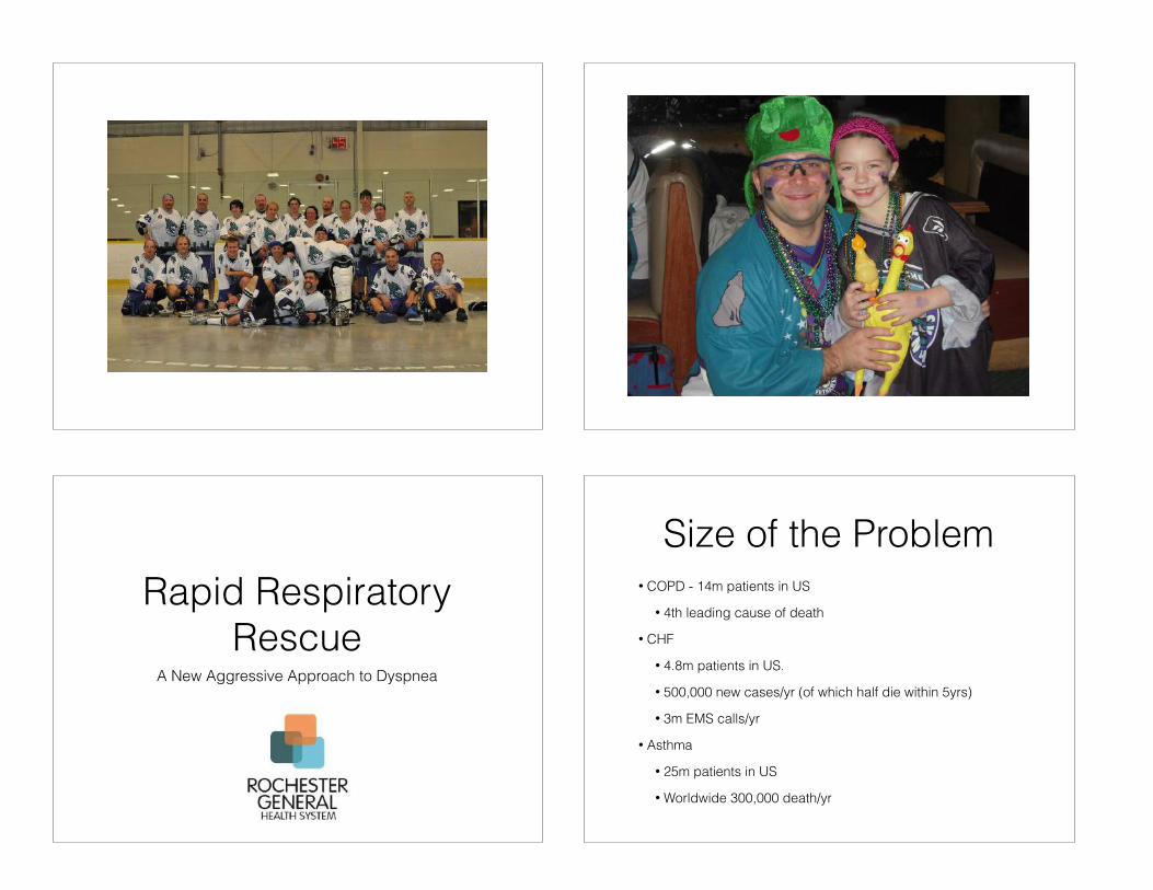
Size of the Problem• COPD - 14m patients in US
• 4th leading cause of death
• CHF
• 4.8m patients in US.
• 500,000 new cases/yr (of which half die within 5yrs)
• 3m EMS calls/yr
• Asthma
• 25m patients in US
• Worldwide 300,000 death/yr

N Engl J Med 2007;356:2156-64.
To EMS
• Symptoms Based Management
• Allows for “Protocol Crossover”
• Allows for non-traditional medication management and a “bundled care” package
Bundled Care
• “If you haven’t done everything in the bundle, you haven’t done everything for the patient”
• Takes Evidence-Based Interventions that have proven benefit to patients to achieve a set of goals
• Doing things that matter and not doing things that don’t (Do no harm)
Disclaimer
• Some of the following is controversial and the use of such responses should fit the clinical situation, with local protocols and be supported by medical direction.

Dyspnea• Types of Dyspnea - What Happens
• Diagnostic Tricks of the Trade
• Traditional Treatments Available
• Non-Traditional Treatments Available
• Unified Progressive Treatment Plan
Mechanical
• The Mechanics of Breathing
• Structures
• Inspiration
• Expiration

Breathing
• Inspiration is an ACTIVE process
• Expiration is a PASSIVE process

Pneumothorax• Tachycardia
• Tachypnea
• Reduced expansion of chest
• Hyper-resonance to percussion
• Quiet or absent breath sounds
• Subcutaneous emphysema
Dyspnea
• Bronchospasm
• Swelling/Obstruction
• Oxygenation/Ventilation restriction
• Structural lung failure
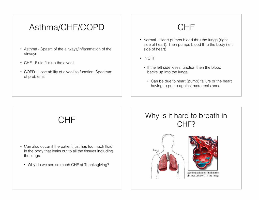
Asthma/CHF/COPD
• Asthma - Spasm of the airways/Inflammation of the airways
• CHF - Fluid fills up the alveoli
• COPD - Lose ability of alveoli to function. Spectrum of problems
CHF• Normal - Heart pumps blood thru the lungs (right
side of heart). Then pumps blood thru the body (left side of heart)
• In CHF
• If the left side loses function then the blood backs up into the lungs
• Can be due to heart (pump) failure or the heart having to pump against more resistance
CHF
• Can also occur if the patient just has too much fluid in the body that leaks out to all the tissues including the lungs
• Why do we see so much CHF at Thanksgiving?
Why is it hard to breath in CHF?

Fluid in the Alveoli….
• Causes
• Greater distance for oxygen to travel into blood
• Thicker fluid for oxygen to diffuse over
• Alveoli lose ability to stay open
Rales
• What is a rale (crackle)?
• Fine rales are due to the alveoli collapsing shut because the surfactant is disrupted. They then stick shut and you have to take a deep breath in to open them up. When they open - they crackle
Rales
• Why is it more common to hear rales at the lung bases?
The CHF patient• Sitting upright
• Tripod position
• Distended neck veins
• Peripheral edema
• ***High Blood Pressure***

Basic Treatment• Position
• Oxygen - Increases the gradient
• Prevent the alveoli collapsing?
• Push the fluid back into the blood?
• Dry out the fluid somewhere else?
CPAP• Continuous Positive Airway Pressure
• For the alveoli to collapse the pressure in the alveoli must drop - CPAP prevents this happening
• For collapsed alveoli - delivering oxygen at pressure helps pop them open - CPAP does that
• To push the fluid in the alveoli back into the blood the fluid must be subjected to pressure - CPAP does that
CPAP• Holds open alveoli - decreases the work the patient
has to do
• Pops open alveoli that were closed - allows more lung to be used (reduces dead space)
• Pushes fluid into the blood stream - decreases the distance that oxygen has to cover to get to the blood
CPAP
• CPAP is like sticking your head out of a window in a moving car and facing into the wind with your mouth open……

Removing the Fluid
• Diuretics
• Dialysis
• Nitroglycerin
Asthma
• Asthma is a 2 part process
• The airways spasm - causing wheezes
• The airways inflame - causing rhonchi
Flash Back
• Inspiration is an ACTIVE process
• Expiration is a PASSIVE process
If the airways spasm…
• Do the patients have a problem getting air in or getting air out?
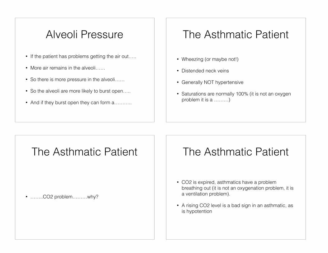
Alveoli Pressure• If the patient has problems getting the air out…..
• More air remains in the alveoli……
• So there is more pressure in the alveoli……
• So the alveoli are more likely to burst open…..
• And if they burst open they can form a………..
The Asthmatic Patient
• Wheezing (or maybe not!)
• Distended neck veins
• Generally NOT hypertensive
• Saturations are normally 100% (it is not an oxygen problem it is a ………)
The Asthmatic Patient
• ……..CO2 problem………why?
The Asthmatic Patient
• CO2 is expired, asthmatics have a problem breathing out (it is not an oxygenation problem, it is a ventilation problem).
• A rising CO2 level is a bad sign in an asthmatic, as is hypotention

The Asthmatic Patient• As asthma progresses……..patient breaths faster,
so despite having problems breathing out CO2 they compensate by breathing faster, so sometimes CO2 drops
• As the asthma gets worse they can no longer compensate……..and the CO2 rises
• As the air gets trapped in the lungs and can’t get out - increases intrapulmonary pressure - presses on the IVC and causes hypotension
The Asthmatic Patient• Albuterol
• Atrovent
• EpiPEN (Controversial)
• CPAP - which works by pressing open all of the airways (the pressure generated by CPAP is not just exerted on the alveoli but on all the airways)
EpiPEN
• Quickly becoming a standard way of treating severe asthma because…………
EpiPEN
• Epi is delivered via the blood, albuterol/atrovent rely on being delivered by breathing

The Inflammation
• Treated with steroids
COPD
• COPD is a mixture of lots of pathologies all related to destruction of lung tissue typically by smoking
What Happens When You Smoke?
• Cilia are paralyzed - debris remains in lungs
• Debris (Tar) remain in the alveoli - and disrupt the surfactant……causing…….and the alveoli are more likely to collapse
• Debris causes inflammation (bronchitis) and breakdown of the alveoli, causing them to join together, lose function and be more likely to collapse (emphysema)
COPD
• Wheezes - from airway irritation and spasm
• Rhonchi - from inflammation of the airways
• Rales - from destruction of the airways

COPD
• How to treat
• Decrease inflammation
• Improve oxygen exchange
• Decrease work of breathing
COPD and CHF
• What happens if you dry out a COPD patient (one of the treatments for CHF)?
• What happens if you give fluids to a CHF patient (one of the treatments for COPD)?
Telling the Difference
• Often you can’t - so you treat what you see and hear.
COPD• Treatment
• Nebs
• Steroids
• EpiPEN
• CPAP - Holds open the airways and the alveoli (and thus decreases the work of breathing)

ALS
• All effective traditional treatments for dyspnea have been moved to the BLS level
• ALS has access to other techniques - etCO2, IV medications, intubation (including RSI)
Case
• RL is a 56M who has previously been diagnosed with COPD and CHF.
• Smokes 2.5ppd and is on 4LNC home oxygen
• 3 months ago he was intubated by EMS for respiratory failure and remained on the vent for 4 weeks (slow wean)
RL
• EMS arrives on scene to find RL in respiratory distress with sats of 82% on oxygen.
• P120. RR40. BP 180/80. etCO2 70.
• Wheezes/Crackles with prolonged expiration globally, pulling off oxygen saying he cant breath
Capno Waveform
45
0

Capno Waveform Case RL
Respiratory Rescue
• Phase 1 - Traditional Response
• Phase 2 - Alternative Response
• Phase 3 - Decompensating Response
• Phase 4 - Failed Response
Traditional
Failed
Decomp
AlternativePhase 1
Traditional Response
Traditional

Hypoxia
• Apply oxygen (to achieve sats 88-97%)
• Start CPAP - Hold mask on face initially
Traditional
Wheeze• Treatment Guided by ETCO2 Waveform
• Continuous Nebulizers
• Albuterol (b2 agonist)
• Atrovent (anticholinergic)
• Magnesium IV (smooth muscle relaxer)
• 1-2 g IV over 10 mins
Traditional
Traditional
Stridor
• Nebulized epinephrine (a and b agonist)
• 5mls 1:1000 epi in nebulizer
• Traditionally used in croup management
Traditional

Asthma/COPD
• Anti-inflammatory action
• Solumedrol 125mg IVP (1.5mg/kg)
Traditional TraditionalIf Hypoxia Present Titrate oxygen to maintain sats 88-97% Initiate CPAP !If Wheeze Present Albuterol 2.5mg Neb Q15 minutes Atrovent 0.5mg Neb Q15 minutes Magnesium 1-2g IV over 10 minutes !If Stridor Present Epinephrine 1:1000 5ml Neb Q15 minutes !If Anaphylaxis is present then manage accordingly !If Asthma/COPD is suspected by history Methylprednisolone up to 125mg IVP (1.5mg/kg)
Phase 2Alternative Response
Alternative
Phase 2• Starting to see evidence of decompensation
• Typical symptoms noted
• Patient discomfort (“looks bad”)
• Rising CO2, dropping O2
• Not moving air/nebs
• Hypotension in COPD/Asthma
Alternative
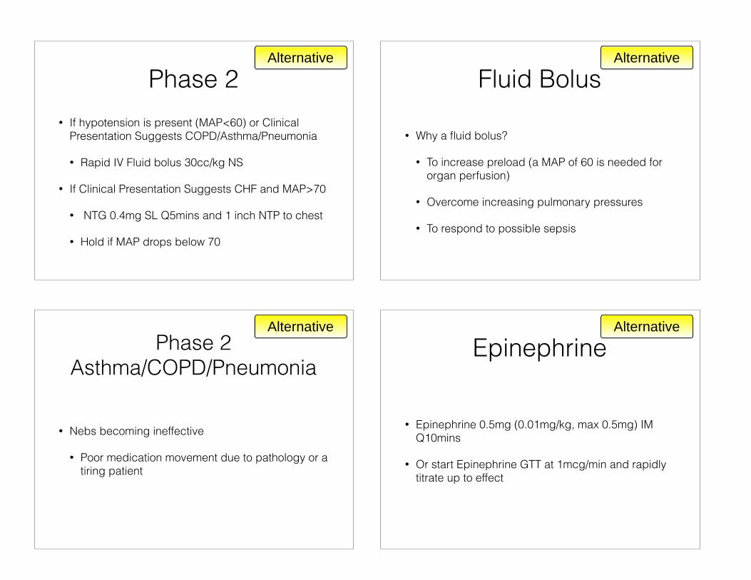
Phase 2• If hypotension is present (MAP<60) or Clinical
Presentation Suggests COPD/Asthma/Pneumonia
• Rapid IV Fluid bolus 30cc/kg NS
• If Clinical Presentation Suggests CHF and MAP>70
• NTG 0.4mg SL Q5mins and 1 inch NTP to chest
• Hold if MAP drops below 70
Alternative
Fluid Bolus
• Why a fluid bolus?
• To increase preload (a MAP of 60 is needed for organ perfusion)
• Overcome increasing pulmonary pressures
• To respond to possible sepsis
Alternative
Phase 2 Asthma/COPD/Pneumonia
• Nebs becoming ineffective
• Poor medication movement due to pathology or a tiring patient
Alternative
Epinephrine
• Epinephrine 0.5mg (0.01mg/kg, max 0.5mg) IM Q10mins
• Or start Epinephrine GTT at 1mcg/min and rapidly titrate up to effect
Alternative
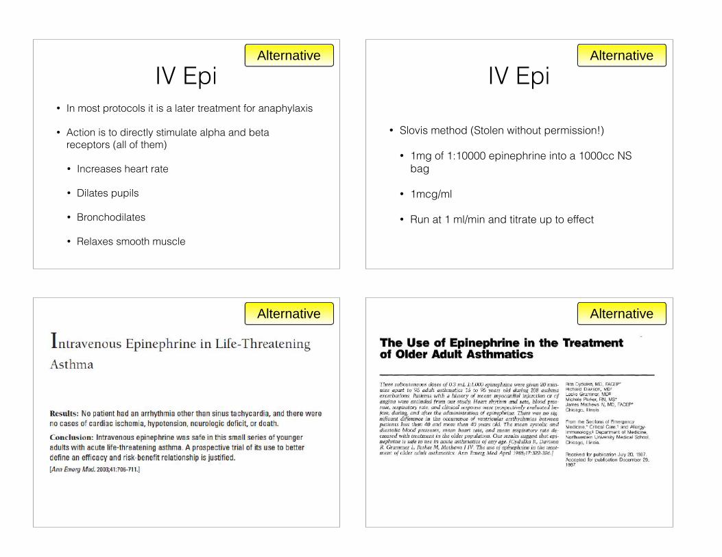
IV Epi• In most protocols it is a later treatment for anaphylaxis
• Action is to directly stimulate alpha and beta receptors (all of them)
• Increases heart rate
• Dilates pupils
• Bronchodilates
• Relaxes smooth muscle
Alternative
IV Epi
• Slovis method (Stolen without permission!)
• 1mg of 1:10000 epinephrine into a 1000cc NS bag
• 1mcg/ml
• Run at 1 ml/min and titrate up to effect
Alternative
Alternative Alternative

Alternative
Takotsubo Cardiomyopathy • Transient cardiac syndrome
• Involves left ventricular apical akinesis and mimics acute coronary syndrome.
• Patients often present with chest pain, have ST-segment elevation on electrocardiogram, and elevated cardiac enzyme levels consistent with a myocardial infarction but clean coronaries on cath
• Also known as Broken Heart Syndrome
Takotsubo Cardiomyopathy
• 2.2% of AMI
• Mean age 67
• 90% are post menopausal females
• The most commonly discussed possible mechanism for TCM is stress-induced catecholamine release, with toxicity to and subsequent stunning of the myocardium.
If hypotension is present (MAP<60) or Clinical Presentation Suggests COPD/Asthma/Pneumonia - Rapid IV Fluid bolus 30cc/kg NS !If Clinical Presentation Suggests CHF and Mean Arterial Pressure>70 - NTG 0.4mg SL Q5mins and 1 inch nitropaste to chest - Hold/remove if MAP drops below 70 !If nebulizer application is failing - Epinephrine 0.3mg 1:1000 IM Q10mins or - 1mcg/min IV and titrate up to effect. (1mg 1:10000 Epi in 1L NS at 1ml/min)
Alternative

Phase 3Decompensating Response
Decomp
Phase 3• Essentially Patient is now in Full Respiratory Failure
• Type 1 Failure - Hypoxic
• Type 2 Failure - Hypercarbic
• Mixed Picture
• Normally are obstructing their care
Decomp
Old Way• Brutane
• Held mask on, held down patient, intubated patient, pushed down gas pedal
• Sedation not an option due to dropping LOC and losing airway
• “Facilitated intubation” - Visiting death!
Decomp
New Way• Ketamine
• Dissociative anesthesia agent
• Bronchodilatory effect
• Does not cause hypotension
• Intubating medication of choice in sepsis and asthma/copd
Decomp

Sequence Intubation• Rapid Sequence Intubation
• Patient is given sedative and paralytic, wait until effect seen, then patient is intubated
• Newer still - Delayed Sequence Intubation (DSI)
• Patient is given sedative that does not suppress respiratory effect and then managed with non-invasive methods to increase saturation, at that point is given paralytic and then intubated
Decomp
Newest• Facilitated Non-Invasive Ventilation with optional
DSI
• Ketamine is given and when it takes effect patient is managed in a non-invasive way
• Reevaluation is done and a determination is made to continue current plan or proceed to paralytic and intubation
Decomp
Phase 3• If able to tolerate CPAP
• CPAP with continuous nebs
• If worsening then Ketamine 1.5mg/kg IV over 30 seconds. Rebolus as needed.
• If no IV then Ketamine 5mg/kg IM
• If unable to tolerate CPAP
• Ketamine 1.5mg/kg IV over 30 seconds. Rebolus as needed.
• If no IV then Ketamine 5mg/kg IM
• Then apply CPAP and give continuous nebs
Decomp
If able to tolerate CPAP and Asthma/COPD/Sepsis is clinically suspected then - Continue CPAP with continuous nebs - If worsening then Ketamine 1.5mg/kg IV over 30 seconds. Rebolus as needed. If no IV then Ketamine 5mg/kg IM. !If unable to tolerate CPAP and Asthma/COPD/Sepsis is clinically suspected then - Ketamine 1.5mg/kg IV over 30 seconds. Rebolus as needed. If no IV then Ketamine 5mg/kg IM. - Then apply CPAP and give continuous nebs !If CHF is clinically suspected then proceed directly to intubation
Decomp

Phase 4Failed Response
Failed
Intubation
• If Asthma/COPD/Sepsis is clinically suspected
• Continue Ketamine for sedation, re-bolus as needed
• Succinylcholine 1-2 mg/kg IVP for paralysis
• Intubate the patient
Failed
Intubation
• If CHF is clinically suspected
• Give etomidate 0.3mg/kg IV for sedation and rocuronium 1mg/kg IV for paralysis
• Intubate the patient
Failed
!If Asthma/COPD/Sepsis is clinically suspected - Continue Ketamine for sedation, re-bolus as needed - Succinylcholine 1-2 mg/kg IVP for paralysis - Intubate the patient !If CHF is clinically suspected - Give etomidate 0.3mg/kg IV for sedation - Rocuronium 1mg/kg IV for paralysis - Intubate the patient
Failed

Patient RL
• BLS was 1st on scene and recognized the patient was critically short of breath
• Initiated 100%NRB immediately
• Started a HF 2.5mg Albuterol Neb
• ALS was en route (RSA level of care)
Patient RL• ALS arrived - Patient remained hypoxic at 85% on
neb
• Continuous albuterol/atrovent was initiated via CPAP
• Patient was given IM 1:1000 EpiPEN
• IV was established
• Patient given 2g IV magnesium
Patient RL• Minimal response to therapy
• Fluid bolus was initiated for presumed COPD/PNA
• IV epi drip was started
• 1mcg/min which was increased at a rate of 1mcg/min every 1 minute
• Patient became more coherent at 6mcg/min and sats improved to 88%
Patient RL• 1 hour ground transport time
• Noted that etCO2 was initially 70 but increased to 85 and patient became more somnolent
• RSA medic determined CPAP was failing to turn patient around and chose to intubate (respiratory failure with AMS and unprotected airway)
• 100mg IV Ketamine given with 100mg of IV Succynylcholine

Patient RL
• Due to continued hypoxia RSA medic chose to use BVM to bring up sats (was able to get sats to 93% prior to intubation)
• Patient was intubated, sedated with versed and paralyzed with vecuronium for transport
Thank [email protected]