Embed Size (px)
Citation preview
Rapidly Progressive (Crescentic) Glomerulonephritis(RPGN)
- Is a syndrome associated with severe glomerular
injury, but does not denote a specific etiologic
form of glomerulonephritis.
- It is characterized by rapid and progressive loss
of renal function associated with severe oliguria
and signs of nephritic syndrome
- If untreated, death from renal failure occurs within
weeks to months.
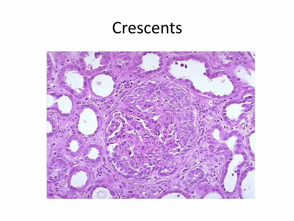
- The most common histologic picture is the
presence of crescents in most of the glomeruli
and these are produced
a. predominantly by the proliferation of the parietal
epithelial cells lining Bowman capsule and
b. by the infiltration of monocytes and
macrophages.
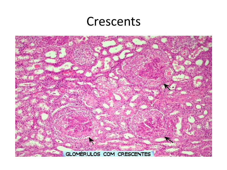
Crescents
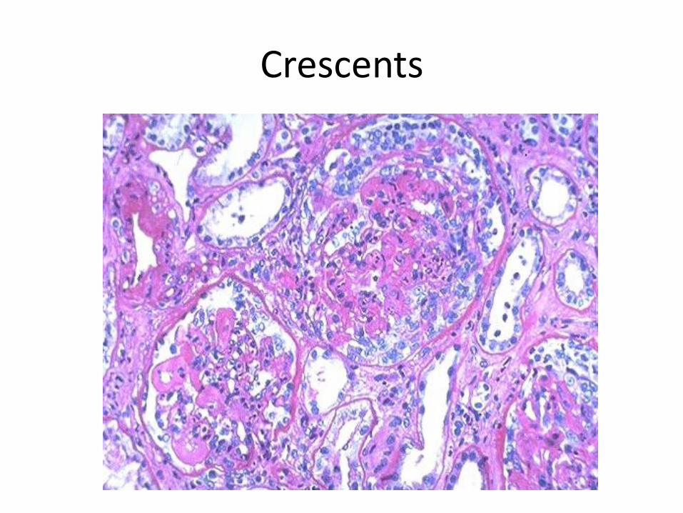
Crescents
Crescents
Classification and Pathogenesis.
- RPGN may be caused by a number of
different diseases, some restricted to the
kidney and others systems.
- Although no single mechanism can explain all
cases, but in most cases the glomerular injury
is immunologically mediated.
classification - RPGN is classified into three groups on the basis
of immunologic findings and In each group the
disease may be
a. Associated with a known disorder,
b. or it may be idiopathic
- The common denominator in all types of
RPGN is severe glomerular injury.
1. Anti-GBM antibody-mediated disease:
- Characterized by linear deposits of IgG and, in
many cases, C3 in the Glomerular basement
membrane(GBM)
- In some of these patients, the anti-GBM
antibodies cross-react with pulmonary alveolar
basement membranes to produce the clinical
picture of pulmonary hemorrhage associated
with renal failure (Goodpasture syndrome)
- The antigen common to the alveoli and GBM is
a peptide within the noncollagenous domain of the α3 chain of collagen type IV
- What triggers the formation of these antibodies
is unclear in most patients.
- Exposure to viruses or hydrocarbon solvents
(found in paints and dyes) and drugs has been
implicated in some patients
2. Diseases caused by immune complex deposition.
- RPGN can be a complication of any of the immune complex nephritides, including :
I. Idiopathic
II. Secondary to
a. Postinfectious glomerulonephritis,
b. Lupus nephritis,
c. IgA nephropathy,
- In this group, immunofluorescence studies
reveal the granular pattern of staining
characteristic of immune complex
deposition.
- These patients usually cannot be helped by
plasmapheresis, and they require treatment
for the underlying disease.
3. Pauci-immune RPGN,
- Defined by the lack of detectable anti-GBM
antibodies or immune complexes by
immunofluorescence and electron microscopy and
can be
1. Idiopathic
- More than 90% of idiopathic cases have c-ANCAs
or p-ANCAs in the sera
2. Most commonly the patients have circulating
antineutrophil cytoplasmic antibodies (ANCAs)
that produce cytoplasmic (c) or perinuclear (p)
staining pattern and are known to play a role in
some vasculitides
- This type of RPGN may be a component of a
systemic vasculitis such as granulomatosis with
polyangiitis (formerly called Wegener
granulomatosis) or microscopic polyangiitis
- .
- The presence of circulating ANCAs in both
idiopathic crescentic glomerulonephritis and
cases that occur as a component of systemic
vasculitis, and the similar pathologic features in
either setting, have led to the idea that these
disorders are pathogenetically related
- According to this concept, all cases of
crescentic glomerulonephritis of the pauci-
immune type are manifestations of small-
vessel vasculitis or polyangiitis, which is limited
to glomerular and perhaps peritubular
capillaries in cases of idiopathic crescentic
glomerulonephritis.
Morphology
Light microscope
- The histologic picture is dominated by distinctive
crescents
- Crescents are formed by proliferation of parietal
cells and by migration of monocytes and
macrophages into the urinary space.
- Neutrophils and lymphocytes may be present.
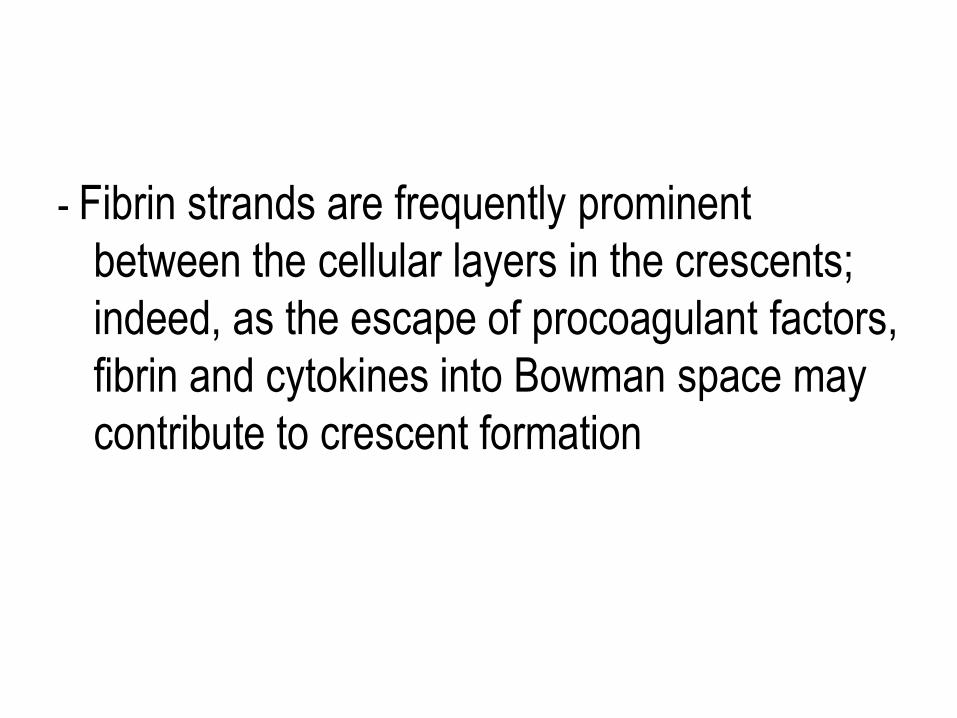
- Fibrin strands are frequently prominent
between the cellular layers in the crescents;
indeed, as the escape of procoagulant factors,
fibrin and cytokines into Bowman space may
contribute to crescent formation
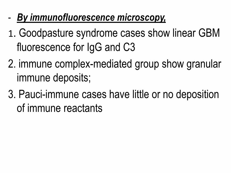
- By immunofluorescence microscopy,
1. Goodpasture syndrome cases show linear GBM
fluorescence for IgG and C3
2. immune complex-mediated group show granular
immune deposits;
3. Pauci-immune cases have little or no deposition
of immune reactants
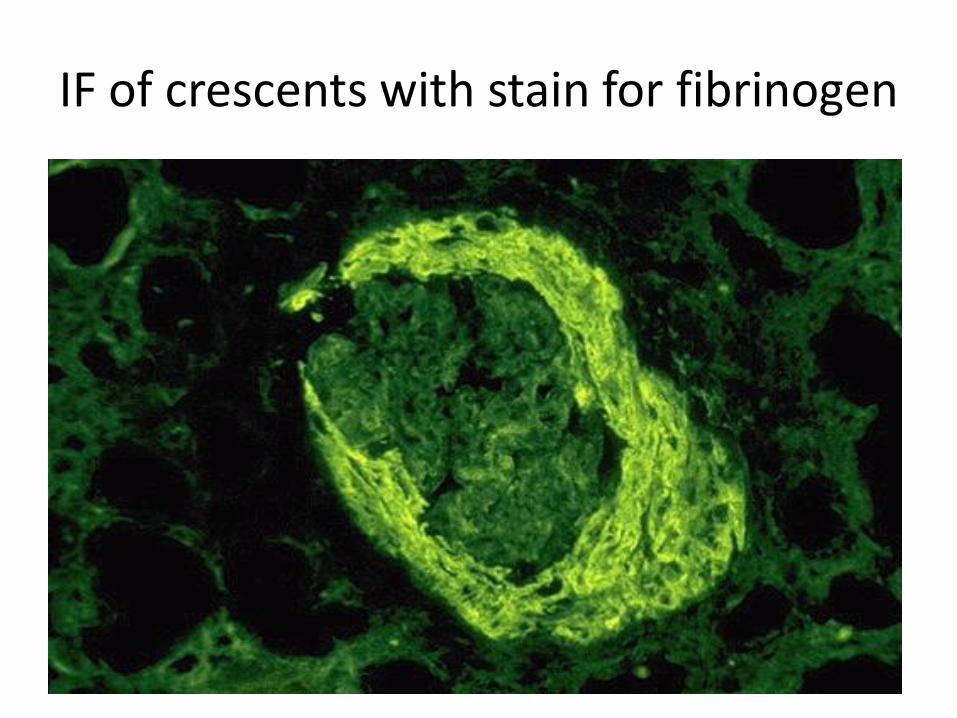
IF of crescents with stain for fibrinogen
Electron microscopy
- Ruptures in the GBM in the three groups
- Shows discloses deposits in those cases due to
immune complex deposition (type II).
Note:
- In time, most crescents undergo organization
and fibrosis
Clinical Course.
- The renal manifestations of all forms of
crescentic glomerulonephritis include:
a. Hematuria with red blood cell casts in the urine,
b. Moderate proteinuria occasionally reaching the
nephrotic range,
c. Variable hypertension
Clinical course
- The renal involvement is usually progressive
over a matter of weeks and culminates in severe
oliguria.
- Recovery of renal function may follow early
intensive plasmapheresis combined with steroids
and cytotoxic agents in Goodpasture syndrome.
which can reverse both pulmonary hemorrhage
and renal failure
- Other forms of RPGN also respond well to
steroids and cytotoxic agents, However, despite
therapy, many patients eventually require
chronic dialysis or transplantation, particularly if the disease is discovered at a late stage.
Note: Serum analyses for anti-GBM antibodies,
antinuclear antibodies, and ANCAs are helpful
in the diagnosis of specific subtypes.
Nephrotic Syndrome
- The manifestations of the syndrome include:
1.Massive proteinuria, with the daily loss of 3.5 gm
or more of protein (less in children)
2. Hypoalbuminemia, with plasma albumin levels
less than 3 gm/dL
3. Generalized edema
4.Hyperlipidemia and lipiduria
- Increased permeability resulting from either
structural or physicochemical alterations in this
barrier allows proteins to escape from the
plasma into the urinary space, resulting in
proteinuria.
- Heavy proteinuria depletes serum albumin
levels at a rate beyond the compensatory
synthetic capacity of the liver, resulting in
hypoalbuminemia.
-
- Increased renal catabolism of filtered albumin
also contributes to the hypoalbuminemia.
- The edema is a consequence of decreased
intravascular colloid osmotic pressure.
- Edema is soft and pitting, and is most marked
in the periorbital regions and dependent
portions of the body.
.
- The largest proportion of protein lost in the urine
is albumin, but globulins are also excreted in
some diseases.
- The ratio of low- to high-molecular-weight
proteins in the urine in various cases of
nephrotic syndrome is a manifestation of the
selectivity of proteinuria
a. A highly selective proteinuria
- Consists mostly of low-molecular-weight
proteins (albumin, and transferrin),
b. A poorly selective proteinuria
- Consists of higher molecular-weight globulins
in addition to albumin.
The genesis of the hyperlipidemia is complex.
- Most patients with nephrotic syndrome have
a. Increased blood levels of cholesterol,
triglyceride, very-low-density lipoprotein, low-
density lipoprotein, Lp(a) lipoprotein, and
apoprotein, and
b. there is a decrease in high-density lipoprotein
concentration in some patients.
• These defects are due to a combination of:
a. increased synthesis of lipoproteins in the liver
b. Abnormal transport of circulating lipid particles,
c. and decreased lipid catabolism
- Lipiduria follows the hyperlipidemia, because lipoproteins also leak across the glomerular capillary wall.
- The lipid appears in the urine either as free fat or as oval fat bodies, representing lipoprotein resorbed by tubular epithelial cells and then shed along with injured tubular cells from the basement membrane.
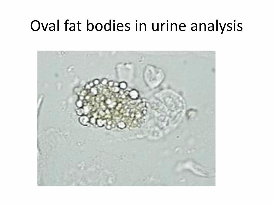
Oval fat bodies in urine analysis
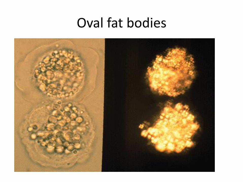
Oval fat bodies
Other manifestations
I. Nephrotic patients are vulnerable to infection,
especially staphylococcal and pneumococcal
infections, probably due to loss of
immunoglobulins in the urine.
II. Thrombotic and thromboembolic complications
due to loss of endogenous anticoagulants (e.g.,
antithrombin III) in the urine.
III. Renal vein thrombosis,
- is a consequence of this hypercoagulable
state, particularly in patients with
membranous nephropathy
Causes of Nephrotic syndrome
- The incidences of the several causes of the
nephrotic syndrome vary according to age and
geography.
1. In children younger than 17 years in North
America, nephrotic syndrome is almost always
caused by a lesion primary to the kidney;
2. Among adults, in contrast, it is often
associated with a systemic disease.
- The most frequent systemic causes of the
nephrotic syndrome are
a. Diabetes,
b. Amyloidosis,
c. SLE.
• The most important of the primary glomerular
lesions are
a. Minimal-change disease : is most common in
children in North America
b. Membranous glomerulopathy, is most
common in older adults :
c. Focal segmental glomerulosclerosis:
occurs at all ages
• Other less common causes of nephrotic
syndrome include:
a. Membranoproliferative glomerulonephritides
b. IgA nephropathy
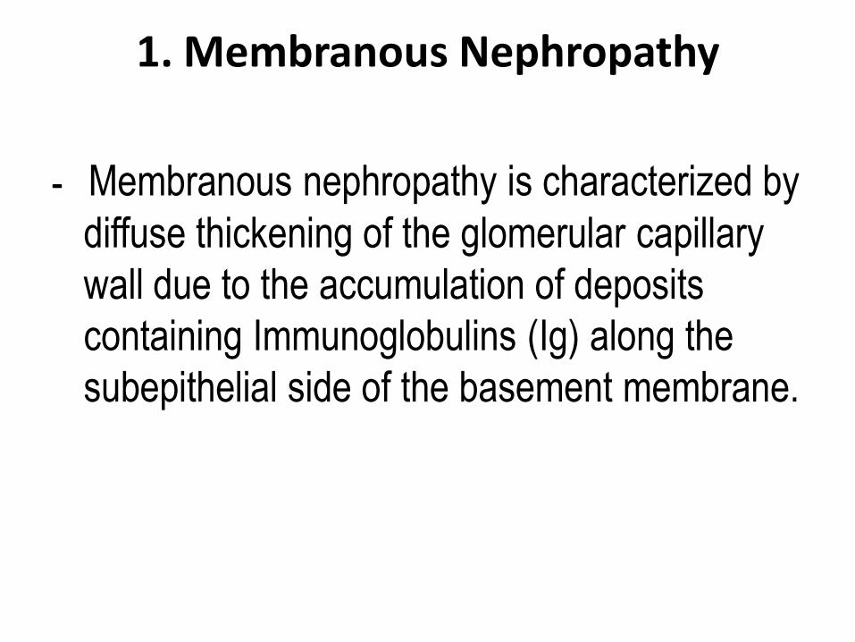
1. Membranous Nephropathy
- Membranous nephropathy is characterized by
diffuse thickening of the glomerular capillary
wall due to the accumulation of deposits
containing Immunoglobulins (Ig) along the
subepithelial side of the basement membrane.
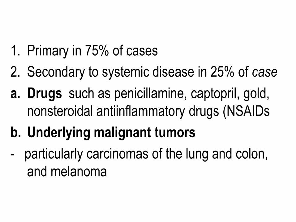
1. Primary in 75% of cases
2. Secondary to systemic disease in 25% of case
a. Drugs such as penicillamine, captopril, gold,
nonsteroidal antiinflammatory drugs (NSAIDs
b. Underlying malignant tumors
- particularly carcinomas of the lung and colon,
and melanoma
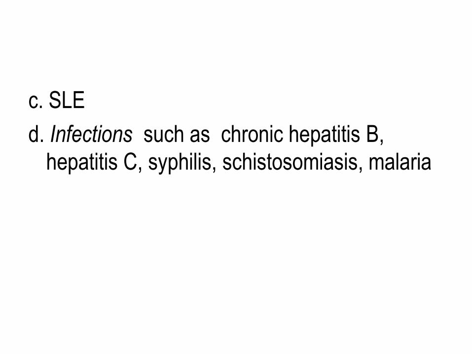
c. SLE
d. Infections such as chronic hepatitis B,
hepatitis C, syphilis, schistosomiasis, malaria
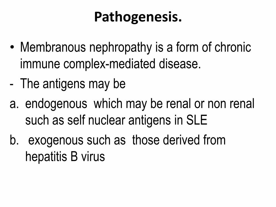
Pathogenesis.
• Membranous nephropathy is a form of chronic
immune complex-mediated disease.
- The antigens may be
a. endogenous which may be renal or non renal
such as self nuclear antigens in SLE
b. exogenous such as those derived from
hepatitis B virus
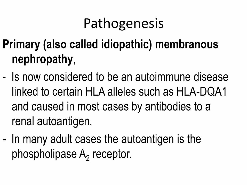
Pathogenesis
Primary (also called idiopathic) membranous
nephropathy,
- Is now considered to be an autoimmune disease
linked to certain HLA alleles such as HLA-DQA1
and caused in most cases by antibodies to a
renal autoantigen.
- In many adult cases the autoantigen is the
phospholipase A2 receptor.
- How does the glomerular capillary wall become
leaky in membranous nephropathy? There is a
paucity of neutrophils, monocytes, or platelets in
glomeruli.
- The presence of complement and corroborating
experimental work suggest that the complement
C5b-C9 membrane attack complex has an
important role.
- It is postulated that C5b-C9 activates
glomerular epithelial and mesangial cells,
inducing them to liberate proteases and
oxidants, which cause capillary wall injury and
increased protein leakage.
- A subclass of IgG, IgG4, which differs from
other IgG subclasses in being a poor activator
of the classical complement pathway, is the
principal immunoglobulin deposited in cases
of primary membranous nephropathy.
- How IgG4 may activate the complement
system is not clear.
Morphology
Light microscopy
- The glomeruli either appear normal in the early
stages of the disease or exhibit uniform, diffuse
thickening of the glomerular capillary wall
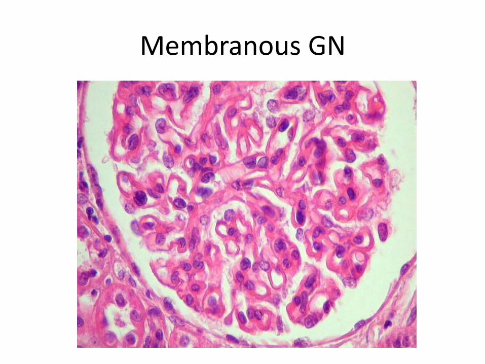
Membranous GN
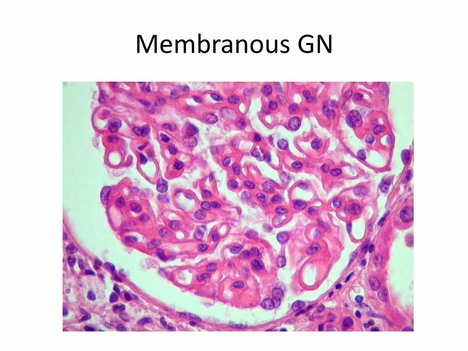
Membranous GN
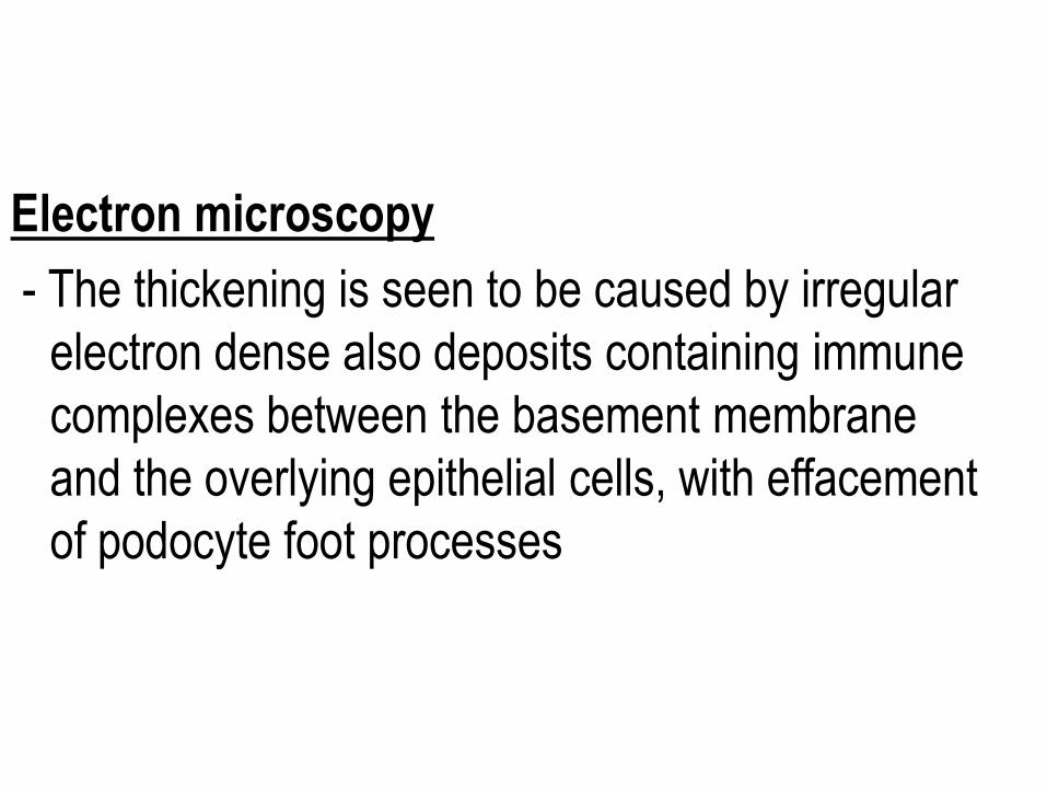
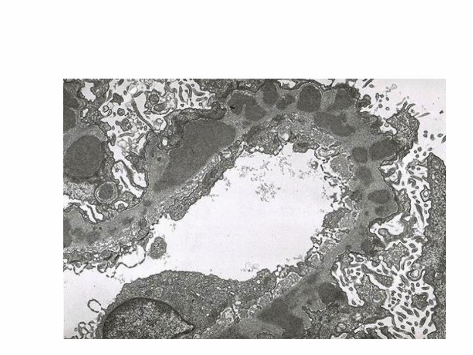
Electron microscopy
- The thickening is seen to be caused by irregular
electron dense also deposits containing immune
complexes between the basement membrane
and the overlying epithelial cells, with effacement
of podocyte foot processes
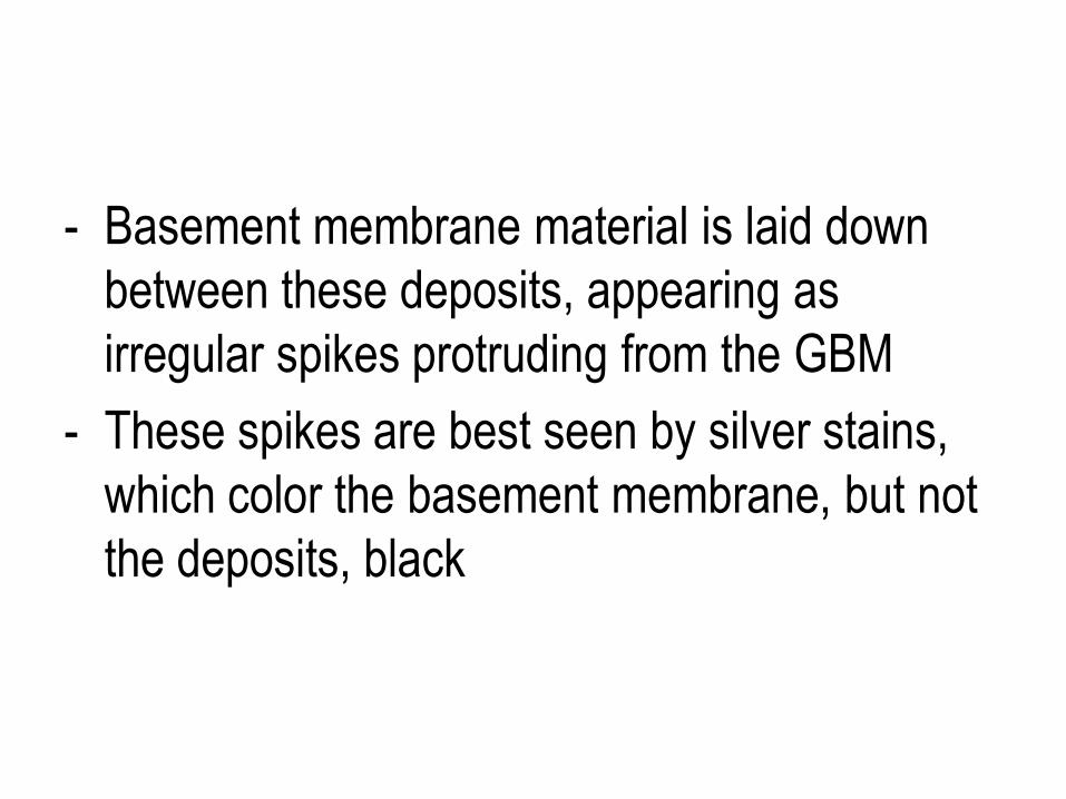
- Basement membrane material is laid down
between these deposits, appearing as
irregular spikes protruding from the GBM
- These spikes are best seen by silver stains,
which color the basement membrane, but not
the deposits, black
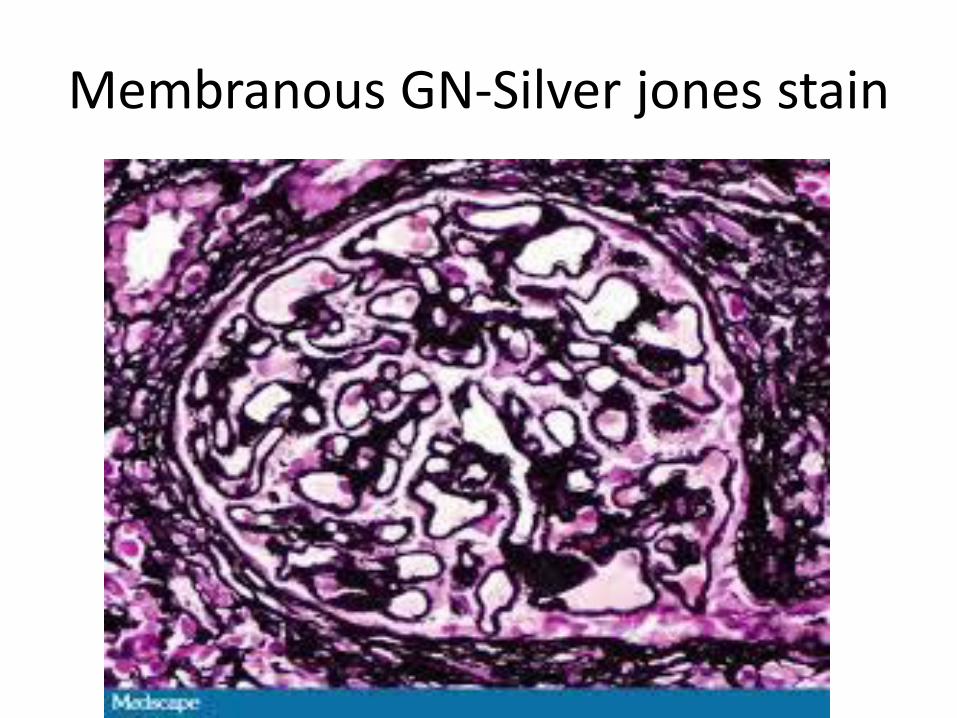
Membranous GN-Silver jones stain
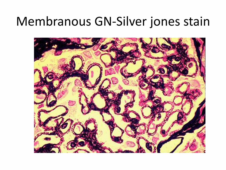
Membranous GN-Silver jones stain
- In time, these spikes thicken to produce
domelike protrusions and eventually close over
the immune deposits, burying them within a
markedly thickened, irregular membrane.
Immunofluorescence microscopy
- Demonstrates that the granular deposits
contain both immunoglobulins and complement
.
- The epithelial cells of the proximal tubules
contain protein reabsorption droplets, and
there may be considerable interstitial
mononuclear cell inflammation.