Embed Size (px)
Citation preview
Rapid Publication
Neutrophil Nicotinamide Adenine Dinucleotide Phosphate Oxidase AssemblyTranslocation of p47-phox and p67-phox Requires Interaction between p47-phox and Cytochrome b.Paul G. Heyworth,* John T. Cumutte,* William M. Nauseef,* Bryan D. Volpp,*Doran W. Pearson,t Henry Rosen,* and Robert A. Clarkt*Department of Molecular and Experimental Medicine, Research Institute of Scripps Clinic, La Jolla, California 92037;tDepartment of Medicine, University of Iowa College of Medicine and Department of Veterans Affairs Medical Center,Iowa City, Iowa 52242; and §Department of Medicine, University of Washington, Seattle, Washington 98195
Abstract
Two of the cytosolic NADPHoxidase components, p47-phoxand p67-phox, translocate to the plasma membrane in normalneutrophils stimulated with phorbol myristate acetate (PMA).Wehave now studied the translocation process in neutrophilsof patients with chronic granulomatous disease (CGD), an in-herited syndrome in which the oxidase system fails to producesuperoxide due to lesions affecting any one of its four knowncomponents: the gp9l-phox and p22-phox subunits of cy-tochrome b5m (the membrane-bound terminal electron trans-porter of the oxidase), p47-phox, and p67-phox. In contrast tonormal cells, neither p47-phox nor p67-phox translocated tothe membrane in PMA-stimulated CGDneutrophils whichlack cytochrome b5!m. In one patient with a rare X-linked formof CGDcaused by a Pro -- His substitution in gp9l-phox, butwhose neutrophils have normal levels of this mutant cy-tochrome bm, translocation was normal. In two patients withp47-phox deficiency, p67-phox failed to translocate, whereasp47-phox was detected in the particulate fraction of PMA-stimulated neutrophils from two patients deficient in p67-phox.Our data suggest that cytochrome b5!4 or a closely linked factorprovides an essential membrane docking site for the cytosolicoxidase components and that it is p47-phox that mediates theassembly of these components on the membrane. (J. Clin. In-vest. 1991. 87:352-356.) Key words: respiratory burst * chronicgranulomatous disease - superoxide - protein phosphorylation-plasma membrane
Introduction
The respiratory burst oxidase of neutrophils and other phago-cytic cells is a multicomponent electron transport system thatcatalyzes the NADPH-dependent, one-electron reduction ofoxygen to produce superoxide- (O°)'. The system is dormant in
This work was published in abstract form in (1990. Clin. Res. 38:434).Address correspondence and reprint requests to Dr. P. G. Hey-
worth, Department of Molecular and Experimental Medicine, BCR-3,Research Institute of Scripps Clinic, La Jolla, CA92037.
Receivedfor publication 8 August 1990 and in revisedform 25 Sep-tember 1990.
1. Abbreviations used in this paper: CGD, chronic granulomatous dis-ease; °2, superoxide.
resting cells but can be activated by several particulate andsoluble stimuli. The dormant oxidase consists of both cytosolicand membrane-bound components, but the active, O-gener-ating complex is confined to the plasma membrane (1, 2). Thisimplies that the cytosolic components must either act in a sig-naling capacity by modifying the membrane components, orthat they must become directly associated with the membranevia a translocation process.
In the inherited syndrome chronic granulomatous disease(CGD), phagocytes fail to generate a respiratory burst and pro-duce O2 (1). The resulting microbicidal defect renders thesepatients highly susceptible to bacterial and fungal infections.CGDis genetically heterogeneous, resulting from lesions af-fecting any one of at least four distinct oxidase components (3).In the most common form a genetic modification of the X-chromosome results in the absence of the 91 -kD transmem-brane glycoprotein subunit of cytochrome b558 (termedgp9 l -phox, for phagocyte oxidase), the terminal electron trans-porter of the oxidase (4, 5). There is also a secondary loss of the22-kD cytochrome subunit (p22-phox) (6, 7). In a much rarerautosomal recessive form of CGD, the primary mutation in-volves the gene encoding this smaller subunit, with an asso-ciated loss of gp9 I -phox (8, 9). These two forms of CGDaretherefore characterized by a total absence of cytochrome b558.In a rare variant form of X-linked CGD, the neutrophils con-tain a cytochrome b558 that appears quantitatively and spec-trally normal, but nonetheless fail to produce O2 (10). In onefamily with this form of the disease, the defect has been identi-fied as a missense mutation in the gene encoding the 9 1-kDsubunit, resulting in a nonconservative Pro -- His substitutionin what is thought to be a junction region between the mem-brane and cytosolic domains of the protein (1 1). In the major-ity of autosomal recessive CGDpatients the levels of cy-tochrome b558 are normal. The genetic defects in these casesinvolve either of the two known cytosolic oxidase components,p47-phox or less commonly p67-phox (12-14).
In intact phagocytic cells a 47-kD protein is phosphorylatedat multiple sites in association with activation of the respiratoryburst (15-17), and it has recently been confirmed that thisphosphoprotein is p47-phox (18-20). Phosphorylated p47-phox has a dual localization in the cytosol and membrane frac-tions of neutrophils activated with phorbol esters (15, 21, 22).Association of the phosphoprotein with the membrane appearsto depend on the presence of cytochrome b558, since phosphor-ylated p47-phox was confined to the cytosol in cytochrome-de-ficient CGDneutrophils (22). Although these findings stronglysupported the idea that translocation of cytosolic oxidase com-ponents to the membrane was involved in the activation pro-
352 Heyworth, Curnutte, Nauseef Volpp, Pearson, Rosen, and Clark
J. Clin. Invest.© The American Society for Clinical Investigation, Inc.0021-9738/91/01/0352/05 $2.00Volume 87, January 1991, 352-356

cess, they did not rule out the possibility that the dual lbcaliza-tion in the activated state represented two pools of p47-phox.
Recently it was conclusively demonstrated that both p47-phox and p67-phox translocate to the membrane during activa-tion of the respiratory burst in intact neutrophils (23). A poly-clonal antiserum, B- 1, which recognizes both components, wasused to probe electrophoretic blots of proteins from neutrophilsoluble and particulate fractions separated by SDS-PAGE. Thetwo proteins were detected in the particulate fraction of acti-vated, but not resting neutrophils. Analytical subcellular frac-tionation revealed that they were present in the plasma mem-brane fraction but not in fractions enriched in either specific orazurophilic granules (23). In an attempt to understand morefully the interactions among the cytosolic oxidase componentsand their roles in oxidase assembly, we have studied the trans-location process in neutrophils from patients with different ge-netic forms of CGD.
Methods
Preparation of neutrophils. Blood was collected by venipuncture fromnormal subjects and patients with CGDafter obtaining informed con-sent. Neutrophils were purified by dextran sedimentation, hypotoniclysis of erythrocytes, and centrifugation through Ficoll-Hypaque (24).After pretreatment with 2 mMdiisopropylfluorophosphate for 20 minat 4VCand at a density of 5 X 10⁷ cells/ml, the neutrophils were washedand resuspended in phosphate-buffered saline with glucose (2.7 mMKCI, 138 mMNaCI, 7.5 mMi-glucose, 8.1 mMNa2HPO4, 1.47 mMKH2PO4, pH 7.3) at the same cell density. All materials were obtainedfrom the sources previously cited (24).
Neutrophil activation andfractionation. Neutrophils (3 X I07 cellsin 0.6 ml) were preincubated at 37°C for 5 min, then either activatedwith PMA(100 ng/ml) or treated with an equal volume (1.5 u1) ofdimethylsulfoxide, and after 5 min transferred to an ice bath. Subse-quent steps are modifications of the method of Clark et al. (23), andwere all performed at 4°C. The cells were pelleted by centrifugation for6 min at 300 g, resuspended in 0.6 ml relaxation buffer (100 mMKCI,3 mMNaCI, 3.5 mMMgCI2, 1.25 mMEGTA, 10 mMpiperazinediethane sulfonic acid, pH 7.3), and disrupted by a single 5-s burst ofsonication, using a microprobe sonicator at low power. This resulted in> 95%breakage. Unbroken cells and nuclei were removed by centrifu-gation for 5 min at 250 g. The resulting supernatant (S1) was centri-fuged for 20 min at 1 5,000 gin the TLA 100.2 rotor of a Beckman TL100 ultracentrifuge (Beckman Instruments, Inc., Palo Alto, CA). Thehigh speed supernatant (S2), representing the cytosolic fraction, wasremoved, and the pellet washed with a further 0.6 ml of relaxationbuffer and recentrifuged at 1 5,000 g for 20 min. The final pellet (P3),referred to here as the membrane fraction, was resuspended in 0.2 ml ofrelaxation buffer and dispersed with a 2-3-s burst of sonication. In eachexperiment, neutrophils from normal control subjects were preparedconcurrently with those from the patients to allow direct comparison ofthe immunoblots. The protein content of the fractions was measuredusing the Pierce bicinchoninic acid assay (Pierce Chemical Co., Rock-ford, IL).
Neutrophils from a patient with the rare X-linked cytochrome b-positive form of CGDwere fractionated using a slightly modified pro-cedure. After activation the cells (5 X I0 in 1 ml) were centrifuged for 3min at 3,600 g, resuspended in 0.5 ml relaxation buffer, and disruptedwith 15-20 0.5-s bursts of sonication. The sonicate was centrifuged for5 min at 400 g to remove unbroken cells and the supernatant diluted to16 ml with relaxation buffer. After centrifugation for 20 min at 150,000g and without further washing, the pellet was resuspended in 0.5 mlrelaxation buffer.
Electrophoresis and immunoblotting. Soluble and particulate frac-tions from resting and activated neutrophils were subjected to SDS-
PAGEon 9% acrylamide gels. The proteins were electrophoreticallytransferred to nitrocellulose and the blots probed with B- 1, a polyclonalantiserum that recognizes p47-phox and p67-phox. Immunoreactiveproteins were detected with '25I-protein A and autoradiography andquantitated, where indicated, by densitometry and calculation of thearea under the curve. These procedures have been described elsewhere(13, 23).
Results
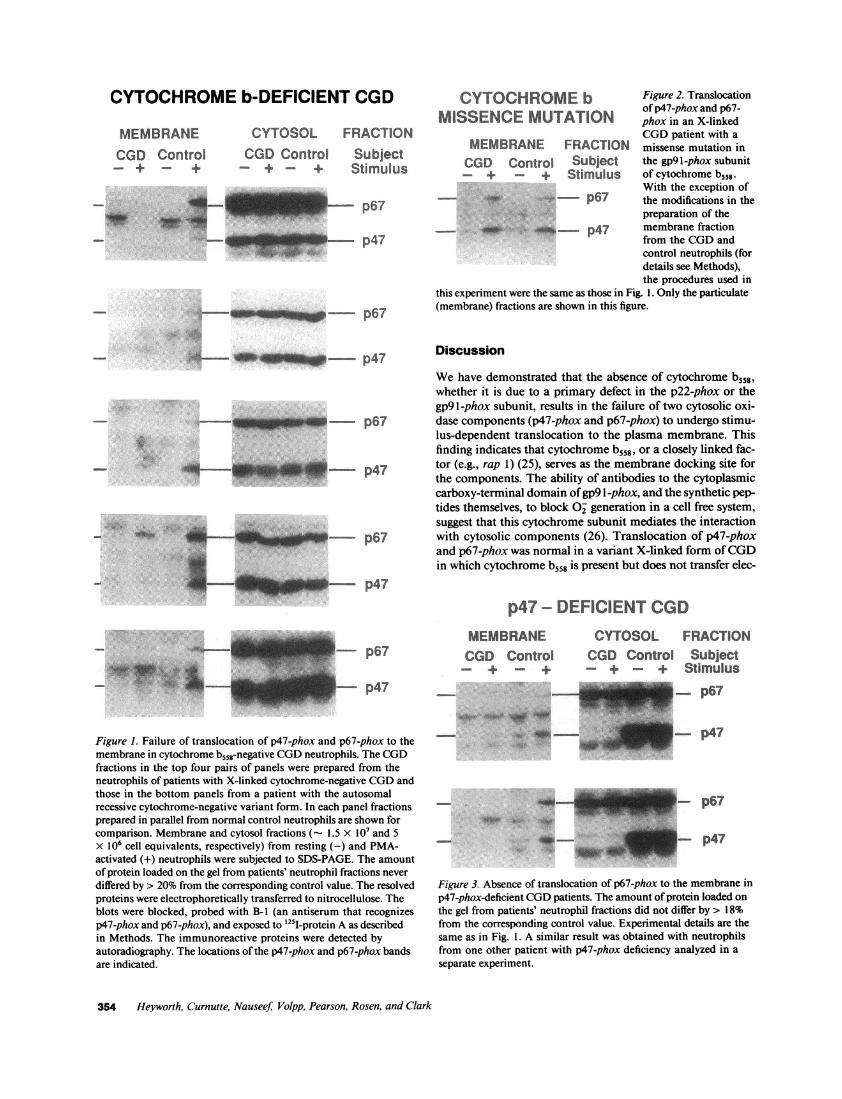
The autoradiographs in Fig. 1 are from CGDneutrophils de-void of cytochrome b558, aligned with their concurrent normalcontrols. The top four sets of panels are from patients withX-linked cytochrome-negative CGD(gp9 l-phox mutation),while the bottom set is from a patient with the much rarerautosomal recessive cytochrome-negative form (p22-phox mu-tation). In both forms the levels of p47-phox and p67-phox inthe cytosol were the same as in the normal controls. In unstim-ulated control neutrophils both p47-phox and p67-phox wereconfined to the cytosol, but after 5-min stimulation with PMAthe two proteins were also detected in the particulate fraction([23] and Fig. 1). In marked contrast, in four of the five subjectswith cytochrome-negative CGD, neither p47-phox nor p67-phox was detected in the membrane fraction. In one patient(Fig. 1, fourth panel) a small amount of immunoreactive pro-tein was detected in the membrane fraction at the position ofp67-phox only, but at a much lower level than was detected incontrol membranes. This probably does not represent contami-nation of the particulate fraction with cytosol, as no p47-phoxwas detected in the same preparation. As far as can be deter-mined, this patient's neutrophils are biochemically and func-tionally the same as those from other patients with X-linkedcytochrome-negative CGD(Curnutte, J. T., unpublished ob-servation). The B- 1 polyclonal antiserum predominantly recog-nizes p47-phox and p67-phox, but also shows minor specifici-ties to other proteins (e.g., at 55 kD) of unknown identity ( 13,23). The intensity of these bands varied from experiment toexperiment depending upon the exposure time of the autora-diograph.
Wealso studied translocation of cytosolic components inone patient with X-linked CGD, in which a Pro -- His substi-tution in gp91-phox renders cytochrome b558 nonfunctional.Translocation in these neutrophils was completely normal;both p47-phox and p67-phox were detected in the membranefraction of PMA-stimulated cells (Fig. 2).
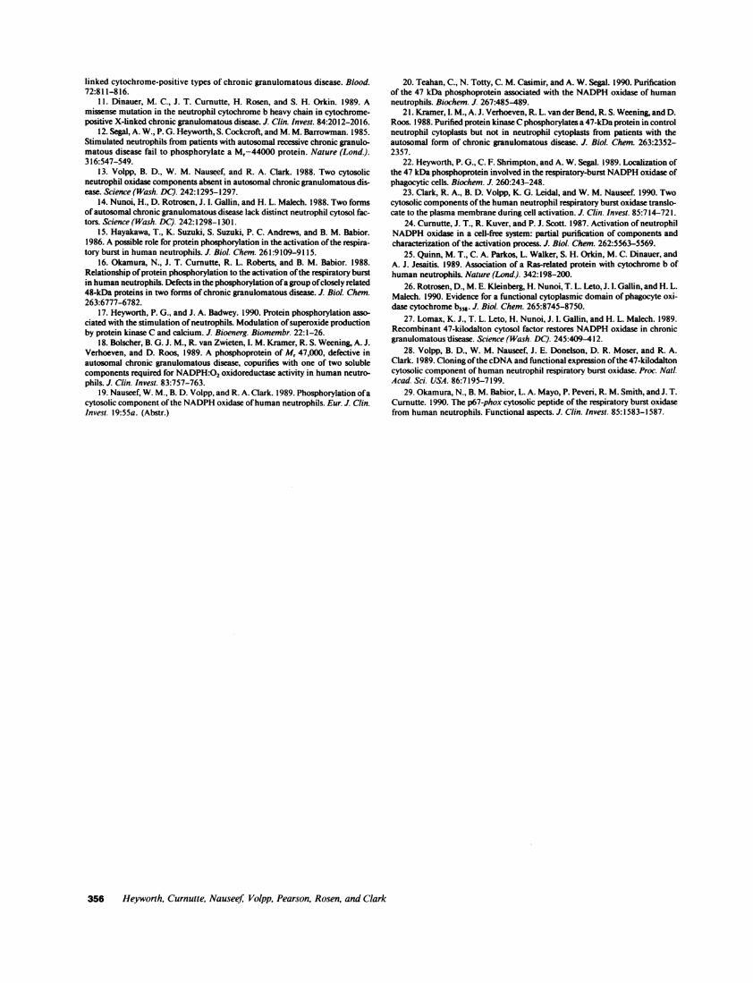
Fig. 3 shows the results of translocation experiments withneutrophils from two patients with p47-phox-deficient CGD.The absence of p47-phox was evident in the autoradiographs ofthe immunoblots of the patients' cytosolic fractions. The levelsof p67-phox were the same as in the control. Despite this, therewas no translocation of p67-phox to the membrane in eitherpatient. In contrast, in two sisters with p67-phox deficiency,p47-phox was detected in the membrane fractions of PMA-stimulated cells (Fig. 4). By densitometry, the relative intensi-ties of these p47-phox bands for the two patients were 1 10 and64, compared with the control band set at 100%. The recoveryof protein in the membrane fraction of the second patient'sPMA-stimulated neutrophils was low (53% of control; see leg-end to Fig. 4), wholly accounting for the lower level of translo-cation.
Neutrophil Nicotinamide Adenine Dinucleotide Phosphate Oxidase Assembly 353
CYTOCHROMEb-DEFICIENT CGD
MEMBRANECGD Control
+
CYTOSOL FRACTIONCGDControl_-+-_ +
SubjectStimulus
- p67
- p47
p67
CYTOCHROMEb Figure 2. Translocationof p47-phox and p67-MISSENCEMUTATION phox in an X-linkedCGDpatient with a
MEMBRANE FRACTION missense mutation inCGD Control Subject the gp9l-phox subunit- + + Stimulus of cytochrome b558-
With the exception of_ -, p67 the modifications in the
preparation of the- _o p47 membrane fraction
from the CGDandcontrol neutrophils (fordetails see Methods),the procedures used in
this experiment were the same as those in Fig. 1. Only the particulate(membrane) fractions are shown in this figure.
4^ _U~E:.m p47
uq 1M --
d-u--.-A
Sif -AILMM-S--
p67
p47
p67
Discussion
Wehave demonstrated that the absence of cytochrome b,58,whether it is due to a primary defect in the p22-phox or thegp9 l -phox subunit, results in the failure of two cytosolic oxi-dase components (p47-phox and p67-phox) to undergo stimu-lus-dependent translocation to the plasma membrane. Thisfinding indicates that cytochrome b558, or a closely linked fac-tor (e.g., rap 1) (25), serves as the membrane docking site forthe components. The ability of antibodies to the cytoplasmiccarboxy-terminal domain of gp9 1 -phox, and the synthetic pep-tides themselves, to block O2 generation in a cell free system,suggest that this cytochrome subunit mediates the interactionwith cytosolic components (26). Translocation of p47-phoxand p67-phox was normal in a variant X-linked form of CGDin which cytochrome b558 is present but does not transfer elec-
p47p47 - DEFICIENT CGD
MEMBRANECGD Control_ + _-+
CYTOSOL FRACTIONCGD Control Subject- + - + Stimulus
Figure 1. Failure of translocation of p47-phox and p67-phox to themembrane in cytochrome b558-negative CGDneutrophils. The CGDfractions in the top four pairs of panels were prepared from theneutrophils of patients with X-linked cytochrome-negative CGDandthose in the bottom panels from a patient with the autosomalrecessive cytochrome-negative variant form. In each panel fractionsprepared in parallel from normal control neutrophils are shown forcomparison. Membrane and cytosol fractions (- 1.5 x 10⁷ and 5X 10⁶ cell equivalents, respectively) from resting (-) and PMA-activated (+) neutrophils were subjected to SDS-PAGE. The amountof protein loaded on the gel from patients' neutrophil fractions never
differed by > 20% from the corresponding control value. The resolvedproteins were electrophoretically transferred to nitrocellulose. Theblots were blocked, probed with B-I (an antiserum that recognizesp47-phox and p67-phox), and exposed to 125I-protein A as describedin Methods. The immunoreactive proteins were detected byautoradiography. The locations of the p47-phox and p67-phox bandsare indicated.
-p67.... ..r"m'
I __ - p47
_-
4.
_
- p67
8- p47
Figure 3. Absence of translocation of p67-phox to the membrane in
p47-phox-deficient CGDpatients. The amount of protein loaded on
the gel from patients' neutrophil fractions did not differ by > 18%from the corresponding control value. Experimental details are thesame as in Fig. 1. A similar result was obtained with neutrophilsfrom one other patient with p47-phox deficiency analyzed in a
separate experiment.
354 Heyworth, Curnutte, NauseefJ Volpp, Pearson, Rosen, and Clark
-- p67
--- p47
I,( .6'1!4
P.. faI'.
'W %,;, si. 1111W.

p67 - DEFICIENT CGDMEMBRANE CYTOSOL FRACTION
CGD Control CGD CGD Control CGD Subject-4+ _+ + -+ - +-+ Stimulus- - - p67
idw -ATE ax __ p47
Figure 4. Translocation of p47-phox to the membrane in p67-phox-deficient CGDpatients. The neutrophils from the two patients inthis experiment were processed simultaneously together with thosefrom a single control subject. The amounts of protein (fig) loaded perlane (reading from left to right) were as follows: Membrane: 175, 139,180, 135, 163, 72; Cytosol: 135, 121, 124, 124, 126, 112.Experimental details are the same as in Fig. 1. A similar result wasobtained with a different p67-phox-deficient patient analyzed in aseparate experiment.
trons as a result of a single amino acid substitution. This is theonly form of CGDstudied in which translocation of both pro-teins was normal, and as such demonstrates that the absence oftranslocation in other forms of CGDis not a secondary effect ofthe failure to generate O°.
Our results are compatible with earlier observations on thephosphorylation of p47-phox in human neutrophils (17). Aftertwo-dimensional electrophoresis of 32P-labeled phosphopro-teins from stimulated neutrophils, p47-phox appeared as achain of six spots, indicating that it contains at least six phos-phorylation sites (10), all of which are on serine residues (16).Moreover, the primary amino acid sequence of p47-phox con-tains six serines in the arginine-rich COOH-terminal domainthat exhibit features of target sites of protein kinases (27,28). Innormal neutrophils activated with PMA, phosphorylated p47-phox was present in both the cytosol and membrane fractions(15, 21, 22). In classic X-linked cytochrome b-negative CGDhowever, the phosphorylated p47-phox was confined to the cy-tosol (22). In addition, two-dimensional electrophoresis re-vealed that in this form of CGDonly four of the six phosphory-lation sites had acquired the 32P label, suggesting that the tworemaining phosphorylation reactions occurred after transloca-tion of the protein to the membrane, in a process that dependedupon cytochrome b558 (10, 16).
The results from our translocation experiments coupledwith the previous phosphorylation studies just discussed, sug-gest the following model as one way to activate the oxidase.Upon neutrophil stimulation the initial steps in phosphoryla-tion of p47-phox occur in the cytosol. The protein then translo-cates to the membrane, binds to a site formed by cytochromeb558, and is phosphorylated at its two remaining sites. In cy-tochrome-negative CGDthe initial phosphorylation steps takeplace, but translocation and further phosphorylation are pre-cluded by the absence of cytochrome b. In X-linked cy-tochrome-positive CGD, in which phosphorylation of p47-phox was normal at all six sites (10), translocation of p47-phoxand p67-phox is normal. This indicates that the Pro Hissubstitution does not interfere with the association of cytosoliccomponents with cytochrome b or the subsequent phosphory-lation of p47-phox. It is important to point out however, thatour data do not necessarily imply a causal relationship between
p47-phox phosphorylation and either oxidase activation ortranslocation of cytosolic components, simply that they canoccur in parallel. Several studies have shown the O° generationcan be activated without apparent p47-phox phosphorylation(reviewed in reference 17). Under these conditions other sig-nals clearly must trigger translocation of the cytosolic compo-nents and activation of the oxidase.
Our finding that translocation of p67-phox is wholly depen-dent on the presence of p47-phox supports the view that cyto-solic components of the NADPHoxidase exist in the form ofcomplexes (13, 24, 29). Translocation of p47-phox, on theother hand, occurred in the absence of p67-phox, suggestingthat the former protein is the ligand that fits into the membranedocking site formed by the cytochrome and that p67-phox fol-lows by virtue of its association with p47-phox. However, it isalso conceivable that p67-phox translocates independently ofp47-phox and recognizes a site formed by the cytochrome/p47-phox complex. In p67-phox deficiency, p47-phox retains theability to bind to the membrane, either alone or as part of asmaller complex. The recent demonstration that phosphoryla-tion of p47-phox was normal (i.e., occurred at all sites) in p67-phox-deficient neutrophils (29), is compatible with our translo-cation data. Howthe membrane complex of cytochrome b558,phosphorylated p47-phox, p67-phox, and perhaps other com-ponents is converted to a catalytically active enzyme systemremains to be determined.
Acknowledgments
This work was supported by grants from the U. S. Public Health Service(AI-24838, RR-00833, AI-28412, HL-34327, and AI-25606), and bygrants from the Department of Veterans Affairs. Dr. Curnutte is anEstablished Investigator of the American Heart Association. Dr. Clarkand Dr. Nauseef are Medical Investigator and Clinical Investigator,respectively, of the Department of Veterans Affairs.
References
1. Curnutte, J. T., and B. M. Babior. 1987. Chronic granulomatous disease.Adv. Hum. Genet. 16:229-297.
2. Clark, R. A. 1990. The human neutrophil respiratory burst oxidase. J.Infect. Dis. 161:1140-1147.
3. Curnutte, J. T. 1988. Classification of chronic granulomatous disease. He-matol. Oncol. Clin. N. Am. 2:241-252.
4. Dinauer, M. C., S. H. Orkin, R. Brown, A. J. Jesaitis, and C. A. Parkos.1987. The glycoprotein encoded by the X-linked chronic granulomatous diseaselocus is a component of the neutrophil cytochrome b complex. Nature (Lond.).327:717-720.
5. Teahan, C., P. Rowe, P. Parker, N. Totty, and A. W. Segal. 1987. TheX-linked chronic granulomatous disease gene codes for the beta-chain of cy-tochrome b-245. Nature (Lond.). 327:720-721.
6. Parkos, C. A., R. A. Allen, C. G. Cochrane, and A. J. Jesaitis. 1987. Purifiedcytochrome b from human granulocyte plasma membrane is comprised of twopolypeptides with relative molecular weights of 91,000 and 22,000. J. Clin. Invest.80:732-742.
7. Segal, A. W. 1987. Absence of both cytochrome b.245 subunits from neutro-phils in X-linked chronic granulomatous disease. Nature (Lond.). 326:88-91.
8. Parkos, C. A., M. C. Dinauer, A. J. Jesaitis, S. H. Orkin, and J. T. Curnutte.1989. Absence of both the 91 kD and 22 kD subunits of human neutrophilcytochrome b in two genetic forms of chronic granulomatous disease. Blood.73:1416-1420.
9. Dinauer, M. C., J. T. Curnutte, and S. H. Orkin. 1989. Gene structure ofneutrophil cytochrome b light chain and mutations in autosomal recessivechronic granulomatous disease. Blood. 74:107a. (Abstr.)
10. Okamura, N., S. E. Malawista, R. L. Roberts, H. Rosen, H. D. Ochs, B. M.Babior, and J. T. Curnutte. 1988. Phosphorylation of the oxidase-related 48 Kphosphoprotein family in the unusual autosomal cytochrome-negative and X-
Neutrophil Nicotinamide Adenine Dinucleotide Phosphate Oxidase Assembly 355
linked cytochrome-positive types of chronic granulomatous disease. Blood.72:811-816.
11. Dinauer, M. C., J. T. Curnutte, H. Rosen, and S. H. Orkin. 1989. Amissense mutation in the neutrophil cytochrome b heavy chain in cytochrome-positive X-linked chronic granulomatous disease. J. Clin. Invest. 84:2012-2016.
12. Segal, A. W., P. G. Heyworth, S. Cockcroft, and M. M. Barrowman. 1985.Stimulated neutrophils from patients with autosomal recessive chronic granulo-matous disease fail to phosphorylate a M,-44000 protein. Nature (Lond.).316:547-549.
13. Volpp, B. D., W. M. Nauseef, and R. A. Clark. 1988. Two cytosolicneutrophil oxidase components absent in autosomal chronic granulomatous dis-ease. Science (Wash. DC). 242:1295-1297.
14. Nunoi, H., D. Rotrosen, J. I. Gallin, and H. L. Malech. 1988. Two formsof autosomal chronic granulomatous disease lack distinct neutrophil cytosol fac-tors. Science (Wash. DC). 242:1298-1301.
15. Hayakawa, T., K. Suzuki, S. Suzuki, P. C. Andrews, and B. M. Babior.1986. A possible role for protein phosphorylation in the activation of the respira-tory burst in human neutrophils. J. Biol. Chem. 261:9109-9115.
16. Okamura, N., J. T. Curnutte, R. L. Roberts, and B. M. Babior. 1988.Relationship of protein phosphorylation to the activation of the respiratory burstin human neutrophils. Defects in the phosphorylation of a group of closely related48-kDa proteins in two forms of chronic granulomatous disease. J. Biol. Chem.263:6777-6782.
17. Heyworth, P. G., and J. A. Badwey. 1990. Protein phosphorylation asso-ciated with the stimulation of neutrophils. Modulation of superoxide productionby protein kinase C and calcium. J. Bioenerg. Biomembr. 22:1-26.
18. Bolscher, B. G. J. M., R. van Zwieten, I. M. Kramer, R. S. Weening, A. J.Verhoeven, and D. Roos, 1989. A phosphoprotein of M, 47,000, defective inautosomal chronic granulomatous disease, copurifies with one of two solublecomponents required for NADPH:02 oxidoreductase activity in human neutro-phils. J. Clin. Invest. 83:757-763.
19. Nauseef, W. M., B. D. Volpp, and R. A. Clark. 1989. Phosphorylation of acytosolic component of the NADPHoxidase of human neutrophils. Eur. J. Clin.Invest. 19:55a. (Abstr.)
20. Teahan, C., N. Totty, C. M. Casimir, and A. W. Segal. 1990. Purificationof the 47 kDa phosphoprotein associated with the NADPHoxidase of humanneutrophils. Biochem. J. 267:485-489.
21. Kramer, I. M., A. J. Verhoeven, R. L. van der Bend, R. S. Weening, and D.Roos. 1988. Purified protein kinase Cphosphorylates a 47-kDa protein in controlneutrophil cytoplasts but not in neutrophil cytoplasts from patients with theautosomal form of chronic granulomatous disease. J. Biol. Chem. 263:2352-2357.
22. Heyworth, P. G., C. F. Shrimpton, and A. W. Segal. 1989. Localization ofthe 47 kDa phosphoprotein involved in the respiratory-burst NADPHoxidase ofphagocytic cells. Biochem. J. 260:243-248.
23. Clark, R. A., B. D. Volpp, K. G. Leidal, and W. M. Nauseef. 1990. Twocytosolic components of the human neutrophil respiratory burst oxidase translo-cate to the plasma membrane during cell activation. J. Clin. Invest. 85:714-72 1.
24. Curnutte, J. T., R. Kuver, and P. J. Scott. 1987. Activation of neutrophilNADPHoxidase in a cell-free system: partial purification of components andcharacterization of the activation process. J. Biol. Chem. 262:5563-5569.
25. Quinn, M. T., C. A. Parkos, L. Walker, S. H. Orkin, M. C. Dinauer, andA. J. Jesaitis. 1989. Association of a Ras-related protein with cytochrome b ofhuman neutrophils. Nature (Lond.). 342:198-200.
26. Rotrosen, D., M. E. Kleinberg, H. Nunoi, T. L. Leto, J. I. Gallin, and H. L.Malech. 1990. Evidence for a functional cytoplasmic domain of phagocyte oxi-dase cytochrome b,,. J. Biol. Chem. 265:8745-8750.
27. Lomax, K. J., T. L. Leto, H. Nunoi, J. 1. Gallin, and H. L. Malech. 1989.Recombinant 47-kilodalton cytosol factor restores NADPHoxidase in chronicgranulomatous disease. Science (Wash. DC). 245:409-412.
28. Volpp, B. D., W. M. Nauseef, J. E. Donelson, D. R. Moser, and R. A.Clark. 1989. Cloning of the cDNAand functional expression ofthe 47-kilodaltoncytosolic component of human neutrophil respiratory burst oxidase. Proc. Natl.Acad. Sci. USA. 86:7195-7199.
29. Okamura, N., B. M. Babior, L. A. Mayo, P. Peveri, R. M. Smith, and J. T.Curnutte. 1990. The p67-phox cytosolic peptide of the respiratory burst oxidasefrom human neutrophils. Functional aspects. J. Clin. Invest. 85:1583-1587.
356 Heyworth, Curnutte, Nauseef; Votpp, Pearson, Rosen, and Clark