Embed Size (px)
Citation preview

www.aana.com/aanajournalonline.aspx AANA Journal April 2012 Vol. 80, No. 2 115
This case report describes a 2.5-year-old girl who was hospitalized with complaints of abdominal pain and vomiting for 2 days. Abdominal ultrasound revealed small bowel–to–small bowel intussusception. Diagnos-tic laparoscopic-assisted exploration of the abdomen revealed 4 separate intestinal intussusceptions along with multiple dark intraluminal masses within the small intestine. Laparoscopic reduction of the intussusceptions was unsuccessful. Laparotomy allowed palpation of the entire small intestine with extraction of the masses, which were found to be human hair (trichobezoars). The intussusceptions were reduced, and the multiple masses were removed through a single enterotomy.
The child recovered following surgery and was dis-charged home to her family. The surgeon counseled the parents before discharging the patient and recom-mended follow-up counseling for their child. The par-ents were given information about trichophagia and strategies to reduce the behavior in their child.
A comprehensive literature review revealed this to be the youngest reported case of intussusception and Rapunzel syndrome due to trichobezoars.
Keywords: Bezoar, pediatric, Rapunzel syndrome, trichobezoar.
Rapunzel Syndrome in a Pediatric Patient: A Case Report
Elizabeth Middleton, CRNA, MSNLynn Fitzgerald Macksey, CRNA, MSNJ. Duncan Phillips, MD
Abezoar is persistent, ingested material that collects within the gastrointestinal tract.1 Bezoars can be made of up vegetable or fruit fibers (phytobezoars), milk curds (lac-tobezoars) or any indigestible material that
is ingested.2 The most common type of bezoar, a gastric trichobezoar, is made up of human hair and found in the stomach.
Trichobezoars are a relatively rare surgical finding. These bezoars can cause intestinal obstruction. Human hair is an indigestible material that is prone to collect between the mucosal folds of the stomach because of the peristaltic contractions and the smooth surface of the hair.3
Trichobezoars are typically due to trichotillomania (the compulsive pulling out of one’s own hair) and trichophagia (swallowing the hair).3 Rarely do adoles-cents chew hair from sources other than their own body.2 In approximately 1% of patients with trichophagia, a trichobezoar develops.2
Case ReportA 2.5-year-old girl was examined because of abdominal pain and vomiting for 2 days. The child was first en-countered in the preoperative area accompanied by her mother, father, and a 15-month old brother. The child lay on her stretcher quietly, eyes open, without any obvious interaction with her family. This behavior was observed for approximately 15 minutes, and neither the child nor family showed a lot of emotion when she was taken from
the preoperative area back to the operating room. This was felt to be different than what is typically anticipated from a 2.5-year-old toddler who clings to the parent, cries, and is afraid to openly go to a “stranger.” She had not received any preoperative sedation or pain medications before coming to the preoperative area. A 22-gauge intra-venous catheter was inserted in the left antecubital space when the child arrived in the preoperative area.
The parents reported that she took no regularly sched-uled medicines at home, had no known drug allergies or chronic illnesses, and that she had been full-term at birth. She had a surgical history of a right eyebrow laceration repair. She weighed 20.4 kg.
An abdominal ultrasound showed intussusception of the small bowel into itself. Air reduction was attempted but unsuccessful. The patient was scheduled for a diag-nostic laparoscopy. General anesthesia was discussed, and parental consent was obtained. The patient was scored as having an ASA II physical status because of her new onset of abdominal pain and vomiting.
Preoperative vital signs included a blood pressure of 104/72 mm Hg, a heart rate of 129/min, and a respiratory rate of 24/min. Before leaving the preoperative area, 1 mg of midazolam was administered intravenously. A pulse oximeter was applied for transport, and oxygen satura-tions were maintained at 100% to the operating room. No supplemental oxygen was given en route.
In the operating room, the child was intermittently sleeping and opening her eyes but did not cry. Standard ASA monitors were applied. Preinduction vital signs were
116 AANA Journal April 2012 Vol. 80, No. 2 www.aana.com/aanajournalonline.aspx
as follows: blood pressure, 107/62 mm Hg; heart rate, 110/min; respiratory rate, 32/min; and oxygen saturation, 100%. An electrocardiogram showed sinus tachycardia.
A smooth rapid-sequence induction was accomplished with 60 mg of propofol and 50 mg of succinylcholine. The child was intubated with a 5.0 oral cuffed endotra-cheal tube using a Macintosh blade size 1. The cuff leak pressure was 21 cm with 0.5 mL of air inflated in the endotracheal tube cuff. The end-tidal carbon dioxide was positive with bilateral breath sounds after 1 attempt at direct laryngoscopy.
Sevoflurane was delivered at an expired concentration level of 3.5%. Anesthetic maintenance was facilitated with sevoflurane, rocuronium, and fentanyl. After induction, the blood pressure was 98/48 mm Hg and the heart rate, 128/min. Ventilator settings were as follows: pressure controlled ventilations, 30/min; tidal volume, 107 mL; and peak inspiratory pressure, 18 cm H2O. A 12F naso-gastric tube was inserted through the left nostril, passed into the stomach without resistance, and secured once the correct internal position was verified by the surgeon.
Laparoscopic examination of the small intestine dem-onstrated multiple areas of the ileum, which were intus-suscepted into each other (Figure 1). The areas were reduced with Hunter bowel graspers, but intussuscep-tion would occur again (Figure 2). At least 4 different areas of intussusception were identified. Laparoscopic examination revealed the terminal ileum had an unusual accordion-type appearance, in that it would not stretch out or lay out normally. At this point, it also became obvious that there were multiple blackish masses within the lumen of the small bowel that could be seen through the lumen wall and could be felt with the Hunter bowel grasper.
Initially, it was thought the multiple dark masses iden-
tified were small enough and might easily pass through the intestinal lumen to be expelled. However, once the number and size of the masses was appreciated, it was decided the child would not be able to pass them on her own and they would have to be surgically removed.
To exteriorize parts of the small intestine from the peritoneal cavity to palpate the intestine, the surgeon reached in through the umbilical trocar, grasped one of the luminal masses, pulled that loop of bowel up to the undersurface of the umbilicus, and then removed the umbilical trocar. The umbilical incision was enlarged, allowing the small bowel loop to be exteriorized through the incision line and exposed for examination. The intus-suscepted bowel was also visualized externally.
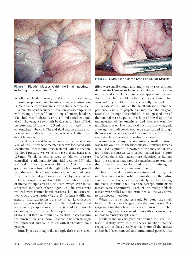
A small enterotomy (incision into the small intestine) was made over one of the black masses. DeBakey forceps were used to pull out a portion of the material; it was found that the masses were balled, matted hair (Figure 3). When the black masses were identified as human hair, the surgeon requested the anesthetist to examine the patient’s scalp for localized areas of missing or thinned hair; however, none were found.
The entire small intestine was eviscerated through the umbilical incision to enable examination of the entire small intestine. Forceps were repeatedly inserted, feeding the small intestine back over the forceps, until black masses were encountered. Each of the multiple black masses were pulled out and examined, all the way down to the ileocecal junction.
When no further masses could be found, the small intestinal lumen was irrigated via the enterotomy. The surgeon noted that a few tiny pieces of hair remained, but it was thought that these would pass without causing the mucosa to “intussuscept” again.
Sterile saline was irrigated all through the small in-testine, distally down to the ileocecal junction into the cecum until it flowed easily to make sure all the masses of hair had been removed and intraluminal patency was
Figure 1. Blackish Masses Within the Small Intestine, Including Intussuscepted Areas
Figure 2. Examination of the Small Bowel for Masses
www.aana.com/aanajournalonline.aspx AANA Journal April 2012 Vol. 80, No. 2 117
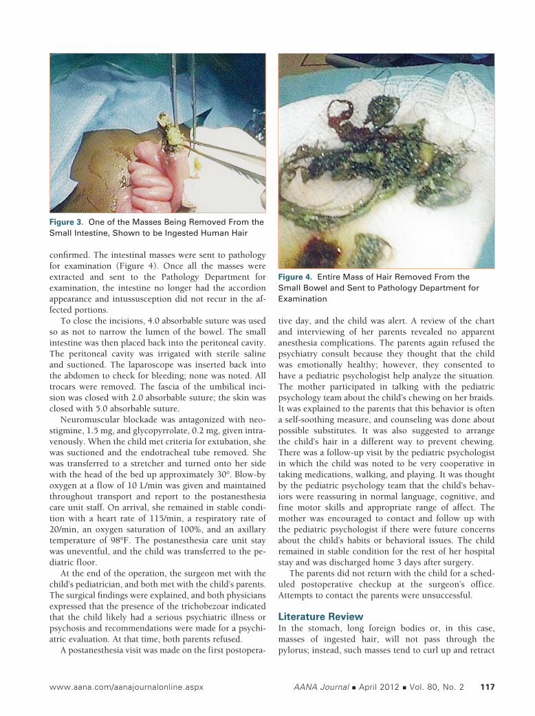
confirmed. The intestinal masses were sent to pathology for examination (Figure 4). Once all the masses were extracted and sent to the Pathology Department for examination, the intestine no longer had the accordion appearance and intussusception did not recur in the af-fected portions.
To close the incisions, 4.0 absorbable suture was used so as not to narrow the lumen of the bowel. The small intestine was then placed back into the peritoneal cavity. The peritoneal cavity was irrigated with sterile saline and suctioned. The laparoscope was inserted back into the abdomen to check for bleeding; none was noted. All trocars were removed. The fascia of the umbilical inci-sion was closed with 2.0 absorbable suture; the skin was closed with 5.0 absorbable suture.
Neuromuscular blockade was antagonized with neo-stigmine, 1.5 mg, and glycopyrrolate, 0.2 mg, given intra-venously. When the child met criteria for extubation, she was suctioned and the endotracheal tube removed. She was transferred to a stretcher and turned onto her side with the head of the bed up approximately 30°. Blow-by oxygen at a flow of 10 L/min was given and maintained throughout transport and report to the postanesthesia care unit staff. On arrival, she remained in stable condi-tion with a heart rate of 115/min, a respiratory rate of 20/min, an oxygen saturation of 100%, and an axillary temperature of 98°F. The postanesthesia care unit stay was uneventful, and the child was transferred to the pe-diatric floor.
At the end of the operation, the surgeon met with the child’s pediatrician, and both met with the child’s parents. The surgical findings were explained, and both physicians expressed that the presence of the trichobezoar indicated that the child likely had a serious psychiatric illness or psychosis and recommendations were made for a psychi-atric evaluation. At that time, both parents refused.
A postanesthesia visit was made on the first postopera-
tive day, and the child was alert. A review of the chart and interviewing of her parents revealed no apparent anesthesia complications. The parents again refused the psychiatry consult because they thought that the child was emotionally healthy; however, they consented to have a pediatric psychologist help analyze the situation. The mother participated in talking with the pediatric psychology team about the child’s chewing on her braids. It was explained to the parents that this behavior is often a self-soothing measure, and counseling was done about possible substitutes. It was also suggested to arrange the child’s hair in a different way to prevent chewing. There was a follow-up visit by the pediatric psychologist in which the child was noted to be very cooperative in taking medications, walking, and playing. It was thought by the pediatric psychology team that the child’s behav-iors were reassuring in normal language, cognitive, and fine motor skills and appropriate range of affect. The mother was encouraged to contact and follow up with the pediatric psychologist if there were future concerns about the child’s habits or behavioral issues. The child remained in stable condition for the rest of her hospital stay and was discharged home 3 days after surgery.
The parents did not return with the child for a sched-uled postoperative checkup at the surgeon’s office. Attempts to contact the parents were unsuccessful.
Literature ReviewIn the stomach, long foreign bodies or, in this case, masses of ingested hair, will not pass through the pylorus; instead, such masses tend to curl up and retract
Figure 3. One of the Masses Being Removed From the Small Intestine, Shown to be Ingested Human Hair
Figure 4. Entire Mass of Hair Removed From the Small Bowel and Sent to Pathology Department for Examination
118 AANA Journal April 2012 Vol. 80, No. 2 www.aana.com/aanajournalonline.aspx
back into the stomach. Continued hair ingestion creates a tightly packed, growing ball of hair forming a large, hard obstruction. The hair then traps thicker materials that hold the hairball together, making removal difficult.4
The churning action of the stomach during attempted digestion helps entangle new hair into an already formed mass. Mucus covers the gastric trichobezoar, giving it a shiny surface. The mass is usually black because of the acidic nature of the stomach that denatures the proteins. Patients often have a foul smelling breath odor because of the decomposition and fermentation of fats.2
The primary symptoms of a gastric trichobezoar include epigastric pain, nausea, and vomiting. In addi-tion to severe halitosis, patients often have patchy areas of hair loss found during physical examination. Patients often remain asymptomatic for many years and remain so until the bezoar increases in size to the point of ob-struction.2 Gastric trichobezoars can also cause gastric ulcers with subsequent gastrointestinal blood loss and anemia.
Rapunzel syndrome is a potential complication of a trichobezoar. It has been defined as a gastric bezoar with a “tail” that can extend through the pylorus all the way to the ileocecal junction.2 This tail consists of the distal ends of long pieces or fragments of hair, some still attached to the growing mass in the stomach.
Rapunzel syndrome remains uncommon, with fewer than 40 cases reported.2 Clinicians find this syndrome most often in emotionally disturbed and/or mentally chal-lenged adolescents, usually between the ages of 13 and 20 years. It reflects an underlying psychiatric disorder, not a problem with gastrointestinal motility.2 Trichotillomania and trichophagia are associated with Rapunzel syndrome and are often indicative of psychosis.2
The name Rapunzel syndrome is from the well-known fairy tale by the Grimm brothers. Vaughan et al5 first reported Rapunzel syndrome and so named it because of the long hair that can extend into the small intestine.
Gonuguntla and Joshi3 reported the youngest docu-mented case of Rapunzel syndrome in the United States: a 5-year-old girl with mental retardation with abdominal pain, vomiting, and a nontender abdominal mass.2
Several methods are used in diagnosis. A contrast upper gastrointestinal series often helps to identify trichobezoars. Because of the characteristic appearance of trichobezoars, they are also easily identifiable by abdomi-nal ultrasound or computed tomography scan.6
Upper endoscopy, considered the “gold standard” for diagnosis, definitively confirms the presence of a tricho-bezoar. Endoscopy can also help a surgeon distinguish between a trichobezoar and another foreign body that can be broken apart and removed endoscopically.3
Several options for the treatment of trichobezoars exist. Treatment focuses on removal of the mass and prevention of recurrence.2 Chemical substances (eg, coca
soft drinks) have been introduced into the stomach to try to dissolve the material. Few successes with this ap-proach have been reported.7
Few successful attempts at endoscopic trichobezoar removal have been documented. In the cases in which endoscopic removal was successful, the trichobezoar was small. Other endoscopic methods identified in the litera-ture include mechanical fragmentation with a modified needle/knife (bezotome) to pulverize bezoars mechanical-ly. A modified lithotripter (bezotriptor) can be used in the endoscopic approach to successfully break apart bezoars with acoustic waves. However, in the case of larger tricho-bezoars, these methods are less likely to be successful.1,3
The advent of minimally invasive surgical techniques has increased the number of laparoscopic attempts to remove trichobezoars, but these procedures are often difficult. One report of a successful removal describes a 10-year-old child who underwent laparoscopic removal through an umbilical incision without any extending in-cision and no substantial complications.4 Advantages of laparoscopic removal are an improved cosmetic appear-ance, fewer postoperative complications, and reduced hospital stay.3
Laparoscopic removal of an entire bezoar is difficult without spillage of hairs into the peritoneal cavity.4 This approach often requires substantially more time because of the complexity of the operation and the need to examine the rest of the bowel for additional pieces that could cause secondary obstructions.3
Open surgery is the most common technique used for trichobezoar removal. Some physicians consider conventional laparotomy to be the treatment of choice.3 In a literature review, 100 cases were identified in which conventional laparotomy was successful in the removal of the trichobezoar(s).3 In the 100 cases, 12% involved 1 or more complications, including perfora-tion of the intestine during removal of the trichobezoar, minor wound infection, pneumonia, paralytic ileus, ileal trichobezoar, and fecal leakage through the lower part of the laparotomy wound.3 Other surgical complications include upper digestive tract bleeding, anemia, bowel intussusception, and, rarely, death.7 If the small bowel loop is damaged, revealing necrosis or perforation, bowel resection may be required.7
After the trichobezoar has been removed by laparoto-my, it is essential to explore the remainder of the small intestine and stomach to look for any retained bezoars.6
DiscussionPrompt diagnosis of a trichobezoar is essential in pre-venting complications. When not recognized, the contin-ued ingestion of hair can cause problems such as gastric mucosal erosion, ulceration, and perforation of the stomach and/or small intestine. Ulceration and perfora-tion generally occur because of the substantial size of an

www.aana.com/aanajournalonline.aspx AANA Journal April 2012 Vol. 80, No. 2 119
unrecognized bezoar, which ultimately decreases blood flow to the stomach and small intestine. Obstructive jaundice, protein-losing enteropathy, and pancreatitis can also develop, and death can occur.3
Trichobezoar should be considered as a possible diagnosis in adolescents with vague symptoms such as epigastric pain, nausea and vomiting, fatigue, weight loss, and an epigastric mass.3 Bezoars have been shown to be the cause of 0.8% of small bowel obstructions that were treated laparoscopically.4
Treatment consists of not only prompt recognition, diagnosis, and removal of the bezoar, but also adequate follow-up. Pharmacotherapy has been suggested to treat trichotillomania; Fluoxetine or other selective serotonin reuptake inhibitors may be used.2
Psychiatric follow-up is important in the treatment and prevention of recurrence of Rapunzel syndrome. The need for adequate counseling should be empha-sized and extended to the family members involved in caring for the child.7 Although infrequent, the trauma of surgery may stop a potential recurrence.7 Documented cases of Rapunzel syndrome are rare; however, they have been linked to early childhood neglect or abuse, psychiatric conditions, or mental retardation.1 The long-term prognosis is excellent, provided that regular follow-up and behavioral therapy are maintained, helping to control trichophagia.2
REFERENCES 1. Macksey L. Aspirated bezoar in a pediatric patient: a case study.
AANA J. 2006;74(4):295-298.
2. Gonuguntla V, Joshi D. Rapunzel syndrome: a comprehensive review of an unusual case of a trichobezoar. Clin Med Res. 2009;7(3):99-102.
3. Gorter RR, Kneepkens CM, Mattens EC, Aronson DC, Heij, HA. Man-agement of trichobezoar: case report and literature review. Pediatr Surg Int. 2010;26(5):457-463.
4. Fraser JD, Leys CM, St Peter SD. Laparoscopic removal of a gastric trichobezoar in a pediatric patient. J Laparoendosc Adv Surg Techn A. 2009;19(6):835-837.
5. Vaughan ED Jr, Sawyers JL, Scott HW Jr. The Rapunzel syndrome: an unusual complication of intestinal bezoar. Surgery. 1968;63(2);339-343.
6. Taori K, Deshmukh A, Rathod J, Sheorain V, Sanyal R. Rapunzel syndrome: a trichobezoar extending into the ileum. Appl Radiol. 2008 March; 34-35.
7. Lopes LR, Oliveira PSS, Pracucho EM, Camargo MA, de Souza Coelho Neto J, Andreollo NA. The Rapunzel syndrome: an unusual trichobezoar presentation. Case Rep Med. 2010;2010:841028 (doi:10.1155/2010/841028.)
AUTHORSElizabeth Middleton, CRNA, MSN, is a nurse anesthetist with American Anesthesiology and practices at Duke Raleigh Hospital, Raleigh, North Carolina. She was a student at the Raleigh School of Nurse Anesthesia at the time this article was written. Email: [email protected].
Lynn Fitzgerald Macksey, CRNA, MSN, is a staff nurse anesthetist with American Anesthesiology. She works at WakeMed Hospital, Raleigh, North Carolina. Email: [email protected].
J. Duncan Phillips, MD, is surgeon in chief at WakeMed’s Children Hospital in Raleigh, North Carolina.