Embed Size (px)
Citation preview
This article was downloaded by: [Jose Maria Aguilar-Camacho]On: 24 July 2013, At: 11:15Publisher: Taylor & FrancisInforma Ltd Registered in England and Wales Registered Number: 1072954 Registeredoffice: Mortimer House, 37-41 Mortimer Street, London W1T 3JH, UK
Journal of Natural HistoryPublication details, including instructions for authors andsubscription information:http://www.tandfonline.com/loi/tnah20
Raspailiidae (Porifera: Demospongiae:Axinellida) from the Mexican PacificOcean with the description of sevennew speciesJose Maria Aguilar-Camacho a & Jose Luis Carballo aa Laboratorio de Ecologia del Bentos , Instituto de Ciencias delMar y Limnología, Universidad Nacional Autónoma de México(Estación Mazatlán) , Mazatlan , MexicoPublished online: 01 May 2013.
To cite this article: Jose Maria Aguilar-Camacho & Jose Luis Carballo (2013) Raspailiidae (Porifera:Demospongiae: Axinellida) from the Mexican Pacific Ocean with the description of seven newspecies, Journal of Natural History, 47:25-28, 1663-1706, DOI: 10.1080/00222933.2013.769642
To link to this article: http://dx.doi.org/10.1080/00222933.2013.769642
PLEASE SCROLL DOWN FOR ARTICLE
Taylor & Francis makes every effort to ensure the accuracy of all the information (the“Content”) contained in the publications on our platform. However, Taylor & Francis,our agents, and our licensors make no representations or warranties whatsoever as tothe accuracy, completeness, or suitability for any purpose of the Content. Any opinionsand views expressed in this publication are the opinions and views of the authors,and are not the views of or endorsed by Taylor & Francis. The accuracy of the Contentshould not be relied upon and should be independently verified with primary sourcesof information. Taylor and Francis shall not be liable for any losses, actions, claims,proceedings, demands, costs, expenses, damages, and other liabilities whatsoever orhowsoever caused arising directly or indirectly in connection with, in relation to or arisingout of the use of the Content.
This article may be used for research, teaching, and private study purposes. Anysubstantial or systematic reproduction, redistribution, reselling, loan, sub-licensing,systematic supply, or distribution in any form to anyone is expressly forbidden. Terms &Conditions of access and use can be found at http://www.tandfonline.com/page/terms-and-conditions

Journal of Natural History, 2013Vol. 47, Nos. 25–28, 1663–1706, http://dx.doi.org/10.1080/00222933.2013.769642
Raspailiidae (Porifera: Demospongiae: Axinellida) from the MexicanPacific Ocean with the description of seven new species
Jose Maria Aguilar-Camacho* and Jose Luis Carballo
Laboratorio de Ecologia del Bentos, Instituto de Ciencias del Mar y Limnología, UniversidadNacional Autónoma de México (Estación Mazatlán), Mazatlan, Mexico
(Received 06 June 2012; final version received 21 January 2013; first published online 1 May 2013)
The taxonomy of the family Raspailiidae has always been controversial. Thefamily was first included in the order Poecilosclerida. It was then allocated tothe order Axinellida and later moved back to Poecilosclerida. Currently with thedevelopment of molecular tools it has been assigned to the order Axinellida.In this contribution we describe 10 species from the Mexican Pacific Ocean. Sevenof them are new to science: Raspailia (Parasyringella) rubra sp. nov., Raspailia(Raspaxilla) hymani (Dickinson 1945), Raspailia (Raspaxilla) hyle (de Laubenfels1930), Aulospongus cerebella (Dickinson 1945), Aulospongus californianus sp. nov.,Aulospongus aurantiacus sp. nov., Eurypon patriciae sp. nov., Eurypon tylospinosumsp. nov., Eurypon diversicolor sp. nov. and Eurypon brunus sp. nov. We discuss thegenus Eurypon and include a table for all the species described worldwide with somecomments about this genus.
http://www.zoobank.org/urn:lsid:zoobank.org:pub:D462084B-EE9C-4C61-884A-C9DB70003B4A
Keywords: Porifera; Raspailiidae; Mexican Pacific; new species; taxonomy
Introduction
The sponges of the family Raspailiidae are characterized by a specialized ectosomalskeleton consisting of small thin styles, oxeas or anisoxeas. The choanosomal skele-ton is reticulate, plumoreticulate, axial, extra-axial or hymedesmoid (Hooper 2002).Monactinal or diactinal spicules are coring the primary fibres. Echinating spiculesare also present (acanthostyles or microspined rhabdostyles). Microscleres are usu-ally absent, although a few genera have raphides in bundles (trichodragmas) (Hooper1991).
Hentschel (1923) allocated sponges with sigmas and chelae as microscleres underthe name of Raspailiidae. Bergquist (1970) considered that the growth form andthe choanosomal skeleton were diagnostic features and recognized two families:Euryponidae and Raspailiidae. These families were allocated to the order Axinellida.
Sponges of the family Euryponidae have a hymedesmoid skeleton, monactines,diactines or tetractines and echinating spicules (genera: Eurypon, Tricheurypon,Acantheurypon). Raspailiidae are characterized by an axial, extra-axial or reticu-late skeleton, with monactinal or diactinal choanosomal spicules and echinatingacanthostyles (genera: Raspailia, Aulospongus, Endectyon and others) (Bergquist1970).
*Corresponding author. Email: [email protected]
© 2013 Taylor & Francis
Dow
nloa
ded
by [
Jose
Mar
ia A
guila
r-C
amac
ho]
at 1
1:15
24
July
201
3

1664 J.M. Aguilar-Camacho and J.L. Carballo
Van Soest et al. (1990) considered the order Axinellida as an artificial group andsynonymized it with the order Halichondrida, but they considered Axinellidae as avalid family.
Hooper (1991) stated that the skeletal organization and the presence of echinatingacanthostyles was a symplesiomorphic character similar to species of the familyMicrocionidae. Due to these morphological features, the family Raspailiidae wasmoved into the suborder Microcionina (order: Poecilosclerida) (Hooper 2002).
Erpenbeck et al. (2007) demonstrated that some genera of the family Raspailiidaewere closely related to the family Axinellidae using ribosomal markers. Morrow et al.(2012) used nuclear and mitochondrial markers and resurrected the order Axinellidaand included the families: Axinellidae Carter 1875, Raspailiidae Nardo 1833 andStelligeridae Lendenfeld 1898.
In the Eastern Pacific there are few taxonomic studies of the family Raspailiidaeand/or of the order Axinellida (de Laubenfels 1932; Dickinson 1945; Desqueyorux-Faundez and van Soest 1997; van Soest et al. 2012a). In this study we describe10 species of this family from the Mexican Pacific Ocean, seven of them are newto science: Raspailia (Parasyringella) rubra sp. nov., Raspailia (Raspaxilla) hymani(Dickinson 1945), Raspailia (Raspaxilla) hyle (de Laubenfels 1930), Aulospongus cere-bella (Dickinson 1945), Aulospongus californianus sp. nov., Aulospongus aurantiacus sp.nov., Eurypon patriciae sp. nov., Eurypon tylospinosum sp. nov., Eurypon diversicolor sp.nov. and Eurypon brunus sp. nov. Based on the literature we discuss the genus Euryponand consider that there are some species assigned to this genus that do not have itsmorphological features.
Material and methods
Specimens from shallow waters were collected by snorkelling, diving and by bot-tom trawling in deeper waters from the Mexican Pacific. Sponges were fixed in4% formaldehyde and transferred to 70% ethanol for preservation. Spicule andskeleton preparation for light and scanning electron microscopy followed the tech-niques described by Boury-Esnault and Rützler (1997). Twenty-five spicules of eachdifferent category chosen at random were measured for each specimen. The minimum-(average)-maximum measurement for each spicule category was calculated.
Holotypes and paratypes were deposited in the Museo de Ciencias Naturales deMadrid (MCNM), and in the “Colección de Esponjas del Pacífico Mexicano” (LEB-ICML-UNAM). Additional material from the Los Angeles County Museum (LACM)and the Allan Hancock Foundation (AHF) was also examined.
Results
Order AXINELLIDA Lévi, 1973Family RASPAILIIDAE Nardo, 1833
Subfamily RASPAILIINAE Nardo, 1833Genus Raspailia Nardo, 1833
Subgenus Parasyringella Topsent, 1928Raspailia (Parasyringella) rubra sp. nov.
(Figures 1A, 2, 3)
Dow
nloa
ded
by [
Jose
Mar
ia A
guila
r-C
amac
ho]
at 1
1:15
24
July
201
3

Journal of Natural History 1665
Figure 1. Photographs of preserved sponges from this study. (A) Raspailia (Parasyringella) rubrasp. nov. (B) Raspailia (Raspaxilla) hymani (Dickinson 1945). (C) Raspailia (Raspaxilla) hyle (deLaubenfels 1930). (D) Aulospongus cerebella (Dickinson 1945). (E) Aulospongus californianus sp.nov. (F) Aulospongus aurantiacus sp. nov. Scale bars: 1 cm (A, D, F); 2 cm (C, E); 4 cm (B).
Dow
nloa
ded
by [
Jose
Mar
ia A
guila
r-C
amac
ho]
at 1
1:15
24
July
201
3
1666 J.M. Aguilar-Camacho and J.L. Carballo
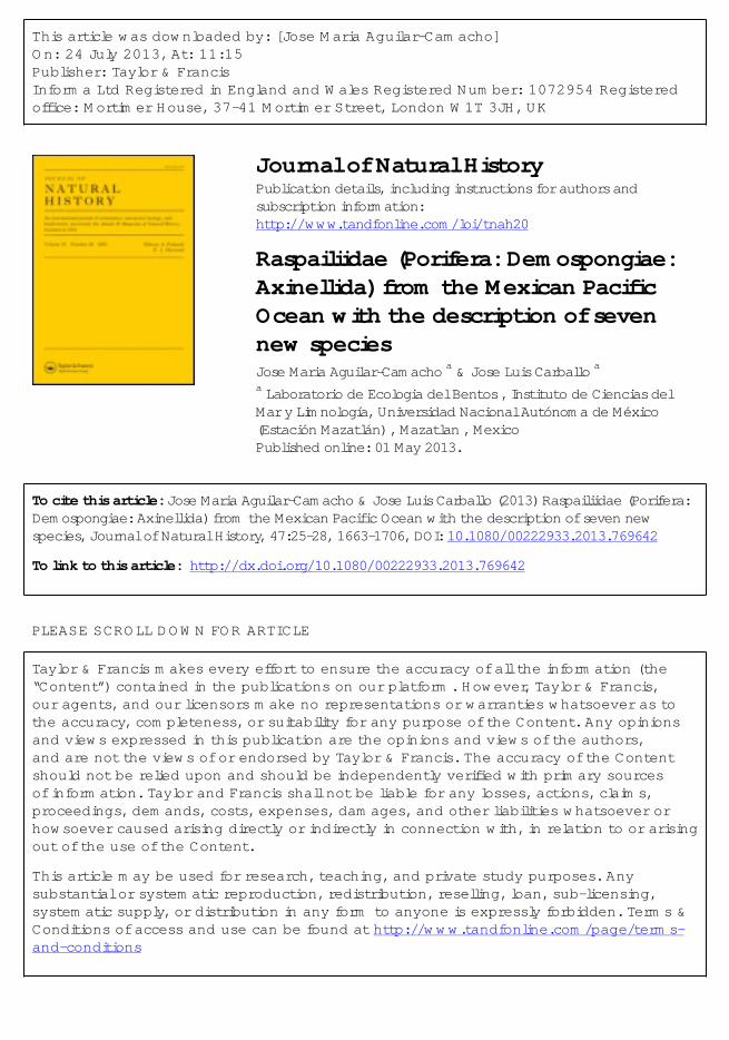
Figure 2. Raspailia (Parasyringella) rubra sp. nov. Scanning electron microscopy images ofspicules. (A) Extra-axial style head; (B) extra-axial style end; (C) choanosomal oxeas; (D)ectosomal style. Scale bars: 4 µm (A),10 µm (B), 50 µm (C), 100 µm (D).
Material examined
Holotype: MCNM 1.01/655, 11/10/2006, Isla Redonda (Marietas Nayarit) 13 m(20◦42′04′′ N, 105◦34′31′′ W). Paratype: 1601-LEB-ICML-UNAM, 11/10/2006, IslaRedonda (Marietas Nayarit) 11 m (20◦42′04′′ N, 105◦34′31′′ W).
Description
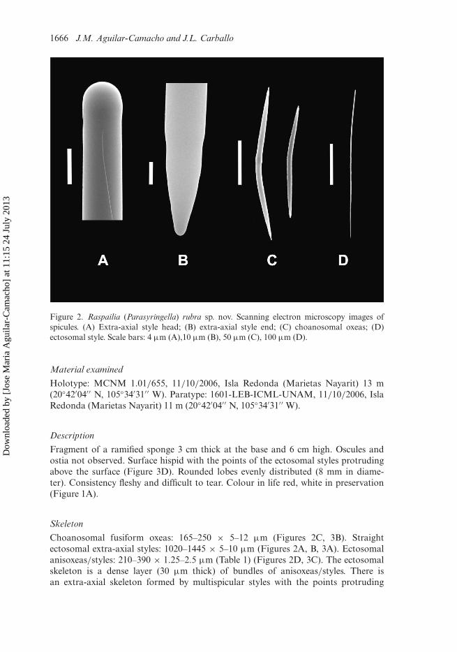
Fragment of a ramified sponge 3 cm thick at the base and 6 cm high. Oscules andostia not observed. Surface hispid with the points of the ectosomal styles protrudingabove the surface (Figure 3D). Rounded lobes evenly distributed (8 mm in diame-ter). Consistency fleshy and difficult to tear. Colour in life red, white in preservation(Figure 1A).
Skeleton
Choanosomal fusiform oxeas: 165–250 × 5–12 µm (Figures 2C, 3B). Straightectosomal extra-axial styles: 1020–1445 × 5–10 µm (Figures 2A, B, 3A). Ectosomalanisoxeas/styles: 210–390 × 1.25–2.5 µm (Table 1) (Figures 2D, 3C). The ectosomalskeleton is a dense layer (30 µm thick) of bundles of anisoxeas/styles. There isan extra-axial skeleton formed by multispicular styles with the points protruding
Dow
nloa
ded
by [
Jose
Mar
ia A
guila
r-C
amac
ho]
at 1
1:15
24
July
201
3
Journal of Natural History 1667
Figure 3. Drawings of Raspailia (Parasyringella) rubra sp. nov. (A) Extra-axial style straight;(B) choanosomal oxea; (C) ectosomal style/anisoxeas; (D) ectosomal skeleton (extra-axial) andchoanosomal skeleton (axial). Scale bars: 80 µm (A–C), 120 µm (D).
Dow
nloa
ded
by [
Jose
Mar
ia A
guila
r-C
amac
ho]
at 1
1:15
24
July
201
3

1668 J.M. Aguilar-Camacho and J.L. Carballo
Table 1. Spicule measurements of Raspailia (Parasyringella) rubra sp. nov. in µm.
Materialexamined
Oxeas(Length × Width)
Extra-axial styles(Length × Width)
Ectosomal anisoxeas/styles(Length × Width)
MCNM1.01/655
175–(200.1)–245 ×5–(7.9)–12.5
1200–(1300.5)–1410 ×5–(7.9)-10
235–(310.6)–340 ×1.25–(1.9)–2.5
LEB-1601 165–(202.7)–250 ×5–(8.3)–12.5
1020–(1289.4)–1445 ×5–(8.6)–10
210–(322.1)–390 ×1.25–(2.1)–2.5
externally in the ectosomal layer. The choanosomal axial skeleton is formed by pri-mary multispicular fibres (60–100 µm thick) interconnected by secondary bispicularor multispicular fibres (15–45 µm thick). The reticulum forms quadrangular irregularmeshes (120–160 µm) (Figure 3D).
Remarks
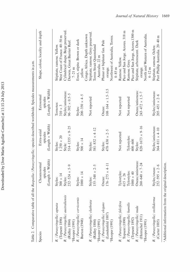
Raspailia (Parasyringella) rubra sp. nov. constitutes the first record of the subgenusin the Eastern Pacific. Species of the subgenus Parasyringella are characterized by anaxial extra-axial skeleton (Hooper 2002) and bearing choanosomal spicules, styles oroxeas (Table 2). The species assigned to this subgenus with oxeas as choanosomalspicules are: R. (Parasyringella) elegans (Lendenfeld 1887) and R. (Parasyringella)nuda Hentschel 1911. Raspailia (Parasyringella) elegans is an orange planar or bipla-nar fan sponge from the north-western coast of Australia (Hooper 1991). It haschoanosomal oxeas (176–275 × 4–11 µm), extra-axial styles (478–830 × 2–5 µm)and ectosomal oxeas (108–164 × 1.5–3.5 µm). The extra-axial styles are longer in R.(Parasyringella) rubra sp. nov. than in R. (Parasyringella) elegans (Table 2). Besides,there are some differences in the morphology and length of the ectosomal spicules(ectosomal oxeas in R. (Parasyringella) elegans versus ectosomal styles/anisoxeas inRaspailia (Parasyringella) rubra sp. nov.). Raspailia (Parasyringella) nuda Hentschel1911 is an orange arborescent or stipitate sponge described from Australia (Hooper1991). It has choanosomal oxeas/anisoxeas (260–640 × 7–24 µm), subectosomal styles(820–1673 × 9–16 µm) and ectosomal oxeas/anisoxeas (243–472 × 1.5–7 µm). Thechoanosomal oxeas are longer in R. (Parasyringella) nuda than in R. (Parasyringella)rubra sp. nov.
Etymology
Named rubra which means red in Latin.
Subgenus Raspaxilla Topsent, 1913Raspailia (Raspaxilla) hymani (Dickinson, 1945)
(Figures 1B, 4, 5)
Hemectyon hymani Dickinson, 1945; Green and Bakus, 1994:41–42.Aulospongus hymani Desqueyroux-Faúndez and van Soest, 1997:442.Raspailia (Raspaxilla) hymani Hooper et al. 1999:685–687.Endectyon (Endectyon) hymani Lee et al. 2007:35.
Dow
nloa
ded
by [
Jose
Mar
ia A
guila
r-C
amac
ho]
at 1
1:15
24
July
201
3
Journal of Natural History 1669
Tab
le2.
Com
para
tive
tabl
eof
allt
heR
aspa
ilia
(Par
asyr
inge
lla)
spec
ies
desc
ribe
dw
orld
wid
e.Sp
icul
esm
easu
rem
ents
inµ
m.
Spec
ies
Cho
anos
omal
spic
ules
(Len
gth
×W
idth
)
Ext
ra-a
xial
spic
ules
(Len
gth
×W
idth
)
Ect
osom
alsp
icul
es(L
engt
h×
Wid
th)
Shap
e,co
lour
,loc
alit
yan
dde
pth
R.(
Par
asyr
inge
lla)
agna
ta(T
opse
nt18
96)
Styl
es:
300
×14
Styl
es:
125
0×
20To
rnot
es:
310
×3
Stip
itat
e,ra
mos
e.Y
ello
w.
Wes
tC
oast
ofF
ranc
e.40
–50
mR
.(P
aras
yrin
gella
)au
stra
liens
is(R
idle
y18
84)
∗ Hoo
per
(199
1)
Styl
es:
232–
524
×5–
9St
yle:
514–
1355
×9–
25St
yles
/an
isox
eas:
96–3
92×
0.5–
5C
ylin
dric
alsh
ape.
Bei
gepr
eser
ved.
Dar
win
,Gre
atB
arri
erR
eef.
7–21
mR
.(P
aras
yrin
gella
)ce
rvic
orni
sB
urto
n(1
948)
Styl
es:
1000
×14
Styl
es:
500
×14
Styl
es:
200–
350
×4–
5E
rect
,stí
pite
.Bro
wn
orda
rkye
llow
.C
ongo
,Afr
ica.
Dep
thun
know
nR
.(P
aras
yrin
gella
)cl
athr
ata
(Rid
ley
1884
)∗ H
oope
r(1
991)
Styl
es:
155–
348
×2–
5St
yles
:58
1–83
2×
4–12
Not
repo
rted
Stip
itat
e,ra
mos
e.G
rey
pres
erve
d.To
rres
Stra
itQ
ueen
slan
dA
ustr
alia
.22
mR
.(P
aras
yrin
gella
)el
egan
s(L
ende
nfel
d18
87)
∗ Hoo
per
(199
1)
Oxe
as:
176–
275
×4–
11St
yles
:47
8–83
0×
2–5
Oxe
as:
108–
164
×1.
5–3.
5P
lana
ror
bipl
anar
fan.
Pal
eor
ange
.N
orth
wes
tof
Aus
tral
ia,T
orre
s.0–
85m
R.(
Par
asyr
inge
lla)
falc
ifera
Tops
ent
(189
0)T
ylos
tyle
s:61
5×
26N
otre
port
edN
otre
port
edR
amos
e.Y
ello
w.
Pic
oan
dSa
nJo
rge.
Azo
res.
318
mR
.(P
aras
yrin
gella
)hu
mili
s(T
opse
nt18
92)
Tyl
osty
les:
1000
×40
Tyl
osty
les:
400–
600
Not
repo
rted
Ram
ose.
Gre
y.P
ico
and
San
Jorg
e.A
zore
s.13
00m
R.(
Par
asyr
inge
lla)
nuda
(Hen
tsch
el19
11)
∗ Hoo
per
(199
1)
Oxe
as/an
isox
eas:
260–
640
×7–
24St
yles
:82
0–16
73×
9–16
Oxe
as/an
isox
eas:
243–
472
×1.
5–7
Stip
itat
e,ar
bore
scen
t.D
ark
oran
ge.
Nor
than
dW
este
rnof
Aus
tral
ia.
6–12
mR
.(P
aras
yrin
gella
)st
ellid
erm
a(C
arte
r18
85)
Styl
es/ox
eas:
252–
595
×2–
6St
yles
/an
isox
eas:
364–
811
×4–
10St
yles
/an
isox
eas:
205–
451
×2–
6St
ipit
ate,
ram
ose.
Och
re.
Port
Phi
llip
Aus
tral
ia.2
0–40
m
∗ Add
itio
nali
nfor
mat
ion
from
the
orig
inal
desc
ript
ion.
Dow
nloa
ded
by [
Jose
Mar
ia A
guila
r-C
amac
ho]
at 1
1:15
24
July
201
3
1670 J.M. Aguilar-Camacho and J.L. Carballo
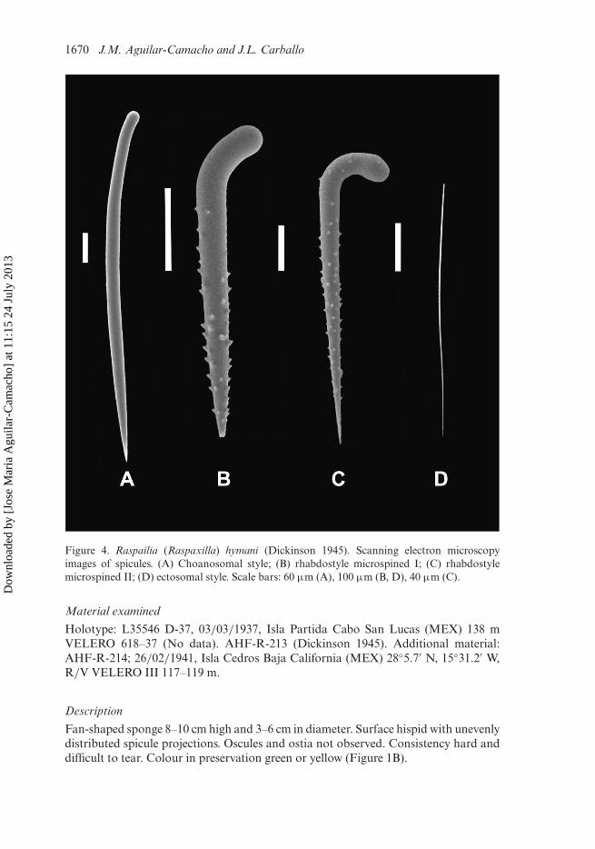
Figure 4. Raspailia (Raspaxilla) hymani (Dickinson 1945). Scanning electron microscopyimages of spicules. (A) Choanosomal style; (B) rhabdostyle microspined I; (C) rhabdostylemicrospined II; (D) ectosomal style. Scale bars: 60 µm (A), 100 µm (B, D), 40 µm (C).
Material examined
Holotype: L35546 D-37, 03/03/1937, Isla Partida Cabo San Lucas (MEX) 138 mVELERO 618–37 (No data). AHF-R-213 (Dickinson 1945). Additional material:AHF-R-214; 26/02/1941, Isla Cedros Baja California (MEX) 28◦5.7′ N, 15◦31.2′ W,R/V VELERO III 117–119 m.
Description
Fan-shaped sponge 8–10 cm high and 3–6 cm in diameter. Surface hispid with unevenlydistributed spicule projections. Oscules and ostia not observed. Consistency hard anddifficult to tear. Colour in preservation green or yellow (Figure 1B).
Dow
nloa
ded
by [
Jose
Mar
ia A
guila
r-C
amac
ho]
at 1
1:15
24
July
201
3

Journal of Natural History 1671
Figure 5. Drawings of Raspailia (Raspaxilla) hymani (Dickinson 1945). (A) Choanosomal style;(B) rhabdostyles microspined I; (C) rhabdostyles microspined II; (D) ectosomal style thin; (E)ectosomal skeleton (extra-axial) and choanosomal skeleton (axial). Scale bars: 100 µm (A–D),250 µm (E).
Skeleton
Straight or curved choanosomal styles: 1150–1720 × 25–50 µm (Figures 4A, 5A).Microspined rhabdostyles in two categories: the first long, curved and with prominentspines: 280–560 × 25–40 µm (Figures 4B, 5B); the second curved, with a pronouncedhead and with short spines: 130–260 × 7.5–15 µm (Figures 4C, 5C). Straight andcurved ectosomal styles: 235–425 × 1.75–2.5 µm (Table 3) (Figures 4D, 5D). Theectosomal skeleton is a dense layer of spongin (100 µm thick). The styles are dispersedwith no special organization. There is a subectosomal extra-axial compressed skeletonformed by the rhabdostyles with the points protruding externally. The choanosomal
Dow
nloa
ded
by [
Jose
Mar
ia A
guila
r-C
amac
ho]
at 1
1:15
24
July
201
3
1672 J.M. Aguilar-Camacho and J.L. Carballo
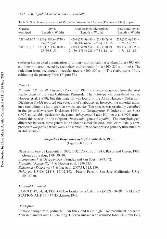
Table 3. Spicule measurements of Raspailia (Raspaxilla) hymani (Dickinson 1945) in µm.
Materialexamined
Styles(Length × Width)
Rhabdostyles microspined(Length × Width)
Ectosomal styles(Length × Width)
AHF-618-37 1150-(1484.6)-1720 ×25-(32.5)-45
1) 280-(371.9)-460 × 25-(30.7)-402) 130-(189.6)-260 × 7.5-(10.6)-15
235-(302.6)-380 ×1.75-(2.2)-2.5
AHF-R-213 1310-(1514.6)-1620 ×25-(36.8)-50
1) 340-(390.9)-560 × 30-(33.8)-402) 130-(171.4)-225 × 7.5-(11.6)-15
360-(393.5)-425 ×1.75-(2.1)-2.5
skeleton has an axial organization of primary multispicular ascendant fibres (300–600µm thick) interconnected by secondary multispicular fibres (100–250 µm thick). Thereticulum forms rectangular irregular meshes (200–300 µm). The rhabdostyles II areechinating the primary fibres (Figure 5E).
Remarks
Raspailia (Raspaxilla) hymani (Dickinson 1945) is a deep-sea species from the WestPacific coast of the Baja California Peninsula. The holotype was considered lost byHooper et al. (1999), but this material was found in the Allan Hancock Collection.Dickinson (1945) reported one category of rhabdostyles; however, the material exam-ined (including the holotype) has two categories. This species was originally describedin the genus Hemectyon (Dickinson 1945), but Desqueyroux-Faúndez and van Soest(1997) moved this species into the genus Aulospongus. Later, Hooper et al. (1999) trans-ferred this species to the subgenus Raspaxilla (genus Raspailia). The morphologicaldifference between these genera is the choanosomal skeleton: axial extra-axially com-pressed in Raspailia (Raspaxilla) and a reticulum of compressed primary fibre bundlesin Aulospongus.
Raspailia (Raspaxilla) hyle (de Laubenfels, 1930)(Figures 1C, 6, 7)
Hemectyon hyle de Laubenfels, 1930, 1932; Dickinson, 1945; Bakus and Green, 1987;Green and Bakus, 1994:39–40.
Aulospongus hyle Desqueyroux-Faúndez and van Soest, 1997:442.Raspailia (Raspaxilla) hyle Hooper et al. 1999:685.Endectyon (Endectyon) hyle Lee et al. 2007:33, 115, 189.Holotype: USNM 21418, 16/02/1924, Puerto Fermín, San José (California, USA)
30–150 m.
Material Examined
L35609 D-17, 04/04/1935, Off Los Frailes Baja California (MEX) 10–28 m VELEROSTATION AHF 751–37 (Dickinson 1945).
Description
Ramose sponge with peduncle 3 cm thick and 8 cm high. Two prominent branches2 cm in diameter and 1–3 cm long. Uneven surface with rounded lobes (1–2 mm long
Dow
nloa
ded
by [
Jose
Mar
ia A
guila
r-C
amac
ho]
at 1
1:15
24
July
201
3

Journal of Natural History 1673
Figure 6. Raspailia (Raspaxilla) hyle (de Laubenfels 1930). Scanning electron microscopyimages of spicules. (A) Subectosomal style curved; (B) choanosomal style straight; (C) rhab-dostyle microspined; (D) ectosomal style curved and thin. Scale bars: 100 µm (A, B, D), 60µm (C).
and 1.3 mm high) evenly distributed. Oscules and ostia not visible. Consistency hardand difficult to tear. Colour in preservation pale beige (Figure 1C).
Skeleton
Straight choanosomal styles: 500–780 × 15–20 µm (Figures 6B, 7A). Curvedsubectosomal styles: 765–1200 × 8–20 µm (Figures 6A, 7C). Rhabdostylesmicrospined curved with prominent spines: 230–400 × 10–20 µm (Figures 6C, 7C).These spines are localized in the terminal third of the rhabdostyles. Curved ectosomalstyles: 230– 395 × 1.75–2.5 µm (Figures 6D, 7D) (Table 4). The ectosomal skele-ton is a dense layer of styles (30–50 µm thick). There is a subectosomal extra-axialskeleton formed by the styles with the points of the spicules protruding externally.
Dow
nloa
ded
by [
Jose
Mar
ia A
guila
r-C
amac
ho]
at 1
1:15
24
July
201
3

1674 J.M. Aguilar-Camacho and J.L. Carballo
Figure 7. Drawings of Raspailia (Raspaxilla) hyle (de Laubenfels 1930). (A) Choanosomal stylestraight; (B) rhabdostyle microspined; (C) subectosomal style curved; (D) ectosomal style thin;.(E) ectosomal skeleton (extra-axial) and choanosomal skeleton (axial). Scale bars: 40 µm (A-E),120 µm (F).
The choanosomal skeleton has an axial compressed skeleton formed by primaryascending multispicular fibres (200–300 µm thick) interconnected by bispicular ormultispicular secondary fibres (20–40 µm thick). The reticulum forms rectangu-lar irregular meshes (100–120 µm). Rhabdostyles are echinating the primary fibres(Figure 7E).
Dow
nloa
ded
by [
Jose
Mar
ia A
guila
r-C
amac
ho]
at 1
1:15
24
July
201
3

Journal of Natural History 1675
Table 4. Spicule measurements of Raspailia (Raspaxilla) hyle (de Laubenfels 1930) in µm. +Spicule measurements from the original description.
Material examined Choanosomal styles(Length × Width)
Subectosomalstyles
(Length × Width)
Rhabdostylesmicrospined
(Length × Width)
Ectosomal styles(Length × Width)
AHF-751-37 500-(630.8)-780 ×15-(17.8)-20
765-(1010)-1200 ×8-(16.4)-20
230-(344.2)-400 ×10-(13.7)-20
210-(323.2)-395 ×1.7-(1.9)-2.5
De Laubenfels(1932)+
1) 430–550 × 15–202) 350–370 × 16–19
800 × 10 180–320 × 12–20 200–330 × 2
Dickinson (1945)+ 480 × 18 >1000 180 × 12 250 × 2Bakus and Green
(1987)+260–620 × 2–7 380–1045 × 12–25 106–420 × 6–34 72–180 × 1–4
Green and Bakus(1994)+
280–680 × 8–31 1125–2200 ×18–34
340–480 × 23–36 310–550 × 1–5
Hooper et al.(1999)+
322–585 × 12–19 715–1560 × 9–16 155–364 × 9–22 165–385 ×0.8–1.5
Remarks
Raspailia (Raspaxilla) hyle (de Laubenfels 1930) is distributed along the West Pacificcoast of Baja California Peninsula and the Pacific coast of the USA. This species wasoriginally described in the genus Hemectyon for bearing rhabodstyles with prominentspines (de Laubenfels 1930). Currently, Hemectyon is considered a subgenus of thegenus Endectyon (Hooper 2002). Hooper et al. (1999) moved this species into thesubgenus Raspaxilla (genus: Raspailia) because of the axial extra-axial skeleton.
Genus Aulospongus Norman, 1878Aulospongus cerebella (Dickinson, 1945)
(Figures 1D, 8, 9)
Heterectya cerebella Dickinson, 1945:22.Aulospongus cerebella Desqueyroux-Faúndez and van Soest, 1997; Hooper et al.
1999:654–656.
Material examined
Holotype: L 355667, 09/03/1936, D-4 Isla Partida Gulf of California (MEX) 83 mVELERO AHF-559–36 (Dickinson 1945). Additional material: LACM, 1941–3;26/02/1941, Isla Cedros, Baja California (MEX) 28◦5.7′ N, 115◦31.2′ W, 117–119 mR/V VELERO III. AHF-1253–41. 2059-LEB-ICML-UNAM, 11/04/2011, Station32 Talud XIV (Gulf of California, MEX) 122 m (27◦56′13′′ N, 111◦19′44′′ W).
Description
Tubular sponge from 3 to 8 cm high and 2 cm thick with an apical oscule (6 mm long).Ostia not visible. Surface hispid with spicule projections (600–1200 µm) evenly dis-tributed in the body. Consistency flexible and difficult to tear. Colour in preservationpale beige (Figure 1D).
Dow
nloa
ded
by [
Jose
Mar
ia A
guila
r-C
amac
ho]
at 1
1:15
24
July
201
3

1676 J.M. Aguilar-Camacho and J.L. Carballo
Figure 8. Aulospongus cerebella (Dickinson 1945). Scanning electron microscopy imagesof spicules. (A) Choanosomal style; (B) rhabdostyle microspined thick; (C) rhabdostylemicrospined thin. Scale bars: 90 µm (A), 100 µm (B, C).
Skeleton
Straight or curved choanosomal styles: 300–720 × 30–45 µm (Figures 8A, 9A).Rhabdostyles microspined with prominent spines: 275–470 × 20–35 µm (Figures 8B,C, 9B) (Table 5). The spines are localized on the terminal third of this spicule.The ectosomal skeleton is a dense layer of spongin (100–120 µm thick) with
Dow
nloa
ded
by [
Jose
Mar
ia A
guila
r-C
amac
ho]
at 1
1:15
24
July
201
3
Journal of Natural History 1677
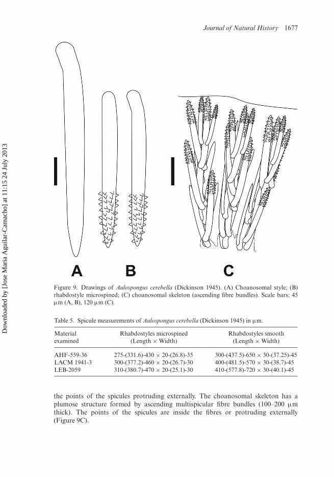
Figure 9. Drawings of Aulospongus cerebella (Dickinson 1945). (A) Choanosomal style; (B)rhabdostyle microspined; (C) choanosomal skeleton (ascending fibre bundles). Scale bars: 45µm (A, B), 120 µm (C).
Table 5. Spicule measurements of Aulospongus cerebella (Dickinson 1945) in µm.
Materialexamined
Rhabdostyles microspined(Length × Width)
Rhabdostyles smooth(Length × Width)
AHF-559-36 275-(331.6)-430 × 20-(26.8)-35 300-(437.5)-650 × 30-(37.25)-45LACM 1941-3 300-(377.2)-460 × 20-(26.7)-30 400-(481.5)-570 × 30-(38.7)-45LEB-2059 310-(380.7)-470 × 20-(25.1)-30 410-(577.8)-720 × 30-(40.1)-45
the points of the spicules protruding externally. The choanosomal skeleton has aplumose structure formed by ascending multispicular fibre bundles (100–200 µmthick). The points of the spicules are inside the fibres or protruding externally(Figure 9C).
Dow
nloa
ded
by [
Jose
Mar
ia A
guila
r-C
amac
ho]
at 1
1:15
24
July
201
3

1678 J.M. Aguilar-Camacho and J.L. Carballo
Remarks
Aulospongus cerebella (Dickinson 1945) is a deep-sea species from the Gulf ofCalifornia and West Pacific coast of Baja Peninsula. Hooper et al. (1999) consideredthat the holotype was lost. However, the type material was found in the Allan HancockCollection.
Aulospongus californianus sp. nov.(Figures 1E, 10, 11)
Material examined
Holotype: MCNM 1.01/656, 11/04/2011, Station 32 Talud XIV (Gulf of California,MEX) 122 m (27◦56′13′′ N, 111◦19′44′′ W). Paratypes: 2060-LEB-ICML-UNAM,
Figure 10. Aulospongus californianus sp. nov. Scanning electron microscopy images of spicules.(A) Choanosomal style straight; (B) rhabdostyle microspined I; (C) rhabdostyle micrsospinedII; (D) ectosomal style. Scale bars: 50 µm (A), 10 µm (B), 100 µm (C, D).
Dow
nloa
ded
by [
Jose
Mar
ia A
guila
r-C
amac
ho]
at 1
1:15
24
July
201
3

Journal of Natural History 1679
Figure 11. Drawings of Aulospongus californianus sp. nov. (A) Choanosomal style straight; (B)rhabdostyle microspined I; (C) rhabdostyle microspined II; (D) ectosomal style/anisoxeas; (E)choanosomal skeleton (ascending fibre bundles). Scale bars: 60 µm (A–D), 250 µm (E).
11/04/2011, Station 32 Talud XIV (Gulf of California, MEX) 122 m (27◦56′13′′ N,111◦19′44′′ W). 2061-LEB-ICML-UNAM, 11/04/2011, Station 32 Talud XIV (Gulfof California, MEX) 122 m (27◦56′13′′ N, 111◦19′44′′ W). 2062-LEB-ICML-UNAM,11/04/2011, Station 32 Talud XIV (Gulf of California, MEX) 122 m (27◦56′13′′ N,111◦19′44′′ W).
Description
Massive or vase-shaped sponge, 1–2 cm in diameter and 3–5 cm high. Surface hispidwith spicule projections evenly distributed. Oscula (6–10 mm) and ostia are circular
Dow
nloa
ded
by [
Jose
Mar
ia A
guila
r-C
amac
ho]
at 1
1:15
24
July
201
3

1680 J.M. Aguilar-Camacho and J.L. Carballo
to oval-shaped (100–150 µm). Consistency hard and difficult to tear. Colour inpreservation pale beige (Figure 1E).
Skeleton
Straight choanosomal styles: 580–1130 × 25–45 µm (Figures 10A, 11A). Microspinedrhabdostyles in two sizes: the first curved and with prominent spines: 340–610 ×22.5–35 µm (Figures 10B, 11B); the second short, with the head pronounced:150–360 × 10–30 µm (Figures 10C, 11C). The spines are localized on the ter-minal third of the spicule. Curved ectosomal styles/anisoxeas: 290–460 × 2.5–5µm (Figures 10D, 11D) (Table 6). The ectosomal skeleton is a dense layer ofstyles/anisoxeas with the points of the spicules protruding externally (30–80 µm thick).Multispicular ascending fibres (480–600 µm thick) (Figure 11E) form a plumosechoanosomal skeleton.
Remarks
Aulospongus californianus sp. nov. is a deep-sea species from the Gulf of California.The only species assigned to this genus in the Eastern Pacific is Aulospongus cere-bella (Dickinson 1945). This is a tubular sponge with straight styles and microspinedrhabdostyles in one category (see above). Aulospongus californianus sp. nov., hasectosomal styles/anisoxeas and rhabdostyles in two categories while A. cerebella lacksthe ectosomal spicules and the rhabdostyles are in one category.
Etymology
Named californianus for the type locality.
Aulospongus aurantiacus sp. nov.(Figures 1F, 12, 13)
Material examined
Holotype: MCNM 1.01/657, 30/10/2003, Isla Venados (Mazatlán, Sinaloa), 7 m(23◦10′15′′ N, 106◦26′42′′ W). Paratype: 962-LEB-ICML-UNAM, 30/10/2003, IslaVenados (Mazatlán, Sinaloa), 4 m (23◦10′15′′ N, 106◦26′42′′ W).
Table 6. Spicule measurements of Aulospongus californianus sp. nov. in µm.
Materialexamined
Styles straight(Length × Width)
Rhabdostyles microspined(Length × Width)
Styles/anisoxeas(Length × Width)
MCNM1.01/656
650-(832.1)-975 ×25-(33.2)-45
1) 360-(498.3)-560 × 25-(26.1)-352) 150-(235.1)-300 × 10-(18.5)-20
300-(398.5)-450 ×2.5-(3.1)-5
LEB-2060 700-(883.6)-1010 ×25-(31.5)-40
1) 410-(478.3)-550 × 25-(29.7)-352) 150-(230.2)-350 × 10-(15.7)-20
350-(388.6)-460 ×2.5-(2.8)-5
LEB-2061 600-(780.6)-900 ×25-(32.5)-40
1) 380-(402.1)-480 × 22.5-(27.9)-352) 150-(201.2)-290 × 15-(20.2)-30
290-(369.2)-425 ×2.5-(3.1)-5
LEB-2062 580-(921.2)-1130 ×25-(33.4)-45
1) 340-(477.5)-610 × 25-(26.8)-302) 150-(248.8)-360 × 10-(17.7)-20
310-(370.2)-420 ×2.5-(3.0)-5
Dow
nloa
ded
by [
Jose
Mar
ia A
guila
r-C
amac
ho]
at 1
1:15
24
July
201
3

Journal of Natural History 1681
Figure 12. Aulospongus aurantiacus sp. nov. Scanning electron microscopy images of spicules.(A) Choanosomal style I; (B) choanosomal style II; (C) rhabdostyle microspined; (D) ectosomalstyle. Scale bars: 50 µm (A), 100 µm (B, D), 10 µm (C).
Description
Encrusting or laminated sponge 3–6 cm long and 1–2.5 cm thick. Oscules and ostianot observed. Surface hispid with conules circular to oval-shaped (300–750 µm long)and evenly distributed. Consistency hard and difficult to tear. Colour in life is orange,pale in preservation (Figure 1F).
Skeleton
Straight or curved styles in two sizes: 1). 440–970 × 10–20 µm (Figures 12A, 13A).2) 135–250 × 5–15 µm (Figures 12B, 13B). Rhabdostyles microspined with prominentspines: 80–125 × 2.5–15 µm (Figures 12C, 13C). Curved or straight subectosomalsubtylostyles/styles: 280–480 × 2.5–7.5 µm (Figures 12D, 13D) (Table 7). Theectosomal skeleton is a dense layer of spongin (40–100 µm thick). The choanosome
Dow
nloa
ded
by [
Jose
Mar
ia A
guila
r-C
amac
ho]
at 1
1:15
24
July
201
3

1682 J.M. Aguilar-Camacho and J.L. Carballo
Figure 13. Drawings of Aulospongus aurantiacus sp. nov. (A) Choanosomal style I; (B)choanosomal style II; (C) ectosomal style/subtylostyle; (D) rhabdostyle microspined; (E)choanosomal skeleton (ascending fibre bundles). Scale bars: 20 µm (A–D), 200 µm (E).
has a plumose structure formed by multispicular fibre bundles (160–260 µm thick).The microspined rhabdostyles are echinating (Figure 13E).
Remarks
Aulospongus aurantiacus sp. nov. is a subtidal sponge from the Gulf of California.This species is characterized by having choanosomal styles and curved rhabdostyles
Dow
nloa
ded
by [
Jose
Mar
ia A
guila
r-C
amac
ho]
at 1
1:15
24
July
201
3

Journal of Natural History 1683
Table 7. Spicule measurements of Aulospongus aurantiacus sp. nov. in µm.
Materialexamined
Choanosomal styles(Length × Width)
Rhabdostylesmicrospined
(Length × Width)
Subtylostyles/styles(Length × Width)
MCNM1.01/657
1) 445-(672.1)-865 × 10-(13.6)-202) 165-(200.1)-240 × 5-(11.7)-15
85-(90.1)-120 ×2.5-(3.4)-5
310-(320.1)-465 ×2.5-(4.8)-7.5
LEB-962 1) 440-(675.2)-970 × 10-(14.2)-202) 135-(189.2)-250 × 5-(12.4)-15
80-(96.5)-125 ×2.5-(3.75)-5
280-(362.5)-480×2.5-(5.6)-7.5
microspined. The species assigned to this genus with these characteristics are:Aulospongus similaustralis Hooper et al. 2008, Aulospongus tubulatus (Bowerbank1873), Aulospongus spinosum (Topsent 1927) and Aulospongus monticularis (Ridleyand Dendy 1886). Aulospongus similaustralis Hooper et al. (2008) is a globularsponge recorded from the western coast of Australia. It has choanosomal styles(150–400 µm), microspined rhabdostyles (70–255 µm) and subectosomal tylostyles(720–1400 µm). Aulospongus aurantiacus sp. nov. has styles in two categories andectosomal subtylostyles/styles while A. similaustralis has one category of stylesand ectosomal tylostyles. Aulospongus tubulatus (Bowerbank 1873) is a massiveor tubular sponge recorded from Sri Lanka. It has choanosomal rhabdostyles(304–462 µm), rhabdostyles microspined (109–126 µm) and ectosomal styles (212–250µm). The rhabdostyles are longer in A. aurantiacus sp. nov. than in A. tubula-tus. Aulospongus spinosum (Topsent 1927) is a bulbous sponge described from CapeVerde at 219 m depth. It has choanosomal rhabdostyles in two sizes (770–1085µm and 90–182 µm), acanthostyles (75–145 µm) and ectosomal oxeas (40–50 µm).The morphology of the ectosomal spicules is the main difference between thesespecies: styles/subtylostyles in A. aurantiacus sp. nov. and oxeas in A. spinosum.Aulospongus monticularis (Ridley and Dendy 1886) is an encrusting or massivesponge described from Cape Verde. This species has choanosomal rhabdostyles(290–518 µm), microspined rhabdostyles (132–275 µm) and subectosomal styles(620–960 µm). Aulospongus aurantiacus has the subectosomal styles shorter thanA. monticularis.
Etymology
Named aurantiacus which means orange in Latin.
Genus Eurypon Gray, 1867Eurypon patriciae sp. nov.
(Figures 14A, 15, 16)
Material examined
Holotype: MCNM 1.01/658, 18/10/2001, Isla Lobos 1 (Mazatlán, Sinaloa)5 m (23◦13′49′′ N, 106◦27′43′′ W). Paratypes: 107-LEB-ICML-UNAM,18/10/2001, Isla Lobos 1 (Mazatlán, Sinaloa) 5 m (23◦13′49′′ N, 106◦27′43′′
Dow
nloa
ded
by [
Jose
Mar
ia A
guila
r-C
amac
ho]
at 1
1:15
24
July
201
3
1684 J.M. Aguilar-Camacho and J.L. Carballo
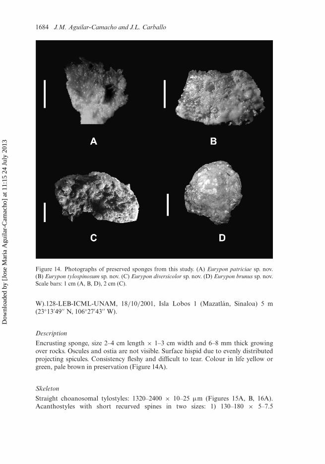
Figure 14. Photographs of preserved sponges from this study. (A) Eurypon patriciae sp. nov.(B) Eurypon tylospinosum sp. nov. (C) Eurypon diversicolor sp. nov. (D) Eurypon brunus sp. nov.Scale bars: 1 cm (A, B, D), 2 cm (C).
W).128-LEB-ICML-UNAM, 18/10/2001, Isla Lobos 1 (Mazatlán, Sinaloa) 5 m(23◦13′49′′ N, 106◦27′43′′ W).
Description
Encrusting sponge, size 2–4 cm length × 1–3 cm width and 6–8 mm thick growingover rocks. Oscules and ostia are not visible. Surface hispid due to evenly distributedprojecting spicules. Consistency fleshy and difficult to tear. Colour in life yellow orgreen, pale brown in preservation (Figure 14A).
Skeleton
Straight choanosomal tylostyles: 1320–2400 × 10–25 µm (Figures 15A, B, 16A).Acanthostyles with short recurved spines in two sizes: 1) 130–180 × 5–7.5
Dow
nloa
ded
by [
Jose
Mar
ia A
guila
r-C
amac
ho]
at 1
1:15
24
July
201
3

Journal of Natural History 1685
Figure 15. Eurypon patriciae sp. nov. Scanning electron microscopy images of spicules. (A)Choanosomal tylostyle head; (B) choanosomal tylostyle end; (C) acanthostyle I; (D) acan-thostyle II; (E) subectosomal style. Scale bars: 2 µm (A), 10 µm (B, D), 20 µm (C),100 µm (D).
µm (Figures 15C, 16C); 2) 55–87.5 × 2.5–5 µm (Figures 15D, 16C). Straightstrongyloxeas/styles: 400–550 × 5–10 µm (Figures 15E, 16B) (Table 8). Theectosomal skeleton is absent. The points of the spicules are protruding exter-nally. The choanosomal skeleton has a hymedesmoid structure. Main tylostyles andacanthostyles are embedded in a spongin layer (20–40 µm thick). The strongyloxeasand styles are dispersed in groups of one to three arranged along the tylostyles in thechoanosome (Figure 16D).
Dow
nloa
ded
by [
Jose
Mar
ia A
guila
r-C
amac
ho]
at 1
1:15
24
July
201
3

1686 J.M. Aguilar-Camacho and J.L. Carballo
Figure 16. Drawings of Eurypon patriciae sp. nov. (A) Choanosomal tylostyles; (B)strongyloxeas/styles; (C) acanthostyles recurved by short spines (two categories); (D)choanosomal skeleton (hymedesmoid). Scale bars: 850 µm (A–C), 140 µm (D).
Remarks
Eurypon patriciae sp. nov. is a subtidal sponge distributed in the Gulf of California.In the Eastern Pacific, there are two species assigned to this genus. Eurypon nigrumBergquist 1967 is a blue encrusting sponge described from Oahu (Hawaii). It hastylostyles in two sizes (1200–2400 µm × 6–12 µm and 170–800 × 6–12 µm) andacanthotylostyles (70–165 × 6–9 µm). The spicule measurements are similar in these
Dow
nloa
ded
by [
Jose
Mar
ia A
guila
r-C
amac
ho]
at 1
1:15
24
July
201
3

Journal of Natural History 1687
Table 8. Spicule measurements of Eurypon patriciae sp. nov in µm.
Materialexamined
Choanosomaltylostyles
(Length × Width)
Acanthostyles(Length × Width)
Styles/strongyloxeas(Length × Width)
MCNM1.01/658
1450-(1985.4)-2225 ×10-(14.6)-25
1) 125-(154.3)-175 × 5-(6.4)-7.52) 60-(70.1)-85 × 2.5-(6.1)-5
450-(434.2)-525 ×5-(6.7)-10
LEB-107 1680-(2000.2)-2400 ×10-(17.5)-25
1) 130-(150.2)-180× 5-(6.6)-7.52) 55-(71.1)-85 × 2.5-(4.5)-5.
400-(485.2)-550 ×5-(5.8)-7.5
LEB-128 1320-(1784.6)-2100 ×10-(16.2)-25
1) 130-(156.4)-180 × 5-(6.8)-7.52) 60-(74.1)-87.5 × 2.5-(3.6)-5.
410-(453.5)-500 ×5-(6.7)-10
two species. However, E. nigrum has tylostyles as ectosomal spicules while E. patri-ciae sp. nov. has strongyloxeas and styles. Eurypon miniaceum Thiele 1905 is a redencrusting sponge described from Calbuco (Chile) at 30 m depth. It has tylostyles inthree categories (2000–3000 × 30 µm; 800 × 30 µm; and >120 µm), acanthostyles(120 µm) and subectosomal styles (550 × 5 µm). Eurypon patriciae sp. nov. has onecategory of styles while E. miniaceum has three.
Etymology
Named for Patricia Bergquist for her contribution to sponge science.
Eurypon tylospinosum sp. nov.(Figures 14B, 17, 18)
Material examined
Holotype: MCNM 1.01/659, 27/11/2002, Cabo Haro, (Guaymas, Sonora) 15 m(27◦52′5′′ N, 110◦57′1′′ W). Paratype: 769-LEB-ICML-UNAM, 27/11/2002, CaboHaro, (Guaymas, Sonora), 15 m (27◦52′5′′ N, 110◦57′1′′ W).
Description
Encrusting sponge, size 2–5 cm length × 1–2 cm width and 3–5 mm thick. Oscules andostia not visible. Surface smooth. Consistency flexible and difficult to tear. Colour inlife red, pale in preservation (Figure 14B).
Skeleton
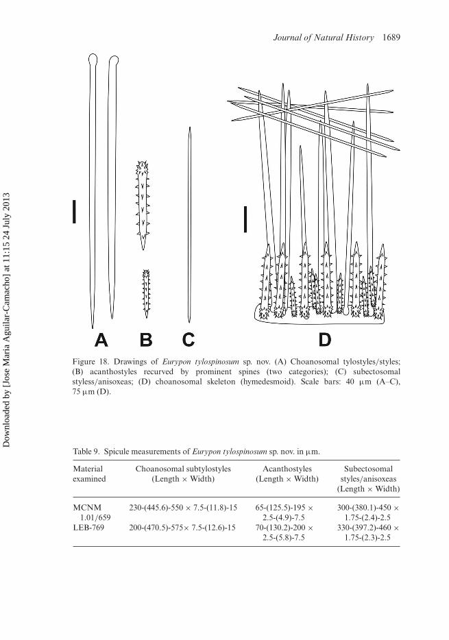
Choanosomal subtylostyles with a pronounced head or modified to style: 200–575 ×7.5–15 µm (Figures 17A, 18A). Acanthostyles with swollen head with prominentspines. These spines are arranged as a crown around the head: 70–200 × 2.5–7.5 µm(Figures 17B, C, 18B). Straight or curved subectosomal styles/anisoxeas: 330–460 ×1.75–2.5 µm (Figures 17D, 18C) (Table 9). The ectosomal skeleton is absent.The choanosomal skeleton has a hymedesmoid structure. Main subtylostyles and
Dow
nloa
ded
by [
Jose
Mar
ia A
guila
r-C
amac
ho]
at 1
1:15
24
July
201
3

1688 J.M. Aguilar-Camacho and J.L. Carballo
Figure 17. Eurypon tylospinosum sp. nov. Scanning electron microscopy images of spicules. (A)Choanosomal tylostyles; (B) acanthostyle with swollen head I; (C) acanthostyle with swollenhead II; (D) subectosomal style. Scale bars: 50 µm (A), 10 µm (B), 20 µm (C), 100 µm (D).
acanthostyles are embedded in a spongin layer (10–25 µm thick). The styles/anisoxeasare dispersed in trichodragmas in the subectosomal region (Figure 18D).
Remarks
Eurypon tylospinosum sp. nov. is a subtidal species distributed from the Gulf ofCalifornia. It is characterized by having acanthostyles with swollen head and withprominent spines, which are arranged as a crown around the head. The only speciesdescribed worldwide that have these features are E. simplex (Bowerbank 1874) andE. coronula (Bowerbank 1874) (Table 12). Eurypon simplex (Bowerbank 1874) is ayellow encrusting sponge described from the Shetland Islands. It has tylostyles (2116 ×27.1 µm) and acanthostyles (105.8–218.9 × 8.4 µm). The tylostyles are shorter inE. tylospinosum sp. nov. than in E. simplex. Besides, E. tylospinosum sp. nov. hassubectosomal styles which are lacking in E. simplex. Eurypon coronula (Bowerbank1874) is a grey encrusting sponge recorded from the Shetland Islands. It has tylostyles(635–1411 µm) and acanthostyles (254 µm). The tylostyles are longer in E. coronula
Dow
nloa
ded
by [
Jose
Mar
ia A
guila
r-C
amac
ho]
at 1
1:15
24
July
201
3
Journal of Natural History 1689
Figure 18. Drawings of Eurypon tylospinosum sp. nov. (A) Choanosomal tylostyles/styles;(B) acanthostyles recurved by prominent spines (two categories); (C) subectosomalstyless/anisoxeas; (D) choanosomal skeleton (hymedesmoid). Scale bars: 40 µm (A–C),75 µm (D).
Table 9. Spicule measurements of Eurypon tylospinosum sp. nov. in µm.
Materialexamined
Choanosomal subtylostyles(Length × Width)
Acanthostyles(Length × Width)
Subectosomalstyles/anisoxeas
(Length × Width)
MCNM1.01/659
230-(445.6)-550 × 7.5-(11.8)-15 65-(125.5)-195 ×2.5-(4.9)-7.5
300-(380.1)-450 ×1.75-(2.4)-2.5
LEB-769 200-(470.5)-575× 7.5-(12.6)-15 70-(130.2)-200 ×2.5-(5.8)-7.5
330-(397.2)-460 ×1.75-(2.3)-2.5
Dow
nloa
ded
by [
Jose
Mar
ia A
guila
r-C
amac
ho]
at 1
1:15
24
July
201
3
1690 J.M. Aguilar-Camacho and J.L. Carballo
than in E. tylospinosum sp. nov. The remaining species assigned to the genus Euryponhave spicules in a different category or length than E. tylospinosum sp. nov. (Table 12).
Etymology
Named tylospinosum by the swollen and spiny head of the acanthostyles.
Eurypon diversicolor sp. nov.(Figures 14C, 19, 20)
Material examined
Holotype: MCNM 1.01/660, 10/06/2003, Isla Redondas (Marietas, Nayarit), 12 m(20◦42′03′′ N, 105◦34′31′′ W). Paratypes: 818-LEB-ICML-UNAM, 10/06/2003, IslaRedondas (Marietas, Nayarit), 12 m (20◦42′03′′ N, 105◦34′31′′ W). 1500-LEB-ICML-UNAM, 11/10/2006, Cueva Marietas (Bahia Banderas, Nayarit), 10 m (20◦42′01′′ N,105◦33′57′′ W).
Description
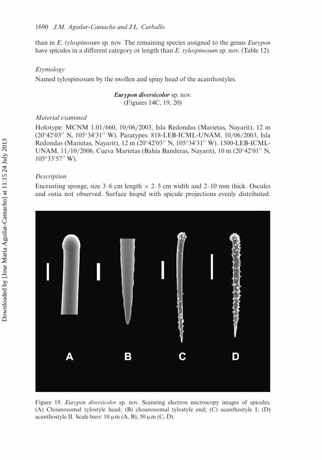
Encrusting sponge, size 3–6 cm length × 2–5 cm width and 2–10 mm thick. Osculesand ostia not observed. Surface hispid with spicule projections evenly distributed.
Figure 19. Eurypon diversicolor sp. nov. Scanning electron microscopy images of spicules.(A) Choanosomal tylostyle head; (B) choanosomal tylostyle end; (C) acanthostyle I; (D)acanthostyle II. Scale bars: 10 µm (A, B), 50 µm (C, D).
Dow
nloa
ded
by [
Jose
Mar
ia A
guila
r-C
amac
ho]
at 1
1:15
24
July
201
3
Journal of Natural History 1691
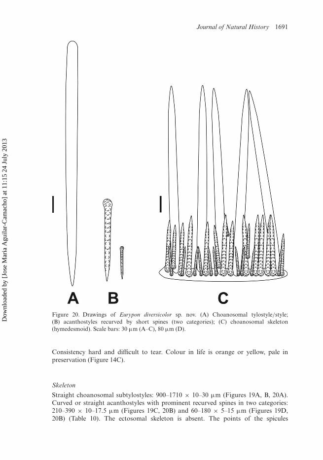
Figure 20. Drawings of Eurypon diversicolor sp. nov. (A) Choanosomal tylostyle/style;(B) acanthostyles recurved by short spines (two categories); (C) choanosomal skeleton(hymedesmoid). Scale bars: 30 µm (A–C), 80 µm (D).
Consistency hard and difficult to tear. Colour in life is orange or yellow, pale inpreservation (Figure 14C).
Skeleton
Straight choanosomal subtylostyles: 900–1710 × 10–30 µm (Figures 19A, B, 20A).Curved or straight acanthostyles with prominent recurved spines in two categories:210–390 × 10–17.5 µm (Figures 19C, 20B) and 60–180 × 5–15 µm (Figures 19D,20B) (Table 10). The ectosomal skeleton is absent. The points of the spicules
Dow
nloa
ded
by [
Jose
Mar
ia A
guila
r-C
amac
ho]
at 1
1:15
24
July
201
3

1692 J.M. Aguilar-Camacho and J.L. Carballo
Table 10. Spicule measurements of Eurypon diversicolor sp. nov. in µm.
Materialexamined
Subtylostyles(Length × Width)
Acanthostyles(Length × Width)
MCNM1.01/660
1120-(1321.5)-1690 × 10-(18.1)-30 1) 200-(302.4)-370 × 10-(15.2)-17.52) 70-(100.6)-175 × 2.5-(8.8)-15
LEB-818 900-(1317.4)-1710 × 10-(17.9)-30 1) 230-(314.2)-390 × 10-(13.1)-17.52) 80-(124.2)-180 × 5-(8.5)-15
LEB-1500 1080-(1279.2)-1580 × 10-(16.2)-25 1) 210-(280.4)-360 × 10-(13.4)-17.52) 60-(98.5)-180 × 2.5-(6.9)-12.5
protrude externally. The choanosomal skeleton has a hymedesmoid structure. Mainsubtylostyles and acanthostyles are erect in a spongin layer (10–20 µm thick)(Figure 20C).
Remarks
Eurypon diversicolor sp. nov. is found in the Mexican Pacific Ocean. The onlysimilar species in the Eastern Pacific is E. nigrum Bergquist 1967. This is a dark-blue encrusting sponge described from Oahu (Hawaii). It has straight tylostylesin two sizes: 1200–2400 × 6–12 µm and 170–800 × 6–12 µm; and acanthostyles(70–165 × 6–9 µm). Eurypon diversicolor sp. nov. has acanthostyles in two cate-gories (Table 10). The acanthostyles I are longer in E. diversicolor sp. nov. thanin E. nigrum. Eurypon duoacanthostyla (Hoshino, 1981) is an orange encrustingsponge described from Mitsusuke (Japan) at 15 m depth. It has straight styles(250–320 × 5–8 µm) and acanthostyles in two categories: 250–280 × 7–9 µm and138–180 × 6–8 µm. Eurypon diversicolor sp. nov. has longer and thicker stylesthan E. duoacanthostyla. The other species belonging to the genus Eurypon fromthe Pacific Ocean have spicules of different length than E. diversicolor sp. nov.(Table 12).
Etymology
Named diversicolor which means different colours in Latin.
Eurypon brunus sp. nov.(Figures 14D, 21, 22)
Material examined
Holotype: MCNM 1.01/661, 31/10/2002, Isla Lobos 1 (Mazatlán, Sinaloa), 5 m(23◦13′49′′ N, 106◦27′43′′ W). Paratypes: 653-LEB-ICML-UNAM, 29/10/2002,Isla Lobos 1 (Mazatlán, Sinaloa) 4 m (23◦13′49′′ N, 106◦27′43′′ W). 655-LEB-ICML-UNAM, 31/10/2002, Isla Lobos 1 (Mazatlán, Sinaloa), 5 m (23◦13′49′′N, 106◦27′43′′ W). 1505-LEB-ICML-UNAM, 11/10/2006, Cueva Marietas (BahiaBanderas, Nayarit), 11 m (20◦42′1′′ N, 105◦33′57′′ W).
Dow
nloa
ded
by [
Jose
Mar
ia A
guila
r-C
amac
ho]
at 1
1:15
24
July
201
3
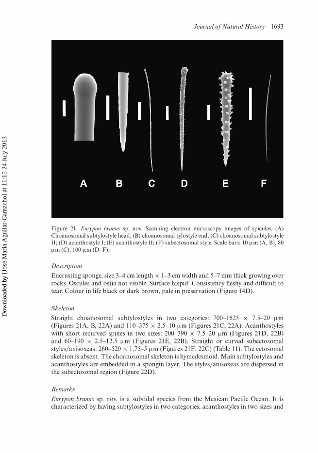
Journal of Natural History 1693
Figure 21. Eurypon brunus sp. nov. Scanning electron microscopy images of spicules. (A)Choanosomal subtylostyle head; (B) choanosomal tylostyle end; (C) choanosomal subtylostyleII; (D) acanthostyle I; (E) acanthostyle II; (F) subectosomal style. Scale bars: 10 µm (A, B), 80µm (C), 100 µm (D–F).
Description
Encrusting sponge, size 3–4 cm length × 1–3 cm width and 5–7 mm thick growing overrocks. Oscules and ostia not visible. Surface hispid. Consistency fleshy and difficult totear. Colour in life black or dark brown, pale in preservation (Figure 14D).
Skeleton
Straight choanosomal subtylostyles in two categories: 700–1625 × 7.5–20 µm(Figures 21A, B, 22A) and 110–375 × 2.5–10 µm (Figures 21C, 22A). Acanthostyleswith short recurved spines in two sizes: 200–390 × 7.5–20 µm (Figures 21D, 22B)and 60–190 × 2.5–12.5 µm (Figures 21E, 22B). Straight or curved subectosomalstyles/anisoxeas: 260–520 × 1.75–5 µm (Figures 21F, 22C) (Table 11). The ectosomalskeleton is absent. The choanosomal skeleton is hymedesmoid. Main subtylostyles andacanthostyles are embedded in a spongin layer. The styles/anisoxeas are dispersed inthe subectosomal region (Figure 22D).
Remarks
Eurypon brunus sp. nov. is a subtidal species from the Mexican Pacific Ocean. It ischaracterized by having subtylostyles in two categories, acanthostyles in two sizes and
Dow
nloa
ded
by [
Jose
Mar
ia A
guila
r-C
amac
ho]
at 1
1:15
24
July
201
3

1694 J.M. Aguilar-Camacho and J.L. Carballo
Figure 22. Drawings of Eurypon brunus sp. nov. (A) Choanosomal tylostyles/styles (twocategories); (B) acanthostyles recurved by short spines (two categories); (C) subectosomalstyle/anisoxeas; (D) choanosomal skeleton (hymedesmoid). Scale bars: 40 µm (A–C), 70µm (D).
Dow
nloa
ded
by [
Jose
Mar
ia A
guila
r-C
amac
ho]
at 1
1:15
24
July
201
3

Journal of Natural History 1695
Table 11. Spicule measurements of Eurypon brunus sp. nov. in µm.
Materialexamined
Choanosomalsubtylostyles
(Length × Width)
Acanthostyles(Length × Width)
Subectosomalstyles/anisoxeas
(Length × Width)
MCNM1.01/661
1) 900-(1190.6)-1525 ×10-(12.1)-15.
2) 240-(295.2)-335 ×2.5-(5.1)-10.
1) 200-(298.1)-340 ×10-(15.4)-20
2) 60-(123.7)-150 × 2.5-(4.6)-10
290-(346.1)-500 ×2.5-(3.5)-5
LEB-653 1) 700-(1123.6)-1625 ×10-(12.1)-15.
2) 110-(255.2)-345 ×5-(7.9)-10.
1) 230-(303.4)-375 ×10-(15.4)-20
2) 60-(123.7)-170 × 2.5-(5.6)-10
325-(366.4)-460 ×2.5-(3.2)-5
LEB-655 1) 900-(1120.4)-1390 ×7.5-(11.5)-17.5.
2) 235-(278.6)-340 ×2.5-(3.8)-5
1) 200-(255.4)-310 ×7.5-(13.4)-20
2) 60-(124.3)-190 × 2.5-(5.7)-10
260-(330.2)-410 ×1.75-(2.9)-5
LEB-1505 1) 800-(1108.3)-1310 ×10-(16.5)-20
2) 280-(332.5)-375 ×2.5-(4.6)-10
1) 215-(302.4)-390 ×7.5-(11.6)-15
2) 70-(128.3)-165 × 5-(8.6)-12.5
260-(392.4)-520 ×2.5-(2.9)-5
subectosomal styles/anisoxeas. The only species assigned to this genus which havethese characteristics are Eurypon miniaceum (Thiele 1905) and Eurypon graphidio-phora (Hentschel 1911) (Table 12). Eurypon miniaceum (Thiele 1905) is a red encrustingsponge described from Calbuco (Chile) at 30 m depth. This species has choanosomaltylostyles in three categories 2000–3000 × 30 µm; 800 × 30 µm; and >120 µm),acanthostyles (120 µm) and subectosomal styles (550 × 5 µm). Eurypon brunus sp.nov. has the acanthostyles I longer than in E. miniaceum. Eurypon graphidiophora(Hentschel 1911) is a grey encrusting sponge described from Australia. This specieshas straight or curved styles (280–1500 × 7–11 µm), acanthostyles (48–88 × 5 µm)and ectosomal rhapides/styles (350–400 × 2–3 µm). The acanthostyles are longer inE. brunus sp. nov. than in E. graphidiophora.
Etymology
Named brunus, which means brown in Latin.
Discussion
This study has revealed seven species new to science and three little knownspecies from the Mexican Pacific Ocean. Previous to this study in the Gulfof California there were four species belonging to the family Raspailiidae(Dickinson 1945). The number has increased from four to 13 (Trikentrium helium,Cyamon argon and Cyamon koltuni were not included in this study). Raspailia(Raspaxilla) hymani (Dickinson 1945) and Raspailia (Raspaxilla) hyle (de Laubenfels1930) are deep-sea species found on the West Pacific coast of Baja Peninsula.
Dow
nloa
ded
by [
Jose
Mar
ia A
guila
r-C
amac
ho]
at 1
1:15
24
July
201
3

1696 J.M. Aguilar-Camacho and J.L. Carballo
Tab
le12
.C
ompa
rati
veta
ble
ofal
lthe
Eur
ypon
spec
ies
desc
ribe
dw
orld
wid
e.Sp
icul
em
easu
rem
ents
inµ
m.
Spec
ies
Cho
anos
omal
spic
ules
(Len
gth
×W
idth
)
Aca
ntho
styl
es(L
engt
h×
Wid
th)
Ect
osom
alsp
icul
es(L
engt
h×
Wid
th)
Col
our,
loca
lity
and
dept
h
E.c
alyp
soi(
Lév
i195
8)T
ylos
tyle
s:20
00×
1075
–90
×8–
9O
xeas
:40
0–47
5×
3B
lue.
Abu
lat,
Saud
iAra
bia.
Lit
tora
lE
.pol
yplu
mos
a(L
évi1
958
such
asP
rora
spai
lia)
Tyl
osty
les:
300–
375
×11
60–3
50×
7–11
Rap
hide
sox
eote
s:28
0–32
0×
0.5–
1O
chre
.Sa
udiA
rabi
a,de
pth
unkn
own
E.c
intu
m(S
arà
1960
)T
ylos
tyle
s:2.
5m
m×
8–30
.14
–16
cabe
za
31–3
16×
7–16
.St
yle
sor
oxea
s:41
5×
510
×5–
9L
ila.
Poin
tIm
pera
tore
,Náp
oles
.70
m
E.c
lava
tella
(Lit
tle
1963
)T
ylos
tyle
s:24
9–47
0×
14–5
175
–145
×5–
9St
yles
:36
1×
4P
urpl
e.F
lori
da,G
ulf
ofM
exic
o.10
mE
.den
isae
(Vac
elet
1969
)T
ylos
tyle
s:31
50×
291)
150–
120
×7–
102)
50.–
90×
7–8
Oxe
as:
250–
300
×4.
5–7.
5B
eige
.C
assi
daig
neC
anyo
n.M
edit
erra
nean
Sea.
300–
350
mE
.obt
usum
Vac
elet
(196
9)T
ylos
tyle
s:¿?
??×
10–1
270
–170
×5–
7.5
Oxe
as:
400–
430
×2.
5–3
Gre
y.Si
cie
Can
yon.
Med
iter
rane
anSe
a.25
0m
E.e
ncru
sta
(Tho
mas
1981
)St
yles
:45
1–67
8×
4–8
63–1
08×
6–8
Tri
chod
ragm
as:
40–5
0×
21R
aphi
des:
40–5
Whi
te.
Seyc
helle
sIs
land
s
(Con
tinu
ed)
Dow
nloa
ded
by [
Jose
Mar
ia A
guila
r-C
amac
ho]
at 1
1:15
24
July
201
3

Journal of Natural History 1697
Tab
le12
.(C
onti
nued
).
E.f
ulvu
m(L
évi1
969)
Tyl
osty
les:
1100
–150
0×
1275
–85
×9
Oxe
as:
475–
530
×7–
8Y
ello
w.
Ven
a,So
uth
Afr
ica.
Dep
thun
know
nE
.top
sent
i(P
ulit
zer-
Fin
ali1
983)
(Syn
onym
yof
E.c
oron
ula,
Tops
ent
1936
)
Subt
ylos
tyle
scu
rved
:19
00–2
500
×12
–14
Styl
es:3
50–6
00×
10.5
–17.
5
55–1
60×
3–11
Styl
es:
370–
430
×1.
5R
ed.
Port
Tri
case
,Poi
ntM
anar
a.M
edit
erra
nean
Sea,
33–6
0m
E.v
esci
cula
ris
(Sar
àan
dSi
ribe
lli19
60)
Tyl
osty
les:
442–
2125
×7–
1798
–105
×3.
5St
yles
:11
00–1
200
×1.
7Y
ello
wN
ápol
es,M
edit
erra
nean
Sea.
30–4
0m
E.m
ajor
(Sar
àan
dSi
ribe
lli19
60)
∗ Pul
itze
r-F
inal
i(19
83)
Tyl
osty
les:
1115
–221
0×
10–1
780
–220
×4–
10.5
Oxe
as:
480–
700
×4–
7.5
Ros
e.N
ápol
es,M
edit
erra
nean
Sea.
14–2
0m
E.l
acaz
ei(T
opse
nt18
91su
chas
Hym
erap
hia)
Tyl
osty
les:
>20
00×
2075
–80
×10
Torn
otes
:23
0×
7Y
ello
w.
Ros
coff
.Dep
thun
know
nE
.gra
phid
ioph
ora
(Hen
tsch
el19
11su
chas
Hym
erap
hia)
∗ Hoo
per
(199
1)
Styl
es:
280–
1500
×7–
1148
–88
×5
Rap
hide
s:35
0–40
0×
2–3
Gre
y.W
este
rnco
ast
ofA
ustr
alia
,Dep
thun
know
n
E.h
ispi
da(B
ergq
uist
1970
)Su
btyl
osty
les:
304–
1150
×3–
1670
–352
×3–
12A
bsen
tO
rang
e.L
eigh
Ree
f,N
ewZ
eala
nd.2
0m
E.n
igru
m(B
ergq
uist
1967
)T
ylos
tyle
s:1)
1200
–240
0×
6–12
2)17
0–80
0×
6–12
70–1
65×
6–9
Abs
ent
Blu
e.O
ahu,
Haw
aii
5–10
m
E.m
inia
ceum
(Thi
ele
1905
)T
ylos
tyle
s:1)
2–3
mm
×30
2)80
0×
303)
asac
anth
osty
les
120
Styl
es:
550
×5
Red
.C
albu
co,C
hile
.30
m
(Con
tinu
ed)
Dow
nloa
ded
by [
Jose
Mar
ia A
guila
r-C
amac
ho]
at 1
1:15
24
July
201
3

1698 J.M. Aguilar-Camacho and J.L. Carballo
Tab
le12
.(C
onti
nued
).
Spec
ies
Cho
anos
omal
spic
ules
(Len
gth
×W
idth
)
Aca
ntho
styl
es(L
engt
h×
Wid
th)
Ect
osom
alsp
icul
es(L
engt
h×
Wid
th)
Col
our,
loca
lity
and
dept
h
E.v
irid
is(T
opse
nt18
89su
chas
Tri
cheu
rypo
n)T
ylos
tyle
s:57
0–16
80×
6–24
113–
365
×7–
14R
aphi
des
intr
icho
drag
mas
:46
–70
×0.
5–3
Whi
te.
Can
aria
sIs
land
s,A
zore
s,M
edit
erra
nean
Sea.
12–4
80m
E.l
ongi
spic
ulum
(Car
ter
1876
such
asM
icro
cion
a)T
ylos
tyle
s:22
57×
28.2
2N
oda
taSt
yles
:56
4B
row
n.C
elti
cSe
a.63
0m
E.s
pinu
laru
m(B
ower
bank
1875
such
asH
ymer
aphi
a)
Styl
es:
529.
16×
1111
8.70
No
data
Yel
low
ochr
e.K
orea
nco
ast.
Dep
thun
know
n
E.s
impl
ex(B
ower
bank
1874
such
asH
ymer
aphi
a)T
ylos
tyle
s:21
16×
27.1
105.
8–21
8.9
×8.
4A
bsen
tP
ale
yello
wpr
eser
ved.
Shet
land
Isla
nds.
Dep
thun
know
nE
.cor
onul
a(B
ower
bank
1874
such
asH
ymer
aphi
a)T
ylos
tyle
:63
5–14
1125
4A
bsen
tG
rey
pres
erve
d.Sh
etla
ndIs
land
sE
.cla
vatu
m(B
ower
bank
1866
such
asH
ymer
aphi
a)
Subt
ylos
tyle
s:68
5–23
10×
11–2
864
–472
×5–
19St
yles
:41
8–69
5×
3–5
Col
our
unkn
own.
Nor
thA
tlan
tic
and
Med
iter
rane
an.3
0–16
00m
E.t
oure
ti(T
opse
nt18
94su
chas
Hym
erap
hia)
Tyl
osty
les:
No
data
50–6
0A
bsen
tB
row
nC
ampe
che,
Gul
fof
Méx
ico.
(dep
thun
know
n)E
.duo
acan
thos
tyla
(Hos
hino
1981
such
asP
rian
os)
Styl
es:
250–
320
×5–
81)
250–
280
×7–
92)
138–
180
×6–
8A
bsen
tO
rang
eor
Pea
ch.
Mit
suku
e,Ja
pan.
15m
E.s
pitz
berg
ensi
s(F
rist
edt
1887
such
asH
ymer
aphi
a)T
ylos
tyle
s:<
2500
Styl
es:
300
Abs
ent
Gre
y.Sp
isbe
rgen
,Art
ic.D
epth
unkn
own
∗ Add
itio
nali
nfor
mat
ion
ofth
eor
igin
alde
scri
ptio
n.
Dow
nloa
ded
by [
Jose
Mar
ia A
guila
r-C
amac
ho]
at 1
1:15
24
July
201
3
Journal of Natural History 1699
Aulospongus cerebella (Dickinson 1945) is a deep-sea species from the Gulf ofCalifornia and the West Pacific coast of Baja Peninsula. Of the remaining species,six are subtidal found in the Mexican Pacific with the exception of Aulosponguscalifornianus sp. nov. which is a deep-sea species from the Gulf of California.
The hymedesmoid skeleton of the genus Eurypon is a homoplasic character inthe order Axinellida (Family Raspailiidae). This choanosomal organization has beenreported in several genera of different orders (such as: Microciona, Acarnus, Timea,Prosuberites and others) (Boury-Esnault et al. 1994). The spicule shape and thepresence of some microscleres are used in the allocation of some genera and families.
For example, in the family Microcionidae the subgenera Clathria and Microcionahave the same spicule elements and the difference between these two subgenera is thetype of choanosomal skeleton (reticulate in Clathria and hymedesmoid in Microciona).However, in the subgenus Thalysias there are species with a hymedesmoid and anaxial or reticulate choanosomal skeleton. The diagnostic features used in allocatingspecies to the genus Thalysias are the presence of ectosomal and subectosomal spicules(Hooper 1996).
The genus Eurypon
The genus Eurypon was originally described by Gray 1867 for the type speciesHymeraphia radiata Bowerbank 1866. The principal characteristics of this genusare the presence of choanosomal styles or tylostyles, echinating acanthostyles andsubectosomal or ectosomal spicules (styles, oxeas and raphides), and an encrustinghabit with a hymedesmoid skeleton (Hooper 2002). The skeleton of the genus Euryponis similar to that found in species belonging to the subgenus Microciona (GenusClathria; Family Microcionidae) (Hooper 1996). Recent molecular studies suggest thatEurypon is polyphyletic and belongs in the Order Axinellida (Morrow et al. 2012).Species of the genus Eurypon have tylostyles in one or two categories as choanosomalspicules. The subectosomal or ectosomal spicules if present are raphides, oxeas orstyles. Table 12 allocates species with these diagnostic features.
Species bearing acanthostyles as choanosomal spicules (genus Acantheurypon)The genus Acantheurypon was created by Topsent (1927) for Hymeraphia pilosella(Topsent 1904). This species has choanosomal acanthostyles, echinating acanthostylesand ectosomal subtylostyles (Table 13). Topsent (1928) described four new species ofthis genus from the Azores. Hooper (1991) synonymized Acantheurypon with Euryponbecause the choanosomal skeleton is hymedesmoid. However, other authors consid-ered the genus Acantheurypon valid (Boury-Esnault et al. 1994). Morrow et al. (2012)demonstrated using molecular tools that the genus Eurypon is polyphyletic and iswithin the order Axinellida. The genus Acantheurypon is monophyletic and grouped inthe order Poecilosclerida. One difference between these two genera is the choanosomalspicule morphology (smooth choanosomal tylostyles in Eurypon and choanosomalacanthostyles in Acantheurypon). Because it has two size classes of acanthostyles,Trachostylea lamellata Lévi 1993 should be included in Acantheurypon. A further mor-phological and molecular examination is required to corroborate whether the genusAcantheurypon should be re-erected. Table 13 indicates species of the genus Euryponbearing choanosomal acanthostyles.
Dow
nloa
ded
by [
Jose
Mar
ia A
guila
r-C
amac
ho]
at 1
1:15
24
July
201
3

1700 J.M. Aguilar-Camacho and J.L. Carballo
Tab
le13
.C
ompa
rati
veta
ble
ofal
lthe
Eur
ypon
spec
ies
desc
ribe
dw
orld
wid
ebe
arin
gch
oano
som
alac
anth
osty
les.
Spic
ule
mea
sure
men
tsin
µm
.
Spec
ies
Cho
anos
omal
acan
thos
tyle
s(L
engt
h×
Wid
th)
Aca
ntho
styl
es(L
engt
h×
Wid
th)
Ect
osom
alsp
icul
es(L
engt
h×
Wid
th)
Col
our,
loca
lity
and
dept
h
Eur
ypon
pilo
sella
(Top
sent
1904
such
asH
ymer
aphi
a)∗ B
oury
-Esn
ault
etal
.(19
94)
350-
1700
-11-
3495
–300
×11
–34
Subt
ylos
tyle
sw
ith
mic
rosp
ined
base
:25
0–66
8×
3.5–
9
Yel
low
orG
reen
.V
ilafr
anca
Isla
nd,
Azo
res.
50–1
740
mE
uryp
onm
ixtu
m(T
opse
nt19
28su
chas
Aca
nthe
uryp
on)
>10
0010
0–32
0Su
btyl
osty
les:
No
data
Gre
y.A
zore
s.90
0–13
30m
Eur
ypon
inic
ipie
ns(T
opse
nt19
28su
chas
Aca
nthe
uryp
on)
770
100–
230
Subt
ylos
tyle
s:N
oda
taC
olou
rno
tre
port
ed.
Azo
res.
1250
mE
uryp
onsc
abio
sum
(Top
sent
1927
such
asA
cant
heur
ypon
)∗ T
opse
nt(1
928)
1068
×20
–24
80–2
50×
6–13
Subt
ylos
tyle
s:47
0–63
0×
3–4
1m
m65
mic
ras×
4–7
mic
ra
Gre
y.A
zore
s.91
4–65
0m
Eur
ypon
muc
rona
le(T
opse
nt19
28su
chas
Aca
nthe
uryp
on)
700–
900
×25
125–
280
×20
370
(Unu
sual
)To
rnot
es:
400–
490
×12
–17
Gre
y.A
zore
s.24
60m
Eur
ypon
hisp
idul
um(T
opse
nt19
04su
chas
Hym
erap
hia)
500
×17
160–
200
×10
–12
Subt
ylos
tyle
s:32
5–36
5×
4G
rey.
Azo
res.
99–8
80m
Eur
ypon
lam
ella
ta(L
évi1
993
such
asT
rach
osty
lea)
1300
–180
0×
8–10
725–
950
×8–
10A
bsen
tC
olou
rno
tre
port
ed.
New
Cal
edon
ia,
965
m
∗ Add
itio
nali
nfor
mat
ion
ofth
eor
igin
alde
scri
ptio
n.
Dow
nloa
ded
by [
Jose
Mar
ia A
guila
r-C
amac
ho]
at 1
1:15
24
July
201
3

Journal of Natural History 1701
Table 14. Species assigned to the genus Eurypon by van Soest et al. (2012b) with a massive orramose form.
Species Choanosomalspicules
(Length × Width)
Acanthostyles(Length × Width)
Ectosomalspicules
(Length × Width)
Shape, colour,locality and depth
Eurypon cactoides(Burton & Rao1932 such asProtoraspailia)
Tylostyles:850 × 16
140 Trichodragmas48 × 8–20
Erect or cactiform.Brown.
Indian Ocean.Depth unknown
Eurypon sessile(Carter 1880 such as
Dictyocylindrus)
Tylostyles:635 × 28.2
Acanthostyles:148 × 8.5
Oxeas:282
Massive, subspheric.Brown.
Gulf of Manar.Depth unknown
Eurypon inuisitatia-canthostyla(Hoshino 1981such as Prianos)
Strongyles:325–430 × 6–12
Acanthostyles:320–482 × 6–16
Absent Massive, Orange.Mitsukue
Japan.15 m
On the presence of sponges with massive form allocated to the genus Eurypon
There are three massive or ramose species of the genus Eurypon (van Soest et al.2012b): Protoraspailia cactoides Burton and Rao 1932, Dictyocylindrus sessile Carter1880, and Prianos inuisitatiacanthostyla Hoshino 1981 (Table 14). The first twohave choanosomal tylostyles, acanthostyles and ectosomal spicules. The skeletonsare of an axial/extra-axial organization. These species are not encrusting or with ahymedesmoid skeleton as in Eurypon. We consider that these two species have moreaffinities with the genus Raspailia. Species assigned to this genus have a more-or-lesscompressed axial skeleton and a radial, plumose or simply reticulate extra-axial skele-ton, with choanosomal spicules consisting of two, three or more different size classes(styles and/or oxeas), and echinating acanthostyles (Hooper 2002). Prianos inuisitati-acanthostyla Hoshino 1981 is a massive sponge described from Japan with strongyles,acanthostyles and an axial compressed choanosomal skeleton. The allocation of thisspecies to one of the current genera from the family Raspailiidae is problematic. Thereare no species with exclusively strongyles and acanthostyles in the skeleton (Hooper2002). Hoshino (1981) did not report the ectosomal spicules. A morphological revi-sion should be undertaken to clarify the status of these three species. They do not havethe morphological features of the genus Eurypon.
The genus Dragmatyle
Topsent (1904) described a new genus for the type species Dragmatyle lictor.This is a white encrusting sponge with tylostyles, ectosomal diactinal spicules andtrichodragmas as microscleres. Hooper (1991) synonymized this genus with Euryponbecause it is encrusting and has tylostyles and ectosomal spicules. Hooper (1991) didnot consider the presence of acanthostyles as a diagnostic feature for this taxonomicdecision. Currently, all the species assigned to the genus Eurypon have this morpho-logical feature. There are currently no genera in the family Raspailiidae [Raspailia(Parasyringella), Ceratopsion or Thrinacophora)], which lack acanthostyles and have
Dow
nloa
ded
by [
Jose
Mar
ia A
guila
r-C
amac
ho]
at 1
1:15
24
July
201
3
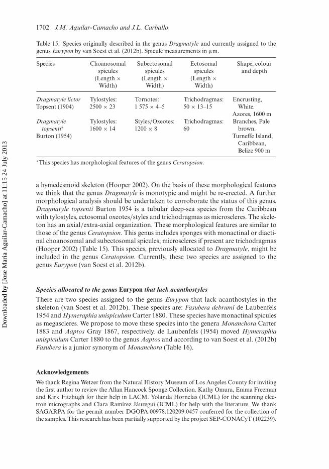
1702 J.M. Aguilar-Camacho and J.L. Carballo
Table 15. Species originally described in the genus Dragmatyle and currently assigned to thegenus Eurypon by van Soest et al. (2012b). Spicule measurements in µm.
Species Choanosomalspicules
(Length ×Width)
Subectosomalspicules
(Length ×Width)
Ectosomalspicules
(Length ×Width)
Shape, colourand depth
Dragmatyle lictorTopsent (1904)
Tylostyles:2500 × 23
Tornotes:1 575 × 4–5
Trichodragmas:50 × 13–15
Encrusting,White.
Azores, 1600 mDragmatyle
topsenti∗
Burton (1954)
Tylostyles:1600 × 14
Styles/Oxeotes:1200 × 8
Trichodragmas:60
Branches, Palebrown.
Turneffe Island,Caribbean,Belize 900 m
∗This species has morphological features of the genus Ceratopsion.
a hymedesmoid skeleton (Hooper 2002). On the basis of these morphological featureswe think that the genus Dragmatyle is monotypic and might be re-erected. A furthermorphological analysis should be undertaken to corroborate the status of this genus.Dragmatyle topsenti Burton 1954 is a tubular deep-sea species from the Caribbeanwith tylostyles, ectosomal oxeotes/styles and trichodragmas as microscleres. The skele-ton has an axial/extra-axial organization. These morphological features are similar tothose of the genus Ceratopsion. This genus includes sponges with monactinal or diacti-nal choanosomal and subectosomal spicules; microscleres if present are trichodragmas(Hooper 2002) (Table 15). This species, previously allocated to Dragmatyle, might beincluded in the genus Ceratopsion. Currently, these two species are assigned to thegenus Eurypon (van Soest et al. 2012b).
Species allocated to the genus Eurypon that lack acanthostylesThere are two species assigned to the genus Eurypon that lack acanthostyles in theskeleton (van Soest et al. 2012b). These species are: Fasubera debrumi de Laubenfels1954 and Hymeraphia unispiculum Carter 1880. These species have monactinal spiculesas megascleres. We propose to move these species into the genera Monanchora Carter1883 and Aaptos Gray 1867, respectively. de Laubenfels (1954) moved Hymeraphiaunispiculum Carter 1880 to the genus Aaptos and according to van Soest et al. (2012b)Fasubera is a junior synonym of Monanchora (Table 16).
Acknowledgements
We thank Regina Wetzer from the Natural History Museum of Los Angeles County for invitingthe first author to review the Allan Hancock Sponge Collection. Kathy Omura, Emma Freemanand Kirk Fitzhugh for their help in LACM. Yolanda Hornelas (ICML) for the scanning elec-tron micrographs and Clara Ramírez Jáuregui (ICML) for help with the literature. We thankSAGARPA for the permit number DGOPA.00978.120209.0457 conferred for the collection ofthe samples. This research has been partially supported by the project SEP-CONACyT (102239).
Dow
nloa
ded
by [
Jose
Mar
ia A
guila
r-C
amac
ho]
at 1
1:15
24
July
201
3
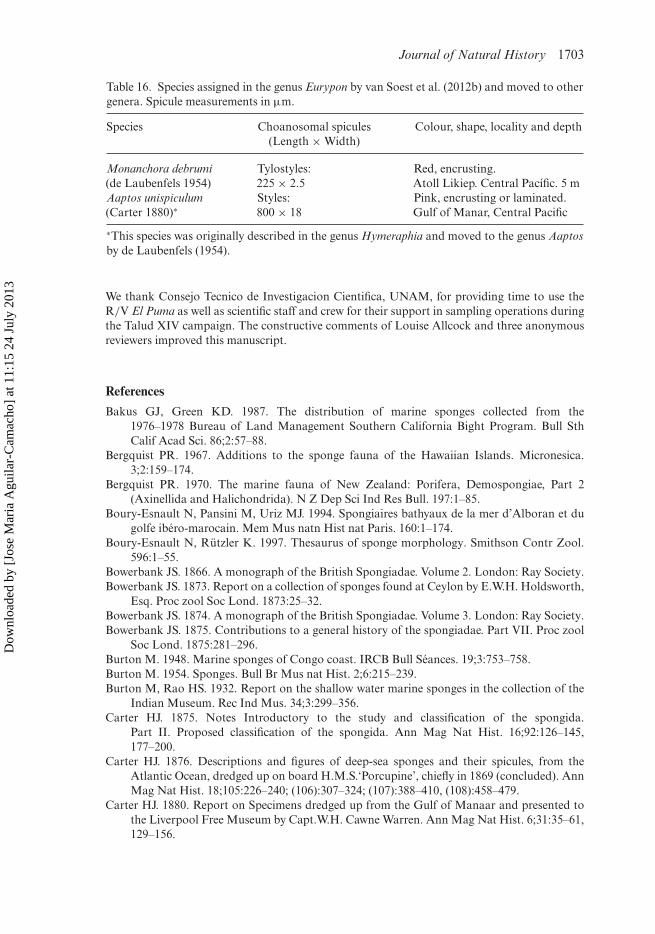
Journal of Natural History 1703
Table 16. Species assigned in the genus Eurypon by van Soest et al. (2012b) and moved to othergenera. Spicule measurements in µm.
Species Choanosomal spicules(Length × Width)
Colour, shape, locality and depth
Monanchora debrumi(de Laubenfels 1954)
Tylostyles:225 × 2.5
Red, encrusting.Atoll Likiep. Central Pacífic. 5 m
Aaptos unispiculum(Carter 1880)∗
Styles:800 × 18
Pink, encrusting or laminated.Gulf of Manar, Central Pacific
∗This species was originally described in the genus Hymeraphia and moved to the genus Aaptosby de Laubenfels (1954).
We thank Consejo Tecnico de Investigacion Cientifica, UNAM, for providing time to use theR/V El Puma as well as scientific staff and crew for their support in sampling operations duringthe Talud XIV campaign. The constructive comments of Louise Allcock and three anonymousreviewers improved this manuscript.
References
Bakus GJ, Green KD. 1987. The distribution of marine sponges collected from the1976–1978 Bureau of Land Management Southern California Bight Program. Bull SthCalif Acad Sci. 86;2:57–88.
Bergquist PR. 1967. Additions to the sponge fauna of the Hawaiian Islands. Micronesica.3;2:159–174.
Bergquist PR. 1970. The marine fauna of New Zealand: Porifera, Demospongiae, Part 2(Axinellida and Halichondrida). N Z Dep Sci Ind Res Bull. 197:1–85.
Boury-Esnault N, Pansini M, Uriz MJ. 1994. Spongiaires bathyaux de la mer d’Alboran et dugolfe ibéro-marocain. Mem Mus natn Hist nat Paris. 160:1–174.
Boury-Esnault N, Rützler K. 1997. Thesaurus of sponge morphology. Smithson Contr Zool.596:1–55.
Bowerbank JS. 1866. A monograph of the British Spongiadae. Volume 2. London: Ray Society.Bowerbank JS. 1873. Report on a collection of sponges found at Ceylon by E.W.H. Holdsworth,
Esq. Proc zool Soc Lond. 1873:25–32.Bowerbank JS. 1874. A monograph of the British Spongiadae. Volume 3. London: Ray Society.Bowerbank JS. 1875. Contributions to a general history of the spongiadae. Part VII. Proc zool
Soc Lond. 1875:281–296.Burton M. 1948. Marine sponges of Congo coast. IRCB Bull Séances. 19;3:753–758.Burton M. 1954. Sponges. Bull Br Mus nat Hist. 2;6:215–239.Burton M, Rao HS. 1932. Report on the shallow water marine sponges in the collection of the
Indian Museum. Rec Ind Mus. 34;3:299–356.Carter HJ. 1875. Notes Introductory to the study and classification of the spongida.
Part II. Proposed classification of the spongida. Ann Mag Nat Hist. 16;92:126–145,177–200.
Carter HJ. 1876. Descriptions and figures of deep-sea sponges and their spicules, from theAtlantic Ocean, dredged up on board H.M.S.‘Porcupine’, chiefly in 1869 (concluded). AnnMag Nat Hist. 18;105:226–240; (106):307–324; (107):388–410, (108):458–479.
Carter HJ. 1880. Report on Specimens dredged up from the Gulf of Manaar and presented tothe Liverpool Free Museum by Capt.W.H. Cawne Warren. Ann Mag Nat Hist. 6;31:35–61,129–156.
Dow
nloa
ded
by [
Jose
Mar
ia A
guila
r-C
amac
ho]
at 1
1:15
24
July
201
3

1704 J.M. Aguilar-Camacho and J.L. Carballo
Carter HJ. 1883. New genus of sponges. Ann Mag Nat Hist. 11;5:369–370.Carter HJ. 1885. Descriptions of Sponges from the Neighbourhood of Port Phillip Heads, South
Australia. Ann Mag Nat Hist. 16;94:277–294, 347–368.de Laubenfels MW. 1930. The sponges of California. (Abstracts of dissertations for the degree
of doctor of philosophy). Stanford Univ Bull. 5;98:24–29.de Laubenfels MW. 1932. The marine and fresh–water sponges of California. Proc US Natl
Mus. 81;27:1–140.de Laubenfels MW. 1936. A discussion of the sponge fauna of the dry tortugas in particular and
the West Indies in General, with material for a revision of the families and orders of theporifera. Carnegie Inst Wash (Tortugas Laboratory 467). 30:1–225.
de Laubenfels MW. 1954. The sponges of the West-Central Pacific. Oregon state monographs.Stud Zool. 7:1–306.
Desqueyroux-Faúndez R, van Soest RWM. 1997. Shallow water demosponges of the galapagosIslands. Rev Suisse Zool. 104;2:379–467.
Dickinson MG. 1945. Sponges on the Gulf of California. Allan Hancock Pac Exp. 11;1:1–251.Erpenbeck D, List-Armitage SE, Alvarez B, Degnan BM, Hooper JNA, Wörheide G. 2007.
The systematics of Raspailiidae (Demospongiae, Poecilosclerida, Microcionina) reanalysedwith a ribosomal marker. J Mar Biol Assoc UK. 87;6:1571–1576.
Fristedt K. 1887. Sponges from the Atlantic and Arctic Oceans and the Behring Sea. Vega-Exped Vetensk Iaktt. 4:401–471.
Gray JE. 1867. Notes on the arrangement of sponges, with the descriptions of some NewGenera. Proc Zool Soc Lond. 1867;2:492–558.
Green KD, Bakus GJ. 1994. Taxonomic atlas of the benthic fauna of the Santa Maria basin andthe western Santa Barbara channel. Santa Barbara Mus Nat Hist. 2:1–87.
Hentschel E. 1911. Tetraxonida. 2. Teil. In: Michaelsen W, Hartmeyer R editor. Die FaunaSüdwest-Australiens. Ergebnisse der Hamburger südwest-australischen Forschungsreise1905. Volume 3. Jena: Fischer; p. 279–393.
Hentschel E. 1923. Erste Unterabteilung der Metazoa: Parazoa, Porifera-Schwämme. In:Kükenthal W, Krumbach T, editor. Handbuch der Zoologie. Eine NaturgeschichtederStämme des Tierreiches. Vol. 1, Protozoa, Porifera, Coelenterata, Mesozoa. Berlin: Walterde Gruyter und Co; p. 307–418.
Hooper JNA. 1991. Revision of the family Raspailiidae (Porifera: Demospongiae), withdescription of Australian species. Invertebr Taxon. 5;6:1179–1418.
Hooper JNA. 1996. Revision of Microcionidae (Porifera: Poecilosclerida: Demospongiae), withdescription of Australian species. Mem Q’ld Mus. 40:1–626.
Hooper JNA. 2002. Family Raspailiidae Hentschel, 1923. In: Hooper JNA, Van SoestRWM, editor. Systema Porifera. Guide to the classification of sponges. London: KluwerAcademic/Plenum Publishers; p. 469–510.
Hooper JNA, Lehnert H, Zea S. 1999. Revision of Aulospongus and other Raspailiidae withrhabdostyles (Porifera: Demospongiae: Poecilosclerida). Mem Q’ld Mus. 43;2:649–707.
Hooper JNA, Sutcliffe P, Schlacher-Hoenlinger MA. 2008. New species of Raspailiidae(Porifera: Demospongiae: Poecilosclerida) from southeast Queensland. Mem Q’ld MusNat. 54;1:1–22.
Hoshino T. 1981. Shallow-water demosponges of Western Japan, 1. J Sci Hiroshima Univ.29;1:47–205.
Lee WL, Elvin DW, Reiswig HM. 2007. The sponges of California. A guide and key to themarine sponges of California. Monterey (CA): Monterey Bay Sanctuary Foundation.
Little FJ. 1963. The sponge fauna of the St. George’s Sound, Apalache Bay, and Panama CityRegións of the Florida Gulf Coast. Tulane Stud Zool. 11;2:31–71.
Lévi C. 1958. Résultats scientifiques des Campagnes de la ‘Calypso’.Campagne 1951–1952 enMer Rouge (suite). Ann Inst Oceanogr. 34;3:3–46.
Dow
nloa
ded
by [
Jose
Mar
ia A
guila
r-C
amac
ho]
at 1
1:15
24
July
201
3
Journal of Natural History 1705
Lévi C. 1969. Spongiaires du Vema Seamount (Atlantique Sud). Bull Mus Hist nat Paris.41;4:952–973.
Lévi C. 1973. Systematique de la Clase Demospongiaria (Demosponges). In: Grassé PP, editor.Traite de Zoologie, Anatomie, Systematique, Biologie, Spongiaires. Paris: Masson et Cie;p. 577–631.
Lévi C. 1993. Porifera Demospongiae: Spongiaires bathyaux de Nouvelle-Calédonie, récoltéspar le ‘Jean Charcot’. Campagne BIOCAL, 1985. In: Crosnier A, editor. Résultatsdes campagnes MUSORSTOM, Volume 11. Mémoires du Muséum national d’Histoirenaturelle (A, Zoologie). p. 9–87.
Morrow CC, Picton BE, Erpenbeck D, Boury-Esnault N, Maggs CA, Allcock AL. 2012.Congruence between nuclear and mitochondrial genes in Demospongiae: a new hypoth-esis for relationships within the G4 clade (Porifera: Demospongiae). Mol Phyl Evol.62:174–190.
Nardo GD. 1833. Auszug aus einem neuen System der Spongiarien,wonach bereitsdie Aufstellung in der Universitäts-Sammlung zu Padua gemacht ist. In: Isis oderEncyclopädische Zeitung Coll. Jena: Oken; p. 519–523.
Pulitzer-Finali G. 1983. A collection of Mediterranean Demospongiae (Porifera) with, inappendix, a list of the Demospongiae hitherto recorded from the Mediterranean Sea.Annali Mus Civ Stor Nat Giacomo Doria. 84:445–621.
Ridley SO. 1884. Spongiida. Report on the zoological collections made in the Indo-Pacific Ocean during the Voyage of H.M.S. ‘Alert’, 1881–82. Brit Mus Nat Hist Pub.1881–2:366–482, 582–630.
Ridley SO, Dendy A. 1886. Preliminary report on the Monaxonida collected by H.M.S.‘Challenger. Ann Mag Nat Hist. 18:325–351, 470–493.
Sarà M. 1960. Poriferi del litorale dell’isola d’lschia e loro ripartizioneper ambienti. Pubbl Stazzool Napoli. 31;3:421–472.
Sarà M, Siribelli L. 1960. La fauna di Poriferi delle ‘secche’ del Golfo di Napoli. La ‘secca’ dellaGaiola. Annuari Ist Mus Zool Univ Napoli. 12;3:1–93.
Thiele J. 1905. Die Kiesel- und Hornschwämme der Sammlung Plate (Fauna Chiliensis III).Zool Jahrb Suppl. 6:407–496.
Thomas PA. 1981. A second collection of marine Demospongiae from Mahe Island in theSeychelles Bank. Ann Mus r Afrique centr Sér in 8 Sci zool. 233:1–54.
Topsent E. 1889. Quelques spongiaires du Banc de Campêche et de la Pointe-à-Pître. Mém Soczool Fr. 2:30–52.
Topsent E. 1890. Notice préliminaire sur les spongiaires recueillis durant les campagnes del’Hirondelle. Bull Soc Zool France. 15:26–32, 65–71.
Topsent E. 1891. Essai sur la faune des spongiaires de Roscoff. Arch Zool Exp Gen. 9;4:523–554.
Topsent E. 1892. Contribution à l’étude des Spongiaires de l’Atlantique Nord (Golfe deGascogne, Terre-Neuve, Açores). Rés Camp Sci Prince Albert I Monaco. 2:1–165.
Topsent E. 1896. Matériaux pour servir à l’étude de la faune des spongiaires de France. MémSoc zool Fr. 9:113–133.
Topsent E. 1904. Spongiaires des Açores. Rés Camp Sci Prince Albert I Monaco. 25:1–280.Topsent E. 1927. Diagnoses d’Éponges nouvelles recueillies par le Prince Albert ler de Monaco.
Bull Inst Océanogr Monaco. 502:1–19.Vacelet J. 1969. Eponges de la Roche du Large et de l’étage bathyal de Méditerranée (Récoltes
de la soucoupe plongeante Cousteau et dragages). Mem Mus natn Hist nat Paris.59;2:145–219.
van Soest RWM, Boury-Esnault N, Hooper JNA, Rützler K, de Voogd NJ, Alvarez de GlasbyB, Hajdu E, Pisera AB, Manconi R, Schoenberg C, et al. 2012b. World Porifera database.[cited 2012 Jun 6]. Available online from: http://www.marinespecies.org/porifera.
Dow
nloa
ded
by [
Jose
Mar
ia A
guila
r-C
amac
ho]
at 1
1:15
24
July
201
3
1706 J.M. Aguilar-Camacho and J.L. Carballo
van Soest R, Carballo JL, Hooper JNA. 2012a. Polyaxone monaxonids: revision of raspailiidsponges with polyactine megascleres (Cyamon and Trikentrion). Zookeys. 239:1–70.
van Soest RWM, Diaz MC, Pomponi SA. 1990. Phylogenetic classification of the Halichondrids(Porifera, Demospongiae). Beaufortia. 40:15–62.
von Lendenfeld R. 1887. Die Chalineen des australischen Gebietes. Zool Jb. 2:723–828.von Lendenfeld R. 1898. Die Clavulina der Adria. Nova Acta Acad Caes Leop Carol Germ
Nat. 69:1–251.
Dow
nloa
ded
by [
Jose
Mar
ia A
guila
r-C
amac
ho]
at 1
1:15
24
July
201
3