Embed Size (px)
Citation preview

ACS SYMPOSIUM SERIES 966
Rational Environmental Management of Agrochemicals
Risk Assessment, Monitoring, and Remedial Action
Ivan R. Kennedy, Editor University ofSydney
Keith R. Solomon, Editor University of Guelph
Shirley J. Gee, Editor University ofCalifornia at Davis
Angus N. Crossan, Editor University ofSydney
Shuo Wang, Editor Tianjin University ofScience and Technology
Francisco Sanchez-Bayo, Editor Chiha University
Sponsored by the ACS Division of Agrochemicals
American Chemical Society, Washington, DC •
+Library of Congress Cataloging-in-Publication Data
Rational environmental management of agrochemicals : risk assessment, monitoring, and remedial action / Ivan R. Kennedy, editor. .. [et al.] ; sponsored by the ACS Division of Agrochemicals.
p. cm.-(ACS symposium series; 966)
Includes bibliographical references and index.
978-0-8412-7420--4 (alk. paper)
1. Agricultural chemicals-Environmental aspects-Congresses.
I. Kennedy, I. R. (Ivan R.), 1938
QH5454.A25R38 2007 628.5 '29-dc22
2007060679
The paper used in this publication meets the rrummum requirements of American National Standard for Infonnation Sciences-Pennanence of Paper for Printed Library Materials, ANSI 239.48-1984.
Copyright © 2007 American Chemical Society
Distributed by Oxford University Press
All Rights Reserved. Reprographic copying beyond that permitted by Sections 107 or 108 of the U.S. Copyright Act is allowed for internal use only, providcd that a per-chapter fee of $36.50 plus $0.75 per page is paid to the Copyright Clearance Center, Inc., 222 Rosewood Drive, Danvers, MA 01923, USA Republication or reproduction for sale of pages in this book is permitted only under license from ACS. Direct these and other permission requests to ACS Copyright Office, Publications Division, 1155 16th Street, N.W., Washinglon, DC 20036.
The citation of trade names and/or names of manufacturers in this publication is not to be construed as an endorsement or as approval by ACS of the conunercial products or services referenced herein; nor should the mere reference herein (0
any drawing, specification, chemical process, or other data be regarded as a license or as a conveyance of any right or permission to the holder, reader, or any other person or corporation, to manufacture, reproduce, use, or sell any palented invention or copyrighted work that may in any way be related thereto. Rcgistercd names, trademarks, etc., used in this publication, even without spcclfic IIHlicatlon thercof, arc not 10 be considered unprotected by law.
PRINTI·:I) IN Till: t INITI':I) STATI·:S OJ' AMFRI('A
Foreword
The ACS Symposium Series was first published in 1974 to provide a mechanism for publishing symposia quickly in book form. The purpose of the series is to publish timely, comprehensive books developed from ACS sponsored symposia based on current scientific research. Occasionally, books are developed from symposia sponsored by other organizations when the topic is of keen interest to the chemistry audience.
Before agreeing to publish a book, the proposed table of contents is reviewed for appropriate and comprehensive coverage and for interest to the audience. Some papers may be excluded to better focus the book; others may be added to provide comprehensiveness. When appropriate, overview or introductory chapters are added. Drafts of chapters are peer-reviewed prior to final acceptance or rejection, and manuscripts are prepared in camera-ready format.
As a rule, only original research papers and original review papers are included in the volumes. Verbatim reproductions of previously published papers are not accepted.
ACS Books Department
170
46. Xu, J. L.; Davis, M. M. 1IIIIIIIII1ily 2000, I J, n. 47. Reynaud, C.-A. ; Quint, L.; Bertocci, B. ; Weill, J. C. Sel1lil1. Immul1ol.
1996,8. 125. 48. Steele, E. 1.; Rothenfluh, H. S.; Blanden, R. V Immunol. Cell Bioi. 1997,
75,82. 49. Jolly, C. J.; Wagner, S. D.; Rada, c.; Klix, N.; Milstein, c.; Neuberger, M.
S. Semil1. Immunol. ]996,8, 159. 50. Green, N. S., Lin; M. M.; Scharff, M, D. Bioessays ]998,20, 227. 51. de Wildt, R. M. Nat. Biotechnol 2000, 18, 989. 52. Sblattero, D.; Bradbury, A. Nat. Biotechnol. 2000, 18, 75, 53, Duenas, M,; Borrebaeck, C. A. Biotechnology ]994,12,999. 54. Desmyter, A.; Decanniere, K.; Muyldermans, S,; Wyns, L. 1. Bioi. Chem.
200],276,26285. 55. Dooley, H.; Flajnik, M. F,; Porter, A. J. R. Mol. Immunol. 2003,40, 25. 56. Xu, L.; Aha, P.; Gu, K.; Kuimelis, R. G.; Kurz, M.; Lam, T.; Lim, A. C.;
Liu, H.; Lohse, P. A.; Sun, L.; Weng, S.; Wagner, R. W.; Lipovsek, D, Chem. Bioi 2002, 9, 933.
57. Hufton, S, E,; van Neer, N,; van den Beuken, T,; Desmet, 1.; Sablon, E.; Hoogenboom, H. R. FEBS Lelf. 2000, 475, 225.
58. Gunneriusson, E.; Nord, K., Uhlen, M.; Nygren, p,-A. Protein Eng. ]999, 12, 873,
59. Beste, G.; Schmidt, F. S,; Stibora, T,; Skerra, A. Proc. Nat. Acad. Sci. U. S. A. ]999,96,1898.
60. Schlehuber, S.; Skerra, A. Drug Discov. Today 2005, 10, 23. 61. Sedgwick, S. G.; Smerdon, S. J. Trends Biochem. Sci. 1999,24,311. 62. Binz, H, K.; Amstutz, P,; Kohl, A.; Stumpp, M. T.; Briand, c.; Forrer, P.;
GrUtter, M. G.; PIUckthun, A. Nat. Biotechnol. 2004,22,575.
Chapter 11
Noncompetitive Fluorescent Immunoassay for the Detection of the Human Urinary Biomarker
3-Phenoxybenzoic Acid with Bench Top Immunosensor KinExA™ 3000
Hee-Joo Kim l, Shirley J. Gee\ Qing X. Le, and Bruce D. Hammock l
'Department of Entomology and UCD Cancer Research Center, University of California, Davis, CA 95616
2Department of Molecular Biosciences and Bioengineering, University of Hawaii at Manoa, 1955 East-West Road, Honolulu, HI 96822
A senSItIve, automated, non-competitive fluorescent immunoassay was developed for quantitative analysis of 3phenoxybenzoic acid (PBA) in human urine sampes as a putative biomarker of exposure to pyrethroid insecticides using the bench top imrnunoanalyzer, KinExA™ 3000 system. The key difference between the KinExA system and the enzymelinked immunosorbent assay (ELISA) is to eliminate the PBAantibody interaction with the coating antigen. This can be achieved by separately capturing the free PBA-antibody onto the hapten-immobilized beads when a constant amount of reaction solution in equilibrium between PBA-antibody and analyte passes through the bead-packed glass capillary column. Optimal dilution of the PBA antibody was determined when fluorescent signals of 0.5-2 were obtained and a sufficient amount of coating antigen was immobilized to ensure the capture of all free antibodies. ICsos of the two KinExA methods (0.3 and 0.6 nglmL for one- and two-step KinExA, respectively) were 3- and 6-fold better than the heterologous ELISA and were approximately 650- and 300-fold lower compared to that of the homologous ELISA (ICso of 200
171© 2007 American Chemical Society
172 173
ng/IllL). The KillLxA assilY was lIl:gligihly <Illc<:lcJ wilhin tested range of pHs (5-10) and ionic strengths (J, 5, and lOX PBS). Similar urine matrix effects were observed in the two KinExA assays with a 5- to la-fold increase in ICsos when 5 and 10% of urine was contained in the reaction bu ffer. A high correlation (r2 = 0.99) was observed between detected and spiked concentrations of PBA standard with average recoveries of 88·160%.
Introduction
Immunoassay has proven to be a sensitive tool to detect environmentally relevant substances such as pesticides or other toxic compounds in a variety of sample matrices (/, 2). The major merit of an immunoassay is the antibodydriven, high selectivity and sensitivity, which enables one to simplify sample preparation leading to rapid analysis of samples and to perform high-throughput analysis with very small sample volume. For immunoassay development, monoclonal antibodies (MAb) have some advantages over polyclonal antibodies (PAb) and are preferable to many researchers, however, PAb also gives comparable selectivity and sensitivity for immunoassay development particularly for the detection of small molecules. Enzyme-linked immunosorbent assay (ELISA) is the most widely accepted immunoassay format reported to date. ELISA methods are divided into two major formats, non-competitive and competitive. Non-competitive ELISA is mostly applicable to the detection of macromolecules such as proteins with at least two antibody binding sites. However, for the detection of small molecules, this type of non-competiti~e
ELISA is not applicable because once the molecule binds to antibody; there is no site available for the binding of reporter molecules. So competitive ELISA is an alternative to non-competitive ELISA for small molecules. Although competitive ELISAs provide satisfactory sensitivities they are limited by the equilibrium between primary antibody and coating antigen. In other words, when antibodies have a higher affinity for the coating antigen than to the analyte during the competition step, high concentrations of analyte must be added to inhibit antibody binding to the coating antigen. This results in increased ICso values. Thus, for the development of a sensitive competitive ELISA for a small molecule, it is essential to synthesize a series of competing haptens that have minor structural modifications of the immunizing hapten. These haptens must be screened to find one with lower affinity to the antibody than the target compound (3-5). The synthesis of competing haptens involves the use of hazardous chemicals and time consuming and laborious procedures. There have been
dTOrlS to develop Iloll-conlpetitive immunoassays for small molecules. Two types of non-competitive immunoassays have been reported. The first is to use anti-idiotype antibodies generated by injecting antibody-analyte complex or recombinant antibody by recombinant DNA techniques (6-8). With these types of antibodies, assays can be conducted in a 96-well plate in a format similar to the sandwich-type ELISA. The second is to remove antibody interaction with the coating antigen by separating the interaction with analyte from the coating antigen so that assay sensitivity is solely dependent on affinity of antibody to target compound (9, fO). For this, the antibody is first allowed to reach equilibrium binding with the analyte, then free antibody is separated from antibody-analyte complex. Detection can be conducted without elution or after elution of captured free antibody. Eluted antibody-analyte complex also can be used for quantification. These methods do not require synthesis of competing haptens. However, they are somewhat complicated, necessitating repeated capture and elution of antibodies for separation and quantification. To further simplify assay procedures and improve assay sensitivity, we report a very sensitive automated non-competitive flow fluorescent immunoassay for the detection of PBA in human urine samples. The PBA PAb used is one that showed a IOO-fold difference in ICsos between heterologous and homologous coating antigens in the ELISA.
Pyrethroids act on the axons of the nervous system by interacting with sodium channels in insects and mammals (l f). The properties of pyrethroids such as high potency in controlling a wide spectrum of insects and low toxicity to birds and mammals have made it accepted worldwide for application in agriculture, forestry, homes, horticulture, and public health (12-14). Although pyrethroids are considered safe for humans, there have been concerns about long-term low level and high exposures, as well as environmental accumulation, and leaching into surface and groundwater (f 5, f 6). Some research has revealed that humans exposed to high levels of pyrethroids may experience suppressive effects on the immune system, endocrine disruption, lymph node and splenic damage, and carcinogenesis (/7-f9). Pyrethroids are metabolized rapidly by oxidation and hydrolytic cleavage of the ester linkage mainly to cis/trans-3-(2,2dichlorovinyl)-2,2-dimethylcyclopropane-l-carboxylic acid (DCCA) and PBA, which are further processed to conjugates such as glucuronide, glycine, taurine, and sulfate (20,2 f). Therefore, the development of a sensitive immunoassay for PBA may be a useful tool to estimate human exposure to pyrethroid insecticides. We have reported several immunoassays for the detection of pyrethroid parent compounds (22-26), their primary and secondary metabolites (27-29).
In this study, we used the Kinetic Exclusion Assay system (Sapidyne Instruments, Boise, 10), a bench top immunoanalyzer, to develop a noncompetitive immunoassay for PBA detection. Limited use of the KinExA has been explored for quantitative analysis (30-33). In this paper, we compared two
175 174
KinExAs to heterologous and homologous ELISAs, and used the KinExA assay to analyze PBA in human urine samples.
Materials and Methods
Reagents
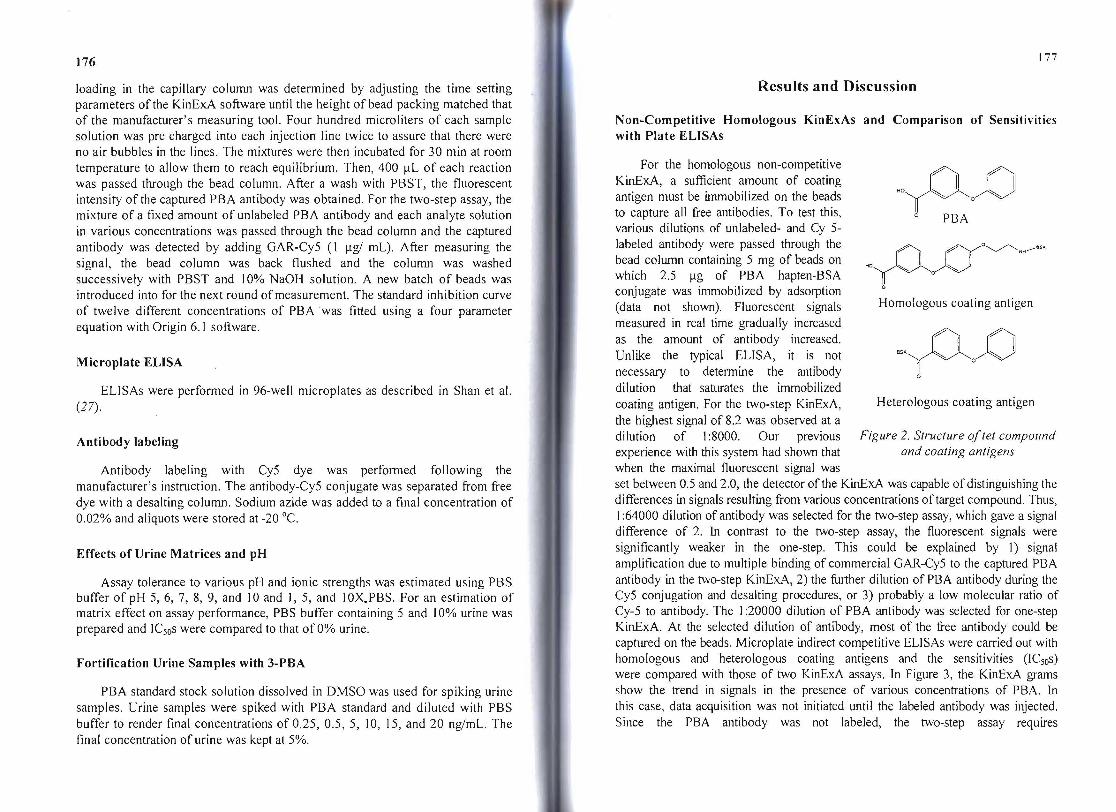
All reagents were of analytical grade unless specified otherwise. Bovine serum albumin (BSA), goat anti-rabbit IgG conjugated with horseradish peroxidase (GAR-HRP), 3,3' ,5,5' -tetramethylbenzidine (TMB), all chemicals for buffer preparation, and PBA were from Sigma (St. Louis, MO, USA). Goat anti-rabbit IgG conjugated with Cy5 (GAR-Cy5) and Cy5 conjugation kit were purchased from Amersham Bioscience (Piscataway, NJ, USA). Desalting columns and the protein A affinity purification kit was purchased from Pierce (Rockford, IL, USA). Polymethylmethacrylate (PMMA) beads were purchased from Sapidyne (Boise, ID, USA). Buffers for ELISA and KinExA assays were normal strength PBS [I X PBS; 8 giL of sodium chloride (NaCI), 0.2 giL of sodium phosphate dibasic anhydrous (Na2HP04), and 0.2 giL of potassium chloride (KCI), pH 7.5], PBST (PBS containing 0.05% Tween 20), carbonate buffer [1.59 gIL sodium carbonate (Na2C03), 2.93 giL sodium hydrogen carbonate (NaHC03), pH9.6], and 0.05M citrate-acetate buffer (14.7 I giL Na3C6Hs07°2H20, pH 5.2). The structures of PBA and its immunizing and coating haptens are shown in Figure 2. Synthesis of the haptens for the production of PAb and assay development were described in our previous paper (29).
Immobilization of PBA Immunizing Hapten-BSA Conjugate to PMMA Beads
Dry PMMA beads (200 mg, 98-/.lm diameter) in a 1.5 mL Eppendorf tube were suspended in I mL of nanopure water and washed two times with PBS buffer by centrifugation and removal of the supernatant solution. Beads were then suspended with I mL of coating buffer, O. I mg of PBA hapten-BSA conjugate added, and the tubes were rolled with an end-over-end rocker for I h at 37 0c. After discarding the coating buffer, I mL of blocking buffer (I % BSA in PBS with 0.05% Tween 20) was added and the tubes rolled again for 2 h at room temperature. After the blocking buffer was discarded, I mL of PBS was added. Bead preparations were stored at 4 °c until use. On the day of use, the contents of two tubes were transferred into the bead reservoir of the KinExA along with 27 mL of PBST to provide a constant supply of beads into the capillary flow column.
KinExA Principles and Non-competitive Assays
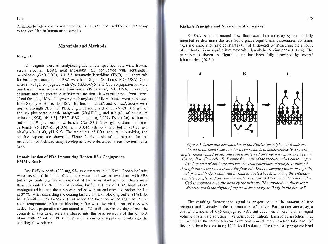
KinExA is an automated flow fluorescent immunoassay system initially intended to determine the true liquid-phase equilibrium dissociation constants (Kd) and association rate constants (koll) of antibodies by measuring the amount of antibodies in an equilibrium state with ligands in solution phase (34-36). The principle is shown in Figure I and has been fully described by several laboratories. (30-36).
A B c
... ..... ~ t,I}I
Ie ~ 1
y~
Figure I. Schematic presentation ofthe KinExA principle. (A) Beads are stirred in the bead reservoir for afew seconds to homogeneously disperse
hapten-immobilized beads and then transferred onto the microporous screen in the capillary flow cell. (B) Sample from one ofthe reaction tubes containing a
fixed amount ofantibody and various concentrations ofanalyte is injected through the rotary selector into the flow cell. While a sample passes through the
cell, free antibody is captured by hapten-coated beads allowing the antibodyanalyte complex to flow into the waste reservoir. (C) The secondary antibody
Cy5 is captured onto the bead by the primary PEA antibody. A fluorescent detector reads the signal ofcaptured secondary antibody in the flow cell.
The resulting fluorescence signal is proportional to the amount of free receptor and inversely to the concentration of analyte. For the one step assay, a constant amount of Cy5-conjugated PBA antibody was mixed with an equal volume of standard solution in various concentrations. Each of 12 injection lines connected to the rotary selector valve was placed into a reaction tube and 13 lh
line into the tube contnining 10% NaOH solution. The time for appropriate bead
177 176
loading in the capillary column was determined by adjusting the time setting parameters of the KinExA software until the height of bead packing matched that of the manufacturer's measuring tool. Four hundred microliters of each sample solution was pre charged into each injection line twice to assure that there were no air bubbles in the lines. The mixtures were then incubated for 30 min at room temperature to allow them to reach equilibrium. Then, 400 fll of each reaction was passed through the bead column. After a wash with PBST, the fluorescent intensity of the captured PBA antibody was obtained. For the two-step assay, the mixture of a fixed amount of unlabeled PBAantibody and each analyte solution in various concentrations was passed through the bead column and the captured antibody was detected by adding GAR-Cy5 (1 flg/ ml). After measuring the signal, the bead column was back flushed and the column was washed successively with PBST and 10% NaOH solution. A new batch of beads was introduced into for the next round of measurement. The standard inhibition curve of twelve different concentrations of PBA was fitted using a four parameter equation with Origin 6.1 software.
Microplate ELISA
ELlSAs were performed in 96-well microplates as described in Shan et al. (27).
Antibody labeling
Antibody labeling with Cy5 dye was performed following the manufacturer's instruction. The antibody-Cy5 conjugate was separated from free dye with a desalting column. Sodium azide was added to a final concentration of 0.02% and aliquots were stored at -20°C.
Effects of Urine Matrices and pH
Assay tolerance to various pH and ionic strengths was estimated using PBS buffer of pH 5,6,7,8,9, and 10 and 1,5, and 10X.PBS. For an estimation of matrix effect on assay performance, PBS buffer containing 5 and 10% urine was prepared and ICsos were compared to that of 0% urine.
Fortification Urine Samples with 3-PBA
PBA standard stock solution dissolved in DMSO was used for spiking urine samples. Urine samples were spiked with PBA standard and diluted with PBS buffer to render final concentrations of 0.25, 0.5, 5, 10, 15, and 20 ng/mL. The final concentration of urine was kept at 5%.
Results and Discussion
Non-Competitive Homologous KinExAs and Comparison of Sensitivities with Plate ELISAs
For the homologous non-competitIve KinExA, a sufficient amount of coating antigen must be immobilized on the beads "yQ~ to capture all free antibodies. To test this,
o PBA various dilutions of unlabeled- and Cy 5labeled antibody were passed through the o~ o"
I Ibead column containing 5 mg of beads on "H NO , 0'p~which 2.5 flg of PBA hapten-BSA
conjugate was immobilized by adsorption o
Homologous coating antigen (data not shown). Fluorescent signals measured in real time gradually increased as the amount of antibody increased. Unlike the typical ELISA, it is not -po~ necessary to determine the antibody o
dilution that saturates the immobilized coating antigen. For the two-step KinExA, Heterologous coating antigen
the highest signal of 8.2 was observed at a dilution of 1:8000. Our previous Figure 2. Structure o/tet compound experience with this system had shown that and coating antigens when the maximal fluorescent signal was set between 0.5 and 2.0, the detector of the KinExA was capable of distinguishing the differences in signals resulting from various concentrations of target compound. Thus, I:64000 dilution of antibody was selected for the two-step assay, which gave a signal difference of 2. In contrast to the two-step assay, the fluorescent signals were significantly weaker in the one-step. This could be explained by 1) signal amplification due to multiple binding of commercial GAR-Cy5 to the captured PBA antibody in the two-step KinExA, 2) the further dilution of PBA antibody during the Cy5 conjugation and desalting procedures, or 3) probably a low molecular ratio of Cy-5 to antibody. The 1:20000 dilution of PBA antibody was selected for one-step KinExA. At the selected dilution of antibody, most of the free antibody could be captured on the beads. Microplate indirect competitive ELlSAs were carried out with homologous and heterologous coating antigens and the sensitivities (ICsos) were compared with those of two KinExA assays. In Figure 3, the KinExA grams show the trend in signals in the presence of various concentrations of PBA. In this case, data acquisition was not initiated until the labeled antibody was injected. Since the PBA antibody was not labeled, the two-step assay requires
179 178
A B
'0 v/"~ 1.1 "
y ;"~'._-:.. ~ U ~ 'M
C 1.0 .5l' i 3.0 ~ 0.'
~ 0.8 ~ g! Li: 1.6g '.1
i:i: O.S . " +I--~--~-~--~-~--~
100 150 200 '" 100 2'00 3~O 4~O Time (second) Time (secondl
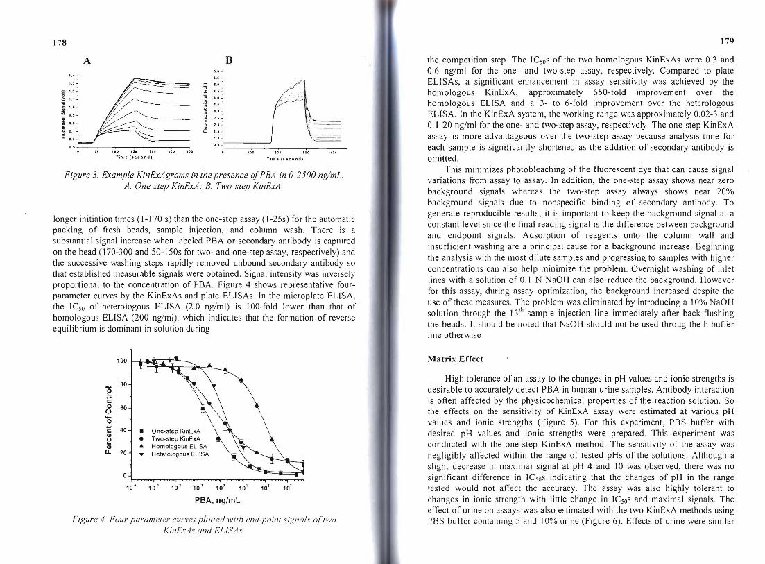
Figure 3. Example KinExAgrams in the presence ofPBA in 0-2500 ng/mL. A. One-step KinExA; B. Two-step KinExA.
longer initiation times (1- 170 s) than the one-step assay (1-25s) for the automatic packing of fresh beads, sample injection, and column wash. There is a substantial signal increase when labeled PBA or secondary antibody is captured on the bead (170-300 and 50- I50s for two- and one-step assay, respectively) and the successive washing steps rapidly removed unbound secondary antibody so that established measurable signals were obtained. Signal intensity was inversely proportional to the concentration of PBA. Figure 4 shows representative fourparameter curves by the KinExAs and plate ELiSAs. In the microplate ELISA, the ICso of heterologous ELISA (2.0 ng/ml) is IOO-fold lower than that of homologous ELISA (200 ng/ml), which indicates that the formation of reverse equilibrium is dominant in solution during
100~~ .._
80 (5 "c -0 60U
0--C • One-step' KinExA Q)
"-40 1• Two-step KinExAU
Q) AI. Homologous ELISA a.. 20 T Hetelologous ELISA
°1 , I i 1 I 1 ~. 10~ 10-' 10" 10" 10' 10' 10' 10'
PBA, ng/mL
Figure 4. Four-parameter curves plol/ed with end-point signals oj'two Kil1ExAs {lnd ELlSAs
the competition step. The ICsos of the two homologous KinExAs were 0.3 and 0.6 ng/ml for the one- and two-step assay, respectively. Compared to plate ELiSAs, a significant enhancement in assay sensitivity was achieved by the homologous KinExA, approximately 650-fold improvement over the homologous ELISA and a 3- to 6-fold improvement over the heterologous ELISA. In the KinExA system, the working range was approximately 0.02-3 and 0.1-20 ng/ml for the one- and two-step assay, respectively. The one-step KinExA assay is more advantageous over the two-step assay because analysis time for each sample is significantly shortened as the addition of secondary antibody is omitted.
This minimizes photobleaching of the fluorescent dye that can cause signal variations from assay to assay. In addition, the one-step assay shows near zero background signals whereas the two-step assay always shows near 20% background signals due to nonspecific binding of secondary antibody. To generate reproducible results, it is important to keep the background signal at a constant level since the final reading signal is the difference between background and endpoint signals. Adsorption of reagents onto the column wall and insufficient washing are a principal cause for a background increase. Beginning the analysis with the most dilute samples and progressing to samples with higher concentrations can also help minimize the problem. Overnight washing of inlet lines with a solution of 0.1 N NaOH can also reduce the background. However for this assay, during assay optimization, the background increased despite the use of these measures_ The problem was eliminated by introducing a 10% NaOH solution through the 13 th sample injection line immediately after back-flushing the beads. It should be noted that NaOH should not be used throug the h buffer line otherwise
Matrix Effect
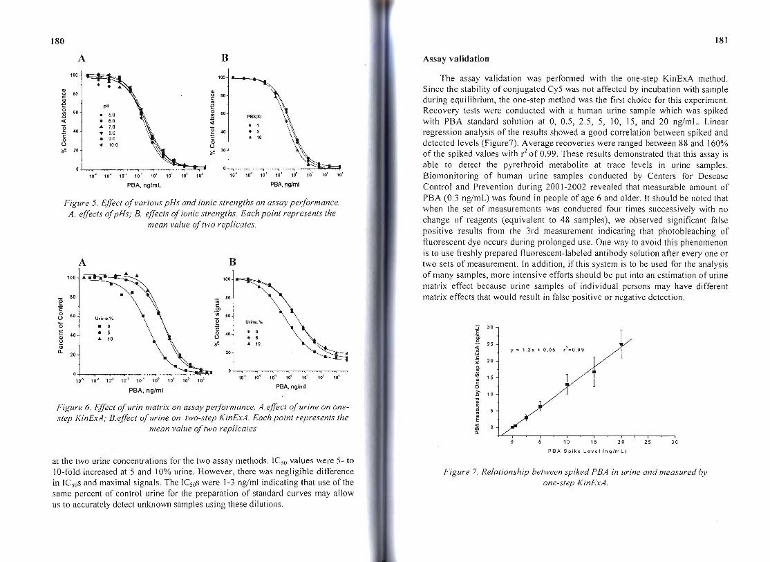
High tolerance of an assay to the changes in pH values and ionic strengths is desirable to accurately detect PBA in human urine samples. Antibody interaction is often affected by the physicochemical properties of the reaction solution. So the effects on the sensitivity of KinExA assay were estimated at various pH values and ionic strengths (Figure 5). For this experiment, PBS buffer with desired pH values and ionic strengths were prepared. This experiment was conducted with the one-step KinExA method. The sensitivity of the assay was negligibly affected within the range of tested pHs of the solutions. Although a sl ight decrease in maximal signal at pH 4 and 10 was observed, there was no significant difference in ICsos indicating that the changes of pH in the range tested would not affect the accuracy. The assay was also highly tolerant to changes in ionic strength with little change in ICsos and maximal signals. The effect of urine on assays was also estimated with the two KinExA methods using PRS buffer conlaining 5 and 10% urine (Figure 6). Effects of urine were similar
181180
A B Assay validation
Figure 5. EjJect ofvarious pHs and ionic strengths on assay performance. when the set of measurements was conducted four times successively with noA. ejJects ofpHs; B. ejJects ofionic strengths. Each point represents the change of reagents (equivalent to 48 samples), we observed significant false mean value oftwo replicates. positive results from the 3rd measurement indicating that photobleaching of fluorescent dye occurs during prolonged use. One way to avoid this phenomenon is to use freshly prepared fluorescent-labeled antibody solution after every one or
A B two sets of measurement. In addition, if this system is to be used for the analysis 1 1 of many samples, more intensive efforts should be put into an estimation of urine100 100
matrix effect because urine samples of individual persons may have different
e 80 matrix effects that would result in false positive or negative detection. iii c: Cl'E
0 'iii U Urine,·/o
Urine, .~60 ~ 0 g'0 :; 30
C '"
40
• • 0 E .. 10
c:40• U C>• 5
() .s J
5 0
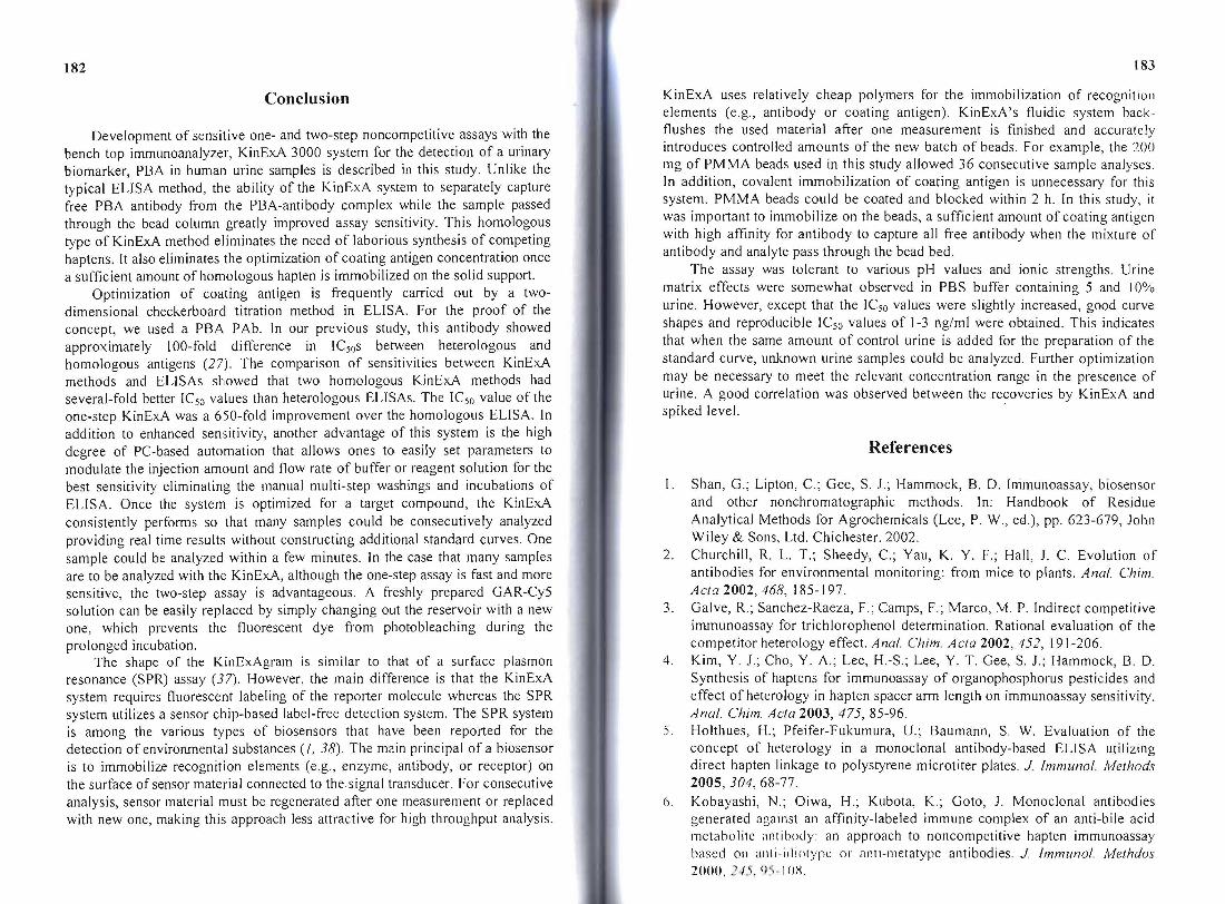
1 .. 10~Q; ":i 25 y = 1.2x + 0.05 r'= 0.990 20 w20 c:
20i: Q.
~ 15 c:
PBA, nglml 0PBA, ng/ml >.c 10
'C
~Figure 6. EjJect ofurin matrix on assay performance. A. ejJect ofurine on one~
step KinExA; B.ejJect ofurine on two-step KinExA. Each point represents the E « IIImean value oftwo replicates 0..
I I I I I
1 0 1 5 20 25 30
PBA Spike Level (ng/mL)
at the two urine concentrations for the two assay methods. ICso values were 5- to 1O-fold· increased at 5 and 10% urine. However, there was negligible difference Figure 7. Relationship between spiked PBA in urine and measured by in ICsos and maximal signals. The ICsos were 1-3 nglml indicating that use of the one-step KinExA. same percent of control urine for the preparation of standard curves may allow us to accurately detect unknown samples using these dilutions.
10.,) 10·l 10·\ 100 10' 101 10~
100 100
" 80 ~ BO'" c:
-ec:
"'-E 0 o 60II) 60
II).c .c« « g "0 4040
Z,c: c:0 oU U 2020~ ~
PBA, nglmL
10'" 10.,) 10.1 10·' 10c 10' 10' 10~
pH
· 5.0
• 6.0 .. 7.0
• B.O
• 9.0
" 10.0
01 1 , , , , ,~ 10-.lo 10'" 10-J 10.' 10·' 100 10' 10J 10 1
The assay validation was performed with the one-step KinExA method. Since the stability of conjugated Cy5 was not affected by incubation with sample during equilibrium, the one-step method was the first choice for this experiment. Recovery tests were conducted with a human urine sample which was spiked
PBSIX) with PBA standard solution at 0, 0.5, 2.5, 5, 10, 15, and 20 ngimL. Linear• 1
• 5 regression analysis of the results showed a good correlation between spiked and .. 10
detected levels (Figure7). Average recoveries were ranged between 88 and 160% of the spiked values with r2 of 0.99. These results demonstrated that this assay is able to detect the pyrethroid metabolite at trace levels in urine samples.
10'" 10-3 10·l 10·' 10c 10' 101 101
Biomonitoring of human urine samples conducted by Centers for Desease PBA, nglml Control and Prevention during 2001-2002 revealed that measurable amount of
PBA (0.3 ng/mL) was found in people of age 6 and older. It should be noted that
182 183
Conclusion
Development of sensitive one- and two-step noncompetitive assays with the bench top immunoanalyzer, KinExA 3000 system for the detection of a urinary biomarker, PBA in human urine samples is described in this study. Unlike the typical ELISA method, the ability of the KinExA system to separately capture free PBA antibody from the PBA-antibody complex while the sample passed through the bead column greatly improved assay sensitivity. This homologous type of KinExA method eliminates the need of laborious synthesis of competing haptens. It also eliminates the optimization of coating antigen concentration once a sufficient amount of homologous hapten is immobilized on the solid support.
Optimization of coating antigen is frequently carried out by a twodimensional checkerboard titration method in ELISA. For the proof of the concept, we used a PBA PAb. In our previous study, this antibody showed approximately 100-fold difference in ICsos between heterologous and homologous antigens (27). The comparison of sensitivities between KinExA methods and ELISAs showed that two homologous KinExA methods had several-fold better ICso values than heterologous ELISAs. The ICso value of the one-step KinExA was a 6S0-fold improvement over the homologous ELISA. In addition to enhanced sensitivity, another advantage of this system is the high degree of PC-based automation that allows ones to easily set parameters to modulate the injection amount and flow rate of buffer or reagent solution for the best sensitivity eliminating the manual multi-step washings and incubations of ELISA. Once the system is optimized for a target compound, the KinExA consistently performs so that many samples could be consecutively analyzed providing real time results without constructing additional standard curves. One sample could be analyzed within a few minutes. In the case that many samples are to be analyzed with the KinExA, although the one-step assay is fast and more sensitive, the two-step assay is advantageous. A freshly prepared GAR-Cy5 solution can be easily replaced by simply changing out the reservoir with a new one, which prevents the fluorescent dye from photobleaching during the prolonged incubation.
The shape of the KinExAgram is similar to that of a surface plasmon resonance (SPR) assay (37). However, the main difference is that the KinExA system requires fluorescent labeling of the reporter molecule whereas the SPR system utilizes a sensor chip-based label-free detection system. The SPR system is among the various types of biosensors that have been reported for the detection of environmental substances (I, 38). The main principal of a biosensor is to immobilize recognition elements (e.g., enzyme, antibody, or receptor) on the surface of sensor material connected to the signal transducer. For consecutive analysis, sensor material must be regenerated after one measurement or replaced with new one, making this approach less attractive for high throughput analysis.
KinExA uses relatively cheap polymers for the immobilization of recognitioll elements (e.g., antibody or coating antigen). KinExA's fluidic system backflushes the used material after one measurement is finished and accurately introduces controlled amounts of the new batch of beads. For example, the 200 mg of PMMA beads used in this study allowed 36 consecutive sample analyses. In addition, covalent immobilization of coating antigen is unnecessary for this system. PMMA beads could be coated and blocked within 2 h. In this study, it was important to immobilize on the beads, a sufficient amount of coating antigen with high affinity for antibody to capture all free antibody when the mixture of antibody and analyte pass through the bead bed.
The assay was tolerant to various pH values and ionic strengths. Urine matrix effects were somewhat observed in PBS buffer containing 5 and 10% urine. However, except that the ICso values were slightly increased, good curve shapes and reproducible ICso values of 1-3 nglml were obtained. This indicates that when the same amount of control urine is added for the preparation of the standard curve, unknown urine samples could be analyzed. Further optimization may be necessary to meet the relevant concentration range in the prescence of urine. A good correlation was observed between the recoveries by KinExA and spiked level.
References
I. Shan, G.; Lipton, c.; Gee, S. 1.; Hammock, B. D. Immunoassay, biosensor and other nonchromatographic methods. In: Handbook of Residue Analytical Methods for Agrochemicals (Lee, P. W., ed.), pp. 623-679, John Wiley & Sons, Ltd. Chichester. 2002.
2. Churchill, R. L. T.; Sheedy, c.; Yau, K. Y. F.; Hall, J. C. Evolution of antibodies for environmental monitoring: from mice to plants. Anal. Chim Acta 2002, 468, 185-197.
3. Galve, R.; Sanchez-Raeza, F.; Camps, F.; Marco, M. P. Indirect competitive immunoassay for trichlorophenol determination. Rational evaluation of the competitor heterology effect. Anal. Chim. Acta 2002,452, 191-206.
4. Kim, Y. 1.; Cho, Y. A.; Lee, H.-S.; Lee, Y. T. Gee, S. 1.; Hammock, B. D. Synthesis of haptens for immunoassay of organophosphorus pesticides and effect of heterology in hapten spacer arm length on immunoassay sensitivity. Anal. Chim. Acta 2003, 475, 85-96.
5. Holthues, H.; Pfeifer-Fukumura, U.; Baumann, S. W. Evaluation of the concept of heterology in a monoclonal antibody-based ELISA utilizing direct hapten linkage to polystyrene microtiter plates. 1. Immunol. Methods 2005, 304, 68-77.
6. Kobayashi, N.; Oiwa, H.; Kubota, K.; Goto, J. Monoclonal antibodies generated against an affinity-labeled immune complex of an anti-bile acid metaoolite i1ntioody: an approach to noncompetitive hapten immunoassay based on i1llli-idiolypc or anti-metatype antibodies. 1. Immunol. Methdos 2000, ]15, ()~·I ox.

I HI
7. SUI.Ukl, c.; Ul:ua, 1-1.; Mahonl:y, W.; Naga11lunl:, T. Open sanuwich elll.ymelinked immunosorbent assay for the qualltitation of small haptens. Anal. Biochem. 2000, 286, 238-246.
8. Pulli, T.; Hoyhty1i, M.; Soderlund, H.; Takkinent, K. One-step homogeneous immunoassay for small analytes. Anal. Chem. 2005, 77,2637-2642.
9. Wang, Q.; Luo, G.; Ou, 1.; Yeung, W. S. B. Noncompetitive immunoassays using protein G affinity capillary chromatography and capillary electrophoresis with laser-induced fluorescence detection. J. Chromalogr. A. 1999,848, 139-148.
10. Lin, J.; Yan, F.; Ju, H. Noncompetitive enzyme immunoassay for carcinoembryonic antigen by flow injection chemiluminescence. C/in. Chim. ACla2004,341,109-115.
II. Miyamoto, J.; Kaneko, H.; Tsuji, R.; Okuno, Y. Pyrethroids, nerve poisons: how their risks to human health should be assessed. Toxicol. Lell. 1995, 82/83, 933-940.
12. Casida, J. E.; Ruzo, L. O. Metabolic chemistry of pyrethroid insecticides. Pestic. Sci. 1980, I 1,257-269.
13. Miyamoto, J.; Beynon, K. I.; Roberts, T. R.; Hemingway, R. I.; Swaine, H. The chemistry, metabolism and residue analysis of synthetic pyrethroids. Pure Appl. Chem. 1981,53, 1967-2022.
14. Schulz, J.; Schmoldt, A.; Schulz, M. Pyrethroids: chemistry and toxicology of an insecticide group. Pharm. Ztg. 1993, 15, 9-24.
15. Cantalamessa, F. Acute toxicity of two pyrethroids, pennethrin and cypermethrin, in neonatal and adult rats. Arch. Toxicol. 1993, 67, 510-513.
16. Environmental Protection Agency. List of Chemicals Evalualed for Carcinogenic Potential; EPA: Washington, DC, 1994.
17. He, F.; Sun, J.; Han, K.; Wu, Y.; Yao, P. Effects of pyrethroid insecticides on subjects engaged in packaging pyrethroids. Br. J. Ind. Med. 1988, 45, 548-551.
18. Hallenbeck, W. H.; Cunningham-Burns, K. M. Pesticides and Human Health, p 118, Springer-Verlag, NY. 1985.
19. Go, V.; Garey, J.; Wolff, M. S.; Pogo, B. G. T. Estrogenic potential of certain pyrethroid compounds in the MCF-7 human breast carcinoma cell line. Environ. Health Perspecl. 1999, 107, 173-177.
20. Eadsforth, C. V.; Bragt, P. c.; Van Sittert, N. 1. Human dose-excretion studies with pyrethroid insecticides cypennethrin and alphacypennethrin: relevance for biological monitoring. Xenobiotica 1988, 18, 603-614.
21. Eadsforth, C. V.; Baldwin, M. K. Human dose-excretion studies with the pyrethroid insecticide, cypennethrin. Xenobiotica 1983, 13, 67-72.
22. Shan, G.; Stoutamire, D. W.; Wengatz, I.; Gee, S. J.; Hammock, B. D. Development of an immunoassay for the pyrethroid insecticide esfenvalerate.J. Agric. FoodChem. 1999,47,2145-2155.
23. Wengatz, I.; Stoutamire, D. W.; Gee, S. J.; Hammock, B. D. Development of an enzyme-linked immunosorbent assay for the detection of the pyrethroid insecticide fenpropathrin. J. Agric. Food Chem. 1998, 46, 22112221.
ISS
24. SIl<i1l Ci.; I.CCIIl<lII, W. IC: S!pul<imire, D. W.; Gee, S. J.; Chang, D. P. Y.; Hammock, 13. D. Lnzymc-linked immunosorbent assay for the pyrethroid permethrin. J. Agric. Food Chem. 2000, 48, 4032-4040.
25. Lee, H.-J.; Shan, G.; Watanabe, T.; Stoutamire, D. W.; Gee, S. J.; Hammock, B. D. Enzyme-linked immunosorbent assay for the pyrethroid deltamethrin.1. Agric. Food Chem. 2002, 50, 5526-5532.
26. Lee, H.-J.; Shan, G.; Ahn, K. c.; Park, E.-K.; Watanabe, T.; Gee, S. J.; Hammock, B. D. Development of an enzyme-linked immunosorbent assay for the pyrethroid cypennethrin.1. Agric. FoodChem. 2004, 52,1039-1043.
27. Shan, G.; Wengatz, I.; Stoutamire, D. W.; Gee, S. J.; Hammock, B. D. An enzyme-linked immunosorbent assay for the detection of esfenvalerate metabolites in human urine. Chem. Res. Toxicol. 1999, 12,1033-1041.
28. Ahn, K. c.; Watanabe, T.; Gee, S. J.; Hammock, B. D. Hapten and antibody production for a human urinary metabolite of the pyrethroid insecticide permethrin.1. Agric. Food Chem. 2004,52,4583-4594.
29. Shan, G.; Huang, H.; Stoutamire, D. W.; Gee, S J.; Leng, G.; Hammock, B. D. A sensitive class specific immunoassay for the detection of pyrethroid metabolites in human urine. Chem. Res. Toxicol. 2004, 17,218-225.
30. Glass, T. R.; Saiki, H.; Joh, T.; Taemi, Y.; Ohmura, N.; Lackie, S. 1. Evaluation of a compact bench top immunoassay analyzer for automatic and near continuous monitoring of a samples for environmental contaminants. Biosens. Bioeleclron 2004, 20, 397-403.
31. O'Connell, K. P.; Valdes, J. J.; Azer, N. L.; Schwartz, R. P. Wright, 1.; Eldefrawi, M. E. Assessment of an automated solid phase competitive fluoroimmunoassay for benzoylecgonine in untreated urine. 1. Immunol. Melhods 1999,225,157-169.
32. Blake, D. A.; Jones, R. M.; Blake II, R. C.; Pavlov, A. R.; Darwish, I. A.; Yu, H. Antibody-based sensors for heavy metal ions. Biosens. Bioeleclron. 2001, /6,799-809.
33. Blake, D. A.; Pavlov, A. R.; Yu, H.; Kohsraviani, M.; Ensley, H. E.; Blake II, R. C. Antibodies and antibody-based assays for hexavalent uranium. Anal Chim. ACla2001, 444,3-11.
34. Drake, A. W.; Myszka, D. G.; Klakamp, S. Characterizing high-affinity antigen/antibody complexes by kinetic-and equilibrium-based methods. Anal. Biochem. 2004, 328, 35-43.
35. Chiu, Y.-W.; Li, Q. X.; Karu, A. E. Selective binding of polychlorinated biphenyl congeners by a monoclonal antibody: analysis by kinetic exclusion fluorescence immunoassay. Anal. Chem. 2001,73,5477-5484.
36. Xie, L.; Jones, R. M.; Glass, T.; Navoa, R.; Wang, Y.; Grace, M. 1. Measurement of the functional affinity constant of a monoclonal antibody for cell surface receptors using kinetic exclusion fluorescence immunoassay. J. Immunol. Method. 2005, 304, 1-14.
37. Marchesini, G. R.; Meulenberg, E.; Haasnoot, W.; Irth, H. Biosensor immunoassays for the detection of bisphenol A. Anal. Chim. ACla 2005, 528,37-45.
38. Rogers, K. R. Recent advances in biosensor techniques for environmental monitoring. Ana!. Chim. Acta 2006, 568, 222-231.