Embed Size (px)
Citation preview
Human Epidermal Langerhans Cells Express the High Affinity Receptor for Immunoglobulin E (rceRI) By Thomas Bieber,* Henri de la Salle,~ Andreas Wollenberg,* John Hakimi,S Richard Chizzonite, S Johannes Ring, II Daniel Hanau,~ and Corinne de la Saller
From the *Laboratory of lmmunodermatology, Department of Dermatology, University of Munich Medical School, 8000 Munich 2, Germany; the ~Laboratoire d'Histocompatibilitd and Institut National de Ia Santd et de la Recherche M~dicale U.311, Centre R~gional de ~ansfusion Sanguine, 67000 Strasbour,~ France; SHoffman-La Roche Ina, Nude),, New Jersey 07110-1199; and the IIDepartment of Dermatology, University of Hamburg Medical School, 2000 Hamburg 20, Germany
Summary It has been suggested that epidermal Langerhans cells (LC) bearing immunoglobulin E (IgE) may be involved in the genesis of atopic disease. The identity of the IgE receptor(s) on LC remained unclear, although it represents a crucial point in understanding cellular events linked to the binding of allergens to LC via IgE. In this report, we demonstrate that epidermal LC express the high affinity receptor for the Fc fragment of IgE (FceRI) which has, so far, only been described on mast cells and basophils. Epidermal LC react with antibodies specific for the u subunit of the tetrameric (oL,~,23') FceRI. Specific transcripts for Fc~RIo~ and FceRI3~ were detected in LC and correspond to those of human basophils and of the human basophil cell line KU812. Furthermore, human basophils, KU812 cells, and LC express the putative B subunit. Thus human LC express the complete structure of FceRI. This finding opens new perspectives in the putative functional role of this structure on antigen-presenting cells.
T he demonstration of IgE molecules on epidermal Lang- erhans cells (LC) 1 in patients with atopic dermatitis has
implied that these cells should perform a major function in the pathophysiology of atopic disease (1, 2). Although ini- tially, only receptors for the Fc fragment of IgG were identified on epidermal LC (3, 4), the low affinity receptor for IgE FceRII/CD23 (5) and the human IgE binding protein (eBP) (6) have now also been found on these cells in lesional, as well as in normal skin. However, attempts to completely block IgE binding on LC by a variety of anti-FceRII/CD23, anti- eBP, and/or anti-Fc3,R reagents remained unsuccessful, sug- gesting the presence of a third IgE-binding structure actu- ally responsible for a part of the IgE-binding capacity of LC. We report here, that normal human LC also express the high a~nity receptor for IgE, Fcelkl, demonstrating that the pres- ence of this structure is not restricted to mast cells and basophils. Our results also document the presence of the puta- tive/~ chain on both human basophils and LC.
1 Abbreviations used in this paper: BP, binding protein; ECL, enhanced chemiluminescence; LC, Langerhans cells.
Materials and Methods Individuals and Biopsy Specimens. Fresh human skin specimens
were obtained during plastic abdominal surgery or reduction mam- moplasty. 6-mm punch biopsies were taken from the buttocks of healthy volunteers. All samples were immediately processed.
Basophils and Basophilic Cell Line. Human basophils were ob- tained by standard isolation technique from the peripheral blood of healthy volunteers (7). The human basophilic KU812 cell line was a kind gift from Dr. Becket (Forschungsinstitut, Borstel, Ger- many). These cells, which constitutively express FceRI (8), were cultured in RPMI 1640 (Gibco, Berlin, Germany) supplemented with 5% FCS (Gibco), antibiotics, and antimycotics (Gibco).
Reagents. Unlabeled mAb IOT6 (IgG1; Immunotech, MarseiUe, France), and PE-labeled T6/RD1 (IgG1, Coulter Corp., Krefeld, Germany) react with CDla which is, in the skin, exclusively ex- pressed on LC (9). Murine mAb 29C6 and 6F7 (both IgG1) were raised from mice immunized with a purified chimeric c~ subunit ofFceRI (FceRIo 0 expressed in Chinese hamster ovarian cells (10). FITC-labeled goat anti-mouse was obtained from Jackson Im- munoResearch Labs, Inc. (West Grove, PA) and unlabeled IgG1 and PE-labeled IgG1 from Becton Dickinson & Co. (Mountain View, CA). For immunohistologic purposes, we also used rabbit anti-mouse Ig antibody and alkaline-phosphatase mouse anti-alkaline-phosphatase complexes (Dakopatts, Hamburg, Ger-
1285 J. Exp. Med. �9 The Rockefeller University Press �9 0022-1007/92/05/1285/06 $2.00 Volume 175 May 1992 1285-1290
many). A peroxidase-conjugated goat anti-mouse Ig antibody (Bio- Pad Laboratories, Richmond, CA) was used for immunoblot anal- ),sis. Oligonucleotides were synthesized by Applig~ne (Illkirch, France).
In Situ Immunolabeling on Cryosections. 6-#m cryosections were prepared from the punch biopsies, air-dried, fixed for 10 min in pure acetone, and then processed for immunohistochemistry using the mAb IOT6a, 29C6, 6F7, or the IgG1 isotype control (all at 10 gg/ml) and the alkaline-phosphatase mouse anti-alkaline- phosphatase technique as described previously in detail (2).
Preparation of Epidermal Cell Suspensions and Enrichment of LC. Isolation and enrichment of LC from fresh human skin specimens was performed as described in detail elsewhere (5).
Flow Cytometric Analysis of FceRI Expression on Epidermal Cells. 10 s I.C-enriched EC were incubated for 30 min with heat- inactivated AB serum. After several washes in PBS supplemented with 1% FCS and 0.1% sodium azide, doublestaining experiments were performed by first incubating (1 h, 4~ the cells with either 29C6 or 6F7 (both at 10 #g/ml). After washing twice in 1% FCS- supplemented PBS, the cells were incubated (1 h, 4~ with FITC- labeled goat anti-mouse antibody. Then, after washing twice, cells were incubated (30 min, 4~ with normal mouse serum (final dilution 1:10) to saturate the second step antibody. The cells were washed twice and incubated (30 min, 4~ with the PE-labeled mAb T6/RD1 (1 #g/ml). Isotype controls were performed with unlabeled IgG1 and PE-labeled mouse IgG1. The cells were then washed twice in 1% FCS-supplemented PBS at 4~ and were ana- lyzed by flow cytometry using a FACScan | (Becton Dickinson & Co.). Fluorescence parameters were collected using a built-in loga- rithmic amplifier, and the data of 10,000 cells obtained with Con- sort 30 software were analyzed with the Lysis-I program (Becton Dickinson & Co.).
lmmunomagnetic Depletion~Purification of CDIa § Cells. The separation of CDla + LC for the preparation of LC-depleted epidermal cells (EC), or for purification of LC was achieved ac- cording to the manufacturer's protocol. The purity of the LC prep- aration was controlled after each application on the magnet by light microscopy, and the procedure was stopped when unbound cells, i.e., keratinocytes and other cells, were completely removed. Addi- tion of the beads and depletion procedure were repeated three to four times. The LC depletion of EC was controlled by anti-CDla staining and flow cytometric analysis.
Imrnunobiochemical Analysis of FceRI on Epidermal Cells. Puri- fied LC or LC-depleted EC were prepared by positive selection as described above. The cells were washed, lysed in NP-40 (Sigma Chemical Co.)-containing buffer and 14 #g of protein were sepa- rated by 12% SDS-PAGE. Then, separated proteins from purified LC or from LC-depleted EC were electroblotted onto nitrocellu- lose (Hybond C; Amersham Corp., Arlington Heights, IL). The strips were blocked with dry milk, and incubated for 1 h with ei- ther the anti-FceRI~ 29C6 or the isotype control (both at 10 /~g/ml). Binding of the primary antibody was revealed by incuba- tion with a peroxidase-conjugated goat anti-mouse Ig antibody followed by an enhanced chemiluminescence (ECL) Western blot detection system, according to the manufacturer's protocol (Amer- sham Corp.).
Amplification of mRNA Transc@ts. Total KNA was isolated from purified LC, 1.C-depleted EC, basophils, and KU812 cells by standard procedure (11), and eDNA was synthesized by extension of 3 pmol of reverse primers specific for tryptase, FceRIo~, or FceRI3, on 100 ng of total RNA by Moloney routine leukemia virus (MoMLV) reverse transcriptase (Gibco). The second-strand synthesis and 30 cycles of amplification were performed directly after the
reverse transcription step by adding 10 pmol of direct primers, 10 pmol of the reverse primers, 1.25 mM of each dNTP, and 1 U of Taq DNA Polymerase (Perkin-Elmer Cetus Corp., Norwalk, CT). For FceRIfl, eDNA was synthesized by extension of 10 pmol of reverse primer on 100 ng of total LC-RNA, or 1/~g of total RNA from basophils. The second-strand synthesis and 35 cycles of amplification were performed by adding 20 pmol of direct primer and 50 pmol of the reverse primer, 1.25 mM of each dNTP, and 1 U ofTaq polymerase. Temperatures were 94~ for denaturation, 55~ for annealing of FceRIc~ and FceRI% 58~ for annealing of FceRIB, and 72~ for polymerizations. Sequences of PCR primers were as follows: for tryptase based on (12): reverse pri- mer 5'-GGATCCAGTCCAAGTAGTAG-3', and direct primer 5'- CTCCCTCATCCACCCCCAGT-Y; for FceKIc~ based on (13): re- verse primer 5'-CTTAGGATGTGGGTTCAGAAGT-Y, and direct primer 5'-GACAGTGGAGAATACAAATGTCA-Y; for FceRI/~ based on (14): reverse primer 5'-TAATTCTTCATAAAGACGAT- CATC(A,T,G,orC)GG-3' and direct primer 5'-ATATGCCTTTGTT- TTGGAACAATIGTITG-Y; for FceRI3, based on (15): reverse primer 5'-TAGGGCCAGCTGGTGTTAATGGCA-3', and direct primer 5'-GATGATTCCAGCAGTGGTCTTGCT-3'. PCR prod- ucts were then electrophoresed on a 2% agarose gel.
Cloning and Sequencing of PCR Amplified Products. After RT- PCR, the fragments were digested with 10 U $1 nuclease (Ap- plig~ne) for 10 min, at room temperature to increase the efficiency of cloning. Then, they were treated with 5 U Kleenow DNA poly- merase (Applig~ne), and the resulting DNA products were cloned in the EcoRV restriction site of pKS + (Stratagene Inc., La Jolla, CA). Three independent clones were sequenced using Sequenase 2.0 sequencing kit (US Biochemical, Cleveland, OH) and Hydro- link sequencing gel matrix (AT Biochem, Malvern, PA). RT-PCR artifacts were excluded by deduction of the nucleotide sequence from at least two identical results.
Southern Blot Analysis. Alkaline blotting and Southern hybridi- zations were performed on Hybond N +TM membranes (Amersham Corp.) according to the manufacturer's conditions. Oligonucleo- tide probes were labeled with T4 polynucleotide kinase and 3/-[32p]ATP, and then hybridized to the blot at 50~ Sequences of the probes for Southern blot are 5'-GAGAGTGAACCTGTGTAC- Y, 5'-AGAATCK}TGAGAAACAGC-Y, 5'-AATTGTCCTCACCC- TCC-3', and 5'-CCTCCCACCGCCATTTC-Y, specific for FceRc~, FceR~, FceR'y and tryptase respectively.
Results and Discussion Immunodetection of FceRI on Epidermal LC. Fc6RI is a tetra-
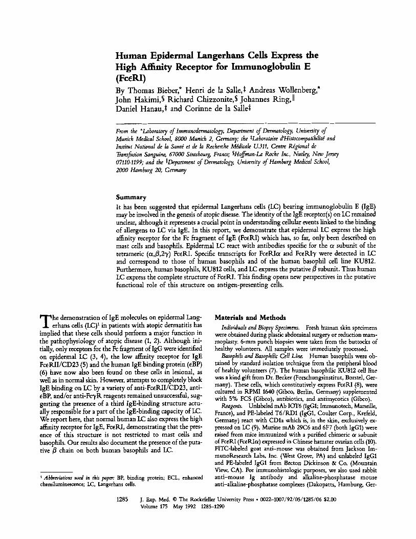
meric structure composed of one 50-55-kD ol chain (FceKIot), one 33-kD fl chain (FceRIfl), and two disulphide-linked iden- tical 7-9-kD 3' chains (FceRI'y) (16). Recently, a series of anti-FceKIoe mAbs were raised from mice immunized with a purified chimeric FceRIol expressed in Chinese hamster ovarian cells (10). Using two of these reagents, the anti-FceRIot antibodies 6F7 and 29C6, we investigated the expression of Fc.eRIo~ on cryosections from normal human skin by immuno- histochemistry. Positive staining was found not only on dermal mast cells but also on some dermal dendritic cells and, most important, on dendritic cells in a suprabasal localization in the epidermis (Fig. 1 A) where mast cells have never been observed. Preliminary experiments on cryosections revealed that, as expected from the characterization of these antibodies (10), 6F7 blocks the IgE binding on epidermal dendritic cells, as well as on dermal mast cells, while 29C6 is ineffective.
1286 Human Epidermal Langerhans Cells Express High Affinity Receptor for IgE
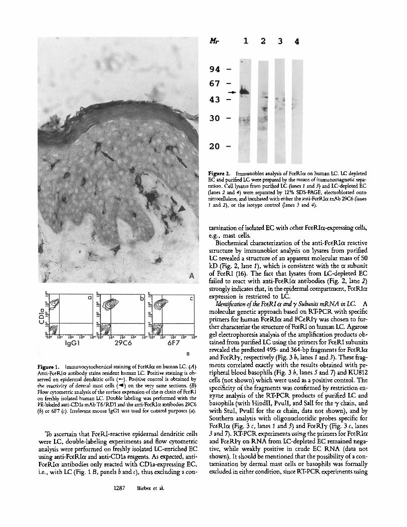
Figure 2. Immunoblot analysis of FceRIa on human LC. LC depleted EC and purified LC were prepared by the means ofimmunomagnetic sepa- ration. Cell lysates from purified I.C (lanes 1 and 3) and LC-depleted EC (lanes 2 and 4) were separated by 12% SDS-PAGE, electroblotted onto nitrocellulose, and incubated with either the anti-FceRIc~ mAb 29C6 (lanes 1 and 2), or the isotype control (lanes 3 and 4).
Figure 1, Immunocytochemical staining of FceRl~ on human LC. (.4) Anti-Fc~Pdc~ antibody stains resident human LC. Positive staining is ob- served on epidermal dendritic calls (~,-). Positive control is obtained by the reactivity of dermal mast cells ( 4 ) on the very same sections. (/3) Flow cytometric analysis of the surface expression of the ~ chain of FcERI on freshly isolated human LC. Double labeling was performed with the PE-labeled anti-CDla mAb T6/RD1 and the anti-FceRIc~ antibodies 29C6 (b) or 6F7 (c). Irrelevant mouse IgG1 was used for control purposes (a).
To ascertain that FceP,.I-reactive epidermal dendritic cells were LC, double-labeling experiments and flow cytometric analysis were performed on freshly isolated LC-enriched EC using anti-FceRIoc and anti-CDla reagents. As expected, anti- FceKIo~ antibodies only reacted with CDla-expressing EC, i.e., with LC (Fig. 1 B, panels b and c), thus excluding a con-
tamination of isolated EC with other FceRIol-expressing cells, e.g., mast cells.
Biochemical characterization of the anti-FcdLIce reactive structure by immunoblot analysis on lysates from purified LC revealed a structure of an apparent molecular mass of 50 kD (Fig. 2, lane 1), which is consistent with the oe subunit of FcetLI (16). The fact that lysates from LC-depleted EC failed to react with anti-FceRIc~ antibodies (Fig. 2, lane 2) strongly indicates that, in the epidermal compartment, FceRIcr expression is restricted to LC.
Identification of the FceRI ol and y Subunits mRNA in LC. A molecular genetic approach based on KT-PCR with specific primers for human FceKIoe and FCeKI3, was chosen to fur- ther characterize the structure of FceRI on human LC. Agarose gel electrophoresis analysis of the amplification products ob- tained from purified LC using the primers for FceKI subunits revealed the predicted 495- and 364-bp fragments for FceKIcx and FceKI% respectively (Fig. 3 b, lanes I and 3). These frag- ments correlated exactly with the results obtained with pe- ripheral blood basophils (Fig. 3 b, lanes 5 and 7) and KU812 cells (not shown) which were used as a positive control. The specificity of the fragments was confirmed by restriction en- zyme analysis of the KT-PCI~ products of purified LC and basophils (with HindlI, PvulI, and Sail for the 3, chain, and with StuI, PvulI for the cx chain, data not shown), and by Southern analysis with oligonucleotidic probes specific for Fc~RIc~ (Fig. 3 c, lanes 1 and 5) and FceKI'y (Fig. 3 c, lanes 3 and 7). KT-PCtL experiments using the primers for Fc~KIc~ and FceRI7 on KNA from LC-depleted EC remained nega- tive, while weakly positive in crude EC KNA (data not shown). It should be mentioned that the possibility of a con- tamination by dermal mast cells or basophils was formally excluded in either condition, since KT-PCIL experiments using
1287 Bieber et al.
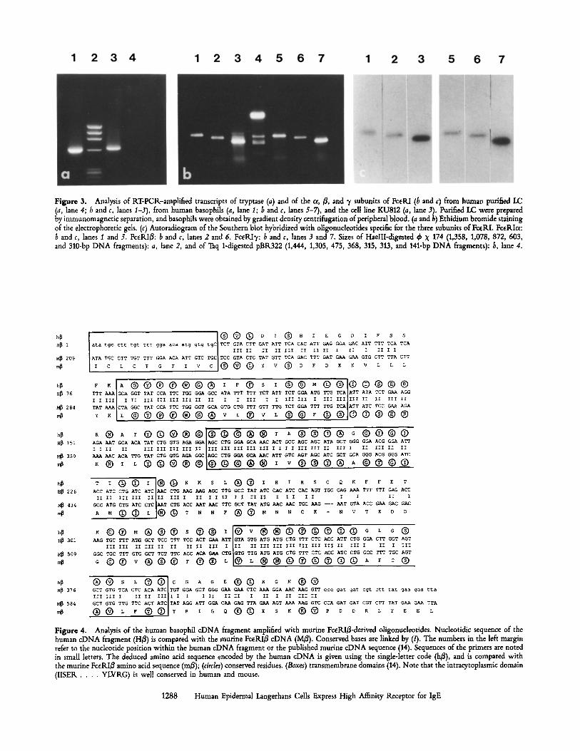
Figure 3. Analysis of KT-PCK-ampLified transcripts of tryptase (a) and of the or, ~, and 7 subunits of FceRI (b and c) from human purified LC (a, lane 4; 6 and c, lanes I-3), from human basophils (a, lane I; b and c, lanes 5-7), and the cell Line KU812 (a, lane 3). Purified LC were prepared by immunomagnetic separation, and basophils were obtained by gradient density centrifugation of peripheral blood. (a and 6) Ethidium bromide staining of the dectrophoretic gds. (c) Autoradiogram of the Southern blot hybridized with oligonudeotides specific for the three subunits of FceKL Fceglc~: b and c, lanes I and 5. FceKI~: b and c, lanes 2 and 6. FceRIT: b and c, lanes 3 and 7. Sizes of HaeIII-digested ~ X 174 (1,358, 1,078, 872, 603, and 310-bp DNA fragments): a, lane 2, and of Taq 1-digested pBR322 (1,444, 1,305, 475, 368, 315, 313, and 141-bp DNA fragments): b, lane 4.
L H~ 1 ata tgc ctt tgt t t t qga aca atg gig tgC TCT GTA CTT GAT ATT TCA CAC ATT ~G ~A GAC ATT TTT TCA TCA III II II II III II II II I II I II I I
209 A~A TGC CTT TGT TTT GGA A~ ATT GTC T~ GTA CTC TAT GTT TCA GAC TTT GAT ~ ~ GTG CTT TTA CTT
C L C F G T I V C @ ~ y V ~ D F D E E V L L L
~ 1 7 6 h~ F K A @ @ @ @ @ @ @ I F @ S I @ @ M @ H~ 76 TTT ~ GCA T TAT CCA TTC T~ G~ GCC ATA TTT TTT TCT ATT TCT GGA ATG TTG TCA ATT ATA G~ A~
I I Ill I III III III III II II I I III I I XII III I III III I~I II III II
284 TAT ~ CTA ~C TAT C~ T~ T~ GGT GCA GTG CTG TTT GTT TTG TCT G~ TTT TTG ATC TCC G~ A~
~ ~ ~ | 1 7 4 1 7 4 1 7 4 1 7 4 1 7 4 1 7 4 ~ ~ | ~ ~ | 1 7 4 �9 | @ | 1 7 4 1 7 4
h, ~| ~ �9 @ | 1 7 4 1 7 4 � 9 1 7 4 | A | 1 7 4 1 7 4 G | 1 7 4 1 6 9 1 7 4 H~ 151 AGA ~T GCA ACA TAT CTG GTG A~ CTG G~ ~C ACT GCC AGC AGC ATA GCT G GGA ACG G~ ATT
I I II II III III III III II III III III III I I ~ I III III II III II III II II
359 ~ ~C ACA TTG TAT CTG G~ A~ ~IA~ CTG ~ ~C ATT GTC AGT A~ ATC ~T GCA G~ ACG GGG ATC
~ | �9 ~ @ | 1 7 4 | 1 7 4 | ~ ~ � 9 1 7 4 1 7 4 1 7 4 ~ � 9 1 7 4 1 7 4 1 7 4
H~ 226 ACC ATC CTG ATC ATC ~C C~ G ~G A~ TTG GCC TAT ATC C ATC CAC AGT TGC G TTT TTT GAG ACC
II II III III II II III II I III X I II II I I I I II I
434 GCC ATG CTG ATC CTC ~T C~ ACC ~T ~C TTC GCT TAT ATG ~C ~C T~ ~G --- ~T GTA ACC G~ GAC GAC
.o H~ 301 ~ G TGC TTT A ~ ~ T TCC T ~ TCC ACT ~ ATT GTA GTG ATG ATG CTG TTT CTC ACC ATT CTG G ~ CTT GGT AGT
III III III II II II II III I lII II III III III III III III II III I II X III
509 GGC TGC TTT GTG ~T TC~ T~ ACC A~ GAA CTG ~GTG TTG ATG ATG CTG TTT CTC ACC ATC CTG GCC TTT T~ AGT
H~ 376 GCT GTG TCA CTC ACA ATC TGT GGA GCT GGG G G~ CTC ~ GGA C ~G GTT ccc gut gut cgt ctt tat gaa gaa tta III ~II I II II III I I I III II I II I III II
584 GCT GTG TTG TTC ACT ATC TAT AGG ATT ~A C~ GAG TTA G~ AGT ~ ~G GTC CCA GAT GAT CGT CTT TAT G~ G~ TTA
| 1 7 4 1 7 4 1 7 4 1 7 4 1 7 4 1 7 4
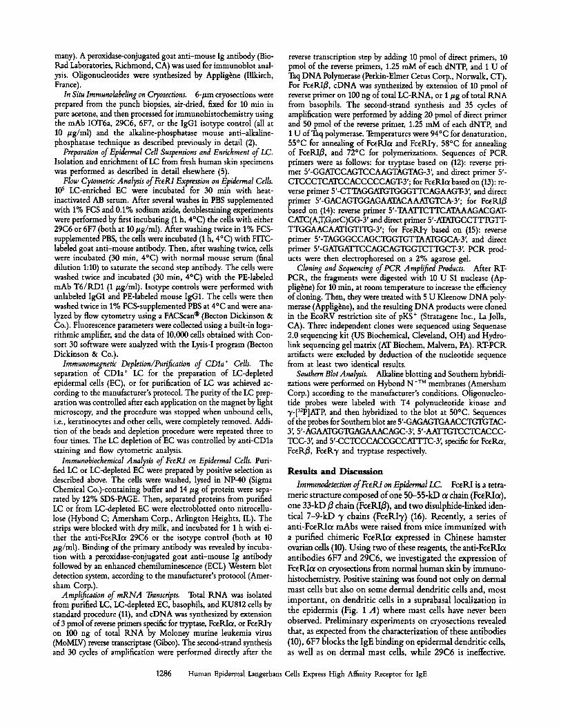
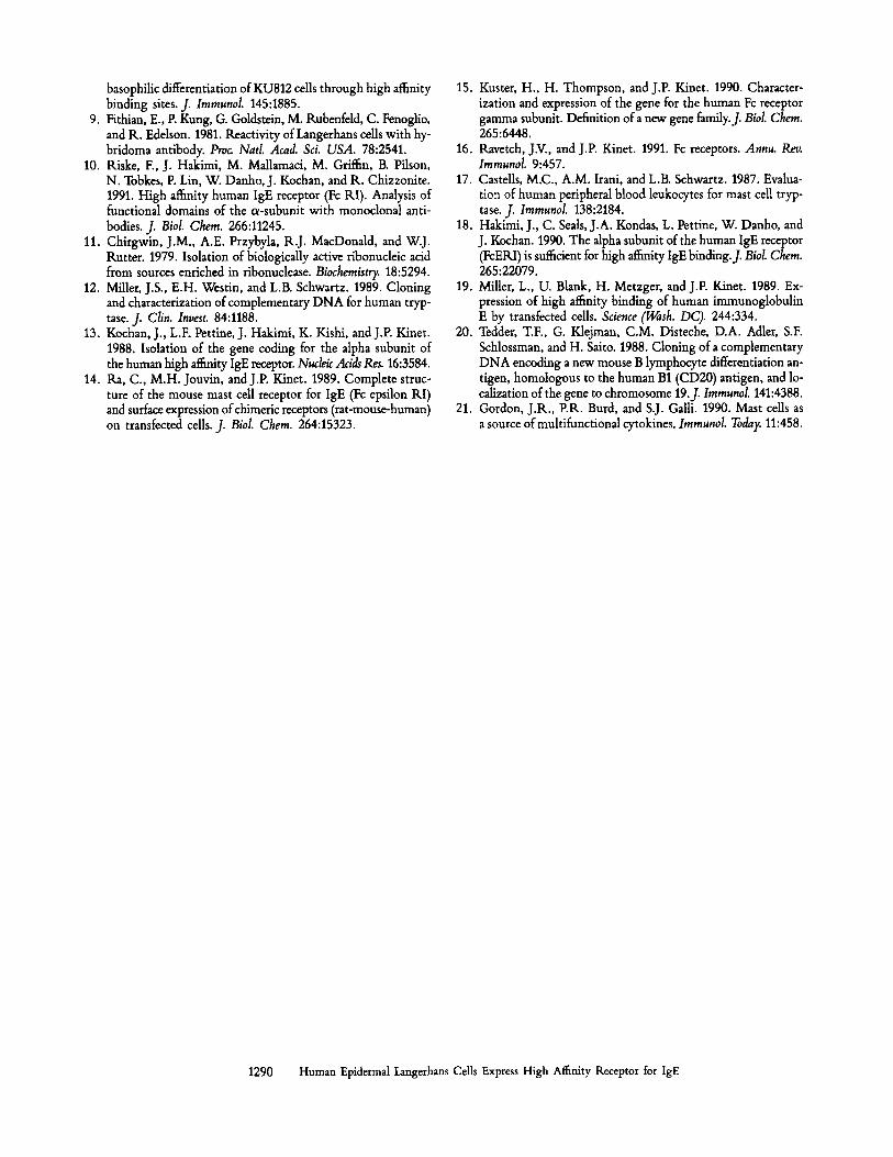
Fibre 4. Ana~is of the human basophil cDNA ~gment ~plified with mufine ~e~B-d~ved oligonuc~ti~s. Nucl~ti~c s~uence of the human cDNA fragment (HB) is compa~ with the murine ~eKI~ cDNA (M~). C o n s e ~ bases a~ linked ~ ~ . The numbem in the le~ ma~n rear to the nucleotide position within the human cDNA ~agment or the p~Lish~ mufine cDNA sequence (14). Sequences of the primers a~ not~ in small lette~. The deduced amino acid sequence encod~ ~ the human cDNA is ~ven using the single-letter code (h~), and is compa~ ~ th the murine ~eKIB amino acid sequence (roB); ( c ~ ) conserved ~sidues. (Boxes) tmnsmemb~ne domains (14). Note that the intmqto~asm~ domain (IISEK . . . . YLVRG) is wen conse~ed in human and mouse.
1288 Human Epidermal Langerhans Cells Express High Affinity Receptor for IgE
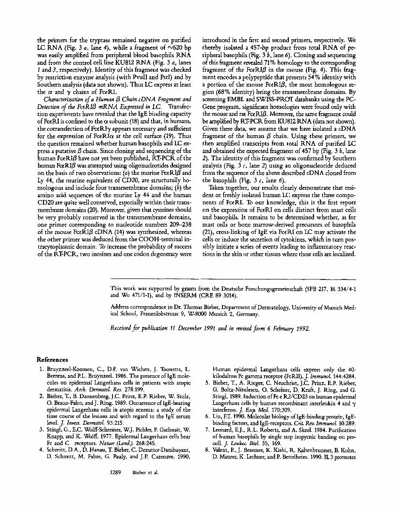
the primers for the tryptase remained negative on purified LC RNA (Fig. 3 a, lane 4), while a fragment of ~620 bp was easily amplified from peripheral blood basophils RNA and from the control cell line KU812 ILNA (Fig. 3 a, lanes 1 and 3, respectively). Identity of this fragment was checked by restriction enzyme analysis (with PvulI and PstI) and by Southern analysis (data not shown). Thus LC express at least the c~ and 3' chains of FceILl.
Characterization of a Human ~ Chain cDNA Fragment and Detection of the Fc6RIB mRNA Expressed in LC. Transfec- tion experiments have revealed that the IgE binding capacity of FceILI is confined to the c~ subunit (18) and that, in humans, the cotransfection of FceRI3' appears necessary and sufficient for the expression of FceRIc~ at the cell surface (19). Thus the question remained whether human basophils and LC ex- press a putative B chain. Since cloning and sequencing of the human FceRI/3 have not yet been published, tLT-PCIL of the human Fc~R/~ was attempted using oligonucleotides designed on the basis of two observations: (a) the murine FceILI~ and Ly 44, the routine equivalent of CD20, are structurally ho- mologous and include four transmembrane domains; (b) the amino acid sequences of the murine Ly 44 and the human CD20 are quite well conserved, especially within their trans- membrane domains (20). Moreover, given that cysteines should be very probably conserved in the transmembrane domains, one primer corresponding to nucleotide numbers 209-238 of the mouse FceRIB cDNA (14) was synthesized, whereas the other primer was deduced from the COOH-terminal in- tracytoplasmic domain. To increase the probability of success of the tLT-PCR, two inosines and one codon degeneracy were
introduced in the first and second primers, respectively. We thereby isolated a 457-bp product from total RNA of pe- ripheral basophils (Fig. 3 b, lane 6). Cloning and sequencing of this fragment revealed 71% homology to the corresponding fragment of the FceRIB in the mouse (Fig. 4). This frag- ment encodes a polypeptide that presents 54% identity with a portion of the mouse FceRI~, the most homologous re- gion (68% identity) being the transmembrane domains. By screening EMBL and SWISS-PILOT databanks using the PC- Gene program, significant homologies were found only with the mouse and rat FcEILI~. Moreover, the same fragment could be amplified by RT-PCIL from KU812 RNA (data not shown). Given these data, we assume that we have isolated a cDNA fragment of the human B chain. Using these primers, we then amplified transcripts from total RNA of purified LC and obtained the expected fragment of 457 bp (Fig. 3 b, lane 2). The identity of this fragment was confirmed by Southern analysis (Fig. 3 c, lane 2) using an oligonucleotide deduced from the sequence of the above described cDNA cloned from the basophils (Fig. 3 c, lane 6).
Taken together, our results clearly demonstrate that resi- dent or freshly isolated human LC express the three compo- nents of FceRI. To our knowledge, this is the first report on the expression of FceRI on cells distinct from mast cells and basophils. It remains to be determined whether, as for mast cells or bone marrow-derived precursors of basophils (21), cross-linking of IgE via FceP, I on LC may activate the cells or induce the secretion of cytokines, which in turn pos- sibly initiate a series of events leading to inflammatory reac- tions in the skin or other tissues where these cells are localized.
This work was supported by grants from the Deutsche Forschungsgemeinschaft (SFB 217, Bi 334/4-1 and Wo 471/1-1), and by INSERM (CRE 89 3014).
Address correspondence to Dr. Thomas Bieber, Department of Dermatology, University of Munich Med- ical School, Frauenlobstrasse 9, W-8000 Munich 2, Germany.
Received for publkation 11 December 1991 and in reuised form 6 February 1992.
References 1. Bruynzeel-Koomen, C., D.F. van Wichen, J. Toonstra, L.
Berrens, and P.L. Bruynzeel. 1986. The presence oflgE mole- cules on epidermal Langerhans cells in patients with atopic dermatitis. Arch. Dermatol. Res. 278:199.
2. Bieber, T., B. Dannenberg, J.C. Prinz, E.P. Rieber, W. Stolz, O. Braun-Falco, and J. Ring. 1989. Occurrence of IgE-bearing epidermal Langerhans cells in atopic eczema: a study of the time course of the lesions and with regard to the IgE serum level. J. Invest. Dermatol. 93:215.
3. Stingl, G., E.C. Wolff-Schreiner, W.J. Pichler, F. Gschnait, W. Knapp, and K. Wolff. 1977. Epidermal Langerhans cells bear Fc and C receptors. Nature (Lond.). 268:245.
4. Schmitt, D.A., D. Hanau, T. Bieber, C. Dezutter-Dambuyant, D. Schmitt, M. Fabre, G. Pauly, and J.P. Cazenave. 1990.
Human epidermal Langerhans ceils express only the 40- kilodalton Fc gamma receptor (FcILII).J. Immunol. 144:4284.
5. Bieber, T., A. Rieger, C. Neuchrist, J.C. Prinz, E.P. Rieber, G. Boltz-Nitulescu, O. Scheiner, D. Kraft, J. Ring, and G. Stingl. 1989. Induction ofFc e R2/CD23 on human epidermal Langerhans cells by human recombinant interleukin 4 and ~/ interferon. J. Extx Med. 170:309.
6. Liu, FT. 1990. Molecular biology oflgE-binding protein, IgE- binding factors, and IgE-receptors. Crit. Rev. Immunol. 10:289.
7. Leonard, E.J., R.L. Roberts, and A. Skeel. 1984. Purification of human basophils by single step isopycnic banding on per- coll. J. Leukoc. Biol. 35, 169.
8. Valent, P., J. Besemer, K. Kishi, R. Kaltenbrunner, B. Kuhn, D. Maurer, K. Lechner, and P. Bettelheim. 1990. IL 3 promotes
1289 Bieber et al.
basophilic differentiation of KU812 cells through high affinity binding sites. J. Immunol. 145:1885.
9. Fithian, E., P. Kung, G. Goldstein, M. Rubenfeld, C. Fenoglio, and R. Edelson. 1981. Reactivity of Langerhans cells with hy- bridoma antibody. Proc. Natl. Acad. Sci. USA. 78:2541.
10. Riske, F., J. Hakimi, M. Mallamaci, M. Griffin, B. Pilson, N. Tobkes, P. Lin, W. Danho, J. Kochan, and R. Chizzonite. 1991. High affinity human IgE receptor (Fc RI). Analysis of functional domains of the c~-subunit with monoclonal anti- bodies. J. Biol. Chem. 266:11245.
11. Chirgwin, J.M., A.E. Przybyla, R.J. MacDonald, and W.J. Rutter. 1979. Isolation of biologically active ribonucleic acid from sources enriched in ribonuclease. Biochemistry. 18:5294.
12. Miller, J.S., E.H. Westin, and L.B. Schwartz. 1989. Cloning and characterization of complementary DNA for human tryp- tase. J. Clin. Invest. 84:1188,
13. Kochan, J., L.F. Pettine, J. Hakimi, K. Kishi, and J.P. Kinet. 1988. Isolation of the gene coding for the alpha subunit of the human high affinity IgE receptor. NudeicAcidsRes. 16:3584.
14. Ra, C., M.H. Jouvin, and J.P. Kinet. 1989. Complete struc- ture of the mouse mast cell receptor for IgE (Fc epsilon KI) and surface expression of chimeric receptors (rat-mouse-human) on transfected cells. J. Biol. Chem. 264:15323.
15. Kuster, H., H. Thompson, and J.P. Kinet. 1990. Character- ization and expression of the gene for the human Fc receptor gamma subunit. Definition of a new gene family.J. Biol. Chem. 265:6448.
16. Ravetch, J.V., and J.P. Kinet. 1991. Fc receptors. Annu. Rev. ImmunoL 9:457.
17. Castells, M.C., A.M. Irani, and L.B. Schwartz. 1987. Evalua- tion of human peripheral blood leukocytes for mast cell tryp- tase. J. Immunol. 138:2184.
18. Hakimi, J., C. Seals, J.A. Kondas, L. Pettine, W. Danho, and J. Kochan. 1990. The alpha subunit of the human IgE receptor (FcERI) is sufficient for high affinity IgE binding.J, Biol. Chem. 265:22079.
19. Miller, L., U. Blank, H. Metzger, and J.P. Kinet. 1989. Ex- pression of high affinity binding of human immunoglobulin E by transfected cells. Science (Wash. DC). 244:334.
20. Tedder, T.F., G. Klejman, C.M. Disteche, D.A. Adler, S.F. Schlossman, and H. Saito. 1988. Cloning of a complementary DNA encoding a new mouse B lymphocyte differentiation an- tigen, homologous to the human B1 (CD20) antigen, and lo- calization of the gene to chromosome 19.J. lmmunol. 141:4388.
21. Gordon, J.R., P.R. Burd, and S.J. GaUl. 1990. Mast cells as a source ofmultifunctional cytokines. Immunol. Today. 11:458.
1290 Human Epidermal Langerhans Cells Express High Affinity Receptor for IgE