Embed Size (px)
Citation preview
Biochem. J. (1979) 179,151-160 151Printed in Great Britain
Reactivities of Hydroxylamine and Sodium Bisulphite with Carbonyl-ContainingHaems and with the Prosthetic Groups of the Erythrocyte Green Haemoproteins
By LOUIS J. DEFILIPPI,* LINDA S. TOLER and DONALD E. HULTQUISTDepartment ofBiological Chemistry, University ofMichigan, Ann Arbor, MI 48109, U.S.A.
(Received 18 August 1978)
The reactivities of alkaline NH20H and neutral NaHSO3 with carbonyl and olefinicgroups conjugated with the tetrapyrrole nucleus of haems were studied. The reactionswere carried out with 2-3 nmol of haem a, spirographis haem, isospirographis haem,2,4-diacetyldeuterohaem and protohaem. Vinyl side chains were found to be insensitiveto the chemical action of both alkaline NH20H and neutral NaHSO3. The formyl-containing haems reacted rapidly with both reagents at room temperature, as evidencedby sizable hypsochromic shifts of the reduced pyridine haemochrome spectrum. In lessalkaline solution, the reactions of these formyl-containing haems with NH20H were muchslower. 2,4-Diacetyldeuterohaem reacted with alkaline NH20H, but not with neutralNaHSO3. These rapid, simple and straightforward tests are readily usable in differentiatingamongformyl, acetyl and other electron-withdrawing side chains conjugated with thetetra-pyrrole ring of haems. We applied these observations to an investigation of the two uniqueprosthetic groups ofthe bovine erythrocyte green haemoproteins. The prosthetic groups ofthese two proteins were isolated and spectrally characterized. Under the conditions used,the haems did not react with either NH20H or NaHSO3, but were altered by dithionite,suggesting that the previous interpretation that a formyl group was present [Hultquist,Dean & Reed (1976) J. Biol. Chem. 251, 3927-3932] may have been premature. Thesestudies also provide evidence that the a-hydroxyfarnesylethyl side chain of haem a affectsthe a-band maximum, but not the /1- or Soret bands of the reduced pyridine haemochromespectrum of haem a.
The reaction of NH20H with carbonyl groups togive the corresponding oxime derivative has classicallybeen used as a test for the detection of carbonyl-containing porphyrins and, less frequently, carbonyl-containing haems (Rawlinson & Hale, 1949; Lemberg& Falk, 1951; Oliver & Rawlinson, 1955; Connellyet al., 1958; Parker, 1959; Morrison et al., 1960;Clezy & Barrett, 1961; Clezy et al., 1964). The magni-tude of the blue-shift of the near-u.v.-visible absorp-tion spectrum that accompanies oxime formationis much larger for formyl-containing tetrapyrrolesthan for acetyl-containing tetrapyrroles. Oximeformation results in a 17-22nm shift of the a-peakof the reduced pyridine haemochrome of haems witha formyl group in conjugation with the tetrapyrrolenucleus, but gives only a 1-2nm shift with haemscontaining a conjugated acetyl group (Lemberg &Falk, 1951). This difference has been used to dis-tinguish between formyl and acetyl substitution on
the periphery of the porphyrin nucleus.A blue-shift of a porphyrin spectrum on reaction
with NaHSO3 has likewise been cited as evidence for* Present address: Biochemistry and Chemicals Re-
search Department, Universal Oil Products Inc., DesPlaines, IL 60016, U.S.A.
Vol. 179
the presence of a formyl group in conjugation with atetrapyrrole nucleus, since the acetyl-containingporphyrins, cryptoporphyrinsp, are reported not toundergo bisulphite-adduct formation (Clezy et al.,1964).The unique prosthetic group of a human erythro-
cyte green haemoprotein (Hultquist et al., 1976) wasfound to undergo reactions with NH2OH andNaHSO3 under the conditions that have been usedwith other haems; characterization of this prostheticgroup and its derivatives distinguished it from allother naturally occurring prosthetic groups andsuggested that it is a complex haem containing botha formyl group and polar acetylatable functionalgroups. Similarly, we have studied two bovineerythrocyte green haemoproteins and shown thatthese proteins differ in terms of the spectral andchemical properties of their prosthetic groups(DeFilippi & Hultquist, 1978a,b).
In attempting to identify unambiguously the sidechains of these haems we further studied the reactionsof NH20H and NaHSO3 with model compounds.We discovered that haem a, which has been shown topossess a formyl group as one of its substituents(Lemberg & Falk, 1951; Lemberg, 1953; Connelly
L. J. DEFILIPPI, L. S. TOLER AND D. E. HULTQUIST
et al., 1958; Caughey et al., 1975), does not rapidlyundergo oxime formation under the neutral or mildlyalkaline conditions at which the reaction was be-lieved to occur. However, we found that the reactionproceeds rapidly in the strongly alkaline conditionsused in pyridine haemochrome formation, a pro-cedure that was believed to be simply a process toassess visually the extent of the reaction. Moreover,we realized that the reaction of formyl-containinghaems with NaHSO3 has received relatively littleattention in the literature (Orii & Washio, 1977;Kitagawa et al., 1977). Reactivity of porphyrins (butnot haems) with HSO3- was apparently first describedby Parker (1959) as yielding alteration in the absorp-tion spectrum of cryptoporphyrin a in dilute coldpyridine; reference was made to unpublished work byR. Lemberg.
In the present paper we report the reactivities ofhaems with alkaline NH2OH and neutral NaHSO3.These reactivities constitute a rapid and straight-forward means of differentiating among formyl,acetyl and other electron-withdrawing side chains ofhaems and have allowed us to re-examine the questionof whether a formyl group is present on the prostheticgroups of the erythrocyte green haemoproteins.
Experimental
MaterialsThe outdated human erythrocytes were obtained
from the University of Michigan Medical Center
Blood Bank. Ox heart was purchased from KapplerPacking Co., Ann Arbor, MI, U.S.A.
Pyridine for spectrophotometry was dried overKOH pellets and distilled from ninhydrin. Purifiedpyridine was stored over 4A Linde molecular sieves(Union Carbide Corp., New York, NY, U.S.A.) andKOH pellets. All other chemicals were reagent gradeand were not further purified.
Silica gel 60 F-254 t.l.c. plates were obtained fromEM Laboratories (Elmsford, NY, U.S.A.); alumina(Woelm, neutral activity, grade I, used for columnchromatography) was obtained from AlupharmChemicals (New Orleans, LA, U.S.A.); polyamideCC6 for column chromatography was obtained fromBrinkmann Instruments (Westbury, NY, U.S.A.).
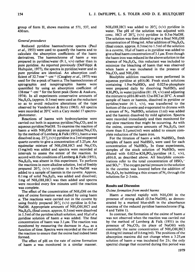
Preparation ofmodel haemsThe structures of the various haems used in this
paper are shown in Fig. 1. Protohaemin IX wasobtained from packed out-dated erythrocytes byusing the acetic acid method of Fischer (1955).Protoporphyrin IX dimethyl ester was prepared fromprotohaemin IX by the method of Caughey et al.(1966). Deuterohaemin IX was prepared and purifiedas described by Falk (1964). 2,4-Diacetyldeutero-haemin IX was prepared from deuterohaemin IX bythe method of Fischer & Orth (1937) and was purified(Lamson et al., 1973) on polyamide columns (2cm x20cm) by using 2.5 % (v/v) acetic acid in methanol.
Spirographis haemin (2-formyl-4-vinyldeutero-haemin IX) and isospirographis haemin (4-formyl-
H3C-
Fig. 1. Schemnatic representation ofthe model haems used in this studyHaem R R' R
Deuterohaem IX -H -H -CH3Protohaem IX -CH=CH2 -CH=CH2 -CH3Spirographis haem -CHO -CH=CH2 -CH3Isospirographis haem -CH=-CH2 -CHO -CH3Haem a a-Hydroxy- -CH=CH2 -CHO
farnesylethyl2,4-Diacetyldeuterohaem IX -COCH3 -COCH3 -CH3
1979
152
REACTIONS OF HAEMS WITH NH2OH AND NaHSO3
A
0.15
0.141i.
0.13
0.12
0.11
0.10
.A \ Il 0.09
\/~~~J ~0.08A:~~~~~0.07
0.06
I\\ I. ~~~~~0.05
0.04
\l 0.03
| s | | k 0.02
\* 0.01
0400 450 500 550 600 650
Wavelength (nm)
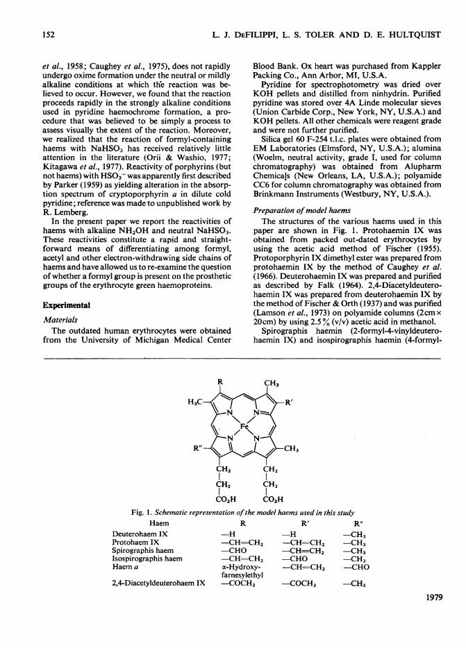
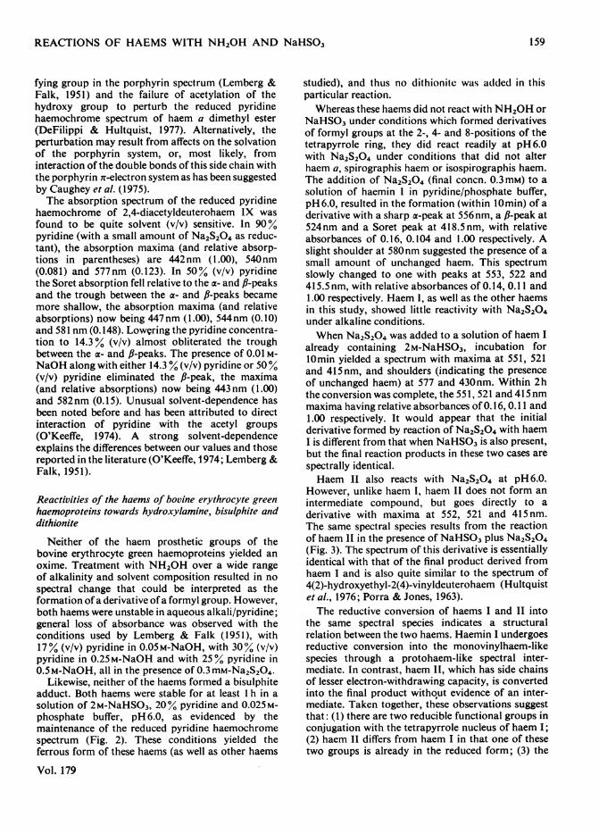
Fig. 2. Spectra ofderivatives ofhaemin I ofbovine erythro-cyte green haemoprotein
Reduced pyridine haemochrome spectra are shownfor freshly prepared haemin I at 20°C. , HaeminI in a solution of 0.025M-potassium phosphatebuffer, pH6.00, containing 20% (v/v) pyridine and 2M-NaHSO3, but no Na2S204. The haem is fully reducedunder these conditions and possesses maxima at579.5, 543 and 433nm. - *-, Haemin I after10min in a solution of 0.025M-potassium phosphatebuffer, pH 6.00, containing 20% pyridine and 0.3mM-Na2S204, but no NaHSO3. The derivative showsmaxima at 556, 524 and 418.5nm. ----, Samesample after 2h of reaction with Na2S204; absorb-ance maxima are at 553, 522 and 415.5nm.
2-vinyldeuterohaemin IX) were prepared as follows.The porphyrin dimethyl esters corresponding to thesehaemins were prepared and purified by the method ofCaughey et al. (1966). The esters of spirographis andisospirographis porphyrins were separated by usingsilica-gel t.l.c. with unwashed chloroform as thesolvent (Sono & Asakura, 1974). Each of the por-phyrin esters was hydrolysed to the free porphyrinat 20°C in 6M-HCI; paper chromatography withlutidine/water (10: 7, v/v) in an atmosphere saturatedwith aq. 7M-NH3 (Chu & Chu, 1955) was used tomonitor the conversion. Haemins were prepared fromeach of the porphyrin isomers by the method ofMorell et al. (1961), and were extracted into ethylacetate by the method of Asakura & Sono (1974).The haemins were purified on polyamide columns(1cm x 6cm) by using 2.5% (v/v) acetic acid inmethanol (Lamson et al., 1973).Haem a was isolated from fresh ox hearts as the
Vol. 179
0.19
0.18
0.17
0.16A
0.15
0.14
0.13
0.12
0400 450 500 550 600 650
Wavelength (nm)
A
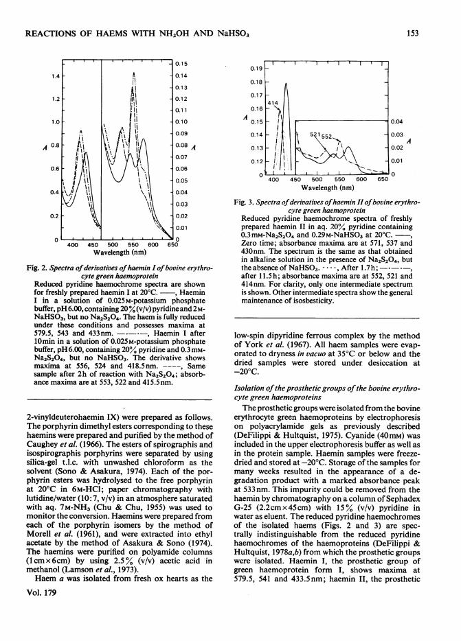
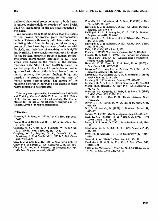
Fig. 3. Spectra ofderivatives ofhaemin II ofbovine erythro-cyte green haemoprotein
Reduced pyridine haemochrome spectra of freshlyprepared haemin II in aq. 20% pyridine containing0.3mM-Na2S204 and 0.29M-NaHSO3 at 20°C.Zero time; absorbance maxima are at 571, 537 and430nm. The spectrum is the same as that obtainedin alkaline solution in the presence of Na2S204, butthe absence of NaHSO3. - -, After 1.7h; *- ,after 11.5h; absorbance maxima are at 552, 521 and414nm. For clarity, only one intermediate spectrumis shown. Other intermediate spectra show the generalmaintenance of isosbesticity.
low-spin dipyridine ferrous complex by the methodof York et al. (1967). All haem samples were evap-orated to dryness in vacuo at 35'C or below and thedried samples were stored under desiccation at-200C.
Isolation ofthe prosthetic groups ofthe bovine erythro-cyte green haemoproteinsThe prosthetic groups were isolated from the bovine
erythrocyte green haemoproteins by electrophoresison polyacrylamide gels as previously described(DeFilippi & Hultquist, 1975). Cyanide (40mM) wasincluded in the upper electrophoresis buffer as well asin the protein sample. Haemin samples were freeze-dried and stored at -20°C. Storage of the samples formany weeks resulted in the appearance of a de-gradation product with a marked absorbance peakat 533 nm. This impurity could be removed from thehaemin by chromatography on a column ofSephadexG-25 (2.2cmx45cm) with 15% (v/v) pyridine inwater as eluent. The reduced pyridine haemochromesof the isolated haems (Figs. 2 and 3) are spec-trally indistinguishable from the reduced pyridinehaemochromes of the haemoproteins (DeFilippi &Hultquist, 1978a,b) from which the prosthetic groupswere isolated. Haemin I, the prosthetic group ofgreen haemoprotein form I, shows maxima at579.5, 541 and 433.5nm; haemin II, the prosthetic
153
L. J. DEFILIPPI, L. S. TOLER AND D. E. HULTQUIST
group of form II, shows maxima at 571, 537, and430nm.
General proceduresReduced pyridine haemochrome spectra (Paul
et al., 1953) were used to quantify the haems and tocalculate the absorption coefficients of the haemderivatives. The haemochrome of haem a wasprepared in pyridine/water (9: 1, v/v) rather than inpure pyridine. As reported previously (DeFilippi &Hultquist, 1977), the spectra in pyridine/water and inpure pyridine are identical. An absorption coef-ficient of 32.7mm-' cm-' (Caughey et al., 1975) wasused for the a-peak of haem a. The haemochromes ofspirographis and isospirographis haems werequantified by using an absorption coefficient of138 mm-' * cm-' for the Soret peak (Sono & Asakura,1974). In all experiments conservative amounts ofNa2S204 (approx. 0.3 mM) were used as the reductantso as to avoid reductive alterations of the typeobserved by Vanderkooi & Stotz (1965). All spectrawere recorded at 20°C with a Cary model 17 spectro-photometer.
Reactions of haems with hydroxylamine werecarried out both in aqueous pyridine/Na2CO3 and inaqueous pyridine/NaOH. To examine the reaction ofhaem a with NH2OH in aqueous pyridine/Na2CO3by the method of Lemberg & Falk (1951), haem a wasdissolved in aq. 25% (v/v) pyridine in a 2ml stopperedquartz cuvette to give a concentration of 3.25,um. Anequimolar mixture of NH20H,HCl and Na2CO3(1 mg/ml) was added and spectra were recorded atintervals to assess the extent of the reaction. Inaccord with the conditions of Lemberg & Falk (1951),Na2S204 was absent in this experiment. To performthe reactions in more alkaline solution, 1 ml of freshlyprepared 20% (v/v) pyridine in 0.5 M-NaOH wasadded to a sample of haemin in the cuvette. Approx.0.1 mg of solid Na2S204 was added and dissolved;1mg of NH2OH,HC1 was then added and spectrawere recorded every few minutes until the reactionwas complete.The effect of the concentration of NH2OH on the
rate of oxime formation was studied by using haema. The reactions were carried out in the cuvette byusing freshly prepared 20% (v/v) pyridine in 0.5M-NaOH. Appropriate amounts of NH2OH,HCl andNa2S204 (final concn. approx. 0.3 mM) were dissolvedin 1.5 ml of the pyridine/alkali solution, and IO,ul of apyridine solution of haem a was added. The finalconcentration of haem was 3.1 pM. The reaction wasmonitored by measuring the decrease in A594 as afunction of time. Spectra were recorded at the end ofthe reaction to ensure that the oxime had indeed beenformed.The effect of pH on the rate of oxime formation
of haem a was monitored in a similar manner.
NH2OH,HCI was added to 20% (v/v) pyridine inwater. The pH of the solution was adjusted withconc. HCI of 20% (v/v) pyridine in 0.5M-NaOH.The solution was then diluted to give a final NH2OHconcentration of 10mg/ml. After addition ofNa2S204(final concn. approx. 0.3 mM) to 1.5 ml of the solutionin a cuvette, 101u of haem a in pyridine was added togive a final haem concentration of 3.1 pm. Even thoughthe haem iron was reduced in the above solution in theabsence of Na2S204, this reductant was included tominimize the bleaching of haem that was observedwhen haem a was incubated in the presence ofNa2CO3 and NH2OH.
Bisulphite addition reactions were performed inaqueous pyridine at pH6.00. Fresh stock solutionscontaining 1.9M-NaHSO3 and 0.025M-phosphatewere prepared daily by dissolving NaHSO3 andKH2PO4 in water/pyridine (81:19, v/v) and adjustingthe mixture to pH 6.00 with 12M-HCl at 20°C. A smallsample of haem or haemin, dissolved in pyridine orpyridine/water (4:1, v/v), was transferred to thebottom of the cuvette and evaporated to dryness witha stream of N2. NaHSO3 solution (1 ml) was addedand the haemin dissolved by mild agitation. Spectrawere recorded immediately and then monitored forany slow reactions that might be occurring. As withthe oxime, conservative amounts of Na2S204 (notmore than 0.3,umol/ml) were added to ensure com-plete reduction of the haem iron.For the titration of haem a with NaHSO3, fresh
samples of haem and NaHSO3 were used for eachconcentration of NaHSO3. In these experiments,samples of the stock solution of NaHSO3 werediluted with 0.025M-KH2PO4/pyridine (4:1, v/v),pH 6.0, as described above. All bisulphite concen-trations refer to the total concentration of HS03-and SO32-. The oxygen partial pressure in the solutionin the cuvettes was lowered before the addition ofNa2S204 by bubbling a thin stream of N2 through thesolutions for 2-3 min.
Results and DiscussionOxime formation from model haemsHaem a reacted rapidly with NH2OH in the
presence of strong alkali (0.5M-NaOH), as demon-strated by a marked blue-shift in the absorbancemaxima of the reduced pyridine haemochrome (Fig.4 and Table 1).
In contrast, the formation of the oxime of haem awas not observed when the reaction was carried outby the method of Lemberg & Falk (1951) in thepresence of Na2CO3 (in place of NaOH) withessentially the same concentration of NH2OH,HCl(0.4mg/ml instead of 0.6mg/ml). The positions of theabsorption maxima did not change when a 3.254uMsolution of haem a was incubated for 2h; the onlyspectral change that occurred during this period was
1979
154

REACTIONS OF HAEMS WITH NH2OH AND NaHSO3
150
1
,I 100
.
E 50)
0
1 51
IE 10
7
.
E 51to
~~~~I I...T..II (a) 30
20 1g20
10CO
~~~~~~I I.10 (b) -30
0 -20
io0 X 10
o I 0400 500 600
150
100
C')IE
50
0
400 500 EWavelength (nm)
600
.C,
EI
Ew
30
20
E
E10
Fig. 4. Reducedpyridine haemochrome spectra ofoximes offormyl-substituted haems
Millimolar absorptions were calculated as describedin the Experimental section. Spectra were recordedafter 15 min of reaction ofhaem with 1 mg ofNH20H/ml of 0.5 M-NaOH/pyridine (4:1, v/v) in the presence
of Na2S204. (a) Spirographis haem oxime; (b)isospirographis haem oxime; (c) haem a oxime.
a general loss of absorption (an 85 % decrease in theabsorbance at the a-peak). Addition of Na2S204(0.3 mM) subsequent to the incubation did not alterthe spectrum. Lemberg & Falk (1951) reported thatafter 30min under similar conditions haem a had
Vol. 179
reacted with NH2OH to give a 17nm blue-shift inthe a-peak of the reduced pyridine haemochromespectrum.
This difference between our findings and those ofLemberg & Falk (1951) might be explained in one ormore of the following ways: (1) NaOH might benecessary to reveal the oxime formation; (2) an
inhibitor of oxime formation might be present in ourreaction mixture; (3) the extent of the reaction mightbe highly dependent on NH2OH concentration;(4) strongly alkaline conditions might be necessaryfor the reaction to proceed at an appreciable rate.The last of these explanations was shown to be
correct by the following experiments. Incubation ofhaem a with NH2OH resulted in no spectral shiftwhen Na2CO3 was eliminated from the reactionmixture or when sodium acetate (Morrison et al.,1960) was substituted for the Na2CO3. However, asshown in Fig. 4(c) the oxime was formed when0.5M-NaOH was used in place of the Na2CO3. Whena mixture of haem a and NH2OH, which had failedto react in the presence of Na2CO3, was then made0.25M in NaOH, a spectral change indicative ofoxime formation was observed. After 1 min, doublea-peaks were observed (587 and 569 nm); within15min, oxime formation was complete. The extentof the reaction at various times was approximatelythe same as when the reaction was carried out inNaOH without prior incubation in the presence ofNa2CO3. This finding rules out the possibility thatoxime formation occurred in the carbonate solution,but that alkali was required to convert it into an
anionic form that is blue-shifted. It also rules out thepossibility that an inhibitor is present in the reactionmixture.
Measurement of the effect of NH2OH concen-tration on the rate of haem a oxime formation in 20%pyridine in 0.5M-NaOH showed that the reactionrate (but not the extent) was indeed concentration-dependent. NH20H concentrations of 0.0144, 0.0719and 0.144M gave observed rate constants of 0.35,1.36 and 2.5min-' respectively. The second-orderrate constant calculated from these values is 50mM-' Mmin-'. This concentration-dependency is not suf-ficiently large, however, to explain the differencebetween our findings and those of Lemberg & Falk(1951).The anomaly can be explained by the effect of
pH on the rate of the reaction. The pH-versus-rateprofile is shown in Fig. 5. The rate of oxime formationin 0.5M-NaOH (calculated pH of 13.7) is 40-150times the observed rates in the pH range 8.0-11.0. ThepH of the carbonate/NH2OH solution, calculated tobe similar to that used by Lemberg & Falk (1951), isin the range where the rate is lowest (pH 8-11), thusexplaining our failure to observe oxime formation.We believe that in previous studies, where the reaction
L 0
400 500 600
Vi
-n--
155
L. J. DEFILIPPI, L. S. TOLER AND D. E. HULTQUIST
Table 1. Absorbance maxinma ofreducedpyridine haemochromesFinal concentrations are: *20% (v/v) pyridine, 0.5M-NaOH, t19% pyridine, 1.9M-NaHSO3; 125% pyridine, 0.5M-NaOH; §90% pyridine.
Absorbance maxima (nm)
Haem
Haem a
Spirographis haem
Isospirographis haem
2,4-Diacetyldeuterohaem
Derivative
Free*Oxime*Bisulphite adducttFree*Oxime*Bisulphite adducttFree*Oxime*Bisulphite adducttFreetFree§Oxime*Bisulphite adductt
Soret II cx
431418 524417 522434 538419 526416 521434 536419 526416 522426442 540421
No reaction
2.5
2.0-
6 1.5 -
1.0
0.5-
6 7 8 9 10 11 12 13 14
pHFig. 5. Rate offormation ofhaem a oxime versuspHReactions were carried ouit at 27°C in aq. 20% (v/v)pyridine with final concentrations of 10mg ofNH20H,HCI/ml and 3.1/pM-haem a. The pH wasadjusted with either dilute HCI or 20%° (v/v) pyridinein 0.5M-NaOH. After addition of a few crystals ofNa2S204 to 1.5 ml of NH20H solution, lOl of apyridine solution of haem a was added and thereaction monitored at 594nm. Apparent rate con-stants (kobs.) were calculated from the slope of theplots of log (A594 at time t - final A594) versus time.
pyridine/aq. 0.5M-NaOH (1:4, v/v). Under theseconditions the reaction proceeds equally well in boththe presence and the absence of Na2S204, but thiscompound is normally included in order both tominimize the bleaching of the haem and to ensurecomplete reduction of the haem iron. The rate offormation of the oxime is independent of Na2S204at the low concentrations tested.Under these conditions spirographis haem and
isospirographis haems, like haem a, react readilywith NH2OH (see Figs. 4a and 4b and Table 1)Diacetyldeuterohaem IX reacts with NH2OH to givea small shift in the absorbance maxima of the reducedpyridine haemochrome derivative. In contrast withthe blue-shift of 20-25nm and 13-15nm observed forthe a- and Soret peaks respectively of the formyl-containing haems, reaction of diacetyldeuterohaemIX with NH2OH gives 8nm and 5nm blue-shifts forthe a- and Soret peaks respectively. Small shifts hadpreviously been reported to result from reaction ofacetyl-containing porphyrins with NH2OH (Lemberg& Falk, 1951). Exposure of protohaem IX to thisreagent resulted in no significant spectral change inthe reduced pyridine haemochrome. Conjugatedvinyl groups of haems are thus unreactive towardsalkaline NH2OH.
was carried out at neutral or slightly alkaline pH,little haem oxime formation occurred until NaOHwas added for the purpose of obtaining the reducedpyridine haemochrome by the method of Paul et al.(1953).Having explained this anomaly, we can now
present a rapid straightforward method for oximeformation with formyl-containing haems. Thereaction conditions are: NH2OH,HCI (>1 mg/ml) in
Bisulphite adductformation from model haemsHaem a was found to react readily with 2M-
NaHSO3 in pyridine/0.025 M-potassium phosphatebuffer (1:4, v/v), pH6.00. Initiation of the reactionresulted in an immediate decrease in absorbance at587 and 431 nm concomitant with the appearance ofan a-peak at 560nm, a fl-peak at 522nm and a Soretpeak at 416.5 nm (see Fig. 6). The addition ofNa2S204did not alter the spectrum, except to give a slightsharpening of the absorption maxima.
1979
587567559583558553581558553581577573
156
REACTIONS OF HAEMS WITH NH20H AND NaHSO3
40500 600 700W h Wavelength (nm)
50 4 -(b)
0 2 4 6
1/1 HSO., +O>-S hr30 I
400 500 600
Wavelength (nm)
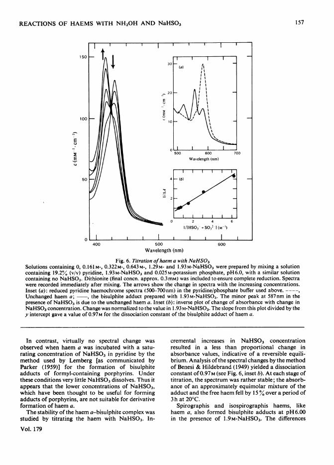
Fig. 6. Titration ofhaem a with NaHS03Solutions containing 0, 0.161 M-, 0.322M-, 0.643M-, 1.29M- and 1.93 M-NaHSO3 were prepared by mixing a solutioncontaining 19.2% (v/v) pyridine, 1.93M-NaHSO3 and 0.025M-potassium phosphate, pH6.0, with a similar solutioncontaining no NaHSO3. Dithionite (final concn. approx. 0.3mM) was included to ensure complete reduction. Spectrawere recorded immediately after mixing. The arrows show the change in spectra with the increasing concentrations.Inset (a): reduced pyridine haemochrome spectra (500-700nm) in the pyridine/phosphate buffer used above.----,Unchanged haem a; , the bisulphite adduct prepared with 1.93M-NaHSO3. The minor peak at 587nm in thepresence of NaHSO3 is due to the unchanged haem a. Inset (b): inverse plot of change of absorbance with change inNaHSO3 concentration. Change was normalized to the value in 1.93 m-NaHSO3. The slope from this plot divided by they intercept gave a value of 0.97M for the dissociation constant of the bisulphite adduct of haem a.
In contrast, virtually no spectral change wasobserved when haem a was incubated with a satu-rating concentration of NaHSO3 in pyridine by themethod used by Lemberg [as communicated byParker (1959)] for the formation of bisulphiteadducts of formyl-containing porphyrins. Underthese conditions very little NaHSO3 dissolves. Thus itappears that the lower concentrations of NaHSO3,which have been thought to be useful for formingadducts of porphyrins, are not suitable for derivativeformation of haem a.The stability of the haem a-bisulphite complex was
studied by titrating the haem with NaHSO3. In-
Vol. 179
cremental increases in NaHSO3 concentrationresulted in a less than proportional change inabsorbance values, indicative of a reversible equili-brium. Analysis ofthe spectral changes by the methodof Benesi & Hildebrand (1949) yielded a dissociationconstant of 0.97M (see Fig. 6, inset b). At each stage oftitration, the spectrum was rather stable; the absorb-ance of an approximately equimolar mixture of theadduct and the free haem fell by 15% over a period of3 h at 20°C.
Spirographis and isospirographis haems, likehaem a, also formed bisulphite adducts at pH 6.00in the presence of 1.9M-NaHSO3. The differences
157
L. J. DEFILIPPI, L. S. TOLER AND D. E. HULTQUIST
I2E
co
30
_
20 E_-
E10 Co
30
EI
20?
E10 X
0
400 500 600 700
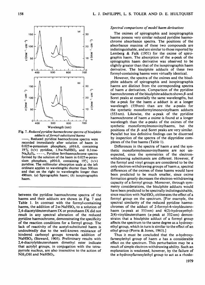
Wavelength (nm)Fig. 7. Reducedpyridine haemochrome spectra ofbisulphite
adducts offormyl-substituted haemsReduced pyridine haemochrome spectra were
recorded immediately after solution of haem in0.025 M-potassium phosphate, pH 6.0, containing19% (v/v) pyridine, l.9M-NaHSO3 and 0.3mM-Na2S204. ----, Pyridine ferrihaemochrome spectraformed by the solution of the haem in 0.025 M-potas-sium phosphate, pH 6.0, containing 19° (v/v)pyridine. The millimolar absorption scale at the leftordinate applies to wavelengths shorter than 500nmand that on the right to wavelengths longer than490nm. (a) Spirographis haem; (b) isospirographishaem.
between the pyridine haemochrome spectra of thehaems and their adducts are shown in Fig. 7 andTable 1. In contrast with the formyl-containinghaems, the addition of 2M-NaHSO3 to a solution of2,4-diacetyldeuterohaem IX or protohaem IX did notresult in any spectral alteration of the reducedpyridine haemochrome, demonstrating the specificityof the reaction conditions for a formyl group. Thelack of reactivity of the acetyl-substituted haem isundoubtedly due to the well-known resistance ofhindered carbonyl groups towards addition ofNaHSO3 (Steward, 1905). Preliminary results with2,4-diacrylyldeuterohaem dimethyl ester indicatethat acrylyl groups, in conjugation with the tetra-pyrrole nucleus, are also insensitive to the action ofNH20H and NaHSO3.
Spectral comparisons ofmodel haem derivativesThe oximes of spirographis and isospirographis
haems possess very similar reduced pyridine haemo-chrome absorbance spectra. The positions of theabsorbance maxima of these two compounds areindistinguishable, and are similar to those reported byLemberg & Falk (1951) for the oxime of spiro-graphis haem. The absorption of the a-peak of thespirographis haem derivative was observed to beslightly greater than that of the isospirographis haemderivative. The bisulphite adducts of these twoformyl-containing haems were virtually identical.
However, the spectra of the oximes and the bisul-phite adducts of spirographis and isospirographishaems are distinct from the corresponding spectraof haem a derivatives. Comparison of the pyridinehaemochromes of the bisulphite adducts shows ,B- andSoret peaks at essentially the same wavelengths, butthe a-peak for the haem a adduct is at a longerwavelength (559 nm) than are the a-peaks forthe synthetic monoformylmonovinylhaem adducts(553 nm). Likewise, the a-peak of the pyridinehaemochrome of haem a oxime is found at a longerwavelength than the a-peaks of the oximes of thesynthetic monoformylmonovinylhaems, but thepositions of the ,B- and Soret peaks are very similar.Parallel but less definitive findings can be discernedby inspection of the spectra of the dipyridine com-plexes of the free haems (Table 1).
Differences in the spectra of haem a and the syn-thetic monoformylmonovinylhaems are not un-expected, since the symmetries of the electron-withdrawing substituents are different. However, ifthe formyl and vinyl groups are considered to be theonly electron-withdrawing groups of haem a, spectraldifferences of the oximes of these haems would havebeen predicted to be much smaller, since oximeformation greatly decreases the electron-withdrawingcapacity of a formyl group. Moreover, through sym-metry considerations, the bisulphite adducts wouldhave been predicted to be spectrally indistinguishable,since reaction with NaHS03 obliterates the effect of aformyl group on the spectrum. [For example, thespectral similarity of the reduced pyridine haemo-chromes of the adduct of 2-formyl-4-vinyldeutero-haem (a-peak at 553 nm) and 4(2)-hydroxyethyl-2(4)-vinyldeuterohaem (a-peak at 552nm) demon-strates that a bisulphite adduct of a formyl groupaffects the spectrum to the same degree as a hydroxy-ethyl group, which in turn is similar to the effect of anethyl group (Porra & Jones, 1963).]Thus it must be concluded that the a-hydroxy-
farnesylethyl group of haem a has a considerableeffect on the spectrum. This perturbation may be aresult of simple electron-withdrawing ability. Such anexplanation is weakened, however, by the failure ofthe a-hydroxyfarnesylethyl group to act as a rhodo-
1979
158
REACTIONS OF HAEMS WITH NH2OH AND NaHSO3
fying group in the porphyrin spectrum (Lemberg &Falk, 1951) and the failure of acetylation of thehydroxy group to perturb the reduced pyridinehaemochrome spectrum of haem a dimethyl ester(DeFilippi & Hultquist, 1977). Alternatively, theperturbation may result from affects on the solvationof the porphyrin system, or, most likely, frominteraction of the double bonds of this side chain withthe porphyrin 7-electron system as has been suggestedby Caughey et al. (1975).The absorption spectrum of the reduced pyridine
haemochrome of 2,4-diacetyldeuterohaem IX wasfound to be quite solvent (v/v) sensitive. In 90%pyridine (with a small amount of Na2S204 as reduc-tant), the absorption maxima (and relative absorp-tions in parentheses) are 442nm (1.00), 540nm(0.081) and 577nm (0.123). In 50% (v/v) pyridinethe Soret absorption fell relative to the a- and fl-peaksand the trough between the a- and f,-peaks becamemore shallow, the absorption maxima (and relativeabsorptions) now being 447nm (1.00), 544nm (0.10)and 581 nm (0.148). Lowering the pyridine concentra-tion to 14.3 % (v/v) almost obliterated the troughbetween the a- and fl-peaks. The presence of 0.01 M-NaOH along with either 14.3% (v/v) pyridine or 50%(v/v) pyridine eliminated the fl-peak, the maxima(and relative absorptions) now being 443nm (1.00)and 582nm (0.15). Unusual solvent-dependence hasbeen noted before and has been attributed to directinteraction of pyridine with the acetyl groups(O'Keeffe, 1974). A strong solvent-dependenceexplains the differences between our values and thosereported in the literature (O'Keeffe, 1974; Lemberg &Falk, 1951).
Reactivities of the haems of bovine erythrocyte greenhaemoproteins towards hydroxylamine, bisulphite anddithionite
Neither of the haem prosthetic groups of thebovine erythrocyte green haemoproteins yielded anoxime. Treatment with NH2OH over a wide rangeof alkalinity and solvent composition resulted in nospectral change that could be interpreted as theformation ofa derivative ofa formyl group. However,both haems were unstable in aqueous alkali/pyridine;general loss of absorbance was observed with theconditions used by Lemberg & Falk (1951), with17% (v/v) pyridine in 0.05M-NaOH, with 30% (v/v)pyridine in 0.25M-NaOH and with 25% pyridine in0.5M-NaOH, all in the presence of 0.3 mM-Na2S204.
Likewise, neither of the haems formed a bisulphiteadduct. Both haems were stable for at least 1 h in asolution of 2M-NaHSO3, 20% pyridine and 0.025M-phosphate buffer, pH 6.0, as evidenced by themaintenance of the reduced pyridine haemochromespectrum (Fig. 2). These conditions yielded theferrous form of these haems (as well as other haems
Vol. 179
studied), and thus no dithionite was a(lded in thisparticular reaction.
Whereas these haems did not react with NH2OH orNaHSO3 under conditions which formed derivativesof formyl groups at the 2-, 4- and 8-positions of thetetrapyrrole ring, they did react readily at pH6.0with Na2S204 under conditions that did not alterhaem a, spirographis haem or isospirographis haem.The addition of Na2S204 (final concn. 0.3mM) to asolution of haemin 1 in pyridine/phosphate buffer,pH6.0, resulted in the formation (within 10min) of aderivative with a sharp a-peak at 556nm, a fl-peak at524nm and a Soret peak at 418.5nm, with relativeabsorbances of 0.16, 0.104 and 1.00 respectively. Aslight shoulder at 580nm suggested the presence of asmall amount of unchanged haem. This spectrumslowly changed to one with peaks at 553, 522 and415.5 nm, with relative absorbances of 0.14, 0.11 and1.00 respectively. Haem 1, as well as the other haemsin this study, showed little reactivity with Na2S204under alkaline conditions.When Na2S204 was added to a solution of haem I
already containing 2M-NaHSO3, incubation for10min yielded a spectrum with maxima at 551, 521and 415 nm, and shoulders (indicating the presenceof unchanged haem) at 577 and 430nm. Within 2hthe conversion was complete, the 551, 521 and 415 nmmaxima having relative absorbances of 0.16, 0.11 and1.00 respectively. It would appear that the initialderivative formed by reaction of Na2S204 with haemI is different from that when NaHSO3 is also present,but the final reaction products in these two cases arespectrally identical.Haem II also reacts with Na2S204 at pH 6.0.
However, unlike haem I, haem II does not form anintermediate compound, but goes directly to aderivative with maxima at 552, 521 and 415nm.The same spectral species results from the reactionof haem II in the presence of NaHSO3 plus Na2S204(Fig. 3). The spectrum of this derivative is essentiallyidentical with that of the final product derived fromhaem I and is also quite similar to the spectrum of4(2)-hydroxyethyl-2(4)-vinyldeuterohaem (Hultquistet al., 1976; Porra & Jones, 1963).The reductive conversion of haems I and II into
the same spectral species indicates a structuralrelation between the two haems. Haemin I undergoesreductive conversion into the monovinylhaem-likespecies through a protohaem-like spectral inter-mediate. In contrast, haem 1I, which has side chainsof lesser electron-withdrawing capacity, is convertedinto the final product witho,ut evidence of an inter-mediate. Taken together, these observations suggestthat: (1) there are two reducible functional groups inconjugation with the tetrapyrrole nucleus of haem I;(2) haem II differs from haem I in that one of thesetwo groups is already in the reduced form; (3) the
159
160 L. J. DEFILIPPI, L. S. TOLER AND D. E. HULTQUIST
unaltered functional group common to both haemsis reduced preferentially on reaction of haem I withNa2S204, accounting for the two-stage reduction ofthis haem.We conclude from these findings that the haems
of the bovine erythrocyte green haemoproteinscontain electron-withdrawing side chains, which canbe distinguished from the formyl, acetyl and acrylylgroups of other haems by their ease of reduction withNa2S204 and their lack of reactivity with NH20Hand NaHSO3. These conclusions contrast with thosefor the isolated prosthetic group of human erythro-cyte green haemoprotein (Hultquist et al., 1976),which were based on the results of the classicalreactions with NH20H and NaHSO3. Since thespectral properties of haem I from the bovine proteinagree well with those of the isolated haem from thehuman protein, the present findings bring intoquestion the structure proposed for the haem ofhuman green haemoprotein. The nature of thereducible electron-withdrawing side chains of thesehaems remains to be elucidated.
This study was supported by Research Grant AM-09250and Training Grant GM-00187 from the U.S. PublicHealth Service. We gratefully acknowledge Dr. VincentMassey for the use of his laboratory facilities and Dr.Richard Lawton for helpful suggestions.
References
Asakura, T. & Sono, M. (1974) J. Biol. Chem. 249, 7087-7093
Benesi, H. A. & Hildebrand, H. J. (1949)J. Am. Chem. Soc.71, 2703-2707
Caughey, W. S., Alben, J. O., Fujimoto, W. Y. & York,J. L. (1966)J. Org. Chem. 31, 2631-2640
Caughey, W. S., Smythe, G. A., O'Keeffe, D. H.,Maskasky, J. E. & Smith, M. L. (1975) J. Biol. Chem.250, 7602-7622
Chu, T. C. & Chu, E. J.-H. (1955) J. Biol. Chem. 212, 1-7Clezy, P. S. & Barrett, J. (1961) Biochemn. J. 78, 798-806Clezy, P., Parker, M. J., Barrett, J. & Lemberg, R. (1964)
Biochimn. Biophys. Acta 82, 361-373
Connelly, J. L., Morrison, M. & Stotz, E. (1958) J. Biol.Chein. 233, 743-747
DeFilippi, L. J. & Hultquist, D. E. (1975) Arch. Biochem.Biophys. 170, 670-675
DeFilippi, L. J. & Hultquist, D. E. (1977) Biochim.Biophys. Acta 498, 395-402
DeFilippi, L. J. & Hultquist, D. E. (1978a) J. Biol. Chem.253, 2946-2953
DeFilippi, L. J. & Hultquist, D. E. (1978b) J. Biol. Chem.253, 2954-2962
Falk, J. E. (1964) BBA Libr. 2, 179Fischer, H. (1955) Org. Synth. Collect. Vol. 3, 442-443Fischer, H. & Orth, H. (1937) Die Chemie des Pyrrols,
vol. 2, part 1, pp. 304-307, Akademische Verlagsgesell-schafts m.b.H., Leipzig
Hultquist, D. E., Dean, R. T. & Reed, D. W. (1976) J.Biol. Chem. 251, 3927-3932
Kitagawa,' T., Kyogoku, Y. & Orii, Y. (1977) Arch.Biochem. Biophys. 181, 228-235
Lamson, D. W., Coulson, A. F. W. & Yonetani, T. (1973)Anal. Chem. 45, 2273-2276
Lemberg, R. (1953) Nature (London) 172, 619-622Lemberg, R. & Falk, J. E. (1951) Biochem. J. 49, 674-683Morell, D. B., Barrett, J. & Clezy, P. S. (1961) Biochem. J.
78, 793-797Morrison, M., Connelly, J., Petix, J. & Stotz, E. (1960)
J. Biol. Cheni. 235, 1202-1205O'Keeffe, D. H. (1974) Ph.D. Thesis, Arizona State
UniversityOliver, 1. T. & Rawlinson, W. A. (1955) Biochem. J. 61,
641-646Orii, Y. & Washio, N. (1977) J. Biochem. (Tokyo) 81,495-503
Parker, M. J. (1959) Biochim. Biophys. Acta 35, 496-509Paul, K. G., Theorell, Hi. & Akeson, A. (1953) Acta
Chemn. Scand. 7, 1284-1287Porra, R. J. & Jones, 0. T. G. (1963) Biochem. J. 87, 186-
192Rawlinson, W. A. & Hale, J. H. (1949) Biochem. J. 45,
247-255Sono, M. & Asakura, T. (1974) Biochem1istry 13, 4386-4394
Steward, A. W. (1905) J. Cheno. Soc. 87, 185-188Vanderkooi, G. & Stotz, E. (1965) J. Biol. Chem. 240,
3418-3424York, J. L., McCoy, S., Taylor, D. N. & Caughey, W. S.
(1967) J. Biol. Cheio. 242, 908-91 1
1979