Embed Size (px)
Citation preview

1
Slide 1 of Chapter 2R. Ward: Spring 2001
Chapter 3: The chromosome theory of inheritance.
Read all sections not crossed out on the chapter outline found on the web
(http://www.msu.edu/course/css/350/) or in the PSSB reading room.

2
Slide 2 of Chapter 2R. Ward: Spring 2001
Genes reside on chromosomes within the
nucleus•“2n” is shorthand for the number of chromosomes in a somatic cell (in plants-all cells except gametes)
•“n” is shorthand for the number of chromosomes in a gamete.
•to count chromosomes, count the centromeres.
•cells carrying one set of chromosomes are haploid (gametes, normally)
•cells carrying two sets of chromosomes are diploid (somatic cells, normally)
•in most animals and some plants, individuals are one sex or the other (dieoecious), and this difference is often controlled by sex chromosomes (X and Y in humans, drosophilla, etc.)
ØWe will not focus on sex chromosomes in our studies.
•2n is generally constant among members of a species, but varies among species. This was evidence of the importance of chromosomes in heredity.
3
Slide 3 of Chapter 2R. Ward: Spring 2001
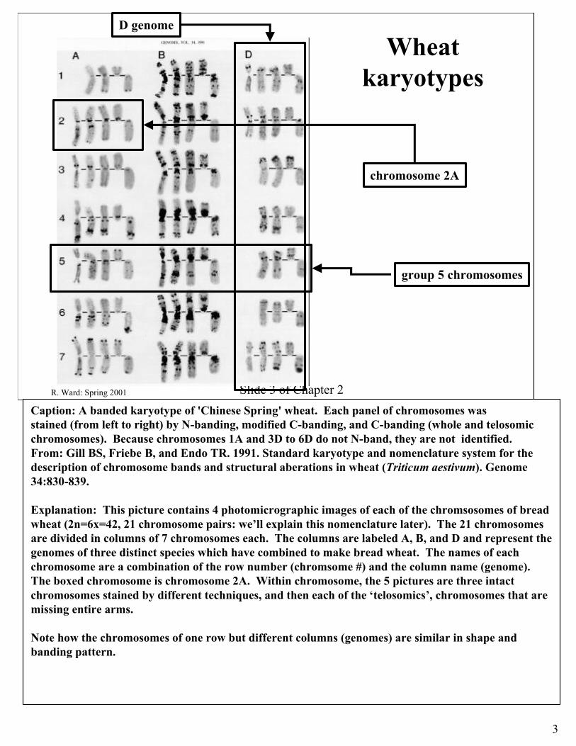
Wheat karyotypes
chromosome 2A
group 5 chromosomes
D genome
Caption: A banded karyotype of 'Chinese Spring' wheat. Each panel of chromosomes was stained (from left to right) by N-banding, modified C-banding, and C-banding (whole and telosomicchromosomes). Because chromosomes 1A and 3D to 6D do not N-band, they are not identified. From: Gill BS, Friebe B, and Endo TR. 1991. Standard karyotype and nomenclature system for the description of chromosome bands and structural aberations in wheat (Triticum aestivum). Genome 34:830-839.
Explanation: This picture contains 4 photomicrographic images of each of the chromsosomes of bread wheat (2n=6x=42, 21 chromosome pairs: we’ll explain this nomenclature later). The 21 chromosomes are divided in columns of 7 chromosomes each. The columns are labeled A, B, and D and represent the genomes of three distinct species which have combined to make bread wheat. The names of each chromosome are a combination of the row number (chromsome #) and the column name (genome). The boxed chromosome is chromosome 2A. Within chromosome, the 5 pictures are three intact chromosomes stained by different techniques, and then each of the ‘telosomics’, chromosomes that are missing entire arms.
Note how the chromosomes of one row but different columns (genomes) are similar in shape and banding pattern.

4
Slide 4 of Chapter 2R. Ward: Spring 2001
chromosome terminology I
•immediately after mitosis each chromsome has one “chromatid” which has one linear molecule of dsDNA.
•immediately before mitosis, each chomosome has two identical “sister” chromatids as a result of DNA replication.
•Sister chromatids are held together by the “centromere”

5
Slide 5 of Chapter 2R. Ward: Spring 2001
general chromosome terminology II
•a chromosome has a centromere (a dark staining body), that is either in the middle of the chromosome (metacentric) or near one end (acrocentric). The regions on either side of the centromere are called ‘arms’.
•sister chromatids are connected at the centromere.
•2n cells usually have two of each type of chromosome, these are homologous (=‘similar by descent’)
•homologous chromosomes carry the same set of loci in the same order. They are descendent from a common progenitor chromosome (possibly thousands of years ago) through mitosis and meiosis.
•Chromosomes that are not of the same type are called non-homologous

6
Slide 6 of Chapter 2R. Ward: Spring 2001
A Human male’s karyotype
•this is a purposefully arranged picture of a human male’s 2n karyotype. Note there are two of each chromosome, where different chromosomes are distinquished by where the centromere is, how long they are, how long their arms are, and the patterns of dark and light along their length.
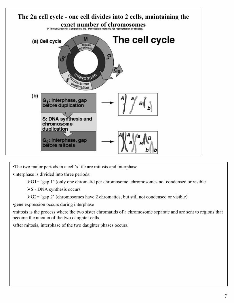
7
Slide 7 of Chapter 2R. Ward: Spring 2001
The 2n cell cycle - one cell divides into 2 cells, maintaining the exact number of chromosomes
•The two major periods in a cell’s life are mitosis and interphase
•interphase is divided into three periods:
ØG1= ‘gap 1’ (only one chromatid per chromosome, chromosomes not condensed or visible
ØS - DNA synthesis occurs
ØG2= ‘gap 2’ (chromosomes have 2 chromatids, but still not condensed or visible)
•gene expression occurs during interphase
•mitosis is the process where the two sister chromatids of a chromosome separate and are sent to regions that become the nuculei of the two daughter cells.
•after mitosis, interphase of the two daughter phases occurs.

8
Slide 8 of Chapter 2R. Ward: Spring 2001
Mitosis
•Mitosis: equational division of a set of chromosomes in one nuclei into two daughter nuclei
•Mitosis begins after the G2 phase of the cell cycle.
ØAfter G2, Chromosomes have two sister chromatids ; two centrosomes exist (organizing centers with attached microtubules) outside the nuclear envelope;
ØProphase- chromosomes condense and can be seen with light microscopy and appropriate stains. Centrosomes move apart two opposite ends (poles) of the cell.
ØPrometaphase-
Ønuclear envelope disappears;
Ønew microtubules grow from centrosomes and attach to the kinetochore (at the centromere) of one sister chromatid, pulling the chromosome (with both sister chromatids) towards that centrosome;
ØMicrotubules from the centrosome at the opposite pole attach to the kinetochore of the second sister chromatid
ØAttachment of both sister chromatid kinetochores to the same centrosome is unstable and will not persist, reducing the chance that both sister chromatids migrate to one pole
ØMetaphase-opposing tension from the opposite poles (caused by the molecular motors of the two kinetochores of a chromosome pulling in opposite directions) move the chromosomes to the metaphase plate, an imaginary midline halfway between the poles.
ØAnaphase- all centomere connections between sister chromatids severe and the kinetochores pull the individual sister chromatids (now chromosomes) towards opposite poles. Each pole recieves one copy of each chromosome.
ØTelophase- reverse of prophase- nuclear envelopes form around the centrosomes, microtubules disappear, chromosomes de-condense.
ØMitosis is followed (usually) by cytokinesis. In plants, a membrane enclosed disk called the cell plate forms inside the cell near the equator, and then grows outward until it forms a new cell wall between the two daughter cells.

9
Slide 9 of Chapter 2R. Ward: Spring 2001
Regulation of the cell cycle
•Successful cell division (mitosis and cytokinesis) requires rigorous adherence to a specific sequence of events.
•Cell accomplish this through a set of “checkpoints” where the completion one set of events is confirmed before the next set of events can occur.
•This figure illustrates this concept, but the details of this regulation is incredibly complex.

10
Slide 10 of Chapter 2R. Ward: Spring 2001
Meiosis overview
•Cells which descend from the zygote via mitosis/cytokinesis differentiate into the various parts of the plant and are called somatic cells. The number of chromosomes in these cells is represented by ‘2n’. These cells are all contain exact copies of the original genetic information that was in the zygote. Each somatic cell has two of each chromosome.
•Meiosis is the process where the number of chromosomes in a somatic cell is reduced by 1/2. This is done in a manner that ensures that each resulting nuclei (4) contains one and only one of each chromosome. These nuclei (chromosome number =n); form cells that undergo a process called gametogenosis resulting in either eggs within an ovule or pollen.

11
Slide 11 of Chapter 2R. Ward: Spring 2001
Meiosis overview-2
•Cells undergoing either mitosis or meiosis have already finished the “S” phase where chromosomes synthesize a second dsDNA molecule so that they go from a single chromatid state to a two chromatid state, where the two sister chromatids are joined at the centromere. In both mitosis and meiosis, no futher DNA replication occurs until the division process is finished.
•Both mitosis and meiosis parcel out chromatids to daughter nuclei. If 2n=4, then there are 8 chromatids prior to either type of division.
ØMitosis- each of the TWO daughter nuclei receives one chromatid from all chromosomes in the mother cell. In this example, 2n=4, and each daughter nuclei receives 1 chromatid (which are considered chromosomes once separated from their sister chromatids) from each of the 4 chromosomes, preserving the full chromosome complement.
ØMeiosis- FOUR daughter nuclei are created. In our example, the 8 original chromatids are parceled out to the 4 daughter so that each meiotic product contains 2 chromatids, one for each type of chromosome.

12
Slide 12 of Chapter 2R. Ward: Spring 2001
meiosis I
•Meiosis (figure 3.4)
•Chromosomes have already replicated and have two chromatids each
•Prophase I (you don’t need to memorize the substages of Prophase I (i.e., leptotene, zygotene, pachytene, diplotene, and diakinesis.
ØChromosomes condense and become visible under light microscope
ØHomologous chromosomes form pairs called bivalents
align longitudinally (they “synapse”, or “pair”). Each pair of homologous chromosomes is tied together by the synaptonemal complex, appear as a ‘tetrad’ of four aligned chromatids.
ØCrossing over occurs, reciprocal exchange of pieces of chromatids involving two or more chromatids.
•Metaphase I
ØSpindle appartus forms
ØBivalents (paired chromosomes held together by chiasmata- sites of crossing over) migrate to the metaphase plate as pairs.
Ømicrotubules (spindle fibers) attach to a single kinetochore for each chromosome.
•Anaphase I
ØIntact chromosomes (with two chromatids) migrate to poles
Øtwo members of a bivalent go to opposite poles
*This is segregation!
•Telophase I and interphase I
Øtwo daughter nuclei form around centrosomes of Meiosis I spindle apparatus
Øthese nuclei have half as many chromosomes as the mother cell had
•Prophase II (within each of the two daughter nuclei already formed)
•Metaphase II
Øchromosomes migrate to metaphase plate, connected to spindle apparatus as in mitosis, i.e., each sister chromatid is attached to a single pole that is the opposite to the one its sister is attached to.
•Anaphase II
ØSister chromatids separate and travel to different poles
•Telophase II
ØFour nuclei form
•Cytokinesis-> four daughter cells

13
Slide 13 of Chapter 2R. Ward: Spring 2001
Prophase I in detail

14
Slide 14 of Chapter 2R. Ward: Spring 2001
Crossing over and recombined chromosomes

15
Slide 15 of Chapter 2R. Ward: Spring 2001
meiosis II
•Telophase I and interphase I
Øtwo daughter nuclei form around centrosomes of Meiosis I spindle apparatus
Øthese nuclei have half as many chromosomes as the mother cell had
•Prophase II (within each of the two daughter nuclei already formed)
•Metaphase II
Øchromosomes migrate to metaphase plate, connected to spindle apparatus as in mitosis, i.e., each sister chromatid is attached to a single pole that is the opposite to the one its sister is attached to.
•Anaphase II
ØSister chromatids separate and travel to different poles
•Telophase II
ØFour nuclei form
•Cytokinesis-> four daughter cells

16
Slide 16 of Chapter 2R. Ward: Spring 2001
mitosis and meiosis compared (a)

17
Slide 17 of Chapter 2R. Ward: Spring 2001
mitosis and meiosis compared (b)