Embed Size (px)
Citation preview
Tetrahedron Letters 51 (2010) 2228–2231
Contents lists available at ScienceDirect
Tetrahedron Letters
journal homepage: www.elsevier .com/ locate / tet let
Real-time colorimetric screening of endopeptidase inhibitors usingadenosine triphosphate (ATP)-stabilized gold nanoparticles
Mi Hee Kim a, Soo Suk Lee b, Sang J. Chung c, Hyun Hye Jang a, Sujung Yi a, Sudeok Kim a, Suk-Kyu Chang a,Min Su Han a,*
a Department of Chemistry, Chung-Ang University, Seoul 156-756, Republic of Koreab Bio & Health Group, Emerging Center, Samsung Advanced Institute of Technology, Mt. 14-1, Nongseo-dong, Giheung-gu, Yongin-si, Gyeonggi-do 446-712, Republic of Koreac BioNanotechnology Research Center, Korea Research Institute of Bioscience and Biotechnology(KRIBB), Yuseong, Daejeon 305-333, Republic of Korea
a r t i c l e i n f o a b s t r a c t
Article history:Received 24 November 2009Revised 8 February 2010Accepted 12 February 2010Available online 20 February 2010
0040-4039/$ - see front matter Crown Copyright � 2doi:10.1016/j.tetlet.2010.02.061
* Corresponding author. Tel.: +82 2 820 5198; fax:E-mail address: [email protected] (M.S. Han).
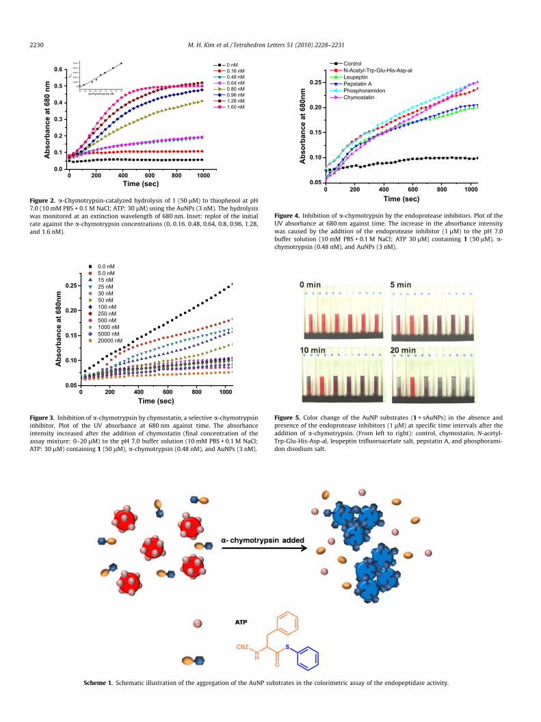
The gold nanoparticles (AuNPs) that were stabilized with adenosine triphosphate (ATP) were stable overa wide range of pHs for the buffer, even in the presence of high concentrations of salt and protein.However, these stabilized AuNPs immediately aggregated when they were exposed to thiol-containingcompounds, such as thiophenol. Endoprotease hydrolyzed the thioester bond in the CBZ–Phe–S–Ph sub-strate, and the hydrolyzed product (thiophenol) reacted with the AuNPs that were stabilized with ATP,causing them to aggregate, which in turn resulted in a visible color change in the AuNPs solution. Thismethod enabled the real-time monitoring of the inhibition potencies of various endopeptidase inhibitorsand the activity of endoprotease. This assay discriminated between the inhibition activities of variousprotease inhibitors for endoprotease on the basis of the color change of the assay solution.
Crown Copyright � 2010 Published by Elsevier Ltd. All rights reserved.
The plasmon absorption bands of gold nanoparticles (AuNPs)depend on their shape and size.1 Typically, discrete AuNPs exhibitan absorption band with a high extinction coefficient, which is 3–5orders higher than the absorption band of organic dye molecules,at around 520 nm. However, the typical absorption band corre-sponding to the aggregated AuNPs appears at longer wavelengthsbecause of the intense color (blue-purple) of the NP solution.AuNPs have widely been used for the colorimetric detection ofDNA sequences, proteins, and metal ions because of their highextinction coefficients and distance-dependent optical properties.2
Colorimetric enzyme assays using AuNPs have gained consider-able attention in bioassay studies because of their simplicity, highsensitivity, and low cost. However, few colorimetric systems havebeen developed for the evaluation of the enzymatic activities.3 A pre-vious study showed that the colorimetric bioassays can be dividedinto two types corresponding to modified and unmodified AuNPs.In the first method, the aggregates of the AuNPs that are modifiedusing the enzyme substrates are used to monitor the enzymaticactivity. The AuNP substrate bond can be cleaved using a suitable en-zyme in order to disperse the AuNPs in the solution.3a–c This processbrings about a change in the color of the AuNPs solution, and there-fore, the real-time monitoring of the enzymatic activities is possible.In the second method, the enzyme substrate and the unmodifiedAuNPs are used to monitor the enzymatic activities.3d–f The sub-strate is pretreated with the target enzyme and then exposed to
010 Published by Elsevier Ltd. All r
+82 2 825 4736.
the unmodified AuNPs. The activity of the target enzyme is moni-tored on the basis of the color change of the AuNPs that results fromthe two-stage process. Although the colorimetric assays that useunmodified AuNPs are simpler than the assays that use modifiedAuNPs, they cannot be used for the real-time monitoring of the activ-ity of the target enzyme because the AuNPs are highly sensitive tovarious factors such as the electrolyte concentration, the pH of thebuffer, and the protein concentration.1,2a Hence, a change in any ofthe above-mentioned parameters results in the irreversible aggrega-tion of the unmodified AuNPs and a significant red shift in the absor-bance spectrum. Therefore, the unmodified AuNPs are not easilyused for the real-time monitoring of the activity of the targetenzyme.
Recently, Zhao et al. reported that a real-time enzyme assaymethod using the ATP-stabilized AuNPs could be prepared by sim-ply mixing the AuNPs with adenosine triphosphate. The stabilizedAuNPs are stable in the buffer solution in the presence of high con-centrations of salt and protein.4 However, the stabilized AuNPsaggregate immediately when they are exposed to the enzyme thathydrolyzes the phosphate bond in ATP because of the change in thesolubility of the stabilized AuNPs in buffer solution at higher saltconcentrations. With this information on the properties of theATP-stabilized AuNPs, an operationally simple colorimetric assaywas examined for the simultaneous real-time monitoring of theendopeptidase activity and the estimation of the potency of theendopeptidase inhibitors. The ATP-stabilized AuNPs (henceforthreferred to as the ‘stabilized AuNPs (sAuNPs)’) and an endoproteasesubstrate (1) were used in this assay. Endoprotease hydrolyzed the
ights reserved.
M. H. Kim et al. / Tetrahedron Letters 51 (2010) 2228–2231 2229
thioester bond in the CBZ–Phe–S–Ph substrate (1).5 The hydrolyzedproduct (thiophenol) reacted with the sAuNPs, causing them toaggregate because of the change in the solubility of the sAuNPs,which in turn resulted in a visible color change in the AuNP solu-tion (Scheme 1). The color change was used to screen the endopep-tidase inhibitors and the endoprotease activity.
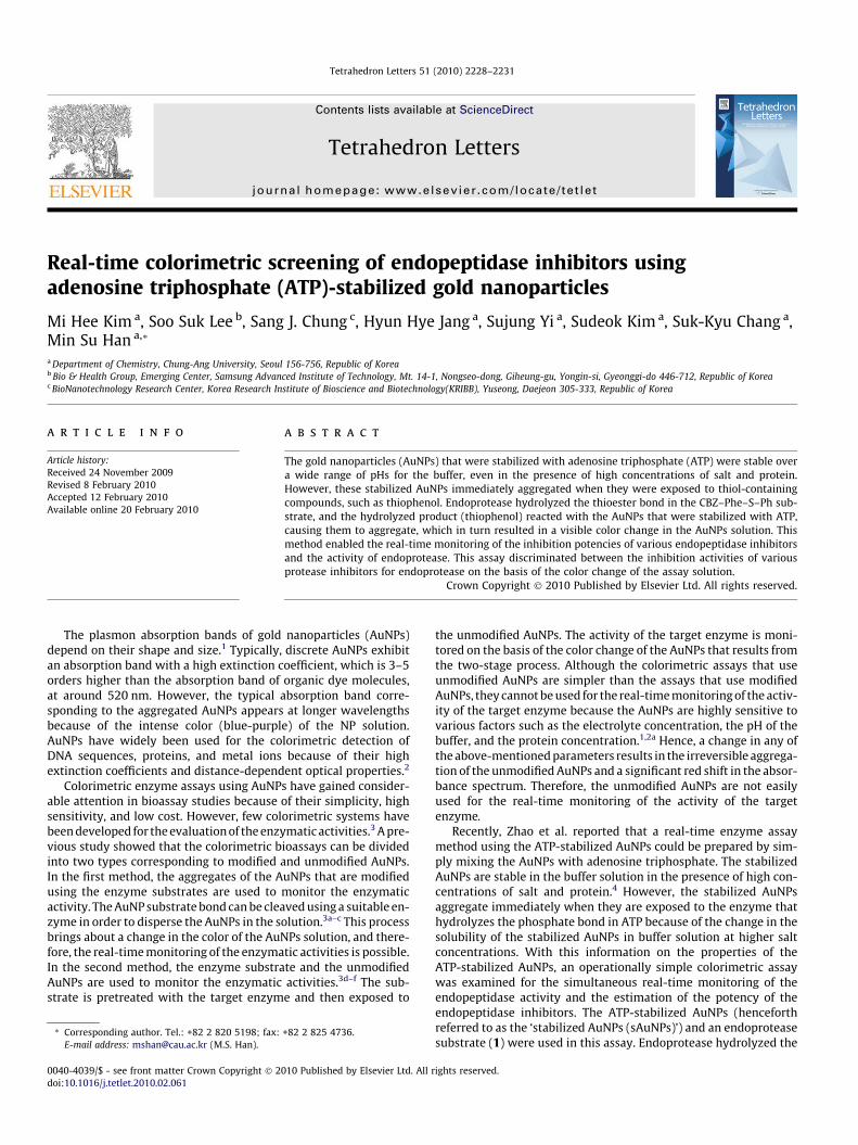
The stability of sAuNPs to 1 was dependent on the concentra-tion of ATP and without a-chymotrypsin, the thioester reactedwith the sAuNPs, causing them to aggregate because the gold sur-face catalyzed the thioester hydrolysis and then the hydrolysisproduct can attach to AuNPs.3f The stability of the sAuNPs to 1was evaluated at various ATP concentrations in order to avoidthe color change that was induced by 1 in the absence of a-chymo-trypsin.3f When AuNPs were stabilized at ATP concentrations thatwere lower than 10 lM in the presence with 1, the surface plas-mon resonance (SPR) band of the sAuNPs changed in 30 min. How-ever, for the AuNPs that were stabilized with ATP concentrations ofmore than 20 lM, the SPR band did not change after 30 min(Fig. 1A). Therefore, the AuNP substrates were prepared for theendoprotease assay by combining 1 (hydrolyzable substrate) withthe sAuNPs that were stabilized by mixing ATP (30 lM) with theAuNPs (diameter: 13 nm).4 The dispersion of the sAuNPs was con-firmed using transmission electron microscopy (TEM; Fig. 1B) andthe appearance of the SPR band at 520 nm in the UV–vis spectrum.However, the sAuNPs aggregated upon the addition of a-chymo-trypsin to the solution (Fig. 1C), and the SPR band red shifted from520 to 680 nm (see Supplementary data). Therefore, the thioesterwas hydrolyzed by the endopeptidase to produce the correspond-ing thiol, which then reacted with the sAuNPs.
This reaction caused the aggregation of the sAuNPs, causing thecolor change to purple and the red shift in the SPR band. This redshift is a well-known phenomenon and is used to confirm the for-mation of the nanoparticle aggregates.1,2 The color change in theAuNPs was detected by the naked eye, and the extinction coeffi-cients were measured from the UV–vis spectrum at 680 nm.
0 200 400 600 800 100.0
0.1
0.2
0.3
0.4
0.5
0.6a)
Abso
rban
ce a
t 680
nm
Time (s
Figure 1. (a) Stabilities of the sAuNPs to 1 in the presence of various concentrations of AT(c) TEM image of the mixture after the addition of a-chymotrypsin. The scale bar repre
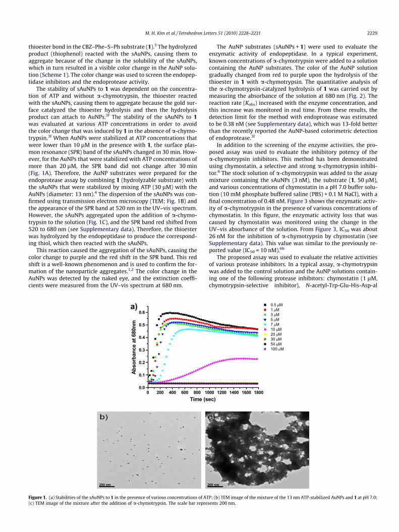
The AuNP substrates (sAuNPs + 1) were used to evaluate theenzymatic activity of endopeptidase. In a typical experiment,known concentrations of a-chymotrypsin were added to a solutioncontaining the AuNP substrates. The color of the AuNP solutiongradually changed from red to purple upon the hydrolysis of thethioester in 1 with a-chymotrypsin. The quantitative analysis ofthe a-chymotrypsin-catalyzed hydrolysis of 1 was carried out bymeasuring the absorbance of the solution at 680 nm (Fig. 2). Thereaction rate (Kobs) increased with the enzyme concentration, andthis increase was monitored in real time. From these results, thedetection limit for the method with endoprotease was estimatedto be 0.38 nM (see Supplementary data), which was 13-fold betterthan the recently reported the AuNP-based colorimetric detectionof endoprotease.3f
In addition to the screening of the enzyme activities, the pro-posed assay was used to evaluate the inhibitory potency of thea-chymotrypsin inhibitors. This method has been demonstratedusing chymostatin, a selective and strong a-chymotrypsin inhibi-tor.6 The stock solution of a-chymotrypsin was added to the assaymixture containing the sAuNPs (3 nM), the substrate (1, 50 lM),and various concentrations of chymostatin in a pH 7.0 buffer solu-tion (10 mM phosphate buffered saline (PBS) + 0.1 M NaCl), with afinal concentration of 0.48 nM. Figure 3 shows the enzymatic activ-ity of a-chymotrypsin in the presence of various concentrations ofchymostatin. In this figure, the enzymatic activity loss that wascaused by chymostatin was monitored using the change in theUV–vis absorbance of the solution. From Figure 3, IC50 was about26 nM for the inhibition of a-chymotrypsin by chymostatin (seeSupplementary data). This value was similar to the previously re-ported value (IC50 = 10 nM).6b
The proposed assay was used to evaluate the relative activitiesof various protease inhibitors. In a typical assay, a-chymotrypsinwas added to the control solution and the AuNP solutions contain-ing one of the following protease inhibitors: chymostatin (1 lM,chymotrypsin-selective inhibitor), N-acetyl-Trp-Glu-His-Asp-al
00 1200 1400 1600 1800ec)
0.5 µM 1 µM 3 µM 5 µM 7 µM 10 µM 20 µM 30 µM 50 µM 100 µM
P; (b) TEM image of the mixture of the 13 nm ATP-stabilized AuNPs and 1 at pH 7.0;sents 200 nm.
0 200 400 600 800 10000.05
0.10
0.15
0.20
0.25
Abs
orba
nce
at 6
80nm
Time (sec)
0.0 nM 5.0 nM 15 nM 25 nM 30 nM 50 nM 100 nM 250 nM 500 nM 1000 nM 5000 nM 20000 nM
Figure 3. Inhibition of a-chymotrypsin by chymostatin, a selective a-chymotrypsininhibitor. Plot of the UV absorbance at 680 nm against time. The absorbanceintensity increased after the addition of chymostatin (final concentration of theassay mixture: 0–20 lM) to the pH 7.0 buffer solution (10 mM PBS + 0.1 M NaCl;ATP: 30 lM) containing 1 (50 lM), a-chymotrypsin (0.48 nM), and AuNPs (3 nM).
0 200 400 600 800 10000.05
0.10
0.15
0.20
0.25
Abs
orba
nce
at 6
80nm
Time (sec)
Control N-Acetyl-Trp-Glu-His-Asp-al Leupeptin Pepstatin A Phosphoramidon Chymostatin
Figure 4. Inhibition of a-chymotrypsin by the endoprotease inhibitors. Plot of theUV absorbance at 680 nm against time. The increase in the absorbance intensitywas caused by the addition of the endoprotease inhibitor (1 lM) to the pH 7.0buffer solution (10 mM PBS + 0.1 M NaCl; ATP 30 lM) containing 1 (50 lM), a-chymotrypsin (0.48 nM), and AuNPs (3 nM).
Figure 5. Color change of the AuNP substrates (1 + sAuNPs) in the absence andpresence of the endoprotease inhibitors (1 lM) at specific time intervals after theaddition of a-chymotrypsin. (From left to right): control, chymostatin, N-acetyl-Trp-Glu-His-Asp-al, leupeptin trifluoroacetate salt, pepstatin A, and phosphorami-don disodium salt.
Scheme 1. Schematic illustration of the aggregation of the AuNP substrates in the colorimetric assay of the endopeptidase activity.
0 200 400 600 800 10000.0
0.1
0.2
0.3
0.4
0.5
0.6
Abs
orba
nce
at 6
80 n
m
Time (sec)
0 nM 0.16 nM 0.48 nM 0.64 nM 0.80 nM 0.96 nM 1.28 nM 1.60 nM
Figure 2. a-Chymotrypsin-catalyzed hydrolysis of 1 (50 lM) to thiophenol at pH7.0 (10 mM PBS + 0.1 M NaCl; ATP: 30 lM) using the AuNPs (3 nM). The hydrolysiswas monitored at an extinction wavelength of 680 nm. Inset: replot of the initialrate against the a-chymotrypsin concentrations (0, 0.16, 0.48, 0.64, 0.8, 0.96, 1.28,and 1.6 nM).
2230 M. H. Kim et al. / Tetrahedron Letters 51 (2010) 2228–2231
M. H. Kim et al. / Tetrahedron Letters 51 (2010) 2228–2231 2231
(1 lM, cysteine protease inhibitor),7 leupeptin trifluoroacetate salt(1 lM, serine protease inhibitor),8 pepstatin A (1 lM, aspartic pro-tease inhibitor),9 and phosphoramidon disodium salt (1 lM, metal-loprotease inhibitor).10 The inhibitor activity was monitored at680 nm as a function of time (sample scan rate: 30 s�1). The slopeof the plot (rate) was used to measure the inhibition. Figure 4shows that only chymostatin effectively inhibited a-chymotrypsin.
High throughput screening (HTS) is widely used to identify thehit compounds from combinatorial libraries.11 The proposed assaywas examined for the HTS of the a-chymotrypsin inhibitors. Thecatalytic hydrolysis of the substrate (1) in the assay solution witha-chymotrypsin brought about a color change from red to purplefor the solution. Therefore, if a-chymotrypsin was successfullyinhibited, the color of the solution remained unchanged. In Figure5, the inhibition activities of various protease inhibitors for a-chy-motrypsin were differentiated on the basis of the color change ofthe assay solution.
In conclusion, a new colorimetric assay was developed forscreening endoprotease activities and determining the relativeinhibitory potencies of the endoprotease inhibitors by monitoringthe kinetics of the sAuNP aggregation. This screening methodwas simpler than the other assays based on the AuNPs and wasused for the easy real-time monitoring of the inhibition potenciesof various endopeptidase inhibitors. Furthermore, this assay wasused for the qualitative and quantitative estimation of the inhibi-tion. This assay can also be adapted to the HTS of potential drugcandidates from large combinatorial libraries and important bio-logical endoproteases by simply changing the thiol-bearingsubstrate.
Acknowledgments
This work was supported by the Korea Research FoundationGrant funded by the Korean Government (MOEHRD, Basic ResearchPromotion Fund) (KRF-2008-331-C00171), the Priority ResearchCenters Program through the National Research Foundation ofKorea(NRF) funded by the Ministry of Education, Science and Tech-nology (2009-0093817), and the Korea Foundation for Interna-tional Cooperation of Science & Technology(KICOS) through a
grant that was provided by the Korean Ministry of Education Sci-ence & Technology(MEST) in 2009 (2008-00656).
Supplementary data
Supplementary data (the experimental details for the colori-metric screening assay, the synthesis of CBZ–Phe–S–Ph (1), theUV–vis spectrum of the SAuNPs versus time in the presence of a-chymotrypsin, the detection limit of the assay method for a-chy-motrypsin, and the IC50 value of chymostatin for the a-chymotryp-sin EDX data of the SAuNPs) associated with this article can befound, in the online version, at doi:10.1016/j.tetlet.2010.02.061.
References and notes
1. (a) Stewart, M. E.; Anderton, C. R.; Thompson, L. B.; Maria, J.; Gray, S. K.; Rogers,J. A.; Nuzzo, R. G. Chem. Rev. 2008, 108, 494–521; (b) Ghosh, S. K.; Pal, T. Chem.Rev. 2007, 107, 4797–4862; (c) Burda, C.; Chen, X.; Narayanan, R.; El-Sayed, M.A. Chem. Rev. 2005, 105, 1025–1102.
2. (a) Rosi, N. L.; Mirkin, C. A. Chem. Rev. 2005, 105, 1547–1562; (b) Lu, Y.; Liu, J.Acc. Chem. Res. 2007, 40, 315–323;.
3. (a) Ghadiali, J. E.; Stevens, M. M. Adv. Mater. 2008, 20, 4359–4363; (b) Xu, X.;Han, M. S.; Mirkin, C. A. Angew. Chem., Int. Ed. 2007, 46, 3468–3470; (c)Laromaine, A.; Koh, L.; Murugesan, M.; Ulijn, R. V.; Stevens, M. M. J. Am. Chem.Soc. 2007, 129, 4156–4157; (d) Choi, Y.; Ho, N. H.; Tung, C. H. Angew. Chem., Int.Ed. 2007, 46, 707–709; (e) Liu, R.; Liew, R.; Zhou, J.; Xing, B. Angew. Chem., Int.Ed. 2007, 46, 8799–8803; (f) Guarise, C.; Pasquato, L.; De Fillippis, V.; Scrimin, P.Proc. Natl. Acad. Sci. U.S.A. 2006, 103, 3978–3982.
4. (a) Zhao, W.; Chiuman, W.; Lam, J. C. F.; Brook, M. A.; Li, Y. Chem. Commun.2007, 3729–3731; (b) Zhao, W.; Brook, M. A.; Li, Y. ChemBioChem 2008, 9,2363–2371.
5. Han, M. S.; Jung, S. O.; Kim, M. J.; Kim, D. H. J. Org. Chem. 2004, 69, 2853–2855.6. (a) Umezawa, H.; Aoyagi, T.; Morishima, H.; Kunimoto, S.; Matsuzaki, M.;
Hamada, M.; Takeuchi, T. J. Antibiot. 1970, 8, 425–427; (b) Özgür, E.; Yücel, M.;Öktem, H. A. Turk. J. Agric. Forum. 2009, 33, 285–294.
7. Garcia-Calvo, M.; Peterson, E. P.; Leiting, B.; Ruel, R.; Nicholson, D. W.;Thornberry, N. A. J. Biol. Chem. 1998, 273, 32608–32613.
8. Kuramochi, H.; Nakata, H.; Ishij, S. J. Biochem. 1979, 86, 1403–1410.9. Rich, D. H.; Bernatowicz, M. S.; Agarwal, N. S.; Kawai, M.; Salituro, F. G.;
Schmidt, P. G. Biochemistry 1985, 24, 3165–3173.10. Kam, C. M.; Nishino, N.; Powers, J. C. Biochemistry 1979, 18, 3032–3038.11. (a) Wang, S.; Sim, T. B.; Kim, Y. S.; Chang, Y. T. Curr. Opin. Chem. Biol. 2004, 8,
371–377; (b) Boger, D. L.; Desharnais, J.; Capps, K. Angew. Chem., Int. Ed. 2003,42, 4138–4176; (c) Johnston, P. A.; Johnston, P. A. Drug Discovery Today 2002, 7,353–363.