Embed Size (px)
Citation preview

REBIN TITUS
Clinical Case Conference

Presenting history
44-year-old, Hispanic male with a history of ESRD, most likely secondary to his diabetes and hypertension, on hemodialysis since March 2008
He also has coronary artery disease status-post stent placement in 2008
Admitted for a scheduled transplant from a living, healthy donor
No complaints on presentation

Past Medical History
ESRD, possibly secondary to diabetes and hypertension.
Diabetic neuropathy. Left arm shunt in November of 2008. Cholecystectomy in 2008. Coronary artery disease status-post
stenting in the left anterior descending with single vessel disease in 2008.
Kidney stones. Erectile dysfunction.

History (contd.)
Social History: The patient does not drink or smoke. He is unemployed. He lives in Lubbock.
Family History: Positive for hypertension in the mother and father, diabetes in the father, and colon cancer in a grandfather. No h/o ESRD

Medications
Aspirin, Atenolol. Sertraline. PhosLo. Calcitriol. Enalapril. Neurontin. Lantus 50 units at night. Allergies: None

PE
Vitals: T 97.8: P 66: R 18 :BP 144/76: Pox: 96% on RA
Gen appearance: Comfortable, in no distress
HEENT: PERRLA, normal conjunctivae, moist MM
Neck: Supple, no lymphadenopathyLungs: CTABCVS: S1, S2, RRR, no M/R/GAbd: Soft, BS +, NT/NDExt: No C/C/E; pulses positive
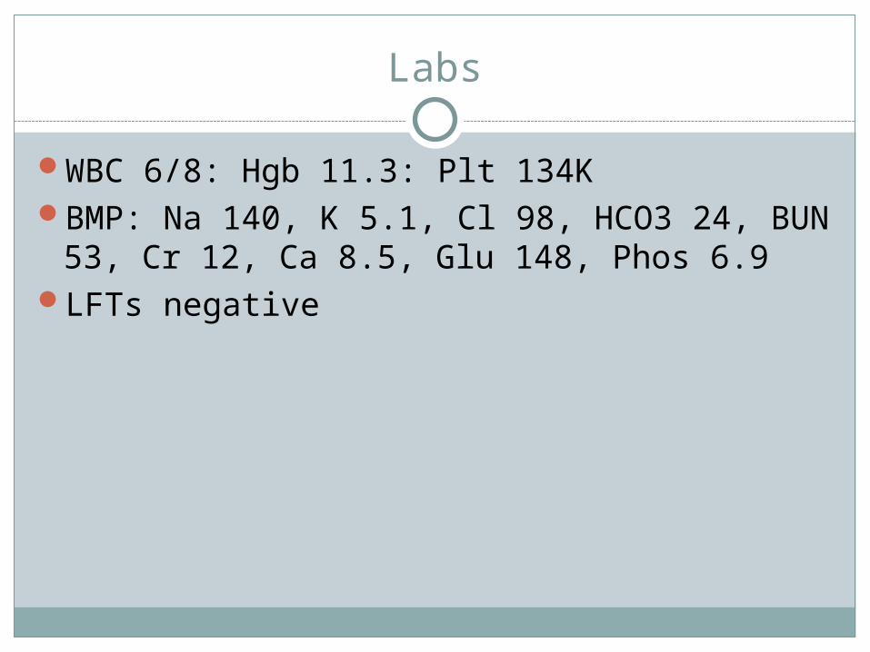
Labs
WBC 6/8: Hgb 11.3: Plt 134KBMP: Na 140, K 5.1, Cl 98, HCO3 24, BUN
53, Cr 12, Ca 8.5, Glu 148, Phos 6.9LFTs negative

Day of Surgery
Was dialysed that night (as per his usual schedule)
Was taken for surgery in the am. Living donor kidney transplanted to the rt
iliac fossa with EBL approx 350 cc and no intraoperative complication

POD # 1, 2
Doing well, good urine output, mild pain at site
Started on sirolimus/mycophenelate/prednisone
Creatinine down to 7.7, then 5.9 the next day

POD # 3
Continues to do wellProducing >3 L of urineCr slightly up to 6.2, BUN 51 Ordered renal ultrasound which showed good
flow with no hydronephrosis, but with elevated resistive indices (1.0) and velocities at the level of the lobar arteries (peak syst vel 47 cm/sec)
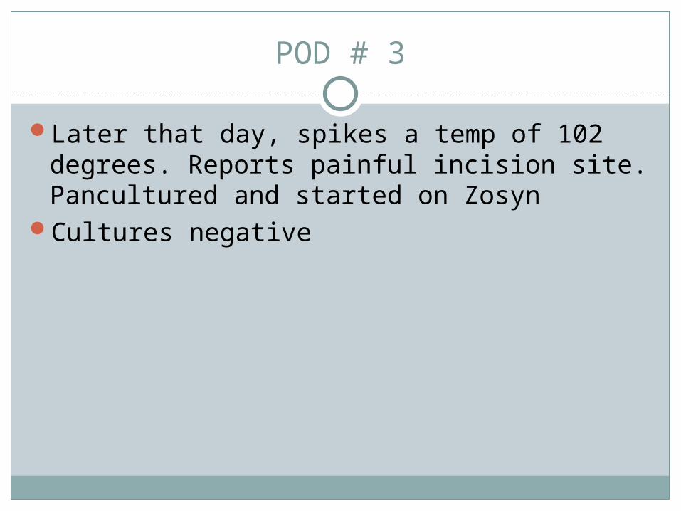
POD # 3
Later that day, spikes a temp of 102 degrees. Reports painful incision site. Pancultured and started on Zosyn
Cultures negative
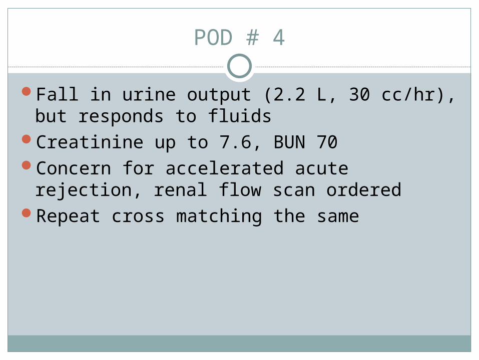
POD # 4
Fall in urine output (2.2 L, 30 cc/hr), but responds to fluids
Creatinine up to 7.6, BUN 70Concern for accelerated acute rejection,
renal flow scan orderedRepeat cross matching the same

POD # 4
Was started on thymoglobulinAcutely developed flash pulmonary edemaThymoglobulin rate cut by half and 100 mg IV
lasix given, but did not respondHe was emergently dialysed, and 5L fluid was
ultrafiltered

POD # 5
Status improved, still on venti maskCreatinine down to 5, BUN 40Urine output not improving, still at about
40cc/hrRenal scan with good flow and questionable
function

POD # 6
Required ultrafiltration again, still on 8L O2Urine output still about 30-100 cc/hour
(1260cc/24 hrs)Continued Rx with thymoglobulin, along with
mycophenalate/methylprednisone (sirolimus held)
Creatinine up to 5.7

POD # 7
Urine output further decreasingRespiratory status now improvedIs now clinically depressedRepeat renal scans and color dopplers
unyieldingDecision made to biopsy

POD # 8
Biopsy unsuccessful, patient refused further attempts
Now oliguric, creatinine 7.5, BUN 96Continued immune suppression with
thymoglobulin

POD # 9-20
Pt’s urine ouput slowly starts to increaseCreatinine continues to rise with peak at
12.2, BUN 88 (though rate of increase less precipitous)
Supportive measures continued, had to be dialyzed one more time secondary to fluid (POD #11)
Immune suppression continues for a total of 11 doses of thymoglobulin along with MMF and prednisone. Tacrolimus started toward the end

POD # 9-20
Renal scan continues to show good flow, but abnormal function
Ultrasound with elevated resistive indices (0.95-1.0), but with normal velocities

POD # 9-20
Provigil and ambien started for his depression
Was transfused 2 units of PRBC’s, amongst other supportive measures
T-cell subsets show adequate immune suppression
Urine output continues to improve, in spite of rising creatinine
No further need for dialysisDischarged on POD # 20

Renal Transplant
The most common complication of renal transplantation is allograft dysfunction.
The overall one year unadjusted survival of a renal allograft is approximately 92 percent for a non-extended criteria deceased donor kidney and approximately 96 percent for a living donor kidney (there is wide inter-center variability)

Risk factors for lower survival
Second or third transplantPrior sensitization with more than 50
percent panel reactivityDelayed graft functionNumber and severity of rejection episodesDonor age less than five or greater than 60
yearsGreater degrees of HLA mismatchingAllograft dysfunction at discharge (plasma
creatinine above 2 mg/dL)

Delayed graft function (DGF)
Any newly transplanted kidney that does not function well
Mostly oliguric and requiring dialysis during the first week of transplantation
Need for accurate and timely recognition and to differentiate from infection and drug toxicity
Rejection can be mimicked by various infections such as BK virus-induced nephropathy

Differential diagnosis of DGF
Acute tubular necrosis (ATN) Intravascular volume contractionHyperacute and acute antibody mediated
rejection (AMR) Accelerated rejectionUrinary tract obstruction due to ureteral necrosis
or hematomaUrine leakThrombosis of the renal artery or vein NephrotoxicityThrombotic microangiopathy (TMA)
ATN
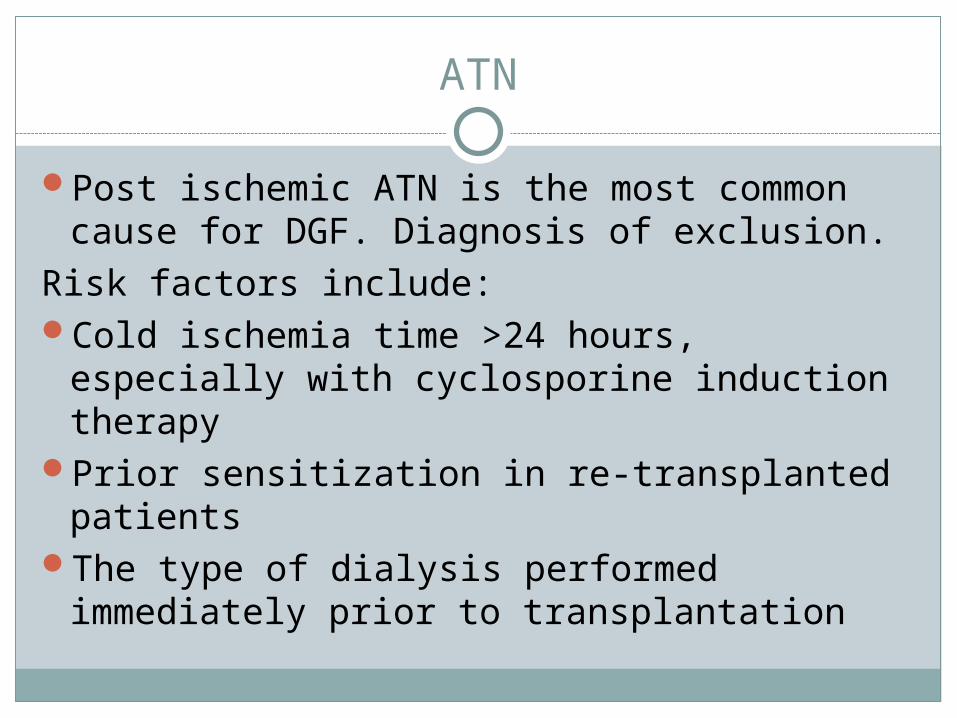
Post ischemic ATN is the most common cause for DGF. Diagnosis of exclusion.
Risk factors include:Cold ischemia time >24 hours, especially
with cyclosporine induction therapyPrior sensitization in re-transplanted patients The type of dialysis performed immediately
prior to transplantation
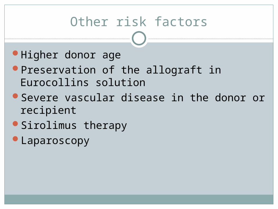
Other risk factors
Higher donor age Preservation of the allograft in Eurocollins
solutionSevere vascular disease in the donor or
recipient Sirolimus therapyLaparoscopy

Pathophysiology of posttransplant ATN
In the short term, it is relatively benign, and resolves spontaneously
Thought to be post ischemic, ischemia-reperfusion injury with increased concentration of oxygen free radicals
Oliguria caused by decreased GFR, tubular obstruction with cellular debris and increased interstitial pressure
May impact long term graft prognosisUsually requires only supportive therapy

Early Transplant Rejection
Accelerated acute rejectionEarly cell-mediated rejectionAntibody-mediated rejection

Accelerated acute rejection
Can occur immediately posttransplant-hyperacute rejection; or it may be delayed several days
Caused by preformed donor specific antibodies, such as ABO isoagglutinins, anti-endothelial antibodies and anti-HLA antibodies
Diagnosis mostly made in the OR and frequently results in allograft loss within the first 24 hours.
Prompt surgical exploration and intra-op biopsy if needed to determine viability

Accelerated acute rejection
Usually oliguric or anuricFever, graft tendernessRenal scan with little or no uptake

Early cell-mediated rejection
Latter part of the first transplant week or typically somewhat later
Differentiated from accelerated by renal scan which shows decreased but persistent flow
Diagnosis by biopsy
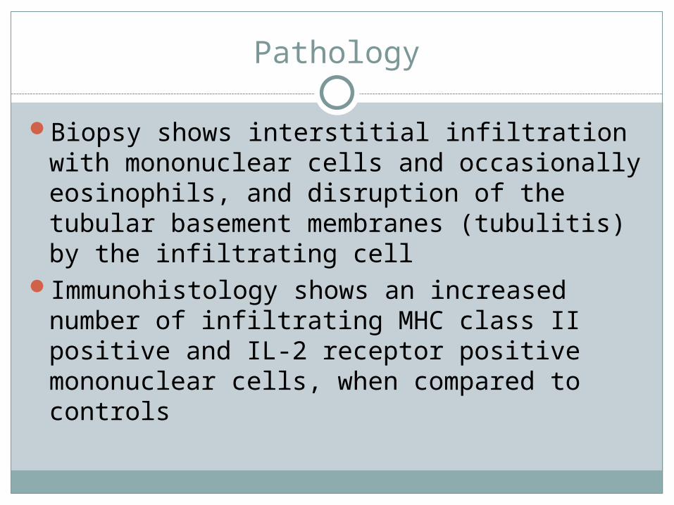
Pathology
Biopsy shows interstitial infiltration with mononuclear cells and occasionally eosinophils, and disruption of the tubular basement membranes (tubulitis) by the infiltrating cell
Immunohistology shows an increased number of infiltrating MHC class II positive and IL-2 receptor positive mononuclear cells, when compared to controls

Histology
Acute cellular rejection in a renal transplant showing diffuse interstitial infiltrate of mononuclear cells, some of which are actively invading the tubules
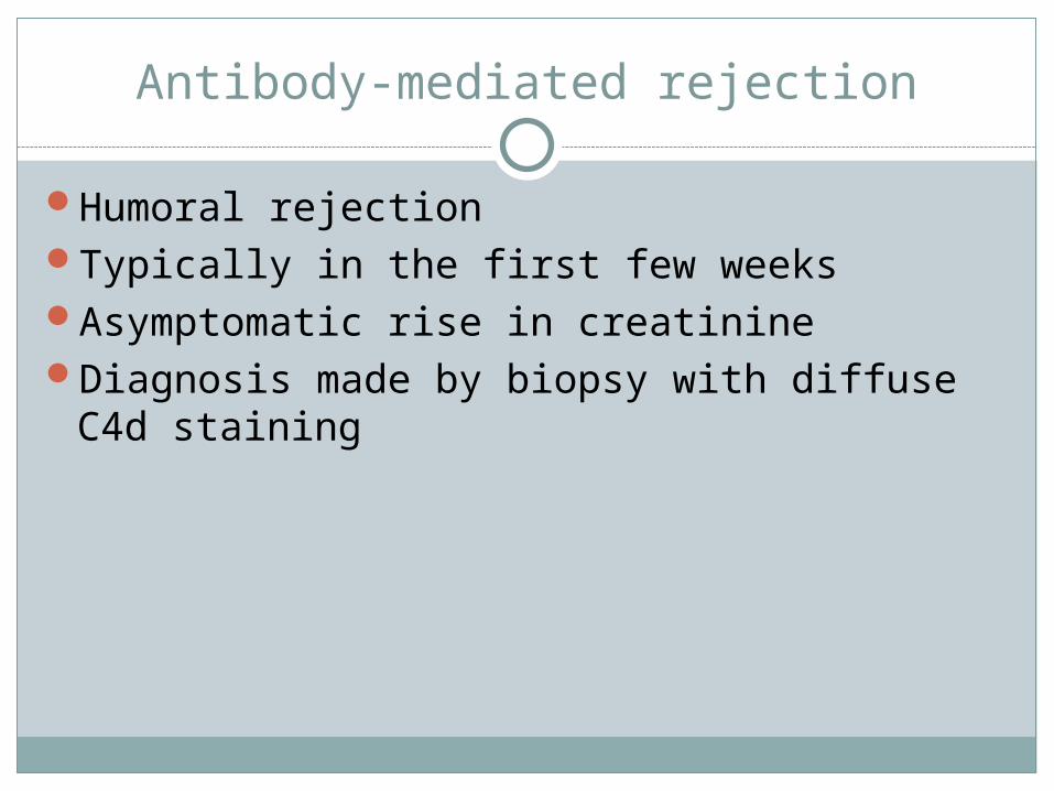
Antibody-mediated rejection
Humoral rejectionTypically in the first few weeksAsymptomatic rise in creatinineDiagnosis made by biopsy with diffuse C4d
staining

Pathology
Biopsy shows capillary endothelial swelling, arteriolar fibrinoid necrosis, fibrin thrombi in glomerular capillaries, and frank cortical necrosis in severe cases.
Differentiated from acute cellular rejection by C4d staining

Renal transplant biopsy with antibody mediated rejection: Immunofluorescence with C4d monoclonal antibody showing diffuse peritubular capillary deposition
Histology

Categories for the Banff classification system
Category 1: Normal — A histologically normal biopsy. Cateogry 2: Antibody-mediated changes —It is due to
documentation of circulating antidonor antibody, and C4d or allograft pathology.
C4d deposition without morphologic evidence of acute rejection
Acute antibody-mediated rejection Histologic type (grade) include the following: Type I - An acute tubular necrosis-like histology (C4d
positive), with minimal inflammation Type II - A capillary-glomerulitis, with margination and/or
thromboses (C4d positive) Type III - Arterial-transmural inflammation/fibrinoid
changes (C4d positive) Chronic active antibody-mediated rejection
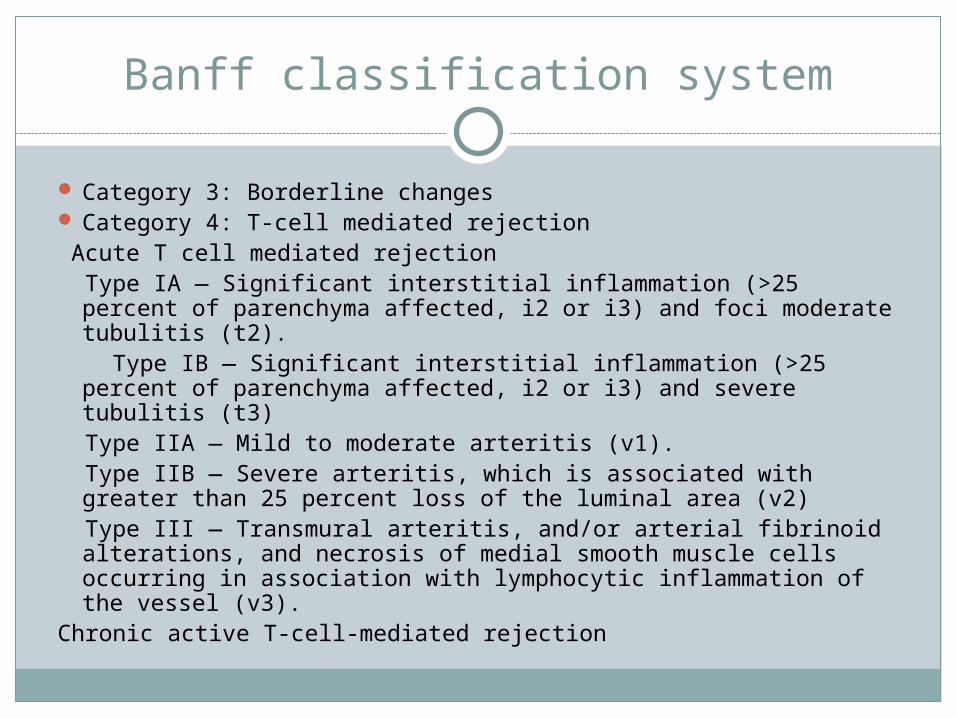
Banff classification system
Category 3: Borderline changes Category 4: T-cell mediated rejection Acute T cell mediated rejection Type IA — Significant interstitial inflammation (>25 percent of
parenchyma affected, i2 or i3) and foci moderate tubulitis (t2).
Type IB — Significant interstitial inflammation (>25 percent of parenchyma affected, i2 or i3) and severe tubulitis (t3)
Type IIA — Mild to moderate arteritis (v1). Type IIB — Severe arteritis, which is associated with greater
than 25 percent loss of the luminal area (v2) Type III — Transmural arteritis, and/or arterial fibrinoid
alterations, and necrosis of medial smooth muscle cells occurring in association with lymphocytic inflammation of the vessel (v3).
Chronic active T-cell-mediated rejection

Banff classification system
Category 5: Interstitial fibrosis and tubular atrophy, without evidence of any specific etiology — a.k.a chronic allograft nephropathy.
Grade I. Mild interstitial fibrosis and atrophy of tubules
(<25 percent of cortical area) II. Moderate interstitial fibrosis and atrophy of tubules (25 to 50 percent of cortical area) III. Severe interstitial fibrosis and atrophy of tubules (>50 percent of cortical area)
Category 6: Other
Treatment of renal allograft rejection
Once acute rejection is confirmed, the possibility of inadequate immunosuppression must be addressed. Factors that can influence:
Aggressive weaningInadequate dosing, especially of
mycophenolate mofetilFailure to recognize drugs that promote
cytochrome P450 metabolism and decrease levels of drugs like tacrolimus, sirolimus, and cyclosporine

Options for treatment
Pulse steroidsAntibodies (monoclonal or polyclonal)Manipulation of baseline immunosuppressionOther therapies

Pulse steroids
Pulse methylprednisolone, 3 to 5 mg/kg, given intravenously for three to five days
Can be used as intensification of maintenance immunosuppression therapy
Usually the only additional treatment added if the rejection is Banff class 1A or 1B
Complications: Increased susceptibility to infection, especially oral candidiasis. Other issues include hyperglycemia, hypertension, peptic ulcers, and psychiatric disturbances including euphoria and depression.

Antibodies
Polyclonal anti T-cell antibodies are prepared by immunizing rabbits or horses with human lymphoid cells derived from the thymus (called antithymocyte globulin [ATG]) or cultured B cell lines.
ATG can be used both for prophylaxis against and for the primary treatment of acute rejection.
The reversal rate has been between 75 and 100 percent in different series, with the plasma creatinine concentration returning to baseline several days to a week after initiating therapy.
In a small study, compared with steroids, antibodies more effectively reversed a first acute rejection (RR of 0.57, 95% CI 0.38-0.87) and prevented allograft loss (RR of 0.74, 95% CI 0.58-0.95

Side effects of ATG
Fever and chills during the initial ATG infusionAnaphylactic reactions, including respiratory distress
and hypotensionPre-ATG administration of the concurrently given
pulse steroids can significantly reduce infusion-related reactions.
Pruritic skin rash (20 percent) Presumed antiplatelet antibody-induced
thrombocytopenia of varying severity (50 percent)CMV and herpes infections Post-transplant lymphoproliferative disease ATG does not induce a host antibody response to the
rabbit or horse serum (like OKT3).

OKT 3
OKT3 was the first mouse antibody licensed for use in humans
Directed against the CD3 antigen that is closely associated with the T cell receptor
Inhibition of cell-mediated immunity via modulation/clearing of CD3+ T cells.
OKT3 has been used as the primary treatment of acute rejection and as rescue therapy for resistant rejection in kidney transplantation

Side effects
Infection Lymphoproliferative disease associated with
Epstein Barr virusFirst-dose reactionPulmonary edemaHemolytic-uremic syndrome

Other therapies
Alemtuzumab (Campath-1H) is a humanized anti-CD52 panlymphocytic (both B and T cells) monoclonal antibody that is approved for the treatment of chronic lymphocytic leukemia. Has been reported in a few limited studies, to be effective in the treatment of acute rejection
IL-2 receptor blockers like basiliximab or daclizumab are indicated for induction in renal transplantation, there are no studies reporting their use in the treatment of acute rejection.
Additional rescue therapy for refractory rejection can be the addition of tacrolimus of MMF to antibody therapy
Antibody-mediated rejection (AMR)
Preliminary data show that patients with AMR may respond to treatment with plasmapheresis, tacrolimus, and MMF, or plasmapheresis plus IVIg, and/or immunoadsorption

Follow up
Continues to improve. Creatinine finally started to downtrend, now
is 7.2Urine output improvedNo need for further dialysis

Thank you for your attention


![Comprehensive ESRD Care Initiative LDO Model€¦ · Comprehensive ESRD Care Initiative LDO Model . July [15], 2015 . ... Comprehensive ESRD Care Initiative Participation Agreement](https://img.pdfslide.net/doc/110x75/5af2cc657f8b9a95468ba91b/comprehensive-esrd-care-initiative-ldo-model-comprehensive-esrd-care-initiative.jpg)