Embed Size (px)
Citation preview

Recent developments in the mechanisticenzymology of the ATP-dependent Lonprotease from Escherichia coli: highlightsfrom kinetic studiesIrene Lee,a Anthony J. Berdisb and Carolyn K. Suzukic
DOI: 10.1039/b609936j
Lon protease, also known as protease La, is one of the simplest ATP-dependentproteases that plays vital roles in maintaining cellular functions by selectivelyeliminating misfolded, damaged and certain short-lived regulatory proteins.Although Lon is a homo-oligomer, each subunit of Lon contains both an ATPaseand a protease active site. This relatively simple architecture compared to otherhetero-oligomeric ATP-dependent proteases such as the proteasome makes Lon auseful paradigm for studying the mechanism of ATP-dependent proteolysis. In thisarticle, we survey some recent developments in the mechanistic characterizationof Lon with an emphasis on the utilization of pre-steady-state enzyme kinetictechniques to determine the timing of the ATPase and peptidase activities of theenzyme.
Introduction
Lon (or protease La) is a serine protease that
selectively degrades abnormal proteins and
short-lived regulatory proteins in various
organisms.1–4 In eubacteria and mitochon-
dria, the Lon holoenzyme is a soluble
cytoplasmic- or matrix-localized complex
respectively, whereas in archaea the pro-
tease is membrane bound. Lon derives its
name from the phenotype of Escherichia coli
that lack the lon gene, which exhibit a cell
division defect and are longer than their wild
type counterparts.5 Since its discovery,
studies have shown that Lon is essential
for cellular homeostasis, mediating protein
quality control and metabolic regulation in
both bacteria and mitochondria.6–8
Pathogenic bacteria such as Brucella abortus
and Salmonella typhimurium, require Lon-
mediated proteolysis for the expression of
virulence genes that promote mammalian
cell infection.9–11 Based upon sequence
homology, Lon belongs to the family of
AAA+ proteins (ATPases Associated with a
variety of cellular Activities), whose mem-
bers are involved in processes including
DNA replication, transcription, membrane
fusion, and proteolysis.12–15 Unlike the
other soluble ATP-dependent proteases
which contain separate oligomeric ATPase
and protease components, Lon exists as a
homo-oligomer, with each polypeptide sub-
unit containing both an ATPase and a
protease domain. Since the two hydrolytic
activities are obligatorily coupled in the
enzyme, Lon provides the simplest
model for studying the mechanism of
ATP-dependent proteolysis.
Mechanistic implications fromexisting studies and questionsto be answered
Electron microscopic imaging of
E. coli Lon demonstrates a hexameric
aDepartment of Chemistry, Case WesternReserve University, Cleveland, OH 44106,USA. E-mail: [email protected];Tel: 216-368-6001bDepartment of Pharmacology, Case WesternReserve University, Cleveland, OH 44106,USA. E-mail: [email protected];Tel: 216-368-4723cCarolyn K Suzuki, Department ofBiochemistry and Molecular Biology, NewJersey Medical School, University of Medicineand Dentistry of New Jersey, Newark,NJ 07103, USA. E-mail: [email protected];Tel: 973-9721555
Irene Lee received her undergraduate degrees inChemistry and Biology at the University of Toledo,Ohio. She then conducted her graduate work withStephen J. Benkovic at the Pennsylvania StateUniversity, University Park, PA on screening anddeveloping antibodies that catalyze amide bondhydrolysis; she received her PhD in 1995. She wasthen employed as a postdoctoral research associate inthe Benkovic lab to study the mechanism of a cell-cycleregulated DNA methyltransferase (CcrM) that hasbeen implicated by genetic studies to be an endogenoussubstrate of Lon protease. She joined the Departmentof Chemsitry at Case Western Reserve University in1998 and is currently an Associate Professor in thedepartment. Her research interests entail the mechan-istic characterization of ATP-dependent proteases andthe design molecules that modulate protein function aswell as nucleic acid metabolism.
Irene Lee
HIGHLIGHT www.rsc.org/molecularbiosystems | Molecular BioSystems
This journal is � The Royal Society of Chemistry 2006 Mol. BioSyst., 2006, 2, 477–483 | 477
Publ
ishe
d on
24
Aug
ust 2
006.
Dow
nloa
ded
by U
nive
rsity
of
Wat
erlo
o on
21/
10/2
014
20:1
6:32
. View Article Online / Journal Homepage / Table of Contents for this issue

ring-shaped structure with a central
cavity16 whereas cryoelectron micro-
scopy shows that Saccharomyces cerevi-
siae Lon (or Pim1p) is a heptameric
ring-shaped protease.17 The X-ray struc-
ture of intact Lon is currently unknown;
however, structures of truncated Lon
protease domains of several bacterial
homologs reveal that the proteolytic
active site contains at least a conserved
catalytic Ser–Lys dyad.18–21 Mutation of
either residue to Ala abolishes protease
but not ATPase activity.22,23 However,
the kinetics of ATP hydrolysis can be
affected by other mutations within the
protease catalytic site.20,22,23 Thus, it
appears the two hydrolytic activities are
perhaps loosely interconnected during
catalysis.
The current paradigm of ATP-depen-
dent proteolysis is that a target substrate
is initially engaged via an unstructured or
loosely folded region after which it is
unfolded and translocated to the prote-
olytic active site where it is cleaved in a
sequential stepwise manner.24–37
Therefore Lon may utilize a similar
mechanism in degrading proteins.
Cleavage site selection by Lon has been
analyzed using both endogenous and
reporter substrates.38–48 Results show
that all Lon proteases cleave substrates
not at defined amino acid sequences
but at sites where hydrophobic residues
are adjacent to the scissile bond. For
endogenous bacterial and mitochondrial
substrates, Lon-mediated cleavage
occurs principally within or adjacent to
a helices and b sheets. In addition, Lon
appears to utilize more than one mechan-
ism to initiate protein degradation. For
example, initial cleavages within SulA, a
bacterial cell division inhibitor are
located at a central functional region
that may rapidly inactivate the protein
and trigger unfolding.45 The major Lon
cleavages sites within the bacterial S2
ribosomal protein are located at the
interior of the molecule;47 by contrast,
human Lon initiates substrate cleavage at
surface exposed sites of the mitochon-
drial processing peptidase a subunit
(MPPa) and the steroidogenic acute
regulatory protein (StAR), which med-
iates steroid hormone biosynthesis.48
Interestingly, mitochondrial Lon
degrades MPPa only when it is folded,
which deviates from the normal ‘‘loosely
structured protein substrate profiles’’
expected for substrates of Lon. These
observations collectively suggest that
there is more to learn about the rules
and mechanism(s) by which folded and
unfolded substrates are recognized,
engaged and degraded by this protease
family.
Lon-mediated protein turnover in vivo
is likely modulated or regulated by
factors that affect the enzymatic activity
of Lon and/or the conformational state
of substrates. For example, unfolded
protein substrates stimulate both the
peptidase and ATPase activities of
Lon.4,49 In addition, the binding of
inorganic polyphosphate (polyP) within
the ATPase domain of E. coli Lon has
been shown to promote the specific
association and degradation of free
ribosomal proteins.47,50,51 On the other
hand, conformational sensitivity of
protein substrates to Lon-mediated pro-
teolysis is influenced by their protein–
protein interactions or binding to cellular
factors. For example, mitochondrial
MPPa is degraded by Lon only when it
is unassembled since heterodimeric
MPPa complexed with MPPb is stable.48
Lon also belongs to a unique group of
proteases that also bind to DNA and
RNA.50,52–59 Bacterial Lon binds to
double-stranded DNA with little
sequence specificity whereas mitochon-
drial Lon binds preferentially to
single-stranded DNA and RNA in a
sequence-dependent manner. Further
experiments are required to elucidate
the physiological importance of DNA
and RNA binding by Lon and the
functional relationship between nucleic
binding and enzymatic activity.
Despite efforts to elucidate the
reaction mechanism of Lon by the
aforementioned techniques, fundamental
questions remain as to how ATP binding
and hydrolysis, as well as the product,
Anthony ‘‘TB’’ Berdis receivedhis BS in Chemistry in 1990from Gannon University inErie, Pennsylvania. He thene a r n e d h i s P h D i nB i o c h e m i s t r y f r o m t h eUniversity of North Texas in1993. His thesis work per-formed under the direction ofProfessor Paul F. Cookfocused on mechanistic enzy-mology of 6-phosphogluconatedehydrogenase. From 1993 to1998, Dr Berdis was a post-doctoral fellow under the direc-tion of Professor Stephen J.
Benkovic in the Department of Chemistry at The PennsylvaniaState University. During that time, Dr Berdis published severalpapers in diverse research projects including catalytic antibodies,the mechanism and regulation of bacteriophage T4 DNAreplication, and bacterial DNA methyltransferases, which is atarget of the ATP-dependent protease Lon. In 2001, Dr Berdisbecame an Assistant Professor of Pharmacology at CaseWestern Reserve University in Cleveland, Ohio. His current
research focuses on the develop-ment of non-natural nucleotidesas probes for understanding themechanism and dynamics ofDNA polymerase fidelity andthe mechanism of ATPase pro-teins. In addition, studies areaimed at implementing thesenovel analogs as adjunctive che-motherapeutic agents.
Carolyn K. Suzuki received herPhD (1992) from the JohnsHopkins University conductingher thesis research with RichardD. Klausner, MD at the
National Institutes of Health, USA. She was a Damon Runyonpost-doctoral fellow with Gottfried Schatz, PhD at theBiozentrum in Basel, Switzerland. She is currently an AssistantProfessor at the University of Medicine and Dentistry of NewJersey–New Jersey Medical School. Her research focuses on thecellular role of ATP-dependent proteases and chaperones inmitochondrial homeostasis and mitochondrial DNA metabolismand the mechanism(s) underlying these functions.
Anthony ‘‘TB’’ Berdis Carolyn K. Suzuki
478 | Mol. BioSyst., 2006, 2, 477–483 This journal is � The Royal Society of Chemistry 2006
Publ
ishe
d on
24
Aug
ust 2
006.
Dow
nloa
ded
by U
nive
rsity
of
Wat
erlo
o on
21/
10/2
014
20:1
6:32
. View Article Online
ADP, affect the kinetic activity of Lon
protease. Since polypeptide cleavage by
Lon is accompanied by ATP hydrolysis,
understanding the kinetic connection
between the two hydrolytic activities
will provide unique mechanistic insights
into Lon and other ATP-dependent
proteases.
Utilization of a kineticapproach to study Lonprotease
Lon belongs to a diverse group of enzymes
that transform the chemical energy
derived from ATP hydrolysis into
mechanical force associated with changes
in protein conformation, translocation
and/or movement along biopolymers such
as polypeptides or nucleic acids.12–15 Pre-
steady-state enzyme kinetic techniques are
often used to decipher the kinetic mechan-
ism that coordinates nucleotide binding
and hydrolysis with the execution of
physical work. For example, kinetic char-
acterization of T4 DNA replication in the
presence of the clamp loader protein (gp
45) and polymerase (gp 44/62) reveal how
ATP consumption is used to sustain
processive DNA replication.60–62 In these
studies, a defined sequence of DNA
primer/template is used as substrate for
monitoring enzyme activities such that the
rate constants associated with substrate
binding, chemical conversion and product
release could be determined from the
reaction time courses. Using these kinetic
constants, one can establish the sequence
of events existing along the enzymatic
reaction pathway and identify rate-limit-
ing steps. Since Lon possesses both
ATPase and peptidase activities that are
functionally connected, a similar kinetic
approach should benefit the mechanistic
characterization of this protease. The
fluorogenic peptide substrate system
described below provides a key experi-
mental tool for studying the mechanism
of Lon.
A fluorescence peptidaseassay developed to study theATP-dependent peptidasereaction mechanism of lon
Kinetic characterization of E. coli Lon
was initially performed using either large
proteins or hydrophobic fluorogenic
tetrapeptides as substrates.42,63–65 These
studies reveal that while ATP binding
minimally supports cleavage of tetrapep-
tides, protein degradation requires ATP
hydrolysis. Interestingly, the degradation
of proteins stimulates the ATPase activ-
ity of Lon whereas tetrapeptides do not.4
The difference in degradation profiles
towards various polypeptides using var-
ious adenine nucleotides lead to the
proposal that ATP hydrolysis is used to
unfold and/or translocate polypeptide
substrates prior to their degradation.30
Since ADP, the nucleotide product of the
reaction, inhibits the proteolytic activity
of Lon, it is proposed that polypeptide
substrate promotes ADP release to
cause an overall stimulation in the ATP
hydrolysis cycle.49,66,67
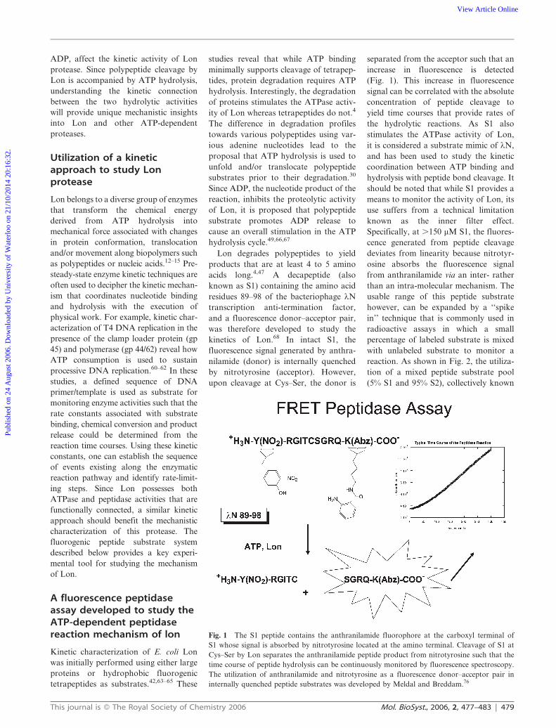
Lon degrades polypeptides to yield
products that are at least 4 to 5 amino
acids long.4,47 A decapeptide (also
known as S1) containing the amino acid
residues 89–98 of the bacteriophage lN
transcription anti-termination factor,
and a fluorescence donor–acceptor pair,
was therefore developed to study the
kinetics of Lon.68 In intact S1, the
fluorescence signal generated by anthra-
nilamide (donor) is internally quenched
by nitrotyrosine (acceptor). However,
upon cleavage at Cys–Ser, the donor is
separated from the acceptor such that an
increase in fluorescence is detected
(Fig. 1). This increase in fluorescence
signal can be correlated with the absolute
concentration of peptide cleavage to
yield time courses that provide rates of
the hydrolytic reactions. As S1 also
stimulates the ATPase activity of Lon,
it is considered a substrate mimic of lN,
and has been used to study the kinetic
coordination between ATP binding and
hydrolysis with peptide bond cleavage. It
should be noted that while S1 provides a
means to monitor the activity of Lon, its
use suffers from a technical limitation
known as the inner filter effect.
Specifically, at .150 mM S1, the fluores-
cence generated from peptide cleavage
deviates from linearity because nitrotyr-
osine absorbs the fluorescence signal
from anthranilamide via an inter- rather
than an intra-molecular mechanism. The
usable range of this peptide substrate
however, can be expanded by a ‘‘spike
in’’ technique that is commonly used in
radioactive assays in which a small
percentage of labeled substrate is mixed
with unlabeled substrate to monitor a
reaction. As shown in Fig. 2, the utiliza-
tion of a mixed peptide substrate pool
(5% S1 and 95% S2), collectively known
Fig. 1 The S1 peptide contains the anthranilamide fluorophore at the carboxyl terminal of
S1 whose signal is absorbed by nitrotyrosine located at the amino terminal. Cleavage of S1 at
Cys–Ser by Lon separates the anthranilamide peptide product from nitrotyrosine such that the
time course of peptide hydrolysis can be continuously monitored by fluorescence spectroscopy.
The utilization of anthranilamide and nitrotyrosine as a fluorescence donor–acceptor pair in
internally quenched peptide substrates was developed by Meldal and Breddam.76
This journal is � The Royal Society of Chemistry 2006 Mol. BioSyst., 2006, 2, 477–483 | 479
Publ
ishe
d on
24
Aug
ust 2
006.
Dow
nloa
ded
by U
nive
rsity
of
Wat
erlo
o on
21/
10/2
014
20:1
6:32
. View Article Online
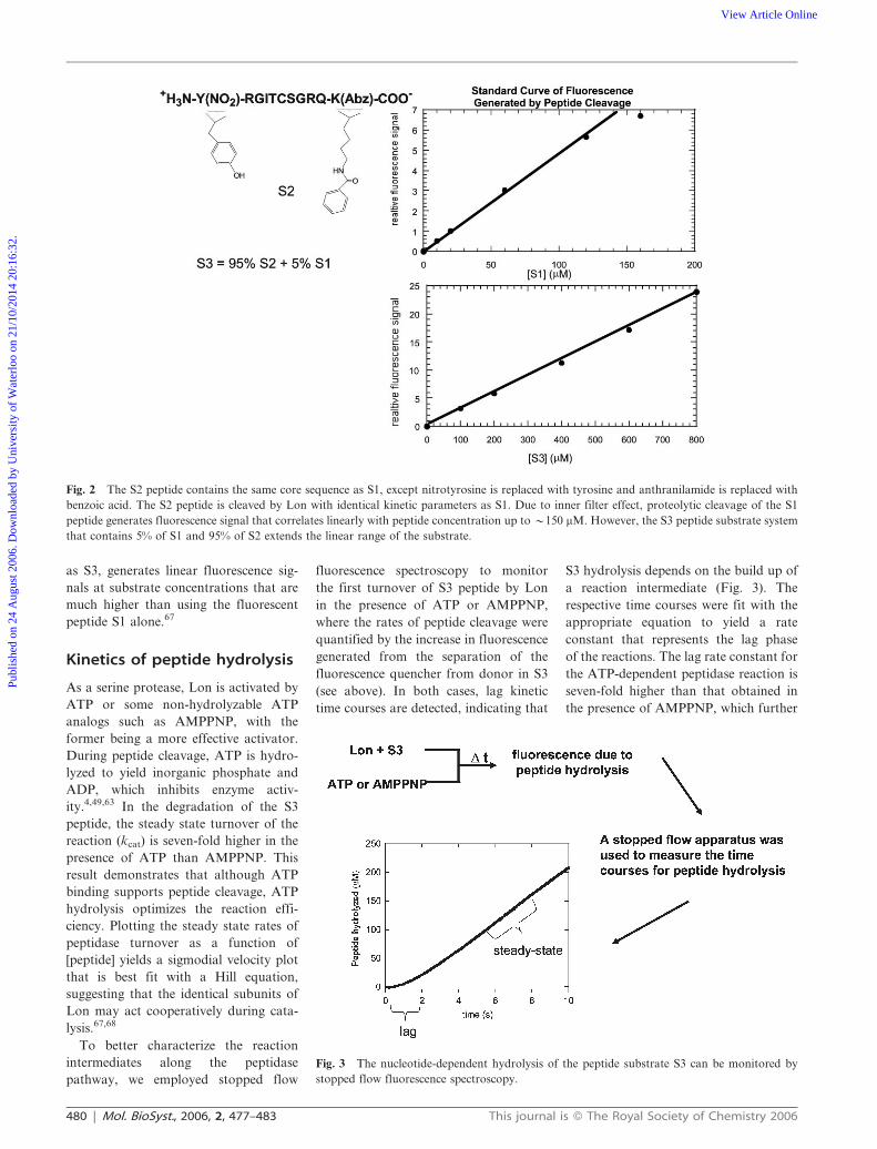
as S3, generates linear fluorescence sig-
nals at substrate concentrations that are
much higher than using the fluorescent
peptide S1 alone.67
Kinetics of peptide hydrolysis
As a serine protease, Lon is activated by
ATP or some non-hydrolyzable ATP
analogs such as AMPPNP, with the
former being a more effective activator.
During peptide cleavage, ATP is hydro-
lyzed to yield inorganic phosphate and
ADP, which inhibits enzyme activ-
ity.4,49,63 In the degradation of the S3
peptide, the steady state turnover of the
reaction (kcat) is seven-fold higher in the
presence of ATP than AMPPNP. This
result demonstrates that although ATP
binding supports peptide cleavage, ATP
hydrolysis optimizes the reaction effi-
ciency. Plotting the steady state rates of
peptidase turnover as a function of
[peptide] yields a sigmodial velocity plot
that is best fit with a Hill equation,
suggesting that the identical subunits of
Lon may act cooperatively during cata-
lysis.67,68
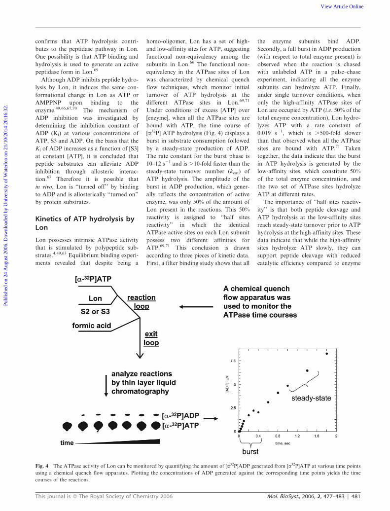
To better characterize the reaction
intermediates along the peptidase
pathway, we employed stopped flow
fluorescence spectroscopy to monitor
the first turnover of S3 peptide by Lon
in the presence of ATP or AMPPNP,
where the rates of peptide cleavage were
quantified by the increase in fluorescence
generated from the separation of the
fluorescence quencher from donor in S3
(see above). In both cases, lag kinetic
time courses are detected, indicating that
S3 hydrolysis depends on the build up of
a reaction intermediate (Fig. 3). The
respective time courses were fit with the
appropriate equation to yield a rate
constant that represents the lag phase
of the reactions. The lag rate constant for
the ATP-dependent peptidase reaction is
seven-fold higher than that obtained in
the presence of AMPPNP, which further
Fig. 2 The S2 peptide contains the same core sequence as S1, except nitrotyrosine is replaced with tyrosine and anthranilamide is replaced with
benzoic acid. The S2 peptide is cleaved by Lon with identical kinetic parameters as S1. Due to inner filter effect, proteolytic cleavage of the S1
peptide generates fluorescence signal that correlates linearly with peptide concentration up to y150 mM. However, the S3 peptide substrate system
that contains 5% of S1 and 95% of S2 extends the linear range of the substrate.
Fig. 3 The nucleotide-dependent hydrolysis of the peptide substrate S3 can be monitored by
stopped flow fluorescence spectroscopy.
480 | Mol. BioSyst., 2006, 2, 477–483 This journal is � The Royal Society of Chemistry 2006
Publ
ishe
d on
24
Aug
ust 2
006.
Dow
nloa
ded
by U
nive
rsity
of
Wat
erlo
o on
21/
10/2
014
20:1
6:32
. View Article Online
confirms that ATP hydrolysis contri-
butes to the peptidase pathway in Lon.
One possibility is that ATP binding and
hydrolysis is used to generate an active
peptidase form in Lon.69
Although ADP inhibits peptide hydro-
lysis by Lon, it induces the same con-
formational change in Lon as ATP or
AMPPNP upon binding to the
enzyme.49,66,67,70 The mechanism of
ADP inhibition was investigated by
determining the inhibition constant of
ADP (Ki) at various concentrations of
ATP, S3 and ADP. On the basis that the
Ki of ADP increases as a function of [S3]
at constant [ATP], it is concluded that
peptide substrates can alleviate ADP
inhibition through allosteric interac-
tion.67 Therefore it is possible that
in vivo, Lon is ‘‘turned off’’ by binding
to ADP and is allosterically ‘‘turned on’’
by protein substrates.
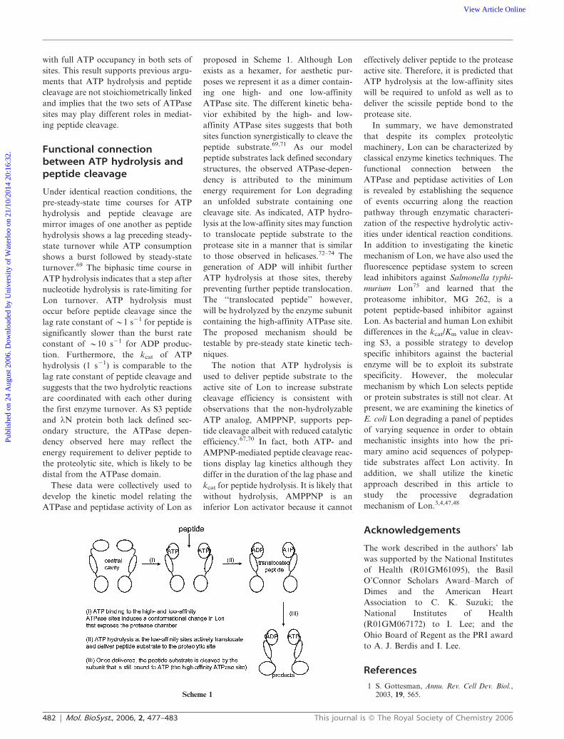
Kinetics of ATP hydrolysis byLon
Lon possesses intrinsic ATPase activity
that is stimulated by polypeptide sub-
strates.4,49,63 Equilibrium binding experi-
ments revealed that despite being a
homo-oligomer, Lon has a set of high-
and low-affinity sites for ATP, suggesting
functional non-equivalency among the
subunits in Lon.66 The functional non-
equivalency in the ATPase sites of Lon
was characterized by chemical quench
flow techniques, which monitor initial
turnover of ATP hydrolysis at the
different ATPase sites in Lon.69,71
Under conditions of excess [ATP] over
[enzyme], when all the ATPase sites are
bound with ATP, the time course of
[a32P] ATP hydrolysis (Fig. 4) displays a
burst in substrate consumption followed
by a steady-state production of ADP.
The rate constant for the burst phase is
10–12 s21 and is .10-fold faster than the
steady-state turnover number (kcat) of
ATP hydrolysis. The amplitude of the
burst in ADP production, which gener-
ally reflects the concentration of active
enzyme, was only 50% of the amount of
Lon present in the reactions. This 50%
reactivity is assigned to ‘‘half sites
reactivity’’ in which the identical
ATPase active sites on each Lon subunit
possess two different affinities for
ATP.69,71 This conclusion is drawn
according to three pieces of kinetic data.
First, a filter binding study shows that all
the enzyme subunits bind ADP.
Secondly, a full burst in ADP production
(with respect to total enzyme present) is
observed when the reaction is chased
with unlabeled ATP in a pulse–chase
experiment, indicating all the enzyme
subunits can hydrolyze ATP. Finally,
under single turnover conditions, when
only the high-affinity ATPase sites of
Lon are occupied by ATP (i.e. 50% of the
total enzyme concentration), Lon hydro-
lyzes ATP with a rate constant of
0.019 s21, which is .500-fold slower
than that observed when all the ATPase
sites are bound with ATP.71 Taken
together, the data indicate that the burst
in ATP hydrolysis is generated by the
low-affinity sites, which constitute 50%
of the total enzyme concentration, and
the two set of ATPase sites hydrolyze
ATP at different rates.
The importance of ‘‘half sites reactiv-
ity’’ is that both peptide cleavage and
ATP hydrolysis at the low-affinity sites
reach steady-state turnover prior to ATP
hydrolysis at the high-affinity sites. These
data indicate that while the high-affinity
sites hydrolyze ATP slowly, they can
support peptide cleavage with reduced
catalytic efficiency compared to enzyme
Fig. 4 The ATPase activity of Lon can be monitored by quantifying the amount of [a32P]ADP generated from [a32P]ATP at various time points
using a chemical quench flow apparatus. Plotting the concentrations of ADP generated against the corresponding time points yields the time
courses of the reactions.
This journal is � The Royal Society of Chemistry 2006 Mol. BioSyst., 2006, 2, 477–483 | 481
Publ
ishe
d on
24
Aug
ust 2
006.
Dow
nloa
ded
by U
nive
rsity
of
Wat
erlo
o on
21/
10/2
014
20:1
6:32
. View Article Online
with full ATP occupancy in both sets of
sites. This result supports previous argu-
ments that ATP hydrolysis and peptide
cleavage are not stoichiometrically linked
and implies that the two sets of ATPase
sites may play different roles in mediat-
ing peptide cleavage.
Functional connectionbetween ATP hydrolysis andpeptide cleavage
Under identical reaction conditions, the
pre-steady-state time courses for ATP
hydrolysis and peptide cleavage are
mirror images of one another as peptide
hydrolysis shows a lag preceding steady-
state turnover while ATP consumption
shows a burst followed by steady-state
turnover.69 The biphasic time course in
ATP hydrolysis indicates that a step after
nucleotide hydrolysis is rate-limiting for
Lon turnover. ATP hydrolysis must
occur before peptide cleavage since the
lag rate constant of y1 s21 for peptide is
significantly slower than the burst rate
constant of y10 s21 for ADP produc-
tion. Furthermore, the kcat of ATP
hydrolysis (1 s21) is comparable to the
lag rate constant of peptide cleavage and
suggests that the two hydrolytic reactions
are coordinated with each other during
the first enzyme turnover. As S3 peptide
and lN protein both lack defined sec-
ondary structure, the ATPase depen-
dency observed here may reflect the
energy requirement to deliver peptide to
the proteolytic site, which is likely to be
distal from the ATPase domain.
These data were collectively used to
develop the kinetic model relating the
ATPase and peptidase activity of Lon as
proposed in Scheme 1. Although Lon
exists as a hexamer, for aesthetic pur-
poses we represent it as a dimer contain-
ing one high- and one low-affinity
ATPase site. The different kinetic beha-
vior exhibited by the high- and low-
affinity ATPase sites suggests that both
sites function synergistically to cleave the
peptide substrate.69,71 As our model
peptide substrates lack defined secondary
structures, the observed ATPase-depen-
dency is attributed to the minimum
energy requirement for Lon degrading
an unfolded substrate containing one
cleavage site. As indicated, ATP hydro-
lysis at the low-affinity sites may function
to translocate peptide substrate to the
protease site in a manner that is similar
to those observed in helicases.72–74 The
generation of ADP will inhibit further
ATP hydrolysis at those sites, thereby
preventing further peptide translocation.
The ‘‘translocated peptide’’ however,
will be hydrolyzed by the enzyme subunit
containing the high-affinity ATPase site.
The proposed mechanism should be
testable by pre-steady state kinetic tech-
niques.
The notion that ATP hydrolysis is
used to deliver peptide substrate to the
active site of Lon to increase substrate
cleavage efficiency is consistent with
observations that the non-hydrolyzable
ATP analog, AMPPNP, supports pep-
tide cleavage albeit with reduced catalytic
efficiency.67,70 In fact, both ATP- and
AMPNP-mediated peptide cleavage reac-
tions display lag kinetics although they
differ in the duration of the lag phase and
kcat for peptide hydrolysis. It is likely that
without hydrolysis, AMPPNP is an
inferior Lon activator because it cannot
effectively deliver peptide to the protease
active site. Therefore, it is predicted that
ATP hydrolysis at the low-affinity sites
will be required to unfold as well as to
deliver the scissile peptide bond to the
protease site.
In summary, we have demonstrated
that despite its complex proteolytic
machinery, Lon can be characterized by
classical enzyme kinetics techniques. The
functional connection between the
ATPase and peptidase activities of Lon
is revealed by establishing the sequence
of events occurring along the reaction
pathway through enzymatic characteri-
zation of the respective hydrolytic activ-
ities under identical reaction conditions.
In addition to investigating the kinetic
mechanism of Lon, we have also used the
fluorescence peptidase system to screen
lead inhibitors against Salmonella typhi-
murium Lon75 and learned that the
proteasome inhibitor, MG 262, is a
potent peptide-based inhibitor against
Lon. As bacterial and human Lon exhibit
differences in the kcat/Km value in cleav-
ing S3, a possible strategy to develop
specific inhibitors against the bacterial
enzyme will be to exploit its substrate
specificity. However, the molecular
mechanism by which Lon selects peptide
or protein substrates is still not clear. At
present, we are examining the kinetics of
E. coli Lon degrading a panel of peptides
of varying sequence in order to obtain
mechanistic insights into how the pri-
mary amino acid sequences of polypep-
tide substrates affect Lon activity. In
addition, we shall utilize the kinetic
approach described in this article to
study the processive degradation
mechanism of Lon.3,4,47,48
Acknowledgements
The work described in the authors’ lab
was supported by the National Institutes
of Health (R01GM61095), the Basil
O’Connor Scholars Award–March of
Dimes and the American Heart
Association to C. K. Suzuki; the
National Institutes of Health
(R01GM067172) to I. Lee; and the
Ohio Board of Regent as the PRI award
to A. J. Berdis and I. Lee.
References
1 S. Gottesman, Annu. Rev. Cell Dev. Biol.,2003, 19, 565.Scheme 1
482 | Mol. BioSyst., 2006, 2, 477–483 This journal is � The Royal Society of Chemistry 2006
Publ
ishe
d on
24
Aug
ust 2
006.
Dow
nloa
ded
by U
nive
rsity
of
Wat
erlo
o on
21/
10/2
014
20:1
6:32
. View Article Online
2 J. A. Maupin-Furlow, M. A. Gil, M. A.Humbard, P. A. Kirkland, W. Li, C. J.Reuter and A. J. Wright, Curr. Opin.Microbiol., 2005, 8, 720.
3 M. R. Maurizi, Experientia, 1992, 48, 178.4 A. L. Goldberg, R. P. Moerschell,
C.H. Chung and M. R. Maurizi,Methods Enzymol., 1994, 244, 350.
5 P. Howard-Flanders, E. Simson andL. Theriot, Genetics, 1964, 49, 237.
6 M. Rep and L. A. Grivell, Curr. Genet.,1996, 30, 367.
7 C. K. Suzuki, M. Rep, J. M. van Dijl,K. Suda, L. A. Grivell and G. Schatz,Trends Biochem. Sci., 1997, 22, 118.
8 L. Van Dyck and T. Langer, Cell. Mol.Life Sci., 1999, 56, 825.
9 G. T. Robertson, M. E. Kovach, C. A.Allen, T. A. Ficht and R. M. Roop, 2nd,Mol. Microbiol., 2000, 35, 577.
10 A. Takaya, T. Tomoyasu, A. Tokumitsu,M. Morioka and T. Yamamoto,J. Bacteriol., 2002, 184, 224.
11 H. Matsui, M. Suzuki, Y. Isshiki,C. Kodama, M. Eguchi, Y. Kikuchi,K. Motokawa, A. Takaya, T. Tomoyasuand T. Yamamoto, Infect. Immun., 2003,71, 30.
12 T. Ogura and A. J. Wilkinson, Genes Cells,2001, 6, 575.
13 P. I. Hanson and S. W. Whiteheart, Nat.Rev. Mol. Cell Biol., 2005, 6, 519.
14 T. C. He, N. Jiang, H. Zhuang andD. M. Wojchowski, J. Biol. Chem., 1995,270, 11055.
15 A. F. Neuwald, L. Aravind, J. L. Spougeand E. V. Koonin, Genome Res., 1999, 9,27.
16 S. C. Park, B. Jia, J. K. Yang, D. L. Van,Y. G. Shao, S. W. Han, Y. J. Jeon,C. H. Chung and G. W. Cheong, Mol.Cells, 2006, 21, 129.
17 H. Stahlberg, E. Kutejova, K. Suda,B. Wolpensinger, A. Lustig, G. Schatz,A. Engel and C. K. Suzuki, Proc. Natl.Acad. Sci. U. S. A., 1999, 96, 6787.
18 I. Botos, E. E. Melnikov, S. Cherry, J. E.Tropea, A. G. Khalatova, F. Rasulova,Z. Dauter, M. R. Maurizi, T. V. Rotanova,A. Wlodawer and A. Gustchina, J. Biol.Chem., 2004, 279, 8140.
19 I. Botos, E. E. Melnikov, S. Cherry,S. Kozlov, O. V. Makhovskaya, J. E.Tropea, A. Gustchina, T. V. Rotanovaand A. Wlodawer, J. Mol. Biol., 2005, 351,144.
20 T. V. Rotanova, E. E. Melnikov, A. G.Khalatova, O. V. Makhovskaya, I. Botos,A. Wlodawer and A. Gustchina, Eur. J.Biochem., 2004, 271, 4865.
21 Y. J. Im, Y. Na, G. B. Kang, S. H. Rho,M. K. Kim, J. H. Lee, C. H. Chung andS. H. Eom, J. Biol. Chem., 2004, 279,53451.
22 N. N. Starkova, E. P. Koroleva,L. D. Rumsh, L. M. Ginodman andT. V. Rotanova, FEBS Lett., 1998, 422,218.
23 H. Fischer and R. Glockshuber, J. Biol.Chem., 1993, 268, 22502.
24 N. Benaroudj, P. Zwickl, E. Seemuller,W. Baumeister and A. L. Goldberg, Mol.Cell, 2003, 11, 69.
25 R. E. Burton, S. M. Siddiqui, Y. I. Kim,T. A. Baker and R. T. Sauer, EMBO J.,2001, 20, 3092.
26 J . R. Hosk ins , K. Yanag ihara ,K. Mizuuchi and S. Wickner, Proc. Natl.Acad. Sci. U. S. A., 2002, 99, 11037.
27 J. A. Kenniston, T. A. Baker, J. M.Fernandez and R. T. Sauer, Cell, 2003,114, 511.
28 Y. I. Kim, R. E. Burton, B. M. Burton,R. T. Sauer and T. A. Baker, Mol. Cell,2000, 5, 639.
29 Y. A. Lam, T. G. Lawson, M. Velayutham,J. L. Zweier and C. M. Pickart, Nature,2002, 416, 763.
30 C. Lee, M. P. Schwartz, S. Prakash,M. Iwakura and A. Matouschek, Mol.Cell, 2001, 7, 627.
31 A. Navon and A. L. Goldberg, Mol. Cell,2001, 8, 1339.
32 J. Ortega, H. S. Lee, M. R. Maurizi andA. C. Steven, EMBO J., 2002, 21, 4938.
33 C. M. Pickart and R. E. Cohen, Nat. Rev.Mol. Cell Biol., 2004, 5, 177.
34 S. Prakash and A. Matouschek, TrendsBiochem. Sci., 2004, 29, 593.
35 B. G. Reid, W. A. Fenton, A. L. Horwichand E. U. Weber-Ban, Proc. Natl. Acad.Sci. U. S. A., 2001, 98, 3768.
36 R. T. Sauer, D. N. Bolon, B. M. Burton,R. E. Burton, J. M. Flynn, R. A. Grant,G. L. Hersch, S. A. Joshi, J. A. Kenniston,I. Levchenko, S. B. Neher, E. S. Oakes,S. M. Siddiqui, D. A. Wah and T. A. Baker,Cell, 2004, 119, 9.
37 S. K. Singh, R. Grimaud, J. R. Hoskins,S. Wickner and M. R. Maurizi, Proc. Natl.Acad. Sci. U. S. A., 2000, 97, 8898.
38 L. Van Melderen, M. H. Thi, P. Lecchi,S. Gottesman, M. Couturier and M. R.Maurizi, J. Biol. Chem., 1996, 271, 27730.
39 J. E. Laachouch, L. Desmet, V. Geuskens,R. Grimaud and A. Toussaint, EMBO J.,1996, 15, 437.
40 M. Gonzalez, E. G. Frank, A. S. Levineand R. Woodgate, Genes Dev., 1998, 12,3889.
41 M. R. Maurizi, J. Biol. Chem., 1987, 262,2696.
42 L. Waxman and A. L. Goldberg, Proc.Natl. Acad. Sci. U. S. A., 1982, 79, 4883.
43 D. A. Bota, H. Van Remmen andK. J. Davies, FEBS Lett., 2002, 532, 103.
44 D. A. Bota and K. J. Davies, Nat. CellBiol., 2002, 4, 674.
45 W. Nishii, T. Maruyama, R. Matsuoka,T. Muramatsu and K. Takahashi, Eur. J.Biochem., 2002, 269, 451.
46 E. Dervyn, D. Canceill and O. Huisman,J. Bacteriol., 1990, 172, 7098.
47 W. Nishii, T. Suzuki, M. Nakada, Y. T.Kim, T. Muramatsu and K. Takahashi,FEBS Lett., 2005, 579, 6846.
48 G. Ondrovicova, T. Liu, K. Singh, B. Tian,H. Li, O. Gakh, D. Perecko, J. Janata,Z. Granot, J. Orly, E. Kutejova andC. K. Suzuki, J. Biol. Chem., 2005, 280,25103.
49 A. S. Menon and A. L. Goldberg, J. Biol.Chem., 1987, 262, 14929.
50 K. Nomura, J. Kato, N. Takiguchi,H. Ohtake and A. Kuroda, J. Biol.Chem., 2004, 279, 34406.
51 A. Kuroda, K. Nomura, R. Ohtomo,J. Kato, T. Ikeda, N. Takiguchi,H. Ohtake and A. Kornberg, Science,2001, 293, 705.
52 G. K. Fu, M. J. Smith and D. M.Markovitz, J. Biol. Chem., 1997, 272, 534.
53 G. K. Fu and D. M. Markovitz,Biochemistry, 1998, 37, 1905.
54 T. Liu, B. Lu, I. Lee, G. Ondrovicova,E. Kutejova and C. K. Suzuki, J. Biol.Chem., 2004, 279, 13902.
55 M. F. Charette, G. W. Henderson,L. L. Doane and A. Markovitz,J. Bacteriol., 1984, 158, 195.
56 C. H. Chung and A. L. Goldberg, Proc.Natl. Acad. Sci. U. S. A., 1982, 79, 795.
57 B. Lu, T. Liu, J. A. Crosby, J. Thomas-Wohlever, I. Lee and C. K. Suzuki, Gene,2003, 306, 45.
58 R. Maas, Cell, 2001, 105, 945.59 A. Y. Lee, C. H. Hsu and S. H. Wu, J. Biol.
Chem., 2004, 279, 34903.60 A. J. Berdis and S. J. Benkovic,
Biochemistry, 1996, 35, 9253.61 A. J. Berdis and S. J. Benkovic,
Biochemistry, 1997, 36, 2733.62 D. J. Sexton, B. F. Kaboord, A. J. Berdis,
T. E. Carver and S. J. Benkovic,Biochemistry, 1998, 37, 7749.
63 A. S. Menon, L. Waxman and A. L.Goldberg, J. Biol. Chem., 1987, 262, 722.
64 A. L. Goldberg and L. Waxman, J. Biol.Chem., 1985, 260, 12029.
65 L. Waxman and A. L. Goldberg, J. Biol.Chem., 1985, 260, 12022.
66 A. S. Menon and A. L. Goldberg, J. Biol.Chem., 1987, 262, 14921.
67 J. Thomas-Wohlever and I. Lee,Biochemistry, 2002, 41, 9418.
68 I. Lee and A. J. Berdis, Anal. Biochem.,2001, 291, 74.
69 D. Vineyard, J. Patterson-Ward, A. J.Berdis and I. Lee, Biochemistry, 2005, 44,1671.
70 J. Patterson, D. Vineyard, J. Thomas-Wohlever, R. Behshad, M. Burke andI. Lee, Biochemistry, 2004, 43, 7432.
71 D. Vineyard, J. Patterson-Ward and I. Lee,Biochemistry, 2006, 45, 4602.
72 S. S. Patel and K. M. Picha, Annu. Rev.Biochem., 2000, 69, 651.
73 R. L. Eoff and K. D. Raney, Biochem. Soc.Trans., 2005, 33, 1474.
74 T. M. Lohman and K. P. Bjornson, Annu.Rev. Biochem., 1996, 65, 169.
75 H. Frase, J. Hudak and I. Lee,Biochemistry, 2006, 45, 8264.
76 M. Meldal and K. Breddam, Anal.Biochem., 1991, 195, 141.
This journal is � The Royal Society of Chemistry 2006 Mol. BioSyst., 2006, 2, 477–483 | 483
Publ
ishe
d on
24
Aug
ust 2
006.
Dow
nloa
ded
by U
nive
rsity
of
Wat
erlo
o on
21/
10/2
014
20:1
6:32
. View Article Online


![Enzymology [Compatibility Mode]](https://img.pdfslide.net/doc/110x75/577d1ec81a28ab4e1e8f3d6e/enzymology-compatibility-mode.jpg)