Embed Size (px)
Citation preview

Institute of Materia Medica, Chinese Academy of Medical Sciences
Chinese Pharmaceutical Association
www.elsevier.com/locate/apsb
Acta Pharmaceutica Sinica B
Acta Pharmaceutica Sinica B 2019;9(2):258–278
https://doi.org/10.10162211-3835 & 2019 ChElsevier B.V. This is
nCorresponding autE-mail address: g
†These authors mad
Peer review under r
www.sciencedirect.com
REVIEW
Recent progress and challenges in screening andcharacterization of UGT1A1 inhibitors
Xia Lva,b,†, Yangliu Xiac,†, Moshe Fineld, Jingjing Wuc,Guangbo Gea,c,n, Ling Yanga
aInstitute of Interdisciplinary Medicine, Shanghai University of Traditional Chinese Medicine, Shanghai 201203, ChinabKey Laboratory of Biotechnology and Bioresources Utilization, Ministry of Education, College of Life Science,Dalian Minzu University, Dalian 116600, ChinacDalian Institute of Chemical Physics, Chinese Academy of Sciences, Dalian 116023, ChinadDivision of Pharmaceutical Chemistry and Technology, Faculty of Pharmacy, University of Helsinki, Finland
Received 8 June 2018; received in revised form 16 August 2018; accepted 27 August 2018
KEY WORDS
UGT1A1 inhibitors;Drug/herbdruginteractions;
Probe substrates;High-throughputscreening
/j.apsb.2018.09.005inese Pharmaceuticaan open access artic
hor at: Institute of [email protected] equal contribution
esponsibility of Inst
Abstract Uridine-diphosphate glucuronosyltransferase 1A1 (UGT1A1) is an important conjugative enzymein mammals that is responsible for the conjugation and detoxification of both endogenous and xenobioticcompounds. Strong inhibition of UGT1A1 may trigger adverse drug/herb–drug interactions, or result inmetabolic disorders of endobiotic metabolism. Therefore, both the US Food and Drug Administration (FDA)and the European Medicines Agency (EMA) have recommended assaying the inhibitory potential of drugsunder development on the human UGT1A1 prior to approval. This review focuses on the significance,progress and challenges in discovery and characterization of UGT1A1 inhibitors. Recent advances in thedevelopment of UGT1A1 probes and their application for screening UGT1A1 inhibitors are summarized anddiscussed in this review for the first time. Furthermore, a long list of UGT1A1 inhibitors, including informationon their inhibition potency, inhibition mode, and affinity, has been prepared and analyzed. Challenges andfuture directions in this field are highlighted in the final section. The information and knowledge that arepresented in this review provide guidance for rational use of drugs/herbs in order to avoid the occurrence ofadverse effects via UGT1A1 inhibition, as well as presenting methods for rapid screening and characterizationof UGT1A1 inhibitors and for facilitating investigations on UGT1A1–ligand interactions.
& 2019 Chinese Pharmaceutical Association and Institute of Materia Medica, Chinese Academy of MedicalSciences. Production and hosting by Elsevier B.V. This is an open access article under the CC BY-NC-ND
license (http://creativecommons.org/licenses/by-nc-nd/4.0/).
l Association and Institute of Materiale under the CC BY-NC-ND license
nterdisciplinary Medicine, Shanghai Udu.cn (Guangbo Ge).to this work.
itute of Materia Medica, Chinese Ac
Medica, Chinese Academy of Medical Sciences. Production and hosting by(http://creativecommons.org/licenses/by-nc-nd/4.0/).
niversity of Traditional Chinese Medicine, Shanghai 201203, China.
ademy of Medical Sciences and Chinese Pharmaceutical Association.

Review on UGT1A1 inhibitors 259
1. Introduction
Uridine-diphosphate (UDP) glucuronosyltransferases (UGT) are asuperfamily of phase II conjugating enzymes (EC 2.4.1.17) thatcatalyse the covalent addition of glucuronic acid from the high energydonor uridine-diphosphate glucuronic acid (UDPGA) to a wide rangeof lipophilic chemicals that contain a suitable acceptor functionalgroup (Fig. 1)1. In mammals, UGTs play predominant roles in thedetoxification of many exogenous and endogenous compounds bygenerating more polar and water-soluble glucuronides. The glucur-onide metabolites are mostly biologically inactive and readilyexcreted from the cell by efflux transporters and, eventually, fromthe body via bile or urine1. Mammalian UGTs could be divided,based on evolutionary divergence, into two main families, UGT1 andUGT2, which can be further divided into three subfamilies UGT1A,UGT2A and UGT2B2. Another family of UDP-sugar transferasesthat was reported to conjugate xenobiotics is UGT3. This familycontains two members and uses other sugar donor than UDPGA3, butit will not be further discussed in this review.
Mammalian UGTs are membrane-bound enzymes that arelocalized in the endoplasmic reticulum (ER) and expressed in atissue-specific way. Many UGTs are highly expressed in the liver,the most important tissue for xenobiotics metabolism, but some are
Figure 1 A schematic presentation of UGT-catalysed glucuronidationaglycone (for example, a phenol) and a glucuronic acid donor (UDPGA).
Figure 2 The elimination pathways of bilirubin (a), and disorders associatconcentrations and exposure of UGT1A1 substrates, hyperbilirubinemia, a
also expressed in extrahepatic tissues, including intestine, kidney,stomach and lung. In human, ten different UGTs are significantlyexpressed in the liver at the protein level, namely UGT1A1,UGT1A3, UGT1A4, UGT1A6, UGT1A9, UGT2B4, UGT2B7,UGT2B10, UGT2B15 and UGT2B17, of which UGT2B7 isexpressed to the highest level, followed in order of expressionlevel by UGT1A1, UGT2B4, UGT2B15, UGT1A4, UGT2B10,UGT1A9, UGT2B17, UGT1A6, and UGT1A3. Six UGTs weredetected in the human intestine, UGT1A1, UGT1A10, UGT2B7,UGT2B17 and very low levels of UGT1A3 and UGT1A4,whereas in the human kidney only three UGTs were detected atthe protein level, UGT1A9, UGT2B7 and UGT1A64.
Among the human UGTs, UGT1A1 is of particular clinicalsignificance due to its unique activity in the conjugative detoxificationof bilirubin, the endogenous by-product of heme metabolism(Fig. 2)5. Alongside its essential role in bilirubin metabolism,contribution and involvement in the glucuronidation of few otherendobiotics6, UGT1A1 also participates in the metabolism anddetoxification of clinical drugs, such as etoposide, SN-38 (the activemetabolite of CPT-11) and other xenobiotics, including environmen-tal toxicants and chemical carcinogens7. Importantly, many studieshave clearly demonstrated that genetics and environmental factors,could affect the expression or the function of UGT1A1, eventually
reactions. Glucuronidation is a bi-substrate reaction that requires an
ed with UGT1A1 deficiency or absence, including the elevated plasmand kernicterus (b).

Xia Lv et al.260
leading to reduced protein level or activity of UGT1A18. Notably,UGT1A1 is a highly polymorphic enzyme with more than onehundred variants, some of which are within the promoter region, likethe rather common variant UGT1A1*28. Commonly, the poly-morphic variants of UGT1A1 result in lower expression level, loweractivity of the enzyme or even complete activity loss9. As shown inFig. 2, the reduced expression/activity of UGT1A1 may increase theplasma concentrations of unconjugated bilirubin, leading to hyperbi-lirubinemia10 from the mild Gilbert's syndrome up to kernicterus andthe potentially fatal Crigler-Najjar syndrome type I11–14. In addition,polymorphic variants of UGT1A1 may lead to drug-induced liverinjury. It has been reported that patients possessing the UGT1A1*28genotype are at a greater risk for irinotecan-induced toxicities, such assevere diarrhea and grades 4 neutropenia15.
Genetic polymorphisms are important, but are not the onlyfactor and sometimes not the main one, from the generalpopulation point of view. Small-molecule inhibitors of UGT1A1may profoundly influence the catalytic activity of UGT1A1,thereby triggering undesirable effects, like drug/herb–drug inter-actions (D/HDI) or drug/herb–endobiotic interactions16. It hasbeen reported that some therapeutic drugs (e.g., indinavir, nilotiniband sorafenib) or herbal extracts (such as milk thistle, green teaand psoralea corylifolia), as well as several natural compounds inherbs or foods (such as amentoflavone, licochalcone A andemodin) are potent UGT1A1 inhibitors, which could significantlyinhibit their activities and lead to undesirable effects17–25. Notably,subjects with UGT1A1 polymorphic variants that already possesslower activity due to lower expression level of the enzyme, such ashomozygous carriers of the UGT1A1*28 genotype, might beexpected to manifest higher susceptibility to adverse effects whenco-administrated with potent UGT1A1 inhibitors26,27. Thus, themajor regulatory agencies, the Food and Drug Administration(FDA) of US and European Medicines Agency (EMA), have
Figure 3 Recent progresses in the development of fluorescent probe subs1,8-naphthalimide (NHPN) and its proposed mechanism for sensing UGT1NPHN and N-(3-carboxypropyl)-4-hydroxy-1,8-naphthalimide (NCHN) onhuman UGT isoforms. (d) NHPN-O-glucuronidation rate (i.e., UGT1A1 adependent inhibition curve of NHPN-O-glucuronidation by 20(S)-protopaenzyme sources. Adapted with permission from the Ref. 44. © 2017 Ame
recommended to study the inhibition potentials of investigationalnew drugs on human UGT1A1 before the drug is approved28,29.
Considering that UGT1A1 plays a pivotal role in endobiotichomeostasis, in addition to its contribution to xenobiotic disposi-tion and detoxification, it was highly necessary to developpractical methods for precise measurements of UGT1A1 activityin complex biological samples. Probe substrates are often used forsensing the real activities of a target enzyme in complex biologicalsystems under physiological conditions. An ideal probe substratefor a target enzyme is expected to be highly specific and reliable,as well as highly sensitive and capable for high-throughputdetection. Unfortunately, currently commercially available sub-strates for UGT1A1 (such as bilirubin, estradiol, and etoposide)have different limitations, such as poor selectivity, poor chemicalstability or their use for high-throughput screening (HTS) isunfeasible30,31. Thus, it was necessary to develop one or a fewmore practical probe substrates for this key conjugative enzyme.
In contrast to non-fluorescent probes, fluorescent substrates fortarget enzyme(s) have inherent advantages, such as high sensitivityand applicability to HTS32–43. Recently, significant breakthroughshave been made in the development of fluorescent probes forUGT1A1, and several fluorescent probes that are highly selectivefor UGT1A1 activities in complex biological samples have beensuccessfully developed (Fig. 344). These fluorescent substratesprovide novel tools for HTS and characterization of UGT1A1inhibitors using fluorescence-based assays3641,44. Furthermore, recentinvestigations on UGT1A1–ligand interactions have demonstratedthat UGT1A1 has multiple ligand-binding sites45,46. Thus, develop-ing site-specific probes, i.e., different one for each ligand binding sitewithin UGT1A1, would be highly beneficial for deciphering theinteractions between UGT1A1 and its different ligands.
Considering the crucial role of UGT1A1 in bilirubin metabo-lism, strong inhibition of this conjugative enzyme by xenobiotics,
trates for UGT1A1. (a) The structure of N-butyl-4-(4-hydroxyphenyl)-A1 activity. (b) A schematic suggestion for the ligand-binding sites ofUGT1A1. (c) The formation rates of NHPN-O-glucuronide in variousctivity) in 14 individual human liver microsomes (HLMs). (e) Dose-naxatriol (PPT), using both HLM and recombinant UGT1A1 as therican Chemical Society.

Figure 4 Probe substrates for UGT1A1. The blue arrows indicate conjugation sites by UGT1A1.
Review on UGT1A1 inhibitors 261
such as drugs, food/herbal constitutes, and environmental toxins,may trigger hyperbilirubinemia. Furthermore, inhibition ofUGT1A1 may also lead to clinically significant DDI/HDI due tothe key roles of UGT1A1 in the detoxification of several drugswith narrow therapeutic windows, such as SN-38 and etopo-side30,47. Over the past twenty years, a variety of probe substratesfor UGT1A1 have been reported, making the screening andevaluation of UGT1A1 inhibitors more convenient and efficient.
This review focuses on the significance, progress and challenges inthe discovery and characterization of UGT1A1 inhibitors fromtherapeutic drugs, environmental toxins, herbal extracts and naturalcompounds. Recent advances in the development of UGT1A1 probesubstrates and their applications for screening and characterization ofUGT1A1 inhibitors are discussed and summarized for the first time.Furthermore, a long list of UGT1A1 inhibitors, including informationon their inhibition potency, inhibition mode, and affinity, has beenanalysed and summarized. In addition, challenges and future direc-tions in this field are highlighted in the final section. The information
Table 1 Probe substrates for human UGT1A1.
Probe substrate Reaction product Reaction d
4-MU 4-MU-7-O-glucuronide UV
Bilirubin Bilirubin-8-O-glucuronide LC–MS/MBilirubin-12-O-glucuronide
17β-Estradiol β-Estradiol-3-O-glucuronide LC–MS/M
Ethinylestradiol Ethinylestradiol-3-O-glucuronide LC–MSEtoposide Etoposide-40-O-glucuronide LC–MS
3,30,40-Trihydroxyflavone 3,30,40-Trihydroxyflavone-40-O-glucuronide
UV
3,6,40-Trihydroxyflavone 3,6,40-Trihydroxyflavone-40-O-glucuronide
UV
NCHN NCHN-4-O-glucuronide Fluorescen
NHPN NHPN-19-O-glucuronide Fluorescen
and knowledge that are presented in this review provide a goodguidance for rational use of clinical drugs or herbal medicines, inorder to avoid the occurrence of adverse side effects via UGT1A1inhibition. The accumulated knowledge is also expected to facilitateinvestigations on UGT1A1–ligand interactions.
2. Recent progress in the development of UGT1A1 probesubstrates
A successful screening for inhibitors of a specific target enzyme(s),and their subsequent and characterization, requires suitable probesubstrates. Unfortunately, the number of probe substrates for thehuman UGT1A1, particularly good ones, was previously verylimited since most of them were either not specific enough for usein tissue or cell preparations (such as human liver microsomes,HLM), not sufficiently sensitive, chemically unstable, or toodifficult to work with for practical reasons such as poor solubility.The probe substrates that are presented in Fig. 4 and Table 1
etection Feature
Nonspecific, only recombinant UGT1A1 could be as theenzyme source
S Highly specific, difficult to work with, reaction yields bothmono- and di-glucuronides
S Conjugated at the 17-OH by UGT2B7 and UGT2B17 inHLM
Reaction yields another 17-O-glucuronideHighly specific in HLM, reaction yields another twoalcoholic glucuronides in HLM and human intestinemicrosomes (HIM)
Highly specific. Conjugated at both the 3- and 30-OH inHLM
Highly specific. Conjugated at both the 3- and 6-OH inHLM
ce Highly specific in HLM, the specificity in HIM is currentlyunclear
ce Highly specific in HLM, only one conjugation site

Table 2 Kinetic parameters of probe substrates for human UGT1A130,36,41,44,48–53.
Probe substrate Enzyme source Kinetic parameter Kinetic behavior Ref.
Km or S50 (μmol/L) Vmax (pmol/min/mg)
4-MU UGT1A1 113 308 Michaelis-Menten 48Bilirubin UGT1A1 0.1 70 Michaelis-Menten 49
HLM 0.3 210 Michaelis-Menten 4917β-Estradiol UGT1A1 13 1300 Hill 50
HLM 11 820 Hill 50Ethinylestradiol UGT1A1 9.7 600 Hill 51
HLM 13 1200 Hill 51Etoposide UGT1A1 285 124 Michaelis-Menten 30
HLM 530 110 Michaelis-Menten 303,30,40-Trihydroxyflavone UGT1A1 1.53 1920 Substrate Inhibition 53
HLM 1.75 1990 Substrate Inhibition 533,6,40-Trihydroxyflavone UGT1A1 0.76 340 Substrate Inhibition 53
HLM 0.83 340 Substrate Inhibition 532-Me-4-OMe TG UGT1A1 2.7 16.1a Michaelis-Menten 41NCHN UGT1A1 126.7 1303 Substrate Inhibition 36
HLM 364.6 1556 Substrate Inhibition 36NHPN UGT1A1 0.7 561 Hill 44
HLM 4.3 557 Hill
anmol/L/s.
Xia Lv et al.262
include both “classical” substrates, such as bilirubin and ethiny-lestradiol, as well as the new generation of fluorescent substratesprobes, namely N-(3-carboxypropyl)-4-hydroxy-1,8-naphthalimide(NCHN) and N-butyl-4-(4-hydroxyphenyl)-1,8-naphthalimide(NHPN). The kinetic properties of these and the other probes inFig. 4 and Table 1 are listed in Table 230,36,41,44,48–53. As can beseen from Fig. 4, there is a large variability in chemical structureamong the UGT1A1 probe substrates, as it might be expected froman enzyme, like UGT1A1, that has a wide spectrum of substrates.The human UGT1A subfamily members share high amino acidsequence homology (465%), and their substrate specificityfrequently overlap, even if at variable kinetic constants. In fact,many UGT1A1 substrates could be glucuronidated by multipleUGT enzymes, a good example for which is the C-7 phenoliccoumarin derivatives, such as 4-methyl-umbelliferone (4-MU)54. Itis also noteworthy that no high-resolution crystal structure of thesubstrate binding domain of UGT1A1, or any other mammalianUGT, has been reported yet. Some UGT homology models55, werederived from structures of microbial UGTs, but they are notsufficient for the simulation of ligands–UGT1A1 interactions orthe design of specific substrates for a given human UGT enzyme.Thus, the rational design and development of highly specific probesubstrates for UGT1A1, or other UGT enzymes, are very challen-ging undertakings. This is further true when the goal is to designoptical probe substrates with high specificity and sensitivity, whichare suitable for high throughput detection or screening.
Several commercial small molecules that are known to beUGT1A1 substrates, such as bilirubin, estradiol, and ethinylestra-diol, have been used and might still be in use for testing itsactivity, but mostly some disadvantages are associated withthem31,56. The specificity of UGT1A1 for the formation ofestradiol-3-O-glucuronide or ethinyloestradiol-3-O-glucuronide ispoor57. Moreover, when it is tested in a complex system, such asHLM, and the substrate is estradiol, other UGTs conjugate thesame “probe” at the 17-OH more efficiently58. Although bilirubinis a UGT1A1 specific substrate that is hardly conjugated by any
other human UGT, it is difficult to use bilirubin as a probe forscreening and characterization of UGT1A1 inhibitors due to itshigh light-sensitivity and the formation of both mono- and di-glucuronides59. Furthermore, the above-mentioned probe sub-strates for UGT1A1 are non-fluorescent, making them less suitablefor HTS. The glucuronidation rates of these commonly usedprobes were routinely detected by liquid chromatography com-bined with UV or mass spectrometry detectors, requiring relativelylong time for sample preparation and analysis.
In contrast to these commonly used “old generation” probesubstrates for UGT1A1, a new generation of fluorescent probes forUGT1A1 has been emerging in recent years, despite greatchallenges in the design and development of practical fluorescentprobes for any given UGT enzyme. Notably, the fluorescenceproperties of many fluorophores are often “turned-off” or “blueshifted” following O-glucuronidation at the hydroxyl group, whichis not highly beneficial for precise measurement of the targetenzyme in complex biological samples, due to the backgroundsignals from the biological matrix. Five years ago, Terai andcoworkers41 have developed the first “turn-on” fluorescent probesfor UGT1A1 with high sensitivity, which was a landmarkachievement in this field, even if the specificity of these probestoward UGT1A1 of that compound was only partial. This type offluorescent probes is intensity-based compounds, designed to workby a mechanism of donor-excited photoinduced electron transfer(d-PET), using TokyoGreen as the fluorophore. More recently, aratiometric fluorescent probe (NCHN) for UGT1A1 has beendeveloped by us, providing a novel tool for rapid screening ofUGT1A1 modulators using microplate reader-based assays36. InHLM, NCHN was highly specific for UGT1A1, even if its affinityfor this conjugative enzyme was low36. This was probably thereason that the binding site of NCHN on UGT1A1 was not highlyconsistent with that of bilirubin60. Recent investigations oninhibition kinetics of UGT1A1 clearly demonstrated that thebinding site of NCHN on UGT1A1 was distinct from that of thehigh affinity binding site of bilirubin, the most important

Review on UGT1A1 inhibitors 263
physiologically relevant substrate for UGT1A160. Taking intoaccount that UGT1A1 may have multiple ligand binding sites, asecond practical and, this time high affinity fluorescent probe forUGT1A1 (NHPN) was subsequently designed and well-characterized by us. Unlike the previous substrate NCHN, thenew fluorescent probe, NHPN, could serve as a good surrogate forbilirubin to investigate UGT1A1–ligand interactions44. Now bothfluorescent probes, NCHN and NHPN, were successfully appliedfor selectively sensing UGT1A1 activities and for HTS, includingcharacterization of UGT1A1 modulators in complex biologicalsamples. Nevertheless, this is not the end of the work since thesituation in the small intestine, where UGT1A10 is expressed,might be somewhat different61. Hence, further investigations onthe rational design and development of fluorescent probes forUGT1A1 (specific in both liver and intestine) with high specificity,good practicability and excellent optical properties (such as longwavelength probes) are still desirable.
Another way to try and learn about the binding sites of thefluorescence probes NCHN and NHPN was by docking theminto a homology model of the human UGT1A1. Such a modelwas previously constructed using the crystal structure of UDP-glucosyltransferases from other sources as the template55, and itmight help to explore the potential interactions betweenUGT1A1 and ligands, especially potential ligand binding sitesof UGT1A1. Hence, a homology model of the human UGT1A1was constructed using the crystal structure of UDP-glucosyltransferase GtfB (PDB code: 1IIR) as the template,followed by molecular docking simulations in which eitherbilirubin, NCHN or NHPN was allowed to interact withUGT1A144. The results demonstrated that NCHN and NHPNcould bind on UGT1A1, but at two different ligand-bindingsites, whereas the ligand-binding site of NHPN on UGT1A1was identical to that of bilirubin (Fig. 5)44. Nevertheless, it isimportant to remember that the “useful resolution” of suchmodels for substrates design may be limited.
Another approach is the design and synthesis of a series ofderivatives with various hydrophobic, acidic and basic groups. Thealready available UGT1A1 specific probes are very useful in thisrespect since they give both some starting points for the synthesis,as well as easy ways to screen the interactions of the products withUGT1A1. In any case, since UGT1A1 was reported to have morethan one ligand-binding site31,46, it is advantageous to use multipleprobe substrates for screening and characterization of UGT1A1
Figure 5 A structural model showing the proposed binding site of bilirubmolecules are colored in magenta, cyan and white, respectively. UDPGA isof the crystal structure of modelling UGT1A1 and the stereo diagram of bisite. Adapted with permission from the Ref. 44. © 2017 American Chemi
inhibitors (or activators) for deeper understanding of UGT1A1-mediated D/HDI or drug/herb–endobiotic interactions.
3. UGT1A1 inhibitors
With the development of small molecular probes for UGT1A1, anincreasing number of compounds, from different sources and withdiverse structures have been discovered and identified as inhibitorsof UGT1A1. Due to potent inhibition of the human UGT1A1 bydrugs or herb ingredients, several adverse D/HDIs and metabolicdisorders that are related to therapeutic drugs and herbs, have beenreported. In the present review, we have focused on identifiedUGT1A1 inhibitors from xenobiotics and their inhibition potency.Thus, many drugs, environmental toxins, herb extracts and naturalcompounds displaying potent inhibition of UGT1A1 are summar-ized and discussed.
3.1. Therapeutic drugs as UGT1A1 inhibitors
3.1.1. Protease inhibitorsProtease inhibitors that were reported to inhibit UGT1A1 are listedin Table 324,62–66, along with the available inhibition properties.Several human immunodeficiency virus (HIV) protease inhibitors,including atazanavir, and indinavir, are associated with unconju-gated hyperbilirubinemia24,67. The study by Zucker and cow-orkers24 discovered that the underlying mechanism of indinavir-induced hyperbilirubinemia in patients was a strong competitiveinhibition of UGT1A1, including inhibition of bilirubin-O-glucur-onidation, with a Ki value of 183 μmol/L. Subsequently, Zhangand coworkers62 reported that six different HIV protease inhibi-tors, namely atazanavir, indinavir, lopinavir, nelfinavir, ritonavir,and saquinavir, inhibited UGT1A1, at IC50 values ranging from2.3 to 87 μmol/L. A mixed-type inhibition kinetic mechanism wasobserved for UGT1A1 inhibition by atazanavir and indinavir, withKi values of 1.9 and 47.9 μmol/L, respectively. In addition,in vitro–in vivo extrapolation (IVIVE) results indicated thatatazanavir and indinavir are likely to induce hyperbilirubinemia62.Efavirenz, a non-nucleoside reverse transcriptase inhibitor that isused for the treatment of HIV type 1 infection, was demonstratedto moderately inhibit UGT1A1-mediated estradiol-3-O-glucuroni-dation in HLM in a noncompetitive manner, with a Ki value of40.3 μmol/L63. An [I]/Ki ratio of 0.32 suggested that the UGT1A1-
in, NCHN and NHPN in human UGT1A1. Carbon atoms in the threeshown in ball and stick type. (a) A stereo view and (b) a detailed viewlirubin, NCHN and NHPN aligned in its corresponding ligand-bindingcal Society.

Table 3 The inhibitory effects of protease inhibitors on UGT1A124,62–66.
Inhibitor Substrate Enzyme source IC50 (μmol/L) Ki (μmol/L) Inhibition type Ref.
Atazanavir Bilirubin UGT1A1 2.3 1.9 Mixed 24, 62HLM 2.5 1.3 –
Indinavir Bilirubin UGT1A1 87 47.9 MixedHLM 68 34 –
UGT1A1 – 183 CompetitiveSaquinavir Bilirubin UGT1A1 13 360 Competitive
HLM 5.0 2.5 –
Lopinavir Bilirubin UGT1A1 8.6 – –
Ritonavir Bilirubin UGT1A1 19 – –
Nelfinavir Bilirubin UGT1A1 11 – –
HLM 2.7 1.4 –
Paritaprevir Ethinylestradiol HLM 14.5 20 Competitive 64Efavirenz β-Estradiol HLM 45.9 40.3 Non-competitive 63
β-Estradiol UGT1A1 33.8 – –
Faldaprevir β-Estradiol HLM 0.45 – – 66Rifabutin β-Estradiol HLM 35a – – 65Rifampicin β-Estradiol HLM 70a – –
–Not determined.aIncubations with preincubation.
Xia Lv et al.264
mediated DDI is possible when the high doses of efavirenz areused63. A number of protease inhibitors that are available forhepatitis C virus (HCV) treatment, including faldaprevir, telapre-vir, simeprevir, daclatasvir, and asunaprevir, could also triggerunconjugated hyperbilirubinemia in the clinic. Faldaprevir stronglyinhibited UGT1A1-mediated estradiol-3-O-glucuronidation, andsince the IC50 value was 0.45 μmol/L, that inhibition is likely tocontribute significantly to the hyperbilirubinemia that wasobserved in patients that had been treated with faldaprevir (I/IC5041)66. Paritaprevir was reported to competitively inhibitUGT1A1, at a Ki value of 20 μmol/L64. However, due to thelow plasma concentration of paritaprevir, it was unlikely to triggerunconjugated hyperbilirubinemia. Rifabutin and rifampicin, twoanti-tuberculosis drugs, exhibited moderate inhibitory effects onUGT1A1, with IC50 values of 35 and 70 μmol/L, respectively65.However, considerable inhibitory effects of rifabutin and rifampi-cin on UGT1A1 are unlikely since the IC50 values of the drugswere much higher than their maximum plasma concentration(Cmax).
3.1.2. Tyrosine kinase inhibitors (TKIs)Tyrosine kinase inhibitors (TKIs) that exhibit strong to moderateinhibitory effects on UGT1A1 are listed in Table 468–74. Most of theTKIs, including nilotinib, regorafenib, sorafenib, pazopanib, lapatinib,erlotinib, gefitinib and icotinib, have been implicated in the develop-ment of hyperbilirubinemia, for which UGT1A1 inhibition is themost likely cause. Lapatinib, pazopanib, regorafenib and sorafenibwere reported to strongly inhibit UGT1A1-mediated bilirubinglucuronidation, with IC50 values ranging between 0.034 and3.734 μmol/L68. Furthermore, regorafenib and sorafenib displayedcompetitive inhibition of estradiol-3-O-glucuronidation by UGT1A1,and the Ki values in these cases were 0.02 and 0.033 μmol/L,respectively68. IVIVE results indicated that UGT1A1 inhibition byregorafenib and sorafenib, but not lapatinib and pazopanib, probablycontributed significantly to the hyperbilirubinemia that was observedin the patients. Liu and coworkers74 have reported that erlotinib andgefitinib competitively inhibit UGT1A1-mediated 4-MU glucuroni-dation, at Ki values of 0.64 and 2.42 μmol/L, respectively. However,
when bilirubin was the substrate, erlotinib exerted a mixed inhibitionpattern, with a Ki value of 2.97 μmol/L. In addition, IVIVE resultsindicated that coadministration of erlotinib at clinical doses withanother drug that is predominantly cleared by UGT1A1 may trigger asignificant increase in the areas under the plasma drug concentration–time curve (AUC) of the other drug, namely DDI74. Nilotinib wasdemonstrated to inhibit UGT1A1 at Ki values ranging between 0.079and 0.53 μmol/L. Importantly, the inhibition of UGT1A1 by nilotinibcould result in a significant increase in the AUC of SN-38, as well asan increased hyperbilirubinemia at high rate23,69. The increased AUCof SN-38 due to nilotinib coadministration may serve as a goodexample for DDI. Recently, the inhibition of UGT1A1 by icotiniband erlotinib (two compounds with similar chemical structure andphysico-chemical properties) were compared and investigated72. Bothicotinib and erlotinib inhibited NCHN-O-Glucuronidation byUGT1A1 noncompetitively. In this case the Ki value displayed byicotinib, 10.04 μmol/L, was clearly higher than the correspondingvalue of erlotinib, 1.72 μmol/L72. IVIVE results indicated thaterlotinib had a much higher DDI potential, while icotinib is unlikelyto cause a significant DDI via UGT1A1 inhibition72. Bothimatinib and lapatinib were reported to be competitive inhibitorsof UGT1A1-mediated 4-MU glucuronidation, with Ki values of19.1 and 0.5 μmol/L, respectively70. In addition, IVIVE resultsindicated that coadministration of imatinib or lapatinib at clinicaldoses might trigger a significant increase in the AUC of drugs thatare predominantly cleared by UGT1A1. It was also found thatdasatinib and imatinib inhibited UGT1A1-mediated paracetamolglucuronidation, at IC50 values of 2 and 29 μmol/L, respectively73.The [I]/Ki ratio results indicated that at clinical relevant doses,imatinib could result in a 22% increase in the AUC of coadmini-strated paracetamol via UGT1A1 inhibition, while dasatinib couldcause only a slight increase in the AUC (6%)73.
3.1.3. Other drugsOther drugs that display inhibitory effects on UGT1A1 are listed inTable 560,75–83. Tolcapone and entacapone, catechol-O-methyltrans-ferase inhibitors that are used as adjunct in the treatment ofParkinson's disease, have similar skeleton, but the use of tolcapone
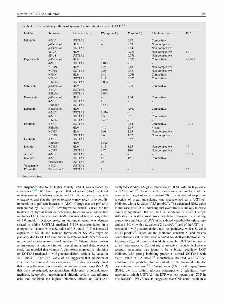
Table 4 The inhibitory effects of tyrosine kinase inhibitors on UGT1A168–74.
Inhibitor Substrate Enzyme source IC50 (μmol/L) Ki (μmol/L) Inhibition type Ref.
Nilotinib 4-MU UGT1A1 – 0.17 Competitiveβ-Estradiol HLM – 0.53 Non-competitiveβ-Estradiol UGT1A1 – 0.14 Non-competitiveSN-38 HLM 0.286 Non-competitive 69SN-38 UGT1A1 0.079 Non-competitive
Regorafenib β-Estradiol HLM – 0.020 Competitive 68,70,714-MU UGT1A1 0.045 – –
NCHN HLM 0.26 0.48 Non-competitiveNCHN UGT1A1 0.25 0.33 Non-competitiveNPHN HLM 0.40 0.048 CompetitiveNPHN UGT1A1 0.17 0.027 CompetitiveBilirubin UGT1A1 0.034 – –
Sorafenib β-Estradiol HLM – 0.033 Competitive4-MU UGT1A1 0.066 – –
Bilirubin UGT1A1 0.048 – –
Pazopanib β-Estradiol HLM – 2.34 Competitive4-MU UGT1A1 1.1 – –
Bilirubin UGT1A1 37.34 – –
Lapatinib β-Estradiol HLM – 0.567 Competitive4-MU UGT1A1 0.536 – –
4-MU UGT1A1 0.5 0.5 CompetitiveBilirubin UGT1A1 0.467 – –
Erlotinib 4-MU UGT1A1 – 0.64 Competitive 72,74Bilirubin HLM 4.19 2.97 MixedNCHN HLM 0.68 1.23 Non-competitiveNCHN UGT1A1 0.69 1.72 Non-competitive
Gefitinib 4-MU UGT1A1 – 2.42 –
Bilirubin HLM >100 – –
Icotinib NCHN HLM 5.15 8.55 Non-competitive 72NCHN UGT1A1 8.76 10.04 Non-competitive
Axitinib 4-MU UGT1A1 – – – 70,73Imatinib 4-MU UGT1A1 11.0 19.1 Competitive
Paracetamol UGT1A1 29 – –
Vandetanib 4-MU UGT1A1 – – –
Dasatinib Paracetamol UGT1A1 2 – – 73
–Not determined.
Review on UGT1A1 inhibitors 265
was suspended due to its higher toxicity, and it was replaced byentacapone84,85. We have reported that tolcapone (also) displayedrelative stronger inhibitory effects on UGT1A1 in comparison withentacapone, and that the use of tolcapone may result in hyperbilir-ubinemia or significant increase in AUC of drugs that are primarilymetabolized by UGT1A160. Levothyroxine, which is used for thetreatment of thyroid hormone deficiency, functions as a competitiveinhibitor of UGT1A1-mediated 4-MU glucuronidation, at a Ki valueof 1.0 μmol/L75. Ketoconazole, an antifungal agent, was demon-strated to inhibit UGT1A1 mediated SN-38 glucuronidation in acompetitive manner, with a Ki value of 3.3 μmol/L76. The increasedexposure of SN-38 and reduced formation of SN-38G might beprimarily due to UGT1A1 inhibition by ketoconazole, when ketoco-nazole and irinotecan were coadministered76. Vitamin A (retinol) isan important micronutrient in both vegetal and animal diets. A recentstudy has revealed that vitamin A also exerts competitive inhibitionof UGT1A1-mediated 4-MU-glucuronidation, with a Ki value of31.1 μmol/L77. The [I]/Ki value of 3.1 suggested that inhibition ofUGT1A1 by vitamin A may exist in vivo77. It was previously foundthat among the seven non-steroidal antiinflammatory drugs (NSAID)that were investigated, acetaminophen, diclofenac, diflunisal, indo-methacin, ketoprofen, naproxen and niflumic acid, it was niflumicacid that exhibited the highest inhibitory effects on UGT1A1-
catalyzed estradiol-3-O-glucuronidation in HLM, with an IC50 valueof 22.2 μmol/L78. More recently, everolimus, an inhibitor of themammalian target of rapamycin (mTOR) that is utilized to preventrejection of organ transplants, was characterized as a UGT1A1inhibitor, with a Ki value of 2.3 μmol/L79. The calculated [I]/Ki valuein this case was 0.004, indicating that everolimus is unlikely to causeclinically significant DDI via UGT1A1 inhibition in vivo79. Diethyl-stilbestrol, a widely used toxic synthetic estrogen, is a strongcompetitive inhibitor of UGT1A1-catalyzed estradiol-3-O-glucuroni-dation in HLM, with a Ki value of 2.1 μmol/L80, and of the UGT1A1-mediated 4-MU-glucuronidation, also competitively, with a Ki valueof 3.7 μmol/L81. Based on the inhibition constant Ki and plasmaconcentrations values that were reported for diethylstilbestrol in theliterature (Cmax 24 μmol/L), it is likely to inhibit UGT1A1 in vivo, ifgiven intravenously. Zafirlukast, a selective peptide leukotrienereceptor antagonist, was found to be a broad specificity UGTinhibitor, with strong inhibiting properties toward UGT1A1 withthe Ki value of 1.2 μmol/L82. Nonetheless, no DDI via UGT1A1inhibition was predicted for zafirlukast, if the unbound inhibitorconcentration was used82. Canagliflozin (CNF) and dapagliflozin(DPF), the first sodium glucose cotransporter 2 inhibitors, werereported to inhibit UGT1A1, but DPF was less potent than CNF inthis respect83. IVIVE results suggested that CNF could result in a

Table 5 The inhibitory effects of other drugs on UGT1A160,75–83.
Inhibitor Substrate Enzyme source IC50 (μmol/L) Ki (μmol/L) Inhibition type Ref.
Entacapone 4-MU UGT1A1 9.10 10.48 Competitive 60Bilirubin HLM 34.97 30.82 MixedNCHN HLM 16.92 14.65 Non-competitiveNCHN UGT1A1 12.24 15.59 Non-competitive
Tolcapone 4-MU UGT1A1 2.38 1.77 CompetitiveBilirubin HLM 1.24 0.68 MixedNCHN HLM 2.07 1.03 Non-competitiveNCHN UGT1A1 1.30 2.39 Non-competitive
Levothyroxine 4-MU UGT1A1 – 1.0 Competitive 75Ketoconazole SN-38 UGT1A1 – 3.3 Competitive 76
Bilirubin HLM 53 – –
Vitamin A 4-MU UGT1A1 31.1 Competitive 77Diclofenac β-Estradiol HLM 60.9 112 Non-competitive 78
4-MU UGT1A1 57.5 – –
Diflunisal β-Estradiol HLM 37.8 – –
Indomethacin β-Estradiol HLM 51.5 – –
Niflumic acid β-Estradiol HLM 22.2 – –
Everolimus 4-MU UGT1A1 – 2.3 Competitive 79Diethylstilbestrol β-Estradiol HLM – 2.1 Competitive 80
4-MU UGT1A1 – 3.7 Competitive 81Zafirlukast 4-MU UGT1A1 0.7 – – 82
SN-38 HLM – 1.2 Non-competitiveCanagliflozin β-Estradiol UGT1A1 – 7.2 Competitive 83
β-Estradiol HLM – 9.1 CompetitiveDapagliflozin β-Estradiol HLM – 81 Competitive
β-Estradiol HLM – 81 Competitive
–Not determined.
Xia Lv et al.266
significant increase in the AUC for exclusive UGT1A1 substrates viaUGT1A1 inhibition, while DNF could not inhibit UGTA1 in vivo83.
3.2. Environmental toxins as UGT1A1 inhibitors
Environmental toxins that display inhibitory effects on UGT1A1 arelisted in Table 686,87. Gossypol is a polyphenolic compound that isfound in cotton seeds and was used as a male anti-fertility drug for along time. However, the clinical utilization of gossypol has alwaysbeen strongly limited due to its toxicity, including hepatotoxicity,pathological changes in rat and human testes, abnormal sperm andenzyme inhibition. In an HLM study, gossypol was found tomoderately inhibit estradiol-3-O-glucuronidation, with an IC50 valueof 23.5 μmol/L. The inhibition kinetic was noncompetitive inhibitionand the Ki value was 34.2 μmol/L86. The [I]/Ki ratio of 0.56 suggestedthat the use of gossypol could cause DDI through inhibition ofUGT1A186. Four uremic toxins, benzyl alcohol, p-cresol, indoxylsulfate, hippuric acid, and a combination of these four uremic toxins,were found to inhibit UGT1A1 to variable extents. Among thefour uremic toxins, p-cresol was the most potent inhibitor ofUGT1A1-mediated estradiol-3-O-glucuronidation in HLM, with aKi value of 43 μmol/L87.
Table 6 The inhibitory effects of environmental toxins on UGT1A1
Inhibitor Substrate Enzyme source IC50(μm
Gossypol β-Estradiol HLM 23.5P-Cresol β-Estradiol HLM –
–Not determined.
3.3. Herbal extracts that inhibit UGT1A1
Many studies have investigated the inhibitory effects of herbextracts on UGT1A1 activity (Table 720,21,88–93). For example,inhibitors were tested among the following eight commonly usedherbal extracts, milk thistle, saw palmetto, echinacea, green teaepigallocatechingallate, garlic, ginseng, black cohosh, and valer-ian, of which milk thistle, saw palmetto, echinacea, and epigallo-catechingallate exhibited the highest inhibition potency ofUGT1A1-mediated estradiol-3-O-glucuronidation, with IC50
values between 7.8 and 211.7 μg/mL21. A volume per dose index(VDI) values suggested that inhibition of intestinal UGT1A1 byepigallocatechingallate and milk thistle, and to a lesser extent bysaw palmetto and echinacea, may be clinically relevant21. Andro-graphis paniculata and Orthosiphon stamineus extracts displayedinhibitory effects on UGT1A1-mediated 4-MU-glucuronidation,with IC50 values of 5.0 and 24.65 μg/mL, respectively88. Poly-gonum multiflorum extracts exhibited strong inhibitory effects onUGT1A1-mediated bilirubin glucuronidation in HLM and in ratliver microsomes (RLM), with Ki values of 1.6 and 0.3 μmol/L,respectively89. Herbal extract of Daio, Kanzo, Keihi, and Ogonstrongly inhibited UGT1A1-mediated estradiol-3-O-
86,87.
ol/L) Ki (μmol/L) Inhibition type Ref.
34.2 Non-competitive 8643 Competitive 87
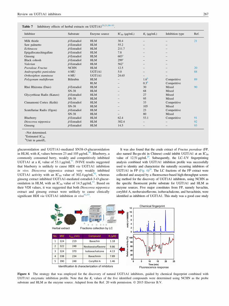
Table 7 Inhibitory effects of herbal extracts on UGT1A120,21,88–93.
Inhibitor Substrate Enzyme source IC50 (μg/mL) Ki (μg/mL) Inhibition type Ref.
Milk thistle β-Estradiol HLM 30.4 – – 21Saw palmetto β-Estradiol HLM 55.2 – –
Echinacea β-Estradiol HLM 211.7 – –
Epigallocatechingallate β-Estradiol HLM 7.8 – –
Ginseng β-Estradiol HLM 603a – –
Black cohosh β-Estradiol HLM 299a – –
Valerian β-Estradiol HLM 562a – –
Psoraleae Fructus NCHN HLM 12.5 – – 20Andrographis paniculata 4-MU UGT1A1 5.0 – – 88Orthosiphon stamineus 4-MU UGT1A1 24.65 – –
Polygonum multiflorum Bilirubin HLM – 1.6b Competitive 89RLM – 0.3b Competitive
Rhei Rhizoma (Daio) β-Estradiol HLM – 30 Mixed 90SN-38 HLM – 68 Mixed
Glycyrrhizae Radix (Kanzo) β-Estradiol HLM – 27 MixedSN-38 HLM – 95 Mixed
Cinnamomi Cortex (Keihi) β-Estradiol HLM – 33 CompetitiveSN-38 HLM – 105 Mixed
Scutellariae Radix (Ogon) β-Estradiol HLM – 23 CompetitiveSN-38 HLM – 80 Mixed
Blueberry β-Estradiol HLM 62.4 53.1 Competitive 91Dioscorea nipponica β-Estradiol HLM 302.4 – – 92Ginseng β-Estradiol HLM 14.5 – – 93
–Not determined.aEstimated IC50.bUnit in μmol/L.
Review on UGT1A1 inhibitors 267
glucuronidation and UGT1A1-mediated SN38-O-glucuronidationin HLM, with Ki values between 23 and 105 μg/mL90. Blueberry, acommonly consumed berry, weakly and competitively inhibitedUGT1A1 at a Ki value of 53.1 μg/mL91. IVIVE results suggestedthat blueberry is unlikely to cause HDI via UGT1A1 inhibitionin vivo. Dioscorea nipponica extract very weakly inhibitedUGT1A1 activity with an IC50 value of 302.4 μg/mL92, whereasginseng extract inhibited UGT1A1-mediated estradiol-3-O-glucur-onidation in HLM, with an IC50 value of 14.5 μg/mL93. Based ontheir VDI values, it was suggested that both Dioscorea nipponicaextract and ginseng extract were unlikely to cause clinicallysignificant HDI via UGT1A1 inhibition in vivo92,93.
Fractions collection by LC Herbal extract
No. MW λmax (nm) Compound Ki (μM)
Identification & characterization of inhibitors
Figure 6 The strategy that was employed for the discovery of naturalUGT1A1 enzymatic inhibition profile. Note that the Ki values of the fivsubstrate and HLM as the enzyme source. Adapted from the Ref. 20 with
It was also found that the crude extract of Fructus psoraleae (FP,also named Bu-gu-zhi in Chinese) could inhibit UGT1A1 at an IC50
value of 12.51 μg/mL20. Subsequently, the LC–UV fingerprintinganalysis combined with UGT1A1 inhibition profile was successfullyused to identity and characterize the naturally occurring inhibitors ofUGT1A1 in FP (Fig. 6)20. The LC fractions of the FP extract werecollected and assayed by a fluorescence-based high-throughput screen-ing method for the discovery of UGT1A1 inhibitors, using NCHN asthe specific fluorescent probe substrate for UGT1A1 and HLM asenzyme sources. Five major constitutes from FP, namely bavachin,corylifol A, neobavaisoflavone, isobavachalcone, and bavachinin, wereidentified as inhibitors of UGT1A1. This study was a good case study
Fluorescence response
Chemical fingerprint
UGT1A1 inhibitors, guided by chemical fingerprint combined withe identified compounds were determined using NCHN as the probepermission. © 2015 Elsevier B.V.

Table 8 The inhibitory effects of fatty acids on UGT1A194.
Inhibitor Substrate Enzyme source IC50 (μmol/L) Ki (μmol/L) Inhibition type Ref.
Oleic acid β-Estradiol UGT1A1 31.6 23.4 Non-competitive 94HLM – 29.3 Non-competitive
Linoleic acid β-Estradiol UGT1A1 33.1 22.1 Non-competitiveHLM – 24.0 Non-competitive
Palmitoleic acid β-Estradiol UGT1A1 37.1 – –
α-Linolenic acid β-Estradiol UGT1A1 26.1 – –
Arachidonic acid β-Estradiol UGT1A1 22.7 – –
DHA β-Estradiol UGT1A1 11.6 1.8 Non-competitiveHLM – 4.3 Non-competitiveHIM – 5.2 Non-competitive
EPA β-Estradiol UGT1A1 19.9 – –
Stearic acid β-Estradiol UGT1A1 450 – –
Decanoic acid β-Estradiol UGT1A1 450 – –
– Not determined.
Xia Lv et al.268
for the discovery of UGT1A1 inhibitors from medicinal plants, whichwould be very helpful for future investigations on UGT1A1-mediatedherb–drug interactions.
3.4. Natural products as UGT1A1 inhibitors
3.4.1. Fatty acidsFatty acids, an important class of natural products, are carboxylicacids with long straight-aliphatic chains, either saturated orunsaturated, ranging from 4 to 28 carbon atoms in length. Theinhibitory effects of 15 saturated and unsaturated fatty acids onUGT1A1-catalyzed estradiol-3-O-glucuronidation were investi-gated. Among the 15 tested fatty acids, 7 displayed stronginhibition, including oleic acid, linoleic acid, docosahexaenoicacid (DHA), eicosapentaenoic acid (EPA), palmitoleic acid,arachidonic acid, and a-linolenic acid, with IC50 values between11.6 and 37.1 μmol/L (Table 8)94. In addition, oleic acid, linoleicacid, and DHA noncompetitively inhibited estradiol-3-O-glucur-onidation mediated by both recombinant UGT1A1 and HLM, withKi values between 1.8 and 29.3 μmol/L. Unlike oleic acid andlinoleic acid, however, DHA has a potency to noncompetitivelyinhibit intestinal estradiol-3-O-glucuronidation, with a Ki value of5.2 μmol/L, probably indicating inhibition of UGT1A1061. Inter-estingly, unsaturated fatty acids exerted strong inhibition againstUGT1A1 activity, whereas saturated fatty acid only poorlyinhibited UGT1A1 activity. An in vivo study demonstrated thatlow concentrations of DHA result in a significant increase inserum bilirubin via UGT1A1 inhibition, while high concentrationsof oleic acid, linoleic acid, and DHA cause a decrease in serumbilirubin via UGT1A1 induction94.
3.4.2. FlavonoidsFlavonoids that display inhibitory effects on UGT1A1 are listed inTable 918,20,25,95–105. Flavonoids are a class of polyphenoliccompounds that are widely distributed in nature and have beendeveloped into drugs, cosmetics and health food due to variouspharmacological properties106. Five major flavonoids componentsof FP, bavachin, corylifol A, neobavaisoflavone, isobavachalcone,and bavachinin (see Fig. 5 for their analysis), exhibited strong tomoderate inhibitory effects on UGT1A1-mediated NCHN-O-glucuronidation in HLM, with Ki values ranging between 1.18and 9.86 μmol/L20. The [I]/Ki value of bavachinin was calculated
to be greater than 0.1, indicating that inhibition of UGT1A1in vivo seems likely. However, the [I]/Ki values of the other fourcompounds that were identified from FP could not be estimateddue to the lack of their plasma concentrations20. In addition,bavachalcone and corylin, two other major bioactive flavonoidsfrom FP, were examined for UGT1A1 inhibition. Bavachalconeinhibited the 4-MU glucuronidation activity of the enzymenoncompetitively, with a Ki value of 5.41 μmol/L, while corylindid not inhibit UGT1A195. Recent studies have demonstrated thatsome important flavonoid ingredients of licorice, including lico-chalcone A (LCA), isoliquiritigenin, and liquiritigenin, inhibitUGT1A1 with Ki values below 10 μmol/L18,96,107. Furthermore,LCA, isoliquiritigenin, and liquiritigenin inhibited the UGT1A1-mediated 4-MU glucuronidation competitively, whereas LCAinhibited it noncompetitively, at least when the substrate wasNCHN18,96,107. In addition, IVIVE results indicated that LCAcould increase the AUC of UGT1A1 substrates by 71%–341% viaUGT1A1 inhibition, while isoliquiritigenin was unlikely to inhibitUGT1A1 in vivo18,98. Several other flavonoids, including wogo-nin, scutellarein, baicalein, alpinetin, genkwanin, apigenin, hesper-etin, and naringenin were reported to strongly inhibit UGT1A1,with Ki values between 0.02 and 16.47 μmol/L. Kinetics analysesof these inhibitions demonstrate that wogonin97, scutellarein98,baicalein99, and alpinetin102 are competitive inhibitors ofUGT1A1-mediated 4-MU glucronidation, while hesperetin, andnaringenin100 are noncompetitive inhibitors. Genkwanin andapigenin are competitive inhibitors of billirubin glucronidation inHLM103. IVIVE results indicated that scutellarein was highlylikely to cause clinically significant HDI via UGT1A1 inhibitionin vivo, while hesperetin and naringenin might not98,100.
Some diet-derived constituents including kaempferol, andepigallocatechin gallate (EGCG) also inhibited UGT1A1, but noneof them was predicted to inhibit UGT1A1 in vivo104. It is worthnoting that deglycosylation of liquiritin into liquiritigenin, ofscutellarein into scutellarin, and of baicalein into baicalin wasshown to significantly increase their inhibitory effects towardsUGT1A1. The inhibition profiles of several other flavonoids,including daidzein, genistein, biochanin A, chrysin, apigenin andnaringenin against UGT1A1-mediated SN38-O-glucuronidationwere examined in UGT1A1-overexpressing Hela cells, resultingin a range of IC50 values between 0.37 and 5.85 μmol/L101. Recentstudies demonstrated that amentoflavone and sciadopitysin, twonatural biflavonoid distributed in many medicinal plants, are strong

Table 9 The inhibitory effects of flavonoids on UGT1A118,20,25,95–105.
Inhibitor Substrate Enzyme source IC50 (μmol/L) Ki (μmol/L) Inhibition type Ref.
Bavachin 4-MU UGT1A1 1.79 1.08 Competitive 20NCHN HLM 1.85 1.18 Non-competitiveNCHN UGT1A1 0.75 0.04 Non-competitive
Neobavaisoflavone 4-MU UGT1A1 1.80 11.96 CompetitiveNCHN HLM 2.42 9.86 Non-competitiveNCHN UGT1A1 2.25 3.95 Non-competitive
Isobavachalcone 4-MU UGT1A1 13.04 10.93 CompetitiveNCHN HLM 4.43 4.13 Non-competitiveNCHN UGT1A1 3.40 4.09 Non-competitive
Bavachinin 4-MU UGT1A1 1.99 2.22 CompetitiveNCHN HLM 4.16 7.89 Non-competitiveNCHN UGT1A1 1.27 4.09 Non-competitive
Corylifol A 4-MU UGT1A1 1.48 0.47 CompetitiveNCHN HLM 1.48 1.46 Non-competitiveNCHN UGT1A1 0.65 0.79 Non-competitive
Licochalcone A 4-MU UGT1A1 0.97 0.78 Competitive 18NCHN HLM 0.84 0.54 Non-competitiveNCHN UGT1A1 0.13 0.23 Non-competitive
Bavachalcone 4-MU UGT1A1 11.3 5.41 Competitive 95Corylin 4-MU UGT1A1 – - –
Liquiritigenin 4-MU UGT1A1 – 9.1 Competitive 107Liquiritin 4-MU UGT1A1 –
a– –
Isoliquiritigenin 4-MU UGT1A1 – 0.7 Competitive 96Wogonin 4-MU UGT1A1 – 1.40 Competitive 97Scutellarein 4-MU UGT1A1 – 0.02 Competitive 98Scutellarin 4-MU UGT1A1 >100 – –
Baicalein 4-MU UGT1A1 – 1.2 Competitive 99Baicalin 4-MU UGT1A1 >100 – –
Hesperetin 4-MU UGT1A1 4.75 9.62 Non-competitive 100Naringenin 4-MU UGT1A1 8.58 7.61 Non-competitive
β-Estradiol UGT1A1 4.89 – – 101Hela1A1 4.24 – –
SN-38 UGT1A1 1.58 – –
Hela1A1 2.63 – –
Alpinetin 4-MU UGT1A1 – 3.0 Competitive 102Genkwanin Bilirubin HLM 23.21 16.47 Competitive 103Apigenin Bilirubin HLM 12.40 4.08 Competitive
β-Estradiol UGT1A1 0.47 – – 101Hela1A1 0.33 – –
Review
onUGT1A
1inhibitors
269
Table 9 (continued )
Inhibitor Substrate Enzyme source IC50 (μmol/L) Ki (μmol/L) Inhibition type Ref.
SN-38 UGT1A1 0.72 – –
Hela1A1 0.48 – –
Naringin 4-MU UGT1A1 14.8 – – 104Kaempferol 4-MU UGT1A1 7.9 – –
EGCG 4-MU UGT1A1 26.2 – –
Daidzein β-Estradiol UGT1A1 52.1 – – 101Hela1A1 67.1 – –
SN-38 UGT1A1 5.01 – –
Hela1A1 5.85 – –
Genistein β-Estradiol UGT1A1 1.83 – –
Hela1A1 0.94 – –
SN-38 UGT1A1 0.98 – –
Hela1A1 1.43 – –
Biochanin A β-Estradiol UGT1A1 1.58 – –
Hela1A1 0.84 – –
SN-38 UGT1A1 0.42 – –
Hela1A1 0.37 – –
Chrysin β-Estradiol UGT1A1 2.02 – –
Hela1A1 0.98 – –
SN-38 UGT1A1 1.16 – –
Hela1A1 1.26 – –
Phloretin β-Estradiol UGT1A1 2.17 – –
Hela1A1 1.66 – –
SN-38 UGT1A1 1.96 – –
Hela1A1 2.84 – –
Amentoflavone 4-MU UGT1A1 0.78 2.21 Competitive 25NCHN HLM 0.21 0.24 Non-competitiveNCHN UGT1A1 0.14 0.27 Non-competitive
Sciadopitysin 4-MU UGT1A1 0.65 0.54 Competitive 105NCHN HLM 0.35 0.41 Non-competitiveNCHN UGT1A1 0.31 0.45 Non-competitive
– Not determined.
Xia
Lvet
al.270

Review on UGT1A1 inhibitors 271
competitive inhibitors of UGT1A1-mediated 4-MU glucuronida-tion, but function as noncompetitive inhibitors in both UGT1A1and HLM when the substrate was NCHN25,105. IVIVE resultssuggested that the use of sciadopitysin could result in a significantincrease in the AUC of UGT1A1 substrates via UGT1A1inhibition107.
3.4.3. QuinonesQuinones are widely distributed in plant species and have multiplepharmacological activities108,109. Quinones that display inhibitoryeffects on UGT1A1 are listed in Table 1019,101,110,111. It was recentlydemonstrated that 10 major quinone constituents of Polygonummultiforum, namely cis-emodindianthrones, trans-emodindianthrones,emodin-8-O-glc, polygonumnolide C2, emodin, polygonumnolideC3, citreorosein, polygonumnolide C4, physcion, and rhein, arenaturally occurring potent inhibitors of UGT1A1, with Ki valuesbetween 0.863 to 127.3 μmol/L19. The inhibition of UGT1A1 activityby these quinones might be one of the reasons for P. multiforum-associated adverse effects, particularly elevated bilirubin levels andliver injury19. Another study demonstrated that emodin competitivelyinhibited UGT1A1 activity in three model systems, HLM, RLMand recombinant UGT1A1, with Ki values of 5.40, 10.02 and4.85 μmol/L, respectively110. In addition, emodin displayed stronginhibitory effects in the UGT1A1/Hela1A1 system when estradiol-3-O-glucuronidation and SN-38-O-glucuronidation activities wereexamined (IC50 o 2 μmol/L)101. It should be added, however, thatemodin is probably a broad specificity inhibitor and it was shown toinhibit UGT1A10 and/or UGT1A8112, and perhaps other UGTsas well. Tanshinone I, tanshinone IIA, cryptotanshinone, anddihydrotanshinone I are major quinone constituents ofDanshen that displayed moderate inhibitory effects on UGT1A1(IC50440 μmol/L)111. Furthermore, since the Cmax values of crypto-tanshinone and dihydrotanshinone I were much lower than theirIC50 values, it was concluded that both of them could not inhibitUGT1A1 in vivo111.
Table 10 The inhibitory effects of quinones on UGT1A119,101,110,11
Inhibitor Substrate Enzyme source IC
cis-Emodin dianthrones Bilirubin RLM –
trans-Emodin dianthrones Bilirubin RLM –
Emodin-8-O-glc Bilirubin RLMPolygonumnolide C2 Bilirubin RLM –
Polygonumnolide C3 Bilirubin RLM –
Polygonumnolide C4 Bilirubin RLM –
Physcion Bilirubin RLM –
Emodin Bilirubin RLM –
Rhein Bilirubin RLM –
Citreorosein Bilirubin RLM –
Emodin Bilirubin UGT1A1 –
Bilirubin HLM –
Bilirubin RLM –
β-Estradiol UGT1A1 1.Hela1A1 0.
SN-38 UGT1A1 0.Hela1A1 0.
Tanshinone I 4-MU UGT1A1 77Tanshinone IIA 4-MU UGT1A1 69Cryptotanshinone 4-MU UGT1A1 43Dihydrotanshinone I 4-MU UGT1A1 67
–Not determined.
3.4.4. LignansLignans that were shown to display inhibitory effects onUGT1A1 are listed in Table 11101,104,113–116. Lignans are a largegroup of natural products that are widely spread in the plantkingdom117. Milk thistle flavonolignans (silybin A, silybin B,isosilybin B, isosilychristin, and silydianin) were demonstrated toinhibit UGT1A1-mediated 4-MU glucuronidation, at IC50 valuesranging between 5.3 and 53.5 μmol/L. This resulted in a predic-tion that none of them was likely to inhibit UGT1A1 in vivo104. Itwas previously reported that silybin inhibits the UGT1A1-mediated 7-hydroxy-4-trifluoromethylcoumarin glucuronidation,at an IC50 value of 1.4 mmol/L114, whereas the metabolite,silibinin-glucuronide was found in another study to inhibit ratUGT1A1 in RLM, with a Ki value of 16 μg/mL113. Podophyllo-toxin, an important lignan that is found in multiple plants,was demonstrated to inhibit the UGT1A1-mediated 4-MU-glucuronidation competitively, with a Ki value of 4.0 μmol/L115.However, 1, 10 and 100 μmol/L of podophyllotoxin didnot really inhibited HLM-catalyzed SN-38 glucuronidation, sincethe residual activities were 109.7%, 103.8%, and 64.1% of thenegative control, respectively. At the same concentrations,podophyllotoxin also had barely a minor effect on HLM-catalyzed estradiol-3-O-glucuronidation, with residual activitiesbeing 95.1%, 89.1%, and 84.1% of the negative control,respectively115. Thus, inhibition of UGT1A1 by podophyllotoxinis substrate-dependent and mostly mild, namely medium andweak inhibition towards HLM-mediated estradiol-3-O-glucuroni-dation and SN-38-O-glucuronidation, respectively115. Honokiol,another plant lignan, was found to slightly inhibit UGT1A1-mediated estradiol-3-O-glucuronidation, with an IC50 value of50.5 μmol/L116. Magnolol and macelignan, two lignans that arefound in multiple plants, exhibited similar inhibitory effects onestradiol-3-O-glucuronidation and SN-38-O-glucuronidation,regardless of whether recombinant UGT1A1, or over-expressingHela cells were the enzyme source101.
1.
50 (μmol/L) Ki (μmol/L) Inhibition type Ref.
0.8630 Competitive 191.083 Competitive3.425 Competitive4.291 Non-competitive12.89 Non-competitive77.42 Un-competitive94.75 Non-competitive10.01 Competitive127.3 Mixed18.56 Mixed4.85 Competitive 1105.40 Competitive10.02 Competitive
27 – – 10177 – –
96 – –
63 – –
.2 – – 111
.8 – –
.5 – –
.3 – –

Table 11 The inhibitory effects of lignans on UGT1A1101,104,113–116.
Inhibitor Substrate Enzyme source IC50 (μmol/L) Ki (μmol/L) Inhibition type Ref.
Silibinin-glucuronide Bilirubin RLM – 16a Competitive 113Silybin A 4-MU HIM 64.8 104
UGT1A1 28.8Silybin B 4-MU HLM 87.3
HIM 46.9UGT1A1 27.5
Isosilybin B 4-MU HLM – – –
HIM 187 – –
UGT1A1 51.1 – –
Isosilychristin 4-MU HLM – – –
HIM – – –
UGT1A1 53.5 – –
Silydianin 4-MU HLM 97.7 – –
HIM – – –
UGT1A1 5.3 – –
Silybin 7-Hydroxy-4-trifluoromethylcoumarin UGT1A1 1.4 – – 114Podophyllotoxin 4-MU UGT1A1 4.0 Competitive 115Honokiol β-Estradiol HLM 50.5 – – 116Magnolol β-Estradiol UGT1A1 36.8 – – 101
Hela1A1 22.6 – –
SN-38 UGT1A1 13.2 – –
Hela1A1 16.4 – –
Macelignan β-Estradiol UGT1A1 7.40 – –
Hela1A1 5.33 – –
SN-38 UGT1A1 4.73 – –
Hela1A1 2.71 – –
–Not determined.aUnit in μg/mL.
Xia Lv et al.272
3.4.5. Other natural compoundsAlongside the above listed, widely occurring plant compounds, thereare other plant compounds that are frequently found, such aspolyphenolic acids, polyphenolics, terpenoids, coumarins and alkaloids,that were reported to inhibit UGT1A1 (Table 1290,93,101,118–123).Salvianolic acids A and B, two major polyphenolic acids ingredientsin Danshen, strongly inhibited UGT1A1-catalyed bilirubin glucuroni-dation, via mixed type inhibition kinetics, with Ki values of 0.22 and4.50 μmol/L, respectively118. Demethylzeylasteral, a triterpenoid that isisolated from Tripterygium wilfordii Hook F, functions as a non-competitive inhibitor of the UGT1A1-mediated 4-MU-glcuronidation,with a Ki value of 21.7 μmol/L119, and PPT, a triterpenoid componentthat is isolated from Ginseng, exerted strong noncompetitive inhibitiontowards UGT1A1, with a Ki value of 8.8 mmol/L121. The [I]/Ki valueof 0.2 that was calculated for PPT inhibiting UGT1A1, suggests thatPPT might also inhibit the enzyme in vivo. Another study found that 20(S)-ginsenoside Rg3 and 20(S)-ginsenoside Rh2, two triterpenoidcomponents that are isolated from Ginseng, exerted potent inhibitoryeffects on UGT1A1-mediated estradiol-3-O-glucuronidation in HLM93.Notably, among the five major xanthophylls that were investigated,astaxanthin, zeaxanthin, β-cryptoxanthin, canthaxanthin and lutein, thestrongest UGT1A1 inhibition was exhibited by β-cryptoxanthin, with aKi value of 12.2 mmol/L
120. Its calculated [I]/Ki value for UGT1A1 was0.012, indicating that β-cryptoxanthin was, however, unlikely to causea clinically significant DDI via UGT1A1 inhibition120. Glycyrrhetinicacid, a triterpenoid component from liquorice, exhibited moderateinhibitory effects on UGT1A1-mediated estradiol-3-O-glucuronidationand SN-38 glucuronidation in HLM, with Ki values of 28.8 and25.4 mmol/L, respectively90. Brachyantheraoside A2, a triterpenoidsaponin from Stauntonia brachyanthera, competitively inhibited
UGT1A1-catalyzed 4-MU glucuronidation, with a Ki value of 9.3mmol/L122. The inhibition profiles of several natural compounds,including gingerol (6-shogaol, 6-, 8-, and 10-gingerol), stilbenoid(resveratrol), capsaicinoid (capsaicin), and coumestan (psoralidin)toward UGT1A1 were tested in UGT1A1-overexpressing Hela cells,revealing IC50 values ranging between 0.86 and 122 μmol/L101.Corydaline, a bioactive isoquinoline alkaloid from Corydalis tubers,was found to moderately inhibit UGT1A1-mediated estradiol-3-O-glucuronidation, at a Ki value of 57.6 mmol/L123.
4. Further challenges and future directions
From the physiological function point of view, UGT1A1 is one ofthe most important mammalian UGTs, due to its essential role inbilirubin metabolism. In most cases, dysfunction or strong inhibi-tion of UGT1A1, either due to inherited mutation(s), or inhibitionby drugs or other xenobiotics, could be detected in the clinic basedon their effects on the plasma levels of unconjugated bilirubin. Thelevels of total blood bilirubin and unconjugated bilirubin are oftendetermined in routine clinical testing. Many UGT1A1 inhibitors,such as several flavonoids and pentacyclic triterpenoids, that wereidentified from in vitro assays, turned out to be ineffective in vivo.Poor cell permeability and poor metabolic stability of these naturalcompounds, together leading to poor bioavailability, are probablythe major causes of their ineffectiveness in vivo. Notably, themajority of the data presented in the review was derived fromin vitro assays, and often the inhibitor concentration was muchhigher than the expected plasma concentration. In other words, theinhibitory effects of these compounds on intracellular UGT1A1,

Table 12 The inhibitory effects of other natural compounds on UGT1A190,93,101,118–123.
Inhibitor Substrate Enzyme source IC50 (μmol/L) Ki (μmol/L) Inhibition type Ref.
Salvianolic Acid A Bilirubin UGT1A1 1.13 0.22 Mixed 118Salvianolic Acid B Bilirubin UGT1A1 10.87 4.50 MixedProtocatechuic aldehyde Bilirubin UGT1A1 738.01 – –
Rosmarinic acid Bilirubin UGT1A1 149.53 – –
Danshensu Bilirubin UGT1A1 340.20 – –
Demethylzeylasteral 4-MU UGT1A1 - 21.70 Non-competitive 119β-Cryptoxanthin β-Estradiol HLM 18.8 12.2 Competitive 120Lutein β-Estradiol HLM 45.5 – –
Canthaxanthin β-Estradiol HLM 38.5 – –
Astaxanthin β-Estradiol HLM 450 – –
Zeaxanthin β-Estradiol HLM 450 – –
20(S)-Protopanaxatriol 4-MU UGT1A1 – 8.8 Non-competitive 121Glycyrrhetinic acid β-Estradiol HLM – 28.8 Mixed 90
SN-38 HLM – 25.4 MixedBrachyantheraoside A2 4-MU UGT1A1 – 9.3 Competitive 12220(S)-Ginsenoside Rg3 β-Estradiol HLM 89.0 – – 9320(S)-Ginsenoside Rh2 β-Estradiol HLM 54.5 – –
Psoralidin β-Estradiol UGT1A1 2.21 – – 101Hela1A1 3.85 – –
SN-38 UGT1A1 0.86 – –
Hela1A1 1.07 – –
6-Shogaol β-Estradiol UGT1A1 8.46 – –
Hela1A1 9.89 – –
SN-38 UGT1A1 1.52 – –
Hela1A1 1.08 – –
6-Gingerol β-Estradiol UGT1A1 135 – –
Hela1A1 80.1 – –
SN-38 UGT1A1 122 – –
Hela1A1 77.3 – –
8-Gingerol β-Estradiol UGT1A1 17.2 – –
Hela1A1 14.2 – –
SN-38 UGT1A1 8.40 – –
Hela1A1 14.1 – –
10-Gingerol β-Estradiol UGT1A1 10.2 – –
Hela1A1 18.5 – –
SN-38 UGT1A1 5.09 – –
Hela1A1 5.76 – –
Resveratrol β-Estradiol UGT1A1 3.42 – –
Hela1A1 1.85 – –
SN-38 UGT1A1 28.3 – –
Hela1A1 19.4 – –
Capsaicin β-Estradiol UGT1A1 51.3 – –
Hela1A1 21.4 – –
SN-38 UGT1A1 23.3 – –
Hela1A1 16.2 – –
Corydaline β-Estradiol HLM 137.1 57.6 Mixed 123
–Not determined.
Review on UGT1A1 inhibitors 273
particularly their in vivo potency against UGT1A, have not beenwell investigated. In order to make in the future such studies moremeaningful, it is necessary to develop new or refined test methods.Among the issues to be considered in future inhibition assays arekeeping the inhibitor concentration low, which is close to itsplasma concentration. Further investigations on the design anddevelopment of fluorescent probes for UGT1A1 with highspecificity, high sensitivity, good practicability and excellentoptical properties (such as long wavelength probes), and theirapplications in HTS of UGT1A1 inhibitors in complex biologicalsystems are still highly desirable.
In contrast to a wide range of structurally diverse UGT1A1inhibitors, UGT1A1 inducers or simulators are rarely reported and
most studies focus on transcriptional regulation of the UGT1A1gene124–126. Induction of UGT1A1 expression by synthetic ornatural compounds in a clinical setting to treat UGT1A1 deficien-cies, such as phenobarbital treatment, is common in neonatals. It isless clear, but an interesting idea, whether having an activitystimulating compound for UGT1A will be effective in preventionof CPT-11/SN-38 toxicity, for patients who are homozygouscarriers of the polymorphic variant UGT1A1*28127. To this end,it is necessary to develop methods for HTS of UGT1A1 inducersor simulators in living systems such as cryo-preserved humanhepatocytes. Although the newly developed fluorescent probes forUGT1A1 (NCHN and NHPN) have been used for rapid screeningof UGT1A1 inducers at the function level36,44, the poor cell

Xia Lv et al.274
permeability of NCHN and the short emission wavelength of theirglucuronides make them unsuitable probe substrates for screeningUGT1A1 inducers or simulators in hepatocytes culture. Hence,cell-based assays in combination with highly sensitive andpractical fluorescence detection for HTS of UGT1A1 inducers orsimulators are one of the challenging objectives in both academicresearch and for drug development.
Besides UGT1A1, other human UGT enzymes also playimportant roles in the metabolism and detoxification of therapeuticdrugs and other xenobiotics. For example, UGT1A4 plays a majorrole in the metabolism of trifluoperazine128, a drug that was firstused for the treatment of schizophrenia and later, more broadly,epilepsy. UGT1A4 can be specifically inhibited by hecogenin(a compound from sisal plant), which may trigger potential risks ofHDI128. UGT2B7 is perhaps the most important human UGTenzyme in drug metabolism, since it is involved in the conjugationof many drugs including the HIV/AIDS (acquired immunodefi-ciency syndrome) drugs (such as zidovudine) and the opioids(such as codeine and morphine)129,130. Therefore, attention shouldbe paid to screening and characterization inhibitors of other humanUGTs, since UGT-mediated DDI and HDI usually involve multi-ple UGT enzymes rather than UGT1A1 only7. In contrast toUGT1A1, the specific substrates for some other human UGTenzymes are rarely reported, due to the common overlappingsubstrate specificity of UGTs.
Considering the inherent advantages of fluorescent probesubstrates, such as highly sensitivity, and applicability to HTSassay131, there is a clear advantage in the design and developmentof practical and highly specific fluorescent probe substrates for atarget UGT enzyme. Notably, the design principles are alreadyavailable and experience in the design and development of specificfluorescent substrates for UGT1A1 will surely assist us and otherresearchers in developing new specific fluorescent substrates forother UGTs. In fact, several groups have already tried in the pastto develop fluorescent substrates for other human UGTs, as well asto construct efficient fluorescence-based assays for HTS ofinhibitors toward target enzyme. For instance, 1-naphthol wasfound to be a good fluorescent substrate for UGT1A6, which canbe used for HTS of UGT1A6 inhibitors using recombinant enzymeas an enzyme source132. However, 1-naphthol can be conjugatedby other human UGTs as well, limiting its applications to therecombinant enzyme, rather than use in more complex system suchas HLM. Very recently, a set of fluorescent 7-hydroxycoumarinederivatives have been developed as specific substrates forUGT1A10 (an extrahepatic UGT) and at least two of them(compound 2 and 4), appear to work well in tissue preparations133.All these findings are very helpful for the design and developmentof fluorescent probes for different human UGTs, and we hope thatmore practical fluorescent probes for human UGT enzymes will besuccessfully developed and used in the near future.
5. Concluding remarks
The key roles of UGT1A1 in both endobiotic homeostasis andxenobiotic metabolism have drawn much attention from bothacademic and drug industry scientists. Now the US FDA and otherregulatory agencies have recommended that the inhibition poten-tials of investigational new drugs on the human UGT1A1 shouldbe evaluated before approval. In this review, the significance,progress and challenges in the discovery and characterization ofUGT1A1 inhibitors, as well as recent advances in the development
of UGT1A1 probe substrates for screening and characterization ofUGT1A1 inhibitors, have been described. The tools for UGT1A1-related investigations, such as probe substrates and specificinhibitors of this key conjugative enzyme, have been summarizedfor the first time. More importantly, lists of UGT1A1 inhibitors,along with detail information that includes their inhibition potency,mode of inhibition, and affinity (when available), have beenprepared and presented. The information and knowledge that aregiven in this review are expected to provide guidance for rationaluse of clinical drugs or herbal medicines in order to avoid theoccurrence of adverse side effects via UGT1A1 inhibition, as wellpresent practical methods for rapid screening and characterizationof UGT1A1 inhibitors and for facilitating the investigations onUGT1A1–ligand interactions. We hope that this review will alsofacilitate the development of more specific and practical tools forother human UGTs in the near future.
Acknowledgments
This work was supported by the NSF of China (81773687,81703606, 81573501, 81473181), the National Key Researchand Development Program of China (2017YFC1700200 and2017YFC1702000), the Fundamental Research Funds for theCentral Universities (wd01185), and the National S&T MajorProjects of China (2017ZX09101004), Program of ShanghaiAcademic/Technology Research Leader (18XD1403600), theInnovative Entrepreneurship Program of High-level Talents inDalian (2016RQ025 & 2017RQ121), and the Doctoral ScientificResearch Foundation of Liaoning Province, China (20170520059).
References
1. Rowland A, Miners JO, Mackenzie PI. The UDP-glucuronosyltrans-ferases: their role in drug metabolism and detoxification. IntJ Biochem Cell Biol 2013;45:1121–32.
2. Oda S, Fukami T, Yokoi T, Nakajima M. A comprehensive review ofUDP-glucuronosyltransferase and esterases for drug development.Drug Metab Pharmacokinet 2015;30:30–51.
3. Meech R, Mackenzie PI. UGT3A: novel UDP-Glycosyltransferasesof the UGT superfamily. Drug Metab Rev 2010;42:45–54.
4. Sato Y, Nagata M, Tetsuka K, Tamura K, Miyashita A, KawamuraA, et al. Optimized methods for targeted peptide-based quantificationof human uridine 50-diphosphate-glucuronosyltransferases in biolo-gical specimens using liquid chromatography-tandem mass spectro-metry. Drug Metab Dispos 2014;42:885–9.
5. Bosma PJ. Inherited disorders of bilirubin metabolism. J Hepatol2003;38:107–17.
6. Bock KW. Roles of human UDP-glucuronosyltransferases in clear-ance and homeostasis of endogenous substrates, and functionalimplications. Biochem Pharmacol 2015;96:77–82.
7. Kiang TK, Ensom MH, Chang TK. UDP-glucuronosyltransferases andclinical drug–drug interactions. Pharmacol Ther 2005;106:97–132.
8. Court MH. Interindividual variability in hepatic drug glucuronida-tion: studies into the role of age, sex, enzyme inducers, and geneticpolymorphism using the human liver bank as a model system. DrugMetab Rev 2010;42:209–24.
9. Sim SC, Kacevska M, Ingelman-Sundberg M. Pharmacogenomics ofdrug-metabolizing enzymes: a recent update on clinical implicationsand endogenous effects. Pharmacogenomics J 2013;13:1–11.
10. Kadakol A, Ghosh SS, Sappal BS, Sharma G, Chowdhury JR,Chowdhury NR. Genetic lesions of bilirubin uridine‐diphosphoglu-curonate glucuronosyltransferase (UGT1A1) causing crigler‐najjarand gilbert syndromes: correlation of genotype to phenotype. HumMutat 2000;16:297–306.

Review on UGT1A1 inhibitors 275
11. McDonagh AF. Bilirubin toxicity to human erythrocytes: a moresanguine view. Pediatrics 2007;120:175–8.
12. Sticova E, Jirsa M. New insights in bilirubin metabolism and theirclinical implications. World J Gastroenterol 2013;19:6398–407.
13. Erlinger S, Arias IM, Dhumeaux D. Inherited disorders of bilirubintransport and conjugation: new insights into molecular mechanismsand consequences. Gastroenterology 2014;146:1625–38.
14. Tukey RH, Strassburg CP. Human UDP-glucuronosyltransferases:metabolism, expression, and disease. Annu Rev Pharmacol Toxicol2000;40:581–616.
15. Ginsberg G, Guyton K, Johns D, Schimek J, Angle K, Sonawane B.Genetic polymorphism in metabolism and host defense enzymes:implications for human health risk assessment. Crit Rev Toxicol2010;40:575–619.
16. Wang L, Chan CE, Wong AL, Wong FC, Lim SW, Chinnathambi A,et al. Combined use of irinotecan with histone deacetylase inhibitorbelinostat could cause severe toxicity by inhibiting SN-38 glucur-onidation via UGT1A1. Oncotarget 2017;8:41572–81.
17. Goon CP, Wang LZ, Wong FC, Thuya WL, Ho PC, Goh BC.UGT1A1 mediated drug interactions and its clinical relevance. CurrDrug Metab 2016;17:100–6.
18. Xin H, Qi XY, Wu JJ, Wang XX, Li Y, Hong JY, et al. Assessmentof the inhibition potential of licochalcone a against human UDP-glucuronosyltransferases. Food Chem Toxicol 2016;90:112–22.
19. Wang Q, Wang Y, Li Y, Wen B, Dai Z, Ma S, et al. Identificationand characterization of the structure–activity relationships involvedin UGT1A1 inhibition by anthraquinone and dianthrone constituentsof Polygonum multiflorum. Sci Rep 2017;7:17952.
20. Wang XX, Lv X, Li SY, Hou J, Ning J, Wang JY, et al.Identification and characterization of naturally occurring inhibitorsagainst UDP-glucuronosyltransferase 1A1 in Fructus Psoraleae (Bu-Gu-Zhi). Toxicol Appl Pharmacol 2015;289:70–8.
21. Mohamed ME, Tseng T, Frye RE. Inhibitory effects of commonly usedherbal extracts on UGT1A1 enzyme activity. Xenobiotica 2010;40:663–9.
22. Peer CJ, Sissung TM, Kim A, Jain L, Woo S, Gardner ER, et al.Sorafenib is an inhibitor of UGT1A1 but is metabolized byUGT1A9: implications of genetic variants on pharmacokinetics andhyperbilirubinemia. Clin Cancer Res 2012;18:2099–107.
23. Singer JB, Shou Y, Giles F, Kantarjian HM, Hsu Y, Robeva AS,et al. UGT1A1 promoter polymorphism increases risk of nilotinib-induced hyperbilirubinemia. Leukemia 2007;21:2311–5.
24. Zucker SD, Qin X, Rouster SD, Yu F, Green RM, Keshavan P, et al.Mechanism of indinavir-induced hyperbilirubinemia. Proc Natl AcadSci U S A 2001;98:12671–6.
25. Lv X, Zhang JB, Wang XX, Hu WZ, Shi YS, Liu SW, et al.Amentoflavone is a potent broad-spectrum inhibitor of human udp-glucuronosyltransferases. Chem Biol Interact 2018;284:48–55.
26. Gammal RS, Court MH, Haidar CE, Iwuchukwu OF, Gaur AH,Alvarellos M, et al. Clinical pharmacogenetics implementationconsortium (Cpic) guideline for UGT1A1 and atazanavir prescribing.Clin Pharmacol Ther 2016;99:363–9.
27. Saif MW, Smith MH, Maloney A, Diasio RB. Imatinib-inducedhyperbilirubinemia with UGT1A1 (*28) promoter polymorphism:first case series in patients with gastrointestinal stromal tumor. AnnGastroenterol 2016;29:551–6.
28. US Food and Drug Administration. Guidance for industry: druginteraction studies—study design, data analysis, implications fordosing and labeling recommendations; Draft guidance. Availablefrom: ⟨http://www.fda.gov/downloads/Drugs/GuidanceComplianceRegulatoryInformation/Guidances/UCM292362.pdf.2012⟩.
29. European Agency Medicines. Guideline on the investigation of druginteractions. Available from: ⟨http://www.ema.europa.eu/docs/en_GB/document_library/Scientific_guideline/2012/07/WC500129606.pdf.2012⟩.
30. Wen Z, Tallman MN, Ali SY, Smith PC. UDP-glucuronosyltransferase1A1 is the principal enzyme responsible for etoposide glucuronidationin human liver and intestinal microsomes: structural characterization ofphenolic and alcoholic glucuronides of etoposide and estimation ofenzyme kinetics. Drug Metab Dispos 2007;35:371–80.
31. Zhou J, Tracy TS, Remmel RP. Correlation between bilirubinglucuronidation and estradiol-3-gluronidation in the presence ofmodel UDP-glucuronosyltransferase 1A1 substrates/inhibitors. DrugMetab Dispos 2011;39:322–9.
32. Zou LW, Wang P, Qian XK, Feng L, Yu Y, Wang DD, et al. Ahighly specific ratiometric two-photon fluorescent probe to detectdipeptidyl peptidase IV in plasma and living systems. BiosensBioelectron 2017;90:283–9.
33. Wang P, Xia YL, Zou LW, Qian XK, Dou TY, Jin Q, et al. Anoptimized two-photon fluorescent probe for biological sensing andimaging of catechol-O-methyltransferase. Chem Eur J 2017;23:10800–7.
34. Dai ZR, Feng L, Jin Q, Cheng H, Li Y, Ning J, et al. A practicalstrategy to design and develop an isoform-specific fluorescent probe fora target enzyme: CYP1A1 as a case study. Chem Sci 2017;8:2795–803.
35. Jin Q, Feng L, Wang DD, Wu JJ, Hou J, Dai ZR, et al. A highlyselective near-infrared fluorescent probe for carboxylesterase 2 andits bioimaging applications in living cells and animals. BiosensBioelectron 2016;83:193–9.
36. Lv X, Ge GB, Feng L, Troberg J, Hu LH, Hou J, et al. An optimizedratiometric fluorescent probe for sensing human UDP-glucuronosyltransferase 1A1 and its biological applications. BiosensBioelectron 2015;72:261–7.
37. Jin Q, Feng L, Wang DD, Dai ZR, Wang P, Zou LW, et al. A two-photon ratiometric fluorescent probe for imaging carboxylesterase 2 inliving cells and tissues. ACS Appl Mater Interfaces 2015;7:28474–81.
38. Feng L, Liu ZM, Hou J, Lv X, Ning J, Ge GB, et al. A highly selectivefluorescent esipt probe for the detection of human carboxylesterase2 and its biological applications. Biosens Bioelectron 2015;65:9–15.
39. Dai ZR, Ge GB, Feng L, Ning J, Hu LH, Jin Q, et al. A highlyselective ratiometric two-photon fluorescent probe for human cyto-chrome P450 1A. J Am Chem Soc 2015;137:14488–95.
40. Liu ZM, Feng L, Ge GB, Lv X, Hou J, Cao YF, et al. A highlyselective ratiometric fluorescent probe for in vitro monitoring andcellular imaging of human carboxylesterase 1. Biosens Bioelectron2014;57:30–5.
41. Terai T, Tomiyasu R, Ota T, Ueno T, Komatsu T, Hanaoka K, et al.Tokyogreen derivatives as specific and practical fluorescent probesfor UDP-glucuronosyltransferase (UGT) 1A1. Chem Commun2013;49:3101–3.
42. Feng L, Liu ZM, Xu L, Lv X, Ning J, Hou J, et al. A highly selectivelong-wavelength fluorescent probe for the detection of humancarboxylesterase 2 and its biomedical applications. Chem Commun2014;50:14519–22.
43. Tong H, Lou K, Wang W. Near-infrared fluorescent probes forimaging of amyloid plaques in Alzheimer's disease. Acta Pharm SinB 2015;5:25–33.
44. Lv X, Feng L, Ai CZ, Hou J, Wang P, Zou LW, et al. A practical andhigh-affinity fluorescent probe for uridine diphosphate glucuronosyltrans-ferase 1A1: a good surrogate for bilirubin. J Med Chem 2017;60:9664–75.
45. Ciotti M, Owens IS. Evidence for overlapping active sites for 17α-ethynlestradiol and bilirubin in the human major bilirubin UDP-glucuronosyltransferase. Biochemistry 1996;35:10119–24.
46. Rios GR, Tephly TR. Inhibition and active sites of UDP-glucuronosyltransferases 2B7 and 1A1. Drug Metab Dispos2002;30:1364–7.
47. Gagné JF, Montminy V, Belanger P, Journault K, Gaucher G,Guillemette C. Common human UGT1A polymorphisms and thealtered metabolism of irinotecan active metabolite 7-ethyl-10-hydroxycamptothecin (SN-38). Mol Pharmacol 2002;62:608–17.
48. Uchaipichat V, Mackenzie PI, Guo XH, Gardner-Stephen D, GaletinA, Houston JB, et al. Human UDP-glucuronosyltransferases: isoformselectivity and kinetics of 4-methylumbelliferone and 1-naphtholglucuronidation, effects of organic solvents, and inhibition bydiclofenac and probenecid. Drug Metab Dispos 2004;32:413–23.
49. Wang Q, Dai Z, Wen BY, Ma SC, Zhang YJ. Estimating thedifferences of UGT1A1 activity in recombinant UGT1A1 enzyme,human liver microsomes and rat liver microsome incubation systemsin vitro. Biol Pharm Bull 2015;38:1910–7.

Xia Lv et al.276
50. Walsky RL, Bauman JN, Bourcier K, Giddens G, Lapham K, NegahbanA, et al. Optimized assays for human UDP-glucuronosyltransferase (UGT)activities: altered alamethicin concentration and utility to screen for UGTinhibitors. Drug Metab Dispos 2012;40:1051–65.
51. Soars MG, Ring BJ, Wrighton SA. The effect of incubation conditionson the enzyme kinetics of UDP-glucuronosyltransferases. Drug MetabDispos 2003;31:762–7.
52. Wang LZ, Ramírez J, Yeo W, Chan MY, Thuya WL, Lau JY, et al.Glucuronidation by UGT1A1 is the dominant pathway of themetabolic disposition of belinostat in liver cancer patients. PLoSOne 2013;8:e54522.
53. Wu B, Zhang S, Hu M. Evaluation of 3,30,40-trihydroxyflavone and3,6,40-trihydroxyflavone (40-O-glucuronidation) as the in vitro func-tional markers for hepatic UGT1A1. Mol Pharm 2011;8:2379–89.
54. Xia YL, Ge GB, Wang P, Liang SC, He YQ, Ning J, et al. Structuralmodifications at the C-4 position strongly affect the glucuronidationof 6,7-Dihydroxycoumarins. Drug Metab Dispos 2015;43:553–60.
55. Laakkonen L, Finel M. A molecular model of the human UDP-glucuronosyltransferase 1A1, its membrane orientation, and theinteractions between different parts of the enzyme. Mol Pharmacol2010;77:931–9.
56. Gradinaru J, Romand S, Geiser L, Carrupt PA, Spaggiari D, Rudaz S.Inhibition screening method of microsomal UGTs using the cocktailapproach. Eur J Pharm Sci 2015;71:35–45.
57. Zhang D, Zhang D, Cui D, Gambardella J, Ma L, Barros A, et al.Characterization of the UDP glucuronosyltransferase activity ofhuman liver microsomes genotyped for the UGT1A1*28 polymorph-ism. Drug Metab Dispos 2007;35:2270–80.
58. Itäaho K, Mackenzie PI, Ikushiro SI, Miners JO, Finel M. Theconfiguration of the 17-hydroxy group variably influencesthe glucuronidation of β-estradiol and epiestradiol byhuman UDP-glucuronosyltransferases. Drug Metab Dispos2008;36:2307–15.
59. Zhou J, Tracy TS, Remmel RP. Bilirubin glucuronidation revisited:proper assay conditions to estimate enzyme kinetics with recombi-nant UGT1A1. Drug Metab Dispos 2010;38:1907–11.
60. Lv X, Wang XX, Hou J, Fang ZZ, Wu JJ, Cao YF, et al. Comparisonof the inhibitory effects of tolcapone and entacapone against humanUDP-glucuronosyltransferases. Toxicol Appl Pharmacol2016;301:42–9.
61. Troberg J, Järvinen E, Ge GB, Yang L, Finel M. UGT1A10 is a highactivity and important extrahepatic enzyme: why has its role inintestinal glucuronidation been frequently underestimated?. MolPharm 2017;14:2875–83.
62. Zhang D, Chando TJ, Everett DW, Patten CJ, Dehal SS, HumphreysWG. In vitro inhibition of UDP glucuronosyltransferases by ataza-navir and other HIV protease inhibitors and the relationship of thisproperty to in vivo bilirubin glucuronidation. Drug Metab Dispos2005;33:1729–39.
63. Ji HY, Lee H, Lim SR, Kim JH, Lee HS. Effect of efavirenz onUDP-glucuronosyltransferase 1A1, 1A4, 1A6, and 1A9 activities inhuman liver microsomes. Molecules 2012;17:851–60.
64. Alam N, Angeli MG, Greenblatt DJ. Mechanism of in-vitro inhibition ofUGT1A1 by paritaprevir. J Pharm Pharmacol 2017;69:1794–801.
65. Cao L, Greenblatt DJ, Kwara A. Inhibitory effects of selectedantituberculosis drugs on common human hepatic cytochromeP450 and UDP-glucuronosyltransferase enzymes. Drug Metab Dis-pos 2017;45:1035–43.
66. Sane RS, Steinmann GG, Huang Q, Li Y, Podila L, Mease K, et al.Mechanisms underlying benign and reversible unconjugatedhyperbilirubinemia observed with faldaprevir administration inhepatitis C virus patients. J Pharmacol Exp Ther 2014;351:403–12.
67. McDonald C, Uy J, Hu W, Wirtz V, Juethner S, Butcher D, et al.Clinical significance of hyperbilirubinemia among HIV-1-infectedpatients treated with atazanavir/ritonavir through 96 weeks in theCASTLE study. AIDS Patient Care STDS 2012;26:259–64.
68. Miners JO, Chau N, Rowland A, Burns K, McKinnon RA,Mackenzie PI, et al. Inhibition of human UDP-
glucuronosyltransferase enzymes by lapatinib, pazopanib, regorafe-nib and sorafenib: implications for hyperbilirubinemia. BiochemPharmacol 2017;129:85–95.
69. Fujita KI, Sugiyama M, Akiyama Y, Ando Y, Sasaki Y. The small-molecule tyrosine kinase inhibitor nilotinib is a potent noncompeti-tive inhibitor of the SN-38 glucuronidation by human UGT1A1.Cancer Chemother Pharmacol 2011;67:237–41.
70. Zhang N, Liu Y, Jeong H. Drug–drug interaction potentials of tyrosinekinase inhibitors via inhibition of UDP-glucuronosyltransferases. SciRep 2015;5:17778.
71. Sun XY, Ge GB, Tang H, Wang YQ, Yao XC, Li L. Inhibition ofregorafenib against UDP-Glucuronosyltransferases. Acta Pharm Sin2017;52:1705–14.
72. Cheng X, Lv X, Qu H, Li D, Hu M, Guo W, et al. Comparison of theinhibition potentials of icotinib and erlotinib against human UDP-glucuronosyltransferase 1A1. Acta Pharm Sin B 2017;7:657–64.
73. Liu Y, Ramírez J, Ratain MJ. Inhibition of paracetamol glucuronidationby tyrosine kinase inhibitors. Br J Clin Pharmacol 2011;71:917–20.
74. Liu Y, Ramírez J, House L, Ratain MJ. Comparison of the drug–druginteractions potential of erlotinib and gefitinib via inhibition of UDP-glucuronosyltransferases. Drug Metab Dispos 2010;38:32–9.
75. Zhao HD, Bao GQ, He XL, Wu T, Wang CG, Wang SZ, et al. Stronginhibition of UDP-glucuronosyltransferase (UGT) 1A1 by levothyr-oxine indicates the potential UGT-inhibition based adverse effect oflevothyroxine. Lat Am J Pharm 2012;31:761–3.
76. Yong WP, Ramirez J, Innocenti F, Ratain MJ. Effects of Ketocona-zole on glucuronidation by UDP-glucuronosyltransferase Enzymes.Clin Cancer Res 2005;11:6699–704.
77. Liu X, Cao YF, Dong PP, Zhu LL, Zhao ZY, Wu X, et al. Theinhibition of UDP-glucuronosyltransferases (UGTs) by vitamin A.Xenobiotica 2017;47:376–81.
78. Mano Y, Usui T, Kamimura H. In vitro inhibitory effects of non‐steroidal antiinflammatory drugs on UDP‐glucuronosyltransferase1A1‐catalysed estradiol 3β‐glucuronidation in human liver micro-somes. Biopharm Drug Dispos 2005;26:35–9.
79. Du Z, Wang G, Cao YF, Hu CM, Yang K, Liu YZ, et al.Everolimus-inhibited multiple isoforms of UDP-glucuronosyltransferases (UGTs). Xenobiotica 2018;48:452–8.
80. Zhu L, Xiao L, Xia Y, Zhou K, Wang H, Huang M, et al.Diethylstilbestrol can effectively accelerate estradiol-17-O-glucuro-nidation, while potently inhibiting estradiol-3-O-glucuronidation.Toxicol Appl Pharmacol 2015;283:109–16.
81. Zhu L, Xiao L, Li W, Zhang Y, Han W, Zhu Y, et al. Human UDP‐glucuronosyltransferases 1A1, 1A3, 1A9, 2B4 and 2B7 are inhibited bydiethylstilbestrol. Basic Clin Pharmacol Toxicol 2016;119:505–11.
82. Oda S, Fujiwara R, Kutsuno Y, Fukami T, Itoh T, Yokoi T, et al.Targeted screen for human UDP-glucuronosyltransferases inhibitorsand the evaluation of potential drug–drug interactions with zafirlu-kast. Drug Metab Dispos 2015;43:812–8.
83. Pattanawongsa A, Chau N, Rowland A, Miners JO. Inhibition ofhuman UDP-glucuronosyltransferase enzymes by canagliflozin anddapagliflozin: implications for drug–drug interactions. Drug MetabDispos 2015;43:1468–76.
84. Nissinen E, Kaheinen P, Penttilä KE, Kaivola J, Lindén IB.Entacapone, a novel catechol-O-methyltransferase inhibitor forParkinson's disease, does not impair mitochondrial energy produc-tion. Eur J Pharmacol 1997;340:287–94.
85. Haasio K, Sopanen L, Vaalavirta L, Lindén IB, Heinonen EH.Comparative toxicological study on the hepatic safety of entacaponeand tolcapone in the rat. J Neural Transm 2001;108:79–91.
86. Zhang YS, Yuan J, Fang ZZ, Tu YY, Hu CM, Li G, et al. Gossypolexhibits a strong influence towards UDP-glucuronosyltransferase(UGT) 1A1, 1A9 and 2B7-mediated metabolism of xenobioticsand endogenous substances. Molecules 2012;17:4896–903.
87. Barnes KJ, Rowland A, Polasek TM, Miners JO. Inhibition of humandrug-metabolising cytochrome P450 and UDP-glucuronosyltransferaseenzyme activities in vitro by uremic toxins. Eur J Clin Pharmacol2014;70:1097–106.

Review on UGT1A1 inhibitors 277
88. Ismail S, Hanapi NA, Ab Halim MR, Uchaipichat V, Mackenzie PI.Effects of Andrographis paniculata and Orthosiphon stamineusextracts on the glucuronidation of 4-methylumbelliferone in humanUGT isoforms. Molecules 2010;15:3578–92.
89. Wang Q, Dai Z, Zhang YJ, Shuangcheng M. The research of thehepatotoxicity of Polygonum multiflorum on the basic of theinhibition of the UGT1A1 enzyme in vivo and in vitro. Chin PharmJ 2016;51:1929–33.
90. Katoh M, Yoshioka Y, Nakagawa N, Yokoi T. Effects of Japaneseherbal medicine, Kampo, on human UGT1A1 activity. Drug MetabPharmacokinet 2009;24:226–34.
91. Choi EJ, Park JB, Yoon KD, Bae SK. Evaluation of the in vitro/in vivo potential of five berries (bilberry, blueberry, cranberry,elderberry, and raspberry ketones) commonly used as herbal supple-ments to inhibit uridine diphospho-glucuronosyltransferase. FoodChem Toxicol 2014;72:13–9.
92. Ji HY, Liu KH, Kong TY, Jeong HU, Choi SZ, Son M, et al.Evaluation of Da-9801, a new herbal drug for diabetic neuropathy,on metabolism-mediated interaction. Arch Pharm Res 2013;36:1–5.
93. Zheng YF, Bae SH, Choi EJ, Park JB, Kim SO, Jang MJ, et al.Evaluation of the in vitro/in vivo drug interaction potential ofBst204, a purified dry extract of ginseng, and its four bioactiveginsenosides through cytochrome P450 inhibition/induction andUDP-glucuronosyltransferase inhibition. Food Chem Toxicol2014;68:117–27.
94. Shibuya A, Itoh T, Tukey RH, Fujiwara R. Impact of fatty acids onhuman UDP-glucuronosyltransferase 1A1 activity and its expressionin neonatal hyperbilirubinemia. Sci Rep 2013;3:2903.
95. Shan L, Yang S, Zhang G, Zhou D, Qiu Z, Tian L, et al. Comparisonof the Inhibitory potential of bavachalcone and corylin against UDP-glucuronosyltransferases. Evid Based Complement Altern Med2014;2014:958937.
96. Lu H, Fang ZZ, Cao YF, Hu CM, Hong M, Sun XY, et al.Isoliquiritigenin showed strong inhibitory effects towards multipleUDP-glucuronosyltransferase (UGT) isoform-catalyzed 4-methylum-belliferone (4-MU) glucuronidation. Fitoterapia 2013;84:208–12.
97. Shan LN, Shi XB. Strong inhibition capability of wogonin towardsUDP-glucuronosyltransferase (UGT) 1A1 and 1A3. Latin AmJ Pharm 2013;32:741–4.
98. Ma GY, Cao YF, Hu CM, Fang ZZ, Sun XY, Hong M, et al.Comparison of inhibition capability of scutellarein and scutellarintowards important liver UDP‐glucuronosyltransferase (UGT) iso-forms. Phytother Res 2014;28:382–6.
99. Teng Y, Nian H, Zhao H, Chen P, Wang G. Biotransformation ofbaicalin to baicalein significantly strengthens the inhibition potentialtowards UDP-glucuronosyltransferases (UGTs) isoforms. Pharmazie2013;68:763–7.
100. Liu D, Wu J, Xie H, Liu M, Takau I, Zhang H, et al. Inhibitory effectof hesperetin and naringenin on human UDP-glucuronosyltransferaseenzymes: implications for herb–drug interactions. Biol Pharm Bull2016;39:2052–9.
101. Sun H, Zhou X, Wu B. Accurate identification of UDP-glucuronosyltransferase 1A1 (UGT1A1) inhibitors using UGT1A1-overexpressing hela cells. Xenobiotica 2015;45:945–53.
102. Gao C, Cao Y, Gao X, Qu Y, Liu H, Liu HL, et al. Significantinhibition of UDP-glucuronosyltransferase (UGT) 1A1 by plantflavonoid alpinetin. Latin Am J Pharm 2012;31:1360–2.
103. Chen H, Miao P, Guo C, Chen N, Ma P, Li H, et al. In vitroinhibitory effect of main flavonoids ingredients of Genkwa Flos onliver microsomal UGTs and UGT1A1 activities. Chin J Mod ApplPharm 2017;34:305–10.
104. Gufford BT, Chen G, Lazarus P, Graf TN, Oberlies NH, Paine MF.Identification of diet-derived constituents as potent inhibitors ofintestinal glucuronidation. Drug Metab Dispos 2014;42:1675–83.
105. Wang XX, Hou J, Ning J, Pan YQ, Hong M, Guo B. Inhibition ofsciadopitysin against UDP-glucuronosyltransferases. Acta Pharm Sin2016;51:749–55.
106. Wang X, Liu Q, Zhu H, Wang H, Kang J, Shen Z, et al. Flavanolsfrom the Camellia sinensis var. assamica and their hypoglycemic andhypolipidemic activities. Acta Pharm Sin B 2017;7:342–6.
107. Guo B, Fan XR, Fang ZZ, Cao YF, Hu CM, Yang JL, et al.Deglycosylation of liquiritin strongly enhances its inhibitory potentialtowards UDP-glucuronosyltransferase (UGT) isoforms. PhytotherRes 2013;27:1232–6.
108. Detremmerie C, Vanhoutte PM, Leung S. Biased activity of solubleguanylyl cyclase: the janus face of thymoquinone. Acta Pharm Sin B2017;7:401–8.
109. Hao DC, Gu X, Xiao P. Anemone medicinal plants: ethnopharmacol-ogy, phytochemistry and biology. Acta Pharm Sin B 2017;7:146–58.
110. Wang Q, Dai Z, Zhang YJ, Ma SC. Hepatotoxicity of emodin basedon UGT1A1 enzyme-mediated bilirubin in liver microsomes. ChinaJ Chin Mater Med 2016;41:4424–7.
111. Zhang XX, Cao YF, Wang LX, Yuan XL, Fang ZZ. Inhibitory effectsof tanshinones towards the catalytic activity of UDP-glucuronosyltransferases (UGTs). Pharm Biol 2017;55:1703–9.
112. Watanabe Y, Nakajima M, Yokoi T. Troglitazone glucuronidation inhuman liver and intestine microsomes: high catalytic activity ofUGT1A8 and UGT1A10. Drug Metab Dispos 2002;30:1462–9.
113. D'Andrea V, Pérez LM, Pozzi EJ. Inhibition of rat liver UDP-glucuronosyltransferase by silymarin and the metabolite silibinin-glucuronide. Life Sci 2005;77:683–92.
114. Sridar C, Goosen TC, Kent UM, Williams JA, Hollenberg PF. Silybininactivates cytochromes P450 3A4 and 2C9 and inhibits majorhepatic glucuronosyltransferases. Drug Metab Dispos 2004;32:587–94.
115. Qi JQ, Cao YF, Sun XY, Hong M, Fang ZZ, Meng DL, et al.Structure-inhibition relationship of podophyllotoxin (PT) analoguestowards UDP-glucuronosyltransferase (UGT) isoforms. Pharmazie2015;70:239–43.
116. Jeong HU, Kong TY, Kwon SS, Hong SW, Yeon SH, Choi JH,et al. Effect of honokiol on cytochrome P450 and UDP-glucuronosyltransferase enzyme activities in human liver micro-somes. Molecules 2013;18:10681–93.
117. Yang Y, An Y, Wang W, Du N, Zhang J, Feng Z, et al. Ninecompounds from the root bark of Lycium chinense and their anti-inflammatory activities. Acta Pharm Sin B 2017;7:491–5.
118. Ma G, Zhang Y, Chen W, Tang Z, Xin X, Yang P, et al. Inhibition ofhuman UGT1A1-mediated bilirubin glucuronidation by polyphenolicacids impact safety of popular salvianolic acid A/B-containing drugsand herbal products. Mol Pharm 2017;14:2952–66.
119. Yu ML, Lin JJ, Yang Y, Zhang WJ, Bai MC, Wang CM, et al.Evaluation of inhibition of UDP-glucuronosyltransferase (UGT) 1A1by demethylzeylasteral. Latin Am J Pharm 2012;31:1067–70.
120. Zheng YF, Min JS, Kim D, Park JB, Choi SW, Lee ES, et al. In vitroinhibition of human UDP-glucuronosyl-transferase (UGT) isoformsby astaxanthin, β-cryptoxanthin, canthaxanthin, lutein, and zeax-anthin: prediction of in vivo dietary supplement–drug interactions.Molecules 2016;21:1052.
121. He YJ, Fang ZZ, Ge GB, Jiang P, Jin HZ, Zhang WD, et al. Theinhibitory effect of 20(S)-protopanaxatriol (PPT) towards UGT1A1and UGT2B7. Phytother Res 2013;27:628–32.
122. Liu D, Li S, Qi JQ, Meng DL, Cao YF. The inhibitory effects of nor-oleanane triterpenoid saponins from Stauntonia brachyantheratowards UDP-glucuronosyltransferases. Fitoterapia 2016;112:56–64.
123. Ji HY, Liu KH, Lee H, Im SR, Shim HJ, Son M, et al. Corydaline inhibitsmultiple cytochrome P450 and UDP-glucuronosyltransferase enzymeactivities in human liver microsomes. Molecules 2011;16:6591–602.
124. Westerink WM, Schoonen WG. Phase II enzyme levels in Hepg2cells and cryopreserved primary human hepatocytes and theirinduction in HepG2 cells. Toxicol Vitr 2007;21:1592–602.
125. Smith CM, Graham RA, Krol WL, Silver IS, Negishi M, Wang H,et al. Differential UGT1A1 induction by chrysin in primaryhuman hepatocytes and HepG2 cells. J Pharmacol Exp Ther2005;315:1256–64.

Xia Lv et al.278
126. Sugatani J. Function, genetic polymorphism, and transcriptionalregulation of human UDP-glucuronosyltransferase (UGT) 1A1. DrugMetab Pharmacokinet 2013;28:83–92.
127. Aoshima N, Fujie Y, Itoh T, Tukey RH, Fujiwara R. Glucose inducesintestinal human UDP-glucuronosyltransferase (UGT) 1A1 to preventneonatal hyperbilirubinemia. Sci Rep 2014;4:6343.
128. Uchaipichat V, Mackenzie PI, Elliot DJ, Miners JO. Selectivity ofsubstrate (trifluoperazine) and inhibitor (amitriptyline, androsterone,canrenoic acid, hecogenin, phenylbutazone, quinidine, quinine, andsulfinpyrazone) “probes” for human UDP- glucuronosyltransferases.Drug Metab Dispos 2006;34:449–56.
129. Barbier O, Turgeon D, Girard C, Green MD, Tephly TR, Hum DW,et al. 30-azido-30-deoxythimidine (Azt) is glucuronidated by humanUDP-glucuronosyltransferase 2B7 (UGT2B7). Drug Metab Dispos2000;28:497–502.
130. Court MH, Krishnaswamy S, Hao Q, Duan SX, Patten CJ, von MoltkeLL, et al. Evaluation of 30-azido-30-deoxythymidine, morphine, andcodeine as probe substrates for UDP-glucuronosyltransferase 2B7(UGT2B7) in human liver microsomes: specificity and influence of theUGT2B7*2 polymorphism. Drug Metab Dispos 2003;31:1125–33.
131. Zhao J, Chen J, Ma S, Liu Q, Huang L, Chen X, et al. Recentdevelopments in multimodality fluorescence imaging probes. ActaPharm Sin B 2018;8:320–38.
132. Soikkeli A, Kurkela M, Hirvonen J, Yliperttula M, Finel M.Fluorescence-based high-throughput screening assay for drug inter-actions with UGT1A6. Assay Drug Dev Technol 2011;9:496–502.
133. Juvonen RO, Rauhamaki S, Kortet S, Niinivehmas S, TrobergJ, Petsalo A, et al. Molecular docking-based design and developmentof a highly selective probe substrate for UDP-glucuronosyltransferase1A10. Mol Pharm 2018;15:923–33.