Embed Size (px)
Citation preview

Sensors 2015, 15, 24374-24396; doi:10.3390/s150924374
sensors ISSN 1424-8220
www.mdpi.com/journal/sensors
Review
Recent Progress in Fluorescent Imaging Probes
Yen Leng Pak, K. M. K. Swamy and Juyoung Yoon *
Department of Chemistry and Nano Sciences, Ewha Womans University, Seoul 120-750, Korea;
E-Mails: [email protected] (Y.L.P.); [email protected] (K.M.K.S.)
* Author to whom correspondence should be addressed; E-Mail: [email protected];
Tel.: +82-2-3277-2400; Fax: +82-2-3277-2384.
Academic Editors: Frances S. Ligler and Brandy J. Johnson
Received: 16 August 2015 / Accepted: 17 September 2015 / Published: 22 September 2015
Abstract: Due to the simplicity and low detection limit, especially the bioimaging ability
for cells, fluorescence probes serve as unique detection methods. With the aid of molecular
recognition and specific organic reactions, research on fluorescent imaging probes has
blossomed during the last decade. Especially, reaction based fluorescent probes have been
proven to be highly selective for specific analytes. This review highlights our recent progress
on fluorescent imaging probes for biologically important species, such as biothiols, reactive
oxygen species, reactive nitrogen species, metal ions including Zn2+, Hg2+, Cu2+ and Au3+,
and anions including cyanide and adenosine triphosphate (ATP).
Keywords: fluorescent probes; imaging probes; fluorescent chemosensors; fluorescent
chemosensors for biothiols; fluorescent chemosensors for ROS; fluorescent chemosensors
for metal ions
1. Introduction
The recognition of biologically and environmentally important species in addition to imaging them
has been an important research task in recent years [1–3]. Compared to other analytical tools, fluorescent
probes have several merits, including simplicity, low detection limit, and most importantly, cell-imaging
with the aid of confocal microscopy [4–10]. Recent developments on near IR (NIR) fluorescent probes
and two-photon fluorescent probes have enabled the visualization of biologically important analytes in
the mouse and in tissues [11–13].
OPEN ACCESS

Sensors 2015, 15 24375
Traditional approaches using molecular recognition and host–guest chemistry have been adopted to
design fluorescent chemosensors for many years [14]. de Silva [15], Czarnik [16,17] et al. reported
pioneering works in this regards. Earlier works on fluorescent chemosensors further extended their
scopes for various biologically important analytes. The most dramatic change in this field was the
appearance of reaction-based fluorescent probes, so-called chemodosimeters [18–20], which react with
specific analytes, resulting in irreversible optical changes, yet, with usually better selectivity than those
originating from host–guest chemistry.
In this review, we will cover our recent contributions to this exciting topic. This review highlights the
recent progress on fluorescent imaging probes for biologically important species, such as biothiols,
reactive oxygen species, reactive nitrogen species, metal ions including Zn2+, Hg2+, Cu2+ and Au3+, and
anions including cyanide and ATP.
2. Fluorescent Probes on Biologically Important Species
2.1. Fluorescent Probes for Biothiols
Biothiols, such as cysteine (Cys), homocysteine (Hcy) and glutathione (GSH), play key roles in
physiological systems. It is known that abnormal intracellular thiols are closely related to various health
problems. Accordingly, fluorescent probes for these biothiols have attracted great attention in recent
years [21].
A few years ago, our group introduced fluorescein-based probe 1 as a fluorescent probe for biological
thiols (Figure 1) [22]. As shown in Figure 1, the spiro lactone ring opening occurred upon the addition
of biothiols (GSH, Cys and Hcy) to the α,β-unsaturated ketone, resulting in fluorescence enhancement
(λmax = 520 nm) in HEPES buffer (20 mM, pH 7.4, 1% CH3CN). To monitor thiols in living cells and
organisms, murine P19 embryonic carcinoma cells and a three-day-old zebrafish were incubated with 1.
Strong fluorescence enhancement was observed inside the cells and zebrafish. When zebrafish and cells
were pretreated with a trapping reagent of thiols, N-methylmaleimide (NMM), significant fluorescence
quenching was observed, which indicates that probe 1 images biothiols, especially GSH, present in the
cells or zebra fish.
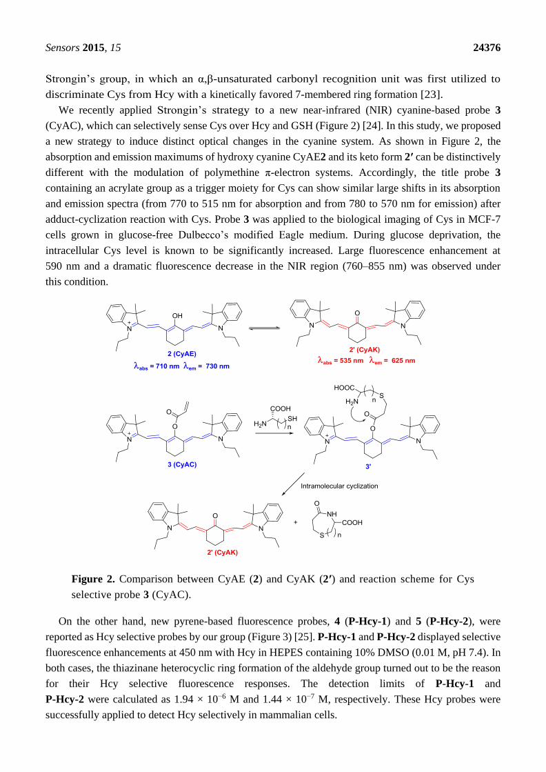
Figure 1. Reaction scheme for biothiol selective probe 1.
Non-selective biothiol probes can usually image GSH, the most abundant biothiol in cells. For Cys
and Hcy, selective recognition of these biothiols is certainly required, which is quite a challenging task
due to the structural similarity of Cys and Hcy. In 2011, a pioneering work was reported by
Sensors 2015, 15 24376
Strongin’s group, in which an α,β-unsaturated carbonyl recognition unit was first utilized to
discriminate Cys from Hcy with a kinetically favored 7-membered ring formation [23].
We recently applied Strongin’s strategy to a new near-infrared (NIR) cyanine-based probe 3
(CyAC), which can selectively sense Cys over Hcy and GSH (Figure 2) [24]. In this study, we proposed
a new strategy to induce distinct optical changes in the cyanine system. As shown in Figure 2, the
absorption and emission maximums of hydroxy cyanine CyAE2 and its keto form 2′ can be distinctively
different with the modulation of polymethine π-electron systems. Accordingly, the title probe 3
containing an acrylate group as a trigger moiety for Cys can show similar large shifts in its absorption
and emission spectra (from 770 to 515 nm for absorption and from 780 to 570 nm for emission) after
adduct-cyclization reaction with Cys. Probe 3 was applied to the biological imaging of Cys in MCF-7
cells grown in glucose-free Dulbecco’s modified Eagle medium. During glucose deprivation, the
intracellular Cys level is known to be significantly increased. Large fluorescence enhancement at
590 nm and a dramatic fluorescence decrease in the NIR region (760–855 nm) was observed under
this condition.
Figure 2. Comparison between CyAE (2) and CyAK (2′) and reaction scheme for Cys
selective probe 3 (CyAC).
On the other hand, new pyrene-based fluorescence probes, 4 (P-Hcy-1) and 5 (P-Hcy-2), were
reported as Hcy selective probes by our group (Figure 3) [25]. P-Hcy-1 and P-Hcy-2 displayed selective
fluorescence enhancements at 450 nm with Hcy in HEPES containing 10% DMSO (0.01 M, pH 7.4). In
both cases, the thiazinane heterocyclic ring formation of the aldehyde group turned out to be the reason
for their Hcy selective fluorescence responses. The detection limits of P-Hcy-1 and
P-Hcy-2 were calculated as 1.94 × 10⁻⁶ M and 1.44 × 10⁻⁷ M, respectively. These Hcy probes were
successfully applied to detect Hcy selectively in mammalian cells.

Sensors 2015, 15 24377
Among the various approaches to design biothiol selective probes, fluorescence enhancement of Cu2+
complexes has been utilized for biothiol imaging. A new bis-pyrene derivative 6 was synthesized and a
large fluorescence quenching effect was observed with Cu2+ at pH 7.4 among the various metal ions
(Figure 3) [26]. The addition of biothiols successfully revived the fluorescence of the 6-Cu2+ ensemble,
which was attributed to the displacement of the ligand to the biothiols for Cu2+. The addition of GSH,
Cys or Hcy effectively induced fluorescence enhancement at 450 nm. The detection limit of this 6-Cu2+
ensemble for GSH was reported to be 0.16 µM. The Cu2+ ensemble could successfully image endogenous
GSH in live cells and with the aid of two-photon microscopy (TPM), the 6-Cu2+ ensemble could also
image GSH in living tissues.
Figure 3. Structures of biothiol probes 4–6.
Compared to Cys and Hcy selective probes, GSH selective fluorescent imaging probes are still
relatively rare. Recently, a new approach to designing GSH selective probes was reported by our group.
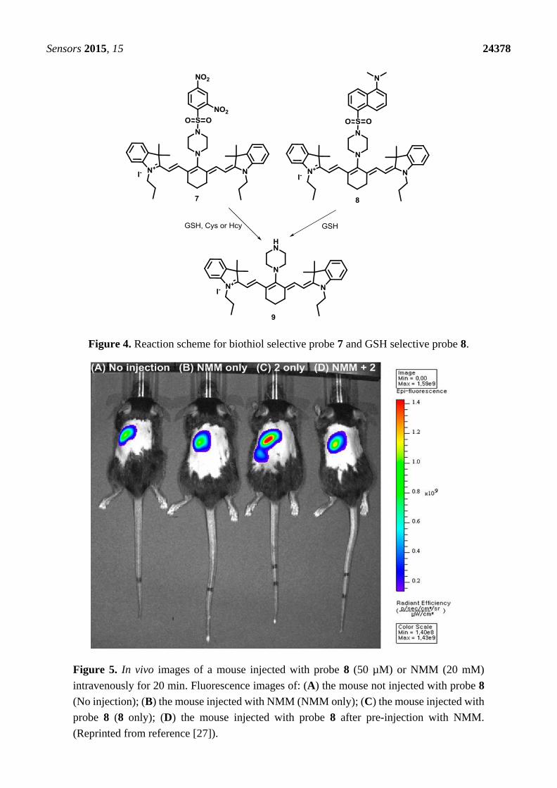
We synthesized two cyanine derivatives containing a 2,4-dinitrobenzene sulfonamide group (7) and a
5-dimethylaminonaphthyl sulfonamide group (8), respectively, as NIR probes for biothiols (Figure 4) [27].
Probe 7 showed large fluorescence enhancements at 736 nm with GSH, Cys and Hcy in HEPES buffer
(10 mM, pH = 7.4) containing 10% DMSO. On the contrary, probe 8 displayed selective turn-on
fluorescence with GSH. After reaction with biothiols, product 9 was confirmed by NMR and mass
spectroscopy. These fluorescence changes with biothiols were further confirmed in HeLa cells. Probe 8
could image the GSH present in HeLa cells and no fluorescence was observed upon treatment with the
thiol blocking agent, N-methylmaleimide (NMM). We also demonstrated that probe 9 can be used to
monitor the intracellular levels of GSH modulated by H2O2 or lipopolysaccharide (LPS) treatment. NIR
probe 8 was further applied to monitor GSH in a mouse model (Figure 5), especially in liver, kidney,
lung, and spleen tissues. It is reported that overdose of the painkiller, acetaminophen, can cause severe
liver damage and a depletion of GSH in liver and kidney cells. The depletion of GSH in mouse tissue
cells was successfully monitored using probe 8.
For Cys and Hcy selective probes, we developed the aryl-thioether substituted nitrobenzothiadiazole
10 (Figure 6) [28]. Only Cys and Hcy induced fluorescence enhancement (λmax = 535 nm) at pH 7.4. The
proposed reaction scheme with Cys and Hcy is illustrated in Figure 6. We also reported that
probe 10 could image these biothiol species in live cells. It is known that the nucleophilicity of Cys (pKa
8.53) is better than that of Hcy (pKa 10.00). In addition, we expect that the p-amino group in probe 10
can manipulate the aryl substitution selectivity at lower pH. Indeed, we observed nice selectivity for Cys
over Hcy in a citric acid-Na2HPO4 (0.01 M, pH 6.0) solution containing 1% DMSO.
Sensors 2015, 15 24378
Figure 4. Reaction scheme for biothiol selective probe 7 and GSH selective probe 8.
Figure 5. In vivo images of a mouse injected with probe 8 (50 µM) or NMM (20 mM)
intravenously for 20 min. Fluorescence images of: (A) the mouse not injected with probe 8
(No injection); (B) the mouse injected with NMM (NMM only); (C) the mouse injected with
probe 8 (8 only); (D) the mouse injected with probe 8 after pre-injection with NMM.
(Reprinted from reference [27]).

Sensors 2015, 15 24379
Figure 6. Structure of probe 10 and its reaction scheme with Cys and Hcy.
2.2. Fluorescent Imaging Probes for Reactive Oxygen Species (ROS) and Reactive Nitrogen
Species (RNS)
Reactive oxygen species (ROS) and reactive nitrogen species (RNS) are active targets for fluorescent
probes due to their significance in human health and disease [29]. We also contributed to the
development of ROS and RNS selective fluorescent probes, especially for hypochlorous acid (HOCl),
hydrogen peroxide (H2O2) and peroxynitrite (ONOO−).
Novel rhodamine derivatives 11–13 were synthesized as fluorescent probes for HOCl (Figure 7) [30].
Fluorescent increase upon the addition of HOCl was attributed to the ring-opening process through
sulfur/selenium oxidation as shown in Figure 7. Probes 11 and 12 showed highly selective fluorescence
enhancement with HOCl among the various ROS, such as H2O2, NO•, •OH, ROO•, ONOO−, 1O2, and
•O2− in KH2PO4 buffer (pH 5.5, 1% CH3CN). On the other hand, probe 13 displayed a lower selectivity
for HOCl, probably due to the higher susceptibility to selenolactone by oxidants.The detection
limits of HOCl probes 11–13 were calculated as 0.4 μM, 0.6 μM and 2 μM, respectively. Furthermore,
probe 11 could image bacteria-mediated microbicidal HOCl production in the mucosal epithelia in
fruit fly.
Figure 7. Structures of HOCl selective fluorescent probes 11–13 and reaction scheme for 11
with HOCl.
We extended this design concept to the so-called “dual-lock” fluorescent probe 14 (FBS) for HOCl
(Figure 8) [31], in which two reacting groups, such as aryl boronate and thiolactone, are introduced.
Sensors 2015, 15 24380
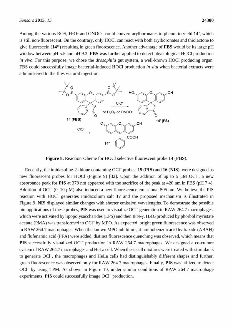
Among the various ROS, H2O2 and ONOO− could convert arylboronates to phenol to yield 14′, which
is still non-fluorescent. On the contrary, only HOCl can react with both arylboronates and thiolactone to
give fluorescein (14″) resulting in green fluorescence. Another advantage of FBS would be its large pH
window between pH 5.5 and pH 9.3. FBS was further applied to detect physiological HOCl production
in vivo. For this purpose, we chose the drosophila gut system, a well-known HOCl producing organ.
FBS could successfully image bacterial-induced HOCl production in situ when bacterial extracts were
administered to the flies via oral ingestion.
Figure 8. Reaction scheme for HOCl selective fluorescent probe 14 (FBS).
Recently, the imidazoline-2-thione containing OCl− probes, 15 (PIS) and 16 (NIS), were designed as
new fluorescent probes for HOCl (Figure 9) [32]. Upon the addition of up to 5 μM OCl−, a new
absorbance peak for PIS at 378 nm appeared with the sacrifice of the peak at 420 nm in PBS (pH 7.4).
Addition of OCl− (0–10 µM) also induced a new fluorescence emissionat 505 nm. We believe the PIS
reaction with HOCl generates imidazolium salt 17 and the proposed mechanism is illustrated in
Figure 9. NIS displayed similar changes with shorter emission wavelengths. To demostrate the possible
bio-applications of these probes, PIS was used to visualize OCl− generation in RAW 264.7 macrophages,
which were activated by lipopolysaccharides (LPS) and then IFN-γ. H2O2 produced by phorbol myristate
acetate (PMA) was transformed to OCl− by MPO. As expected, bright green fluorescence was observed
in RAW 264.7 macrophages. When the known MPO inhibitors, 4-aminobenzoicacid hydrazide (ABAH)
and flufenamic acid (FFA) were added, distinct fluorescence quenching was observed, which means that
PIS successfully visualized OCl− production in RAW 264.7 macrophages. We designed a co-culture
system of RAW 264.7 macrophages and HeLa cell. When these cell mixtures were treated with stimulants
to generate OCl−, the macrophages and HeLa cells had distinguishably different shapes and further,
green fluorescence was observed only for RAW 264.7 macrophages. Finally, PIS was utilized to detect
OCl− by using TPM. As shown in Figure 10, under similar conditions of RAW 264.7 macrophage
experiments, PIS could successfully image OCl− production.

Sensors 2015, 15 24381
Figure 9. Structures of 15 (PIS) and 16 (NIS) and reaction scheme of 15 (PIS) with HOCl.
Figure 10. TPM images of (a–e) PIS and (f) 16 (10 μM, ρDMF = 0.5%) labeled RAW
264.7 cells. (a) Control image; (b) Cells pretreated with NaOCl (200 μM) for 30 min and
then incubated with PIS; (c) Cells pretreated with LPS (100 ng/mL) for 16 h, IFN-γ
(400 U/mL) for 4 h, and PMA (10 nM) for 30 min and then with PIS; (d) Cells pretreated
with LPS, IFN-γ, and 4-ABAH (50 μM) for 4 h and then incubated with PIS; (e) Cells
pretreated with LPS, IFN-γ, and FAA (50 μM) for 4 h and then with PIS; (g) Average TPEF
intensities in (a–f), n = 5. Scale bar: 20 μm. (Reprinted from reference [32]).

Sensors 2015, 15 24382
James and our group recently reported a unique system for sensing peroxynitrite (ONOO−) using
boronate-based fluorescent probe 17 and D-fructose (Figure 11) [33]. When D-fructose was added to
probe 17 at pH 7.3, a large fluorescence enhancement was observed. Among the various ROS and RNS
species, only ONOO− induced a significant fluorescence quenching effect. We believe that the unique
interaction of probe 17 with D-fructose plays two important roles in this study, namely to strengthen the
fluorescence signal and to prevent the oxidation of boronic acid by other ROS/RNS. This system was
also successfully applied to image endogenous and exogenous ONOO− in RAW 264.7 cells and
HeLa cells.
Figure 11. Reaction scheme for probe 17 with ONOO−.
The red emitting probe 18 (CHCN) composed of a linked coumarin-hemicyanine was recently
developed as a dual ratiometric and colorimetric probe for peroxynitrite (ONOO−) (Figure 12) [34].
Among the various ROS and RNS, only ONOO− induced significant ratiometric fluorescence changes
(F515nm/F635nm). The proposed reaction mechanism is shown in Figure 12. The formation of two products,
1,3,3-trimethyloxindole and Coum-CHO was also confirmed. As shown in Figure 13, 18 (CHCN)
displayed ratiometric fluorescence changes for exogenous and endogenous ONOO− during the
phagocytic immune response. More specifically, red fluorescence was reduced with the enhancement of
green fluorescence when RAW 264.7 cells were treated with LPS and IFN-g followed by additional
stimulation with PMA. Treatment with a superoxide scavenger, 2,2,6,6-tetramethyl-1-piperidinyloxy
(TEMPO) or an NO synthase inhibitor, aminoguanidine, caused no changes in the ratios of
red/green fluorescence.
Recently, a new boronate-based naphthalimide derivative 19 was developed as a H2O2 selective
fluorescent probe (Figure 14) [35]. Figure 14 explains the design strategy for probe 19, in which a
morpholine moiety acts as a lysosome-targetable group and a p-dihydroxyborylbenzyloxycarbonyl
group was used as a transponder. Among various ROS and RNS, probe 19 showed a selective fluorescent
enhancement at 528 nm at pH 7.4 (0.1 M PBS containing 1% DMF). Probe 19 was clearly localized in
the lysosomes, which was confirmed by costaining with LysoTracker Blue DND-22.
Probe 19 was further applied to image the level of endogenous and exogenous H2O2 in the lysosome of
the RAW 264.7 cells. Furthermore, time-dependent fluorescence of 19 in the cells was reported.

Sensors 2015, 15 24383
Figure 12. Reaction scheme of peroxinitrite probe 18.
Figure 13. Confocal ratiometric fluorescence images of RAW 264.7 cells for endogenous
ONOO− during phagocytic immune response. The cells were stained with 5 μM 18 (CHCN)
for 30 min and then washed with DPBS before imaging. (a) Control; (e) lipopolysaccharides
(LPS) (1 μg/mL) for 16 h, interferon-γ (50 ng/mL) for 4 h, PMA (10 nM) for 30 min; (i) LPS
(1 μg/mL) for 16 h, interferon-γ (50 ng/mL) for 4 h, PMA (10 nM) for 30 min, and then AG
(1 mM) for 16 h; (m) LPS (1 μg/mL) or 16 h, interferon-γ (50 ng/mL) for 4 h, PMA (10 nM)
for 30 min, and then TEMPO (100 μM) for 16 h. The green channel (a,e,I,m) represents the
fluorescence obtained at 490–540 nm with an excitation wavelength at 473 nm, the red
channel (b,f,j,n) represents the fluorescence obtained at 575–675 nm with an excitation
wavelength at 559 nm, images (c,g,k,o) represent DIC channels (differential interference
contrast), and images (d,h,I,p) represent merged images of red and green channels,
respectively. (Reprinted from reference [34]).

Sensors 2015, 15 24384
Figure 14. Design strategy of H2O2 selective probe 19 and its reaction scheme with H2O2.
2.3. Fluorescent Imaging Probes for Metal Ions
Among the biologically abundant metal ions, Zn2+ is known to play key roles in a variety of
physiological processes, such as Alzheimer’s disease, epilepsy, ischemic stroke, etc. Due to the lower
reactivity of this metal ion, most Zn2+ selective fluorescent probes are based on the design of selective
ligands and effective fluorescence changes [36].
In 2009, we reported a 7-nitrobenz-2-oxa-1,3-diazole (NBD) derivative as a fluorescent and colorimetric
probe for Zn2+ (Figure 15) [37]. Among the various metal ions, probe 20 displayed a selective
fluorescence enhancement only with Zn2+ in 100% aqueous solution (0.1 M HEPES, pH 7.2).
In addition, red to yellow color change with Zn2+ was observed by the naked eye. A large fluorescence
enhancement as well as colorimetric changes was attributed to photoinduced electron transfer (PET) and
internal charge transfer (ICT) mechanisms as shown in Figure 15. The dissociation constant (Kd) for 20
was reported as 1.3 μM. Relatively high concentrations of Zn2+ are present in pancreatic islets, which
are known to play a key role in insulin biosynthesis and storage. The practical-use probe 20 could
successfully detect the intrinsic Zn2+ Ions in pancreatic β-cells.
Figure 15. Proposed binding mode of probe 20 with Zn2+.
We recently proposed a new strategy called “receptor transformer” [38]. As shown in Figure 16,
the naphthalimide derivative (21) binds Zn2+ in an imidic acid tautomeric form of the probe in aqueous
solutions while other heavy transition metal ions, especially Cd2+, bind to probe 21 in an amide
tautomeric form. With the aid of these unique binding modes, probe 21 displayed a selective fluorescence
enhancement for Zn2+ over other metal ions with a red-shift from 483 to 514 nm. On the other hand, the
addition of Cd2+ induced an enhanced blue-shift in emission from 483 to 446 nm in aqueous solution,
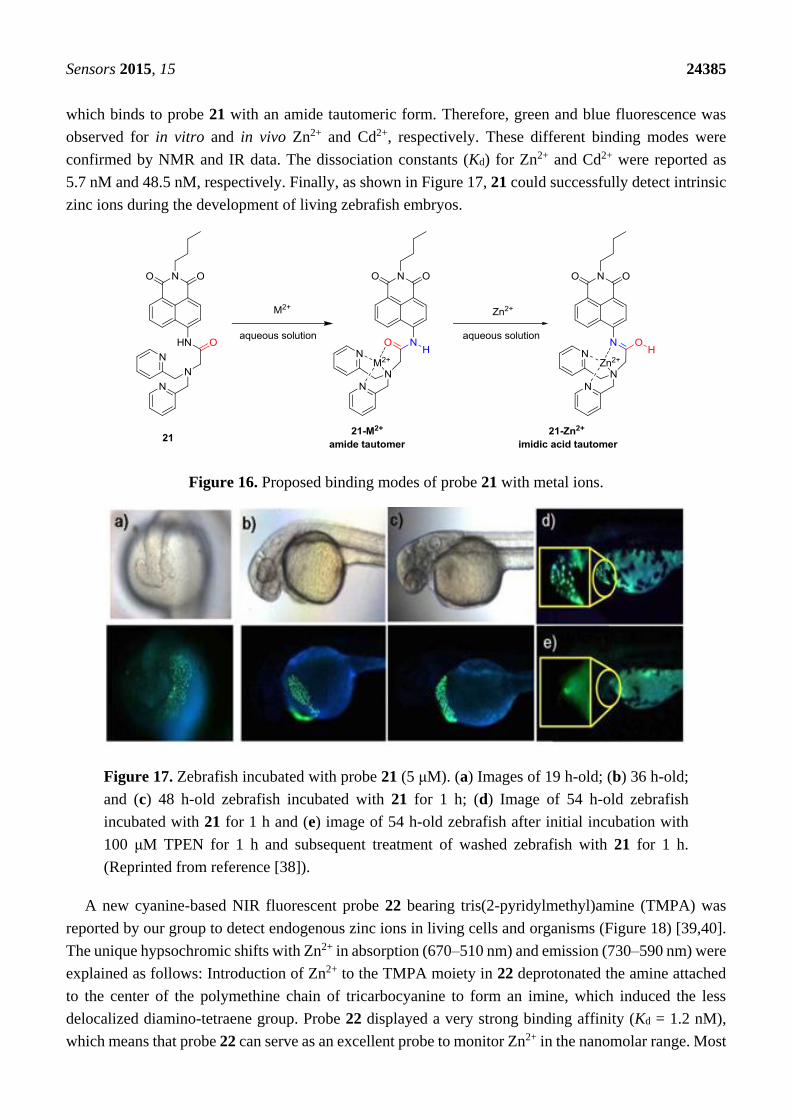
Sensors 2015, 15 24385
which binds to probe 21 with an amide tautomeric form. Therefore, green and blue fluorescence was
observed for in vitro and in vivo Zn2+ and Cd2+, respectively. These different binding modes were
confirmed by NMR and IR data. The dissociation constants (Kd) for Zn2+ and Cd2+ were reported as
5.7 nM and 48.5 nM, respectively. Finally, as shown in Figure 17, 21 could successfully detect intrinsic
zinc ions during the development of living zebrafish embryos.
Figure 16. Proposed binding modes of probe 21 with metal ions.
Figure 17. Zebrafish incubated with probe 21 (5 μM). (a) Images of 19 h-old; (b) 36 h-old;
and (c) 48 h-old zebrafish incubated with 21 for 1 h; (d) Image of 54 h-old zebrafish
incubated with 21 for 1 h and (e) image of 54 h-old zebrafish after initial incubation with
100 μM TPEN for 1 h and subsequent treatment of washed zebrafish with 21 for 1 h.
(Reprinted from reference [38]).
A new cyanine-based NIR fluorescent probe 22 bearing tris(2-pyridylmethyl)amine (TMPA) was
reported by our group to detect endogenous zinc ions in living cells and organisms (Figure 18) [39,40].
The unique hypsochromic shifts with Zn2+ in absorption (670–510 nm) and emission (730–590 nm) were
explained as follows: Introduction of Zn2+ to the TMPA moiety in 22 deprotonated the amine attached
to the center of the polymethine chain of tricarbocyanine to form an imine, which induced the less
delocalized diamino-tetraene group. Probe 22 displayed a very strong binding affinity (Kd = 1.2 nM),
which means that probe 22 can serve as an excellent probe to monitor Zn2+ in the nanomolar range. Most
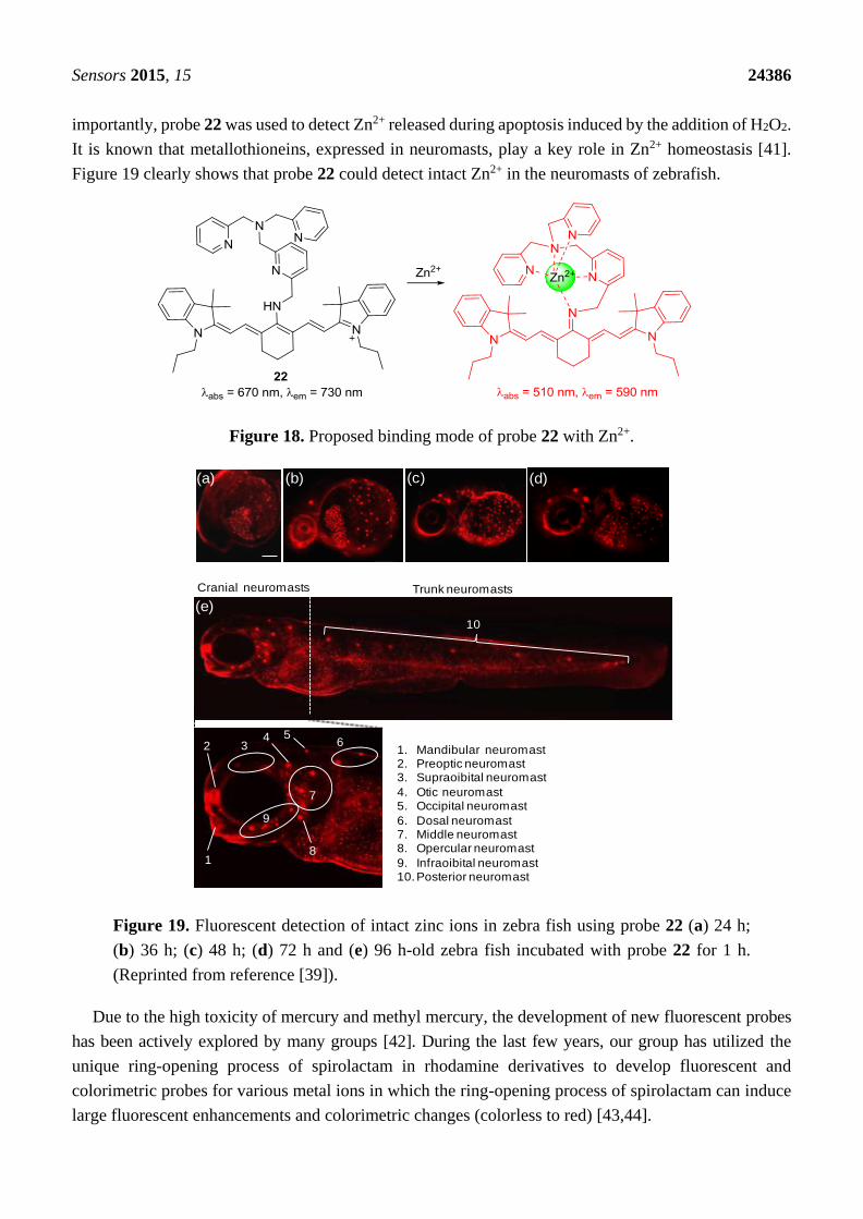
Sensors 2015, 15 24386
importantly, probe 22 was used to detect Zn2+ released during apoptosis induced by the addition of H2O2.
It is known that metallothioneins, expressed in neuromasts, play a key role in Zn2+ homeostasis [41].
Figure 19 clearly shows that probe 22 could detect intact Zn2+ in the neuromasts of zebrafish.
Figure 18. Proposed binding mode of probe 22 with Zn2+.
Figure 19. Fluorescent detection of intact zinc ions in zebra fish using probe 22 (a) 24 h;
(b) 36 h; (c) 48 h; (d) 72 h and (e) 96 h-old zebra fish incubated with probe 22 for 1 h.
(Reprinted from reference [39]).
Due to the high toxicity of mercury and methyl mercury, the development of new fluorescent probes
has been actively explored by many groups [42]. During the last few years, our group has utilized the
unique ring-opening process of spirolactam in rhodamine derivatives to develop fluorescent and
colorimetric probes for various metal ions in which the ring-opening process of spirolactam can induce
large fluorescent enhancements and colorimetric changes (colorless to red) [43,44].
(a) (b) (c) (d)
Cranial neuromasts Trunk neuromasts
10
(e)
1
2 34 5
6
7
9
1. Mandibular neuromast2. Preoptic neuromast3. Supraoibital neuromast
4. Otic neuromast5. Occipital neuromast
6. Dosal neuromast7. Middle neuromast8. Opercular neuromast
9. Infraoibital neuromast10. Posterior neuromast
8
10

Sensors 2015, 15 24387
Two simple rhodamine hydrazone derivatives bearing thiol (23) and carboxylic acid (24) groups were
developed as selective fluorescent and colorimetric probes for Hg2+ (Figure 20) [45]. Upon the addition
of Hg2+, the selective ring-opening process of spirolactams in probes 23 and 24 were observed in
CH3CN-H2O (1:99, v/v) solution, which was accompanied by large fluorescent enhancement and
colorimetric change. The detection limits were calculated as 1 nM for 23 and 4.2 nM for 24. Both probes
could successfully image Hg2+ accumulated in the nematode C. elegans, which was pretreated with
nanomolar concentrations of Hg2+.
Figure 20. Structures of Hg2+ selective probes 23–25.
On the other hand, a rhodamine-based sensor 25 bearing a histidine group was also reported as a
selective probe for Hg2+ (Figure 20) [46]. Upon the addition of 100 equiv Hg2+ in 0.02 M pH 7.4 HEPES:
EtOH (1:9, v/v), turn-on fluorescence (100-fold) was observed due to the spiro-lactam ring opening. We
believe that imidazole nitrogen and two carbonyl oxygenscan provided a nice binding site for Hg2+.
Probe 25 was further applied to visualize Hg2+ in HeLa cells.
A rhodamine derivative bearing the selenolactone group 26 was synthesized for the detection of
inorganic and organic mercury species (Figure 21) [47]. Through the mercury ion-promoted deselenation
reaction, probe 26 displayed highly selective fluorescent and colorimetric changes for mercury species
with high sensitivity in 20 mM HEPES buffer (pH 7.4, 1% CH3CN). We then attempted to monitor the
mercury species that accumulated in living organisms using 26. Zebrafish has been used as a suitable
animal model for the study of fluorescent probes [48]. For our study, adult zebrafish was pre-incubated
with Hg2+ (500 nM) or methylmercury (500 nM) and then with probe 26. As shown in Figure 22, strong
red fluorescence was observed in the gallbladder, eggs and fin due to the presence of inorganic mercury
and methylmercury.
Figure 21. Proposed reaction scheme for probe 26 with Hg2+ and CH3Hg+.

Sensors 2015, 15 24388
Figure 22. Images of zebrafish organs treated with 20 µM 26 (0.2% DMSO) and 500 nM
HgCl2 or 500 nM CH3HgCl. (a) Images of zebrafish organs treated with 26 in the absence
of HgCl2 or CH3HgCl; (b) presence of 500 nM HgCl2 or (c) presence of 500 nM CH3HgCl
(upper, merged images; lower, fluorescence images). (Reprinted from reference [47]).
Rhodamine derivative bearing boronic acid (27) was studied as a probe for Cu2+ for the first time
(Figure 23) [49]. When Cu2+ was introduced to probe 27 in 20 mM HEPES (0.5% CH3CN) at pH 7.4,
selective turn-on fluorescence and distinct colorimetric change was observed, which was also attributed to
the Cu2+-induced spirolactam ring opening process. Probe 27 was applied to visualize Cu2+ in cells and
zebra fish.
Figure 23. Proposed binding mode of probe 27 with Cu2+.
A reaction based fluorescent probe based on rhodamine-alkyne derivative 28 was synthesized
for the detection of Au3+ (Figure 24) [50]. The addition of Au3+ induced a highly selective turn-on
fluorescence and colorimetric change in EtOH-HEPES buffer (0.01 M, pH 7.4) (1:1, v/v). The proposed

Sensors 2015, 15 24389
reaction mechanism from the propargylamide moiety of 28 to the oxazolecarbaldehyde of 29 is
illustrated in Figure 24. The rate constant for the conversion of 28 to 29 was reported to be Kobs = 4.5
(±0.20) × 10−4 s−1 and the detection limit was calculated as 320 nM. In addition, probe 28 was
successfully applied to detect Au3+ in the cell.
Figure 24. Proposed reaction scheme of probe 28 with Au3+.
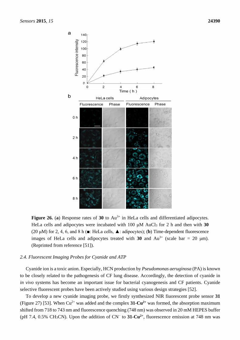
A 4-Amino-1,8-naphthalimide derivative bearing an alkyne group (30) was explored as a fluorescent
probe for Au3+ (Figures 25 and 26) [51]. The addition of Au3+ induced a distinct color change from
yellow to light pink and a large blue shift by about 56 nm in the emission spectra. The detection limit
was calculated to be 8.44 μM. We observed that surfactants could enhance the reaction rate of probe 30
with Au3+. Probe 30 was also applied to detect Au3+ in HeLa cells and differentiated adipocytes.
In particular, probe 30 showed a unique ability to detect Au3+ in lipid droplets in cells. As shown in
Figure 26, the response rates of 30 with Au3+ in differentiated adipocytes were greater than those in
HeLa cells, which is consistent with the rate enhancement in the presence of surfactants.
Figure 25. Structure of the probe 30.
Sensors 2015, 15 24390
Figure 26. (a) Response rates of 30 to Au3+ in HeLa cells and differentiated adipocytes.
HeLa cells and adipocytes were incubated with 100 μM AuCl3 for 2 h and then with 30
(20 μM) for 2, 4, 6, and 8 h (■: HeLa cells, ▲: adipocytes); (b) Time-dependent fluorescence
images of HeLa cells and adipocytes treated with 30 and Au3+ (scale bar = 20 μm).
(Reprinted from reference [51]).
2.4. Fluorescent Imaging Probes for Cyanide and ATP
Cyanide ion is a toxic anion. Especially, HCN production by Pseudomonas aeruginosa (PA) is known
to be closely related to the pathogenesis of CF lung disease. Accordingly, the detection of cyanide in
in vivo systems has become an important issue for bacterial cyanogenesis and CF patients. Cyanide
selective fluorescent probes have been actively studied using various design strategies [52].
To develop a new cyanide imaging probe, we firstly synthesized NIR fiuorescent probe sensor 31
(Figure 27) [53]. When Cu2+ was added and the complex 31-Cu2+ was formed, the absorption maximum
shifted from 718 to 743 nm and fluorescence quenching (748 nm) was observed in 20 mM HEPES buffer
(pH 7.4, 0.5% CH3CN). Upon the addition of CN− to 31-Cu2+, fluorescence emission at 748 nm was

Sensors 2015, 15 24391
recovered since CN− and Cu2+ form the stable [Cu(CN)x]n−. The detection limit for CN− was reported as
5 μM. Probe 31-Cu2+ was applied to visualize the CN− produced by P. aeruginosa (PA) in C. elegans
cells. To confirm the bacterial infection in the intestine of the nematodes, a PA14 strain labeled with
GFP (green fluorescent protein) was introduced for the C. elegans to feed on before the imaging. As
shown Figure 28a, neither green nor NIR fluorescence was observed in the nematodes after C. elegans
exposure to only E. coli OP50 and probe 31-Cu2+. On the contrary, when nematodes were exposed to
PA14 and the probe, both green and NIR fluorescence were observed, which indicates that the probe can
image HCN produced by PA14 in the nematodes (Figure 28b–d). As shown in Figure 29, treatment with
a β-lactam antibiotic, ceftazidime, significantly reduced both the green and NIR fluorescence intensities.
Figure 27. Structure of cyanide selective probe 31 and 31-Cu2+ complex.
Figure 28. NIR imaging of cyanide in C. elegans infected with a P. aeruginosa strain (PA14)
labeled with green fluorescent protein (GFP). Before the imaging, the nematodes fed on
either non-infectious E. coli OP50 (a) or GFP-labeled PA14 for 2 days (b–d);
(b) the anterior end; (c) the medial part; (d) the posterior end of C. elegans. The scale bars
represent 20 μm. (IL = intestinal lumen; I = intestine; E = eggs; PA = PA14-GFP;
A = anus). (Reprinted from reference [54]).
Imidazolium derivatives serve as host molecules for the recognition of various anions since they can
form a unique (C–H)+—anion type ionic hydrogen bond [54–56].
We synthesized an imidazolium receptor bearing two pyrene groups (32) as ATP selective probes, in
which a pyrene excimer acts as a signal source and imidazoliums serve as the triphosphate anion receptor
(Figure 30) [57]. Among the various nucleoside triphosphates, only ATP showed significant ratiometric
changes in its emission. More specifically, only ATP induced an increase in monomer emission at 375
with a sacrifice of excimer emission at 487 nm. We believe this result can be attributed to the different
binding mode of ATP compared to the other four nucleoside triphosphate bases: GTP, CTP,
UTP and TTP. As shown in Figure 30, ATP can form a characteristic sandwich π-π stack of

Sensors 2015, 15 24392
pyrene-adenine-pyrene while GTP, CTP, UTP and TTP interact with probe 32 from the outside, with the
stacked pyrene-pyrene dimer of 32 resulting in only excimer fluorescence quenching. These different
binding modes were proposed based on NMR study and theoretical calculation results. Oligomycin is
reported to decrease cellular ATP levels. Probe 32 showed strong blue fluorescence in HeLa cells and
the fluorescence was quenched upon the addition of oligomycin.
Figure 29. Visualization of antibiotic efficacy against P. aeruginosa infection in C. elegans
with the NIR sensor. The nematodes fed on GFP-labeled P. aerugionosa (PA14) for
two days. They were then incubated with ceftazidime (200 μg/mL) for 2 h before the in vivo
imaging. The scale bars represent 20 μm. (Reprinted from reference [53]).
Figure 30. Proposed binding modes of probe 32 with ATP and GTP.
3. Conclusions and Future Perspectives
Fluorescence is one of the most powerful tools currently available due to its low detection limit and
bioimaging capabilities via confocal microscopy. The application of fluorescent imaging probes for
Sensors 2015, 15 24393
various biologically important species has been widely reported in the last decade. In this review, we
focused on our exciting contributions in this field. This review was categorized by target analytes, such
as biothiols, ROS and RNS, metal ions such as Zn2+ and Hg2+, and finally cyanide and ATP.
Certainly, their application in biology and environmental science has been a strong driving force for
the development of fluorescent probes. We believe that intelligent fluorescent probes will be an
important research area in the future, and the next generation of functional fluorescent probes will
involve imaging and therapeutic agents in vivo [58,59].
Acknowledgments
This study was supported by the National Research Foundation of Korea (NRF) grant funded by the
Korean government (MSIP) (No. 2012R1A3A2048814).
Conflicts of Interest
The authors declare no conflict of interest.
References
1. Zhou, X.; Lee, S.; Xu, Z.; Yoon, J. Recent Progress on the Development of Chemosensors for Gases.
Chem. Rev. 2015, 115, 7944–8000.
2. Wu, J.; Kwon, B.; Liu, W.; Anslyn, E.V.; Wang, P.; Kim, J.S. Chromogenic/Fluorogenic Ensemble
Chemosensing Systems. Chem. Rev. 2015, 115, 7893–7943.
3. Shamirian, A.; Ghai, A.; Snee, P.T. QD-Based FRET Probes at a Glance. Sensors 2015, 15,
13028–13051.
4. Yin, J.; Hu, Y.; Yoon, J. Fluorescence probes and bioimaging: Alkali metals, alkaline earth metals
and protons. Chem. Soc. Rev. 2015, 44, 4619–4644.
5. Miller, E.W.; Chang, C.J. Fluorescent probes for nitric oxide and hydrogen peroxide in cell
signaling. Curr. Opin. Chem. Biol. 2007, 11, 620–625.
6. Guo, Z.; Shin, I.; Yoon, J. Recognition and Sensing of Various Species Using Boronic Acid
Derivatives. Chem. Commun. 2012, 48, 5956–5967.
7. Thomas, J.A. Optical imaging probes for biomolecules: An introductory perspective. Chem. Soc. Rev.
2015, 44, 4494–4500.
8. Zhou, Y.; Zhang, J.F.; Yoon, J. Fluorescent and Colorimetric Chemosensors for Detection of
Fluoride Ion. Chem. Rev. 2014, 114, 5511–5571.
9. Lee, S.; Yuen, K.K.Y.; Jolliffe, K.A.; Yoon, J. Fluorescent and Colorimetric Chemosensors for
Pyrophosphate. Chem. Soc. Rev. 2015, 44, 1749–1762.
10. Chen, X.; Pradhan, T.; Wang, F.; Kim, J.S.; Yoon, J. Fluorescent Chemosensors Based on
Spiroring-Opening of Xanthenes and Related Derivatives. Chem. Rev. 2012, 112, 1910–1956.
11. Guo, Z.; Park, S.; Yoon, J.; Shin, I. Recent Progress on Near-Infrared Fluorescent Probes for
Bioimaging Applications. Chem. Soc. Rev. 2014, 43, 16–29.
12. Amiot, C.L.; Xu, S.; Liang, S.; Pan, L.; Zhao, J.X. Near-Infrared Fluorescent Materials for Sensing
of Biological Targets. Sensors 2008, 8, 3082–3105.
Sensors 2015, 15 24394
13. Kim, H.M.; Cho, B.R. Two-Photon Probes for Intracellular Free Metal Ions, Acidic Vesicles, and
Lipid Rafts in Live Tissues. Acc. Chem. Res. 2009, 42, 863–872.
14. Zhang, X.; Yin, J.; Yoon, J. Recent Advances in Development of Chiral Fluorescent and
Colorimetric Sensors. Chem. Rev. 2014, 114, 4918–4959.
15. De Silva, A.P.; Gunaratne, H.Q.N.; Gunnlaugsson, T.A.; Huxley, T.M.; McCoy, C.P.;
Rademacher J.T.; Rice, T.E. Signaling Recognition Events with Fluorescent Sensors and Switches.
Chem. Rev. 1997, 97, 1515–1566.
16. Czarnik, A.W. Chemical Communication in Water Using Fluorescent Chemosensors. Acc. Chem. Res.
1994, 27, 302–308.
17. Yoon, J.; Czarnik, A.W. Fluorescent Chemosensors of Carbohydrates. A Means of Chemically
Communicating the Binding of Polyols in Water Based on Chelation-Enhanced Quenching. J. Am.
Chem. Soc. 1992, 114, 5874–5875.
18. Tang, Y.; Lee, D.; Wang, J.; Li, G.; Lin, W.; Yoon, J. Development of fluorescent probes based on
protection-deprotection of aldehyde, hydroxyl, and amino functional groups for biological imaging.
Chem. Soc. Rev. 2015, 44, 5003–5015.
19. Jun, M.E.; Roy, B.; Ahn, K.H. Turn-on fluorescent sensing with reactive probes. Chem. Commun.
2011, 47, 7583–7601.
20. Yeung, M.C.-L.; Yam, V.W.-W. Luminescent cation sensors: From host-guest chemistry,
supramolecular chemistry to reaction-based mechanisms. Chem. Soc. Rev. 2015, 44, 4192–4202.
21. Jung, H.S.; Chen, X.; Kim, J.S.; Yoon, J. Recent progress in luminescent and colorimetric
chemosensors for detection of thiols. Chem. Soc. Rev. 2013, 42, 6019–6031.
22. Chen, X.; Ko, S.-K.; Kim, M.J.; Shin, I.; Yoon, J. A Thiol-Specific Fluorescent Probe and Its
Application for Bioimaging. Chem. Commun. 2010, 46, 2751–2753.
23. Yang, X.; Guo, Y.; Strongin, R.M. Conjugate Addition/Cyclization Sequence Enables Selective and
Simultaneous Fluorescence Detection of Cysteine and Homocysteine. Angew. Chem. Int. Ed. 2011,
50, 10690–10693.
24. Guo, Z.; Nam, S.W.; Park, S.; Yoon, J. A Highly Selective Ratiometric Near-Infrared Fluorescent
Cyanine Sensor for Cysteine with Remarkable Shift and Its Application Bioimaging. Chem. Sci.
2012, 3, 2760–2765.
25. Lee, H.Y.; Choi, Y.P.; Kim, S.K.; Yoon, T.; Guo, Z.; Swamy, K.M.K.; Kim, G.; Lee, J.Y.;
Shin, I.; Yoon, J. Selective Homocysteine Turn-on Fluorescent Probes and Their Bioimaging
Applications. Chem. Commun. 2014, 50, 6967–6969.
26. Hu, Y.; Heo, C.H.; Kim, G.; Jun, E.J.; Yin, J.; Kim, H.M.; Yoon, J. One-Photon and Two-Photon
Sensing of Biothiols Using A Bis-Pyrene-Cu(II) Ensemble and Its Application to Image GSH in the
Cells and Tissues. Anal. Chem. 2015, 87, 3308–3313.
27. Yin, J.; Kwon, Y.; Kim, D.; Lee, D.; Kim, G.; Hu, Y.; Ryu, J.-H.; Yoon, J. A Cyanine Based
Fluorescence Probe for Highly Selective Detection of Glutathione in Cell Cultures and Live Mice
Tissues. J. Am. Chem. Soc. 2014, 136, 5351–5358.
28. Lee, D.; Kim, G.; Yin, J.; Yoon, J. Aryl-thioether substituted nitrobenzothiadiazole probe for
selective detection of cysteine and homocysteine. Chem. Commun. 2015, 51, 6518–6520.
29. Chen, X.; Tian, X.; Shin, I.; Yoon, J. Fluorescent and luminescent probes for detection of reactive
oxygen and nitrogen species. Chem. Soc. Rev. 2011, 40, 4783–4804.
Sensors 2015, 15 24395
30. Chen, X.; Lee, K.-A.; Ha, E.-M.; Lee, K.M.; Seo, Y.Y.; Choi, H.K.; Kim, H.N.; Kim, M.J.;
Cho, C.-S.; Lee, S.Y.; et al. A specific and sensitive method for detection of hypochlorous acid for
the imaging of microbe-induced HOCl production. Chem. Commun. 2011, 47, 4373–4375.
31. Xu, Q.; Lee, K.-A.; Lee, S.; Lee, K.M.; Lee, W.-J.; Yoon, J. A Highly Specific Fluorescent Probe
for Hypochlorous Acid and Its Application in Imaging Microbe-Induced HOCl Production. J. Am.
Chem. Soc. 2013, 135, 9944–9949.
32. Xu, Q.; Heo, C.H.; Kim, G.; Lee, H.W.; Kim, H.M.; Yoon, J. Development of Imidazoline-2-
Thiones Based Two-Photon Fluorescence Probes for Imaging Hypochlorite Generation in a
Co-Culture System. Angew. Chem. Int. Ed. 2015, 54, 4890–4894.
33. Sun, X.; Xu, Q.; Kim, G.; Flower, S.E.; Lowe, J.P.; Yoon, J.; Fossey, J.S.; Qian, X.; Bull, S.D.;
James, T.D. A water-soluble boronate-based fluorescent probe for the selective detection of
peroxynitrite and imaging in living cells. Chem. Sci. 2014, 5, 3368–3373.
34. Zhou, X.; Kwon, Y.; Kim, G.; Ryu, J.-H.; Yoon, J. A ratiometric fluorescent probe based on a
coumarin-hemicyanine scaffold for sensitive and selective detection of endogenous peroxynitrite.
Biosens. Bioelectron. 2015, 64, 285–291.
35. Kim, D.; Kim, G.; Nam, S.-J.; Yin, J.; Yoon, J. Visualization of Endogenous and Exogenous
Hydrogen Peroxide Using A Lysosome-Targetable Fluorescent Probe. Sci. Rep. 2015, 5, 8488.
36. Xu, Z.; Yoon, J.; Spring, D.R. Fluorescent Chemosensors for Zn2+. Chem. Soc. Rev. 2010, 39,
1996–2006.
37. Xu, Z.; Kim, G.-H.; Han, S.J.; Jou, M.J.; Lee, C.; Shin, I.; Yoon, J. An NBD-based colorimetric and
fluorescent chemosensor for Zn2+ and its use for detection of intracellular zinc ions. Tetrahedron
2009, 65, 2307–2312.
38. Xu, Z.; Baek, K.-H.; Kim, H.N.; Cui, J.; Qian, X.; Spring, D.R.; Shin, I.; Yoon, J. Zn2+-Triggered
Amide Tautomerization Produces a Highly Zn2+-Selective, Cell–Permeable and Ratiometric
Fluorescent Sensor. J. Am. Chem. Soc. 2010, 132, 601–610.
39. Guo, Z.; Kim, G.-H.; Shin, I.; Yoon, J. A Cyanine-Based Fluorescent Sensor for Detecting
Endogenous Zinc Ions in Live Cells and Organisms. Biomaterials 2012, 33, 7818–7827.
40. Guo, Z.; Kim, G.-H.; Yoon, J.; Shin, I. Synthesis of a highly Zn2+-selective cyanine-based
probe and its use for tracing endogenous zinc ions in cells and organisms. Nat. Protocol. 2014, 9,
1245–1254.
41. Palmiter, R.D. The elusive function of metallothioneins. Proc. Natl. Acad. Sci. USA 1998, 95,
8428–8430.
42. Kim, H.N.; Ren, W.X.; Kim, J.S.; Yoon, J. Fluorescent and Colorimetric Sensors for Detection of
Lead, Cadmium, and Mercury Ions. Chem. Soc. Rev. 2012, 41, 3210–3244.
43. Park, S.; Kim, W.; Swamy, K.M.K.; Jung, J.Y.; Kim, G.; Kim, Y.; Kim, S.-J.; Yoon, J. Rhodamine
Hydrazone Derivatives Bearing Thiophene Group as Fluorescent and Colorimetric Chemosensors
for Hg2+. Dyes Pigm. 2013, 99, 323–328.
44. Kwon, J.Y.; Jang, Y.J.; Lee, Y.J.; Kim, K.-M.; Seo, M.-S.; Nam, W.; Yoon, J. A Highly Selective
Fluorescent Chemosensor for Pb2+. J. Am. Chem. Soc. 2005, 127, 10107–10111.
45. Kim, H.N.; Nam, S.-W.; Swamy, K.M.K.; Jin, Y.; Chen, X.; Kim, Y.; Kim, S.-J.; Park, S.; Yoon, J.
Rhodamine Hydrazone Derivatives as Hg2+ Selective Fluorescent and Colorimetric Chemosensors
and Their Applications to Bioimaging and Microfluidic System. Analyst 2011, 136, 1339–1343.

Sensors 2015, 15 24396
46. Kwon, S.K.; Kim, H.N.; Rho, J.H.; Swamy, K.M.K.; Shanthakumar, S.M.; Yoon, J. Rhodamine
Derivative Bearing Histidine Binding Site as a Fluorescent Chemosensor for Hg2+. Bull. Korean
Chem. Soc. 2009, 30, 719–721.
47. Chen, X.; Baek, K.-H.; Kim, Y.; Kim, S.-J.; Shin, I.; Yoon, J. A Selenolactone-Based Fluorescent
Chemodosimeter to Monitor Mercury/ Methylmercury Species in vitro and in vivo. Tetrahedron
2010, 66, 4016–4021.
48. Ko, S.-K.; Chen, X.; Yoon, J.; Shin, I. Zebrafish as a good vertebrate model for molecular imaging
using fluorescent probes. Chem. Soc. Rev. 2011, 40, 2120–2130.
49. Swamy, K.M.K.; Ko, S.-K.; Kwon, S.K.; Lee, H.N.; Mao, C.; Kim, J.-M.; Lee, K.-H.; Kim, J.;
Shin, I.; Yoon, J. Boronic Acid-Linked Fluorescent and Colorimetric Probes for Copper Ions. Chem.
Commun. 2008, 5915–5917.
50. Jou, M.J.; Chen, X.; Swamy, K.M.K.; Kim, H.N.; Kim, H.-J.; Lee, S.-G.; Yoon, J. Highly Selective
Fluorescent Probe for Au3+ Based on Cyclization of Propargylamide. Chem. Commun. 2009, 14,
7218–7220.
51. Choi, J.Y.; Kim, G.-H.; Guo, Z.; Swamy, K.M.K.; Lee, H.Y.; Pai, J.; Shin, S.; Shin, I.; Yoon, J.
Highly Selective Ratiometric Fluorescent Probe for Au3+ and Its Application to Bioimaging.
Biosens. Bioelectron. 2013, 49, 438–441.
52. Wang, F.; Wang, L.; Chen, X.; Yoon, J. Recent progress in the development of fluorometric and
colorimetric chemosensors for detection of cyanide ion. Chem. Soc. Rev. 2014, 43, 4312–4324.
53. Chen, X.; Nam, S.-W.; Kim, G.-H.; Song, N.; Jeong, Y.; Shin, I.; Kim, S.K.; Kim, J.; Park, S.;
Yoon, J. A Near-Infrared Fluorescent Sensor for Detection of Cyanide in Aqueous Solution and Its
Application for Bioimaging. Chem. Commun. 2010, 46, 8953–8955.
54. Xu, Z.; Kim, S.K.; Yoon, J. Revisit to Imidazolium Receptors for the Recognition of Anions:
Highlighted Research During 2006–2009. Chem. Soc. Rev. 2010, 39, 1457–1466.
55. Kim, S.K.; Singh, N.J.; Kwon, J.; Hwang, I.-C.; Park, S.J.; Kim, K.S.; Yoon, J. Fluorescent
Imidazolium Receptors for the Recognition of Pyrophosphate. Tetrahedron 2006, 62, 6065–6072.
56. Kim, S.K.; Kang, B.-G.; Koh, H.S.; Yoon, Y.J.; Jung, S.J.; Jeong, B.; Lee, K.-D.; Yoon, J. A New
ImidazoliumCavitand for the Recognition of Dicarboxylates. Org. Lett. 2004, 6, 4655–4658.
57. Xu, Z.; Singh, N.J.; Lim, J.; Pan, J.; Kim, H.N.; Park, S.; Kim, K.S.; Yoon, J. Unique Sandwich
Stacking of Pyrene-Adenine-Pyrene for Selective and Ratiometric Fluorescent Sensing of ATP at
Physiological pH. J. Am. Chem. Soc. 2009, 131, 15528–15533.
58. Kolemen, S.; Işık, M.; Kim, G.M.; Kim, D.; Geng, H.; Buyuktemiz, M.; Karatas, T.; Zhang, X.-F.;
Dede, Y.; Yoon, J.; et al. Intracellular Modulation of Excited-State Dynamics in a Chromophore
Dyad: Differential Enhancement of Photocytotoxicity Targeting Cancer Cells. Angew. Chem. Int. Ed.
2015, 54, 5340–5344.
59. Kim, E.-J.; Bhuniya, S.; Lee, H.; Kim, H.M.; Cheong, C.; Maiti, S.; Hong, K.S.; Kim, J.S.
An Activatable Prodrug for the Treatment of Metastatic Tumor. J. Am. Chem. Soc. 2014, 136,
13888–13894.
© 2015 by the authors; licensee MDPI, Basel, Switzerland. This article is an open access article
distributed under the terms and conditions of the Creative Commons Attribution license
(http://creativecommons.org/licenses/by/4.0/).


![[SOLUTIONS] - careerendeavour.in · ated carbonyl compounds to unsaturated amines. Correct option is (c). 10. The reaction given below is an example of MeO NO2 NaOEt NO2 (a) E 2-elimination](https://img.pdfslide.net/doc/110x75/5e6892f8a098c748805e4072/solutions-ated-carbonyl-compounds-to-unsaturated-amines-correct-option-is-c.jpg)