Embed Size (px)
Citation preview
Proc. Nadl. Acad. Sci. USAVol. 87, pp. 3655-3659, May 1990Biochemistry
Receptor-mediated endocytosis of transferrin-polycationconjugates: An efficient way to introduce DNA intohematopoietic cells
(transferrinfection/transferrin receptor/chloroquine/DNA transfection/gene therapy)
MARTIN ZENKE, PETER STEINLEIN, ERNST WAGNER, MATTHEW COTTEN, HARTMUT BEUG,AND MAX L. BIRNSTIELResearch Institute of Molecular Pathology, Dr. Bohr-Gasse 7, A-1030 Vienna, Austria
Contributed by Max L. Birnstiel, February 15, 1990
ABSTRACT Most current gene transfer methods functionsatisfactorily in specialized systems involving established celllines but are often not applicable with nonadherent, primaryhematopoietic cells, which are notoriously difficult to transfect.To approach this problem, we have investigated an alternativemethod of gene transfer, "transferrnfection," in which DNAcomplexed to transferrin-polycation conjugates is introducedinto cells by receptor-mediated endocytosis [Wagner, E.,Zenke, M., Cotten, M., Beug, H. & Birnstiel, M. L. (1990)Proc. Natl. Acad. Sci. USA 87, 3410-3414]. We show here thattransferrin-polylysine and transferrin-protamine, when com-plexed to plasmid DNA containing a luciferase reporter gene,is efficiently bound and moved into avian erythroblasts byendocytosis. Successful transfer and expression of the Iu-ciferase reporter gene depends on specific interaction of thetransferrin-polylysine-DNA complex with the transferrin re-ceptor and occurs in a significant fraction (>95%) of the cells.Gene transfer efficiency by transferrinfection is lower thanwith an optimized DEAE-dextran transfection method butreaches similar efficiencies when the cells are treated withchloroquine. Because the procedure in the absence of chloro-quine is completely nontoxic to cells, a constant expression levelof transferred genes may be maintained by repeated additionsof transferrin-polylysine-DNA complex. In addition, the use-fulness of transferrinfection for gene transfer into primaryhematopoietic cells is demonstrated.
Many techniques are available to introduce and expressforeign DNA in eukaryotic cells (1-4). However, most pro-cedures function well mainly with adherent cells and/orestablished cell lines and are often associated with severecytotoxic side effects. In particular, most available methodsproved to be ineffective or too toxic with nonestablishedhematopoietic cells. Only recently, specific protocols em-ploying electroporation or DEAE-dextran have met withsome success in such cells (5, 6). We have therefore studiedthe utility of receptor-mediated endocytosis as an alternativemeans to introduce and express DNA in hematopoietic cells,thus avoiding cytotoxic effects. A similar approach has beenused by Wu and Wu (7) who employed the asialoglycoproteinreceptor to deliver DNA to liver cells.
In a recent communication (8) we have described trans-ferrin-polycation conjugates that efficiently bind DNA ofwidely different size and can replace native iron transferrin asan efficient iron transporter into the living cell. Becausetransferrin receptors are elevated on the surface of exten-sively proliferating or neoplastic hematopoietic cells (forreview, see ref. 9), we attempted to use these transferrin-polycation conjugates to efficiently introduce DNA into cells
via the transferrin cycle (a method termed "transferrinfec-tion"). Pilot experiments described in the previous commu-nication, showing the principal feasibility of the transferrin-polylysine gene transfer approach (8), encouraged us to studyits usefulness with hematopoietic cells in more detail, byusing both established cell lines and primary hematopoieticcells of avian origin.
MATERIALS AND METHODSTransferrin-Polylysine and Transferrin-Protamine Conju-
gates and Recombinant Plasmids. The origin of polylysine(Sigma), protamine (Sigma), and chicken transferrin (ion-saturated conalbumin; Sigma) and the synthesis of transfer-rin-polylysine and transferrin-protamine conjugates havebeen described (8). The pRSVL plasmid DNA (10) as well asthe pRSV-,8Gal and pB-SK- DNAs (Stratagene) were pre-pared by using the Triton-X lysis procedure (11) followed bycesium chloride/ethidium bromide equilibrium density gra-dient centrifugation, destaining with 1-butanol, and dialysisagainst 10 mM Tris HCl, pH 7.5/1 mM EDTA.
Cells and Cell Culture. The temperature-sensitive v-erbB-transformed chicken erythroblast cell line HD3 (12) aswell as the lymphoid (REVT) transformed clone REV NPB4(13) were grown in standard growth medium (EBM + H; ref.12) at a density of 1-2 x 10⁶ cells per ml at 37°C and 5% Co2.A primary clone of erythroblasts transformed by a retrovirusexpressing the human epidermal growth factor receptor to-gether with a temperature-sensitive myb oncogene (EGFR-ts-myb) was generated as will be described elsewhere (ref. 14and H.B., unpublished work). The cells were grown inCFU-E medium (15) in the presence of 20 ng of epidermalgrowth factor per ml. Normal bone marrow cells wereprepared as described (16) and enriched for immature ery-throid cells by centrifugation through Percoll at 1.072 g/cm3and removal of cells adherent to plastic (17).
Transferrin-Polylysine-DNA or Transferrin-Protamine-DNA Complex Formation and Gene Transfer Reaction. In atypical complex formation reaction 10 ,g of transferrin-polylysine or transferrin-protamine conjugate in 250 ,ul ofH20 was added to 3 ,tg of pRSVL plasmid DNA containedin 250 ,lI of 0.3 M NaCl (added while agitating). Phosphatebuffers should be avoided because precipitates form. Byusing the above conditions, up to 100 ,ug of transferrin-polylysine or transferrin-protamine conjugate and 30 gg ofplasmid DNA could be used per 500 ,ul of final volumewithout precipitation of the transferrin-polylysine-DNA ortransferrin-protamine-DNA complexes. After 30 min atroom temperature, the transferrin-polylysine-DNA or trans-ferrin-protamine-DNA mixture was directly added to 5-10 x106 HD3 cells growing at 0.5-1 x 10⁶ cells per ml in standard
Abbreviation: FITC, fluorescein isothiocyanate.
3655
The publication costs of this article were defrayed in part by page chargepayment. This article must therefore be hereby marked "advertisement"in accordance with 18 U.S.C. §1734 solely to indicate this fact.
Proc. Natl. Acad. Sci. USA 87 (1990)
growth medium (see above) and incubated for 16-48 hr.Other cell types were treated similarly, except being grownin their respective media [see above; CFU-E medium plusREV factor (13) was used for the normal bone marrow cells].
Preparation of Tritiated Transferrin-Polylysine. Tritiatedtransferrin-polylysine was prepared by conjugation of triti-ated polylysine to transferrin in a similar fashion as described(8). Tritiation of poly(L-lysine) (hydrobromide salt, Mr18,000; Sigma) was performed by treatment with formalde-hyde and tritiated sodium borohydride (18).Uptake of Transferrin-Polylysine-DNA Complexes into In-
tracellular Vesicles. Avian erythroblasts (HD3; ref. 12) wereincubated with transferrin-free differentiation medium (8) at370C for 18 hr. After addition of transferrin-polylysine conju-gates [labeled with fluorescein isothiocyanate (FITC) at thepolylysine moiety (8)] that had been complexed with DNA insome of the experiments, cells were further incubated for 18hr. Cells were cytocentrifuged onto slides, fixed with a mixtureof 3.7% formaldehyde (prepared from paraformaldehyde; ref.19)/0.02% glutaraldehyde, washed with phosphate-bufferedsaline (PBS), mounted in Mowiol 4.88 (Hoechst) (19), andexamined using a Zeiss Axiophot fluorescence microscope.FITC-labeled goat antimouse antibody (0.1 mg/ml) was usedin the controls. For quantitative determination of FITC-transferrin or FITC-transferrin-polylysine-DNA, cells wereincubated with the respective transferrin-polylysine prepara-tion for 6 hr at 370C, washed three times in cold PBS/bovineserum albumin, and processed for quantitative fluorescenceactivated cell sorter analysis in a Becton Dickinson FACScan.
Luciferase Assay. Aliquots from cultures receiving trans-ferrin-polylysine/transferrin-protamine-DNA complexeswere harvested by centrifugation (8 min, 1500 X g, 4°C), cellextracts were prepared (10), and bioluminescence was mea-sured with the Clinilumat counter LB 9502 (Berthold, Wild-bach, F.R.G.). For determination of the percentage of cellsexpressing DNA, the cells were allowed to take up transfer-rin-polylysine-pRSV-,BGal complexes or, as controls, trans-ferrin-polylysine-pB-SK- complexes prepared as above for36 hr. Cells were then loaded with fluorescein di-p-D-galacto-pyranoside (Molecular Probes) by osmotic shock, diluted instaining medium, and processed in a Becton DickinsonFACScan exactly as described (20).
RESULTS AND DISCUSSIONEfficient Binding and Internalization of Transferrin-
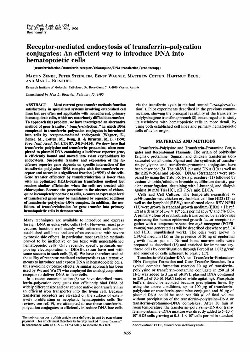
Polylysine-DNA Complexes in Avian Hematopoietic Cells.Binding of transferrin-polylysine and transferrin-polylysine-DNA to cell-surface receptors was measured with tritiatedcompounds (21), whereas internalization was followed byusing fluoresceinated transferrin-polylysine. Fig. 1A showsthat both transferrin-polylysine and transferrin-polylysine-DNA complexes bind to viable HD3 cells in a saturablefashion. Apparent binding constants calculated from thesedata were 22 nM for transferrin-polylysine and 43 nM fortransferrin-polylysine-DNA complexes. Although some-what higher, these Kd values (which rather represent on-ratesthan real dissociation constants; ref. 21) were still in reason-able agreement with those determined for native transferrin(15 nM; I. Killisch, P.S., K. Roemisch, H.B., and G. Griffith,unpublished data).To study internalization, transferrin-polylysine conjugates
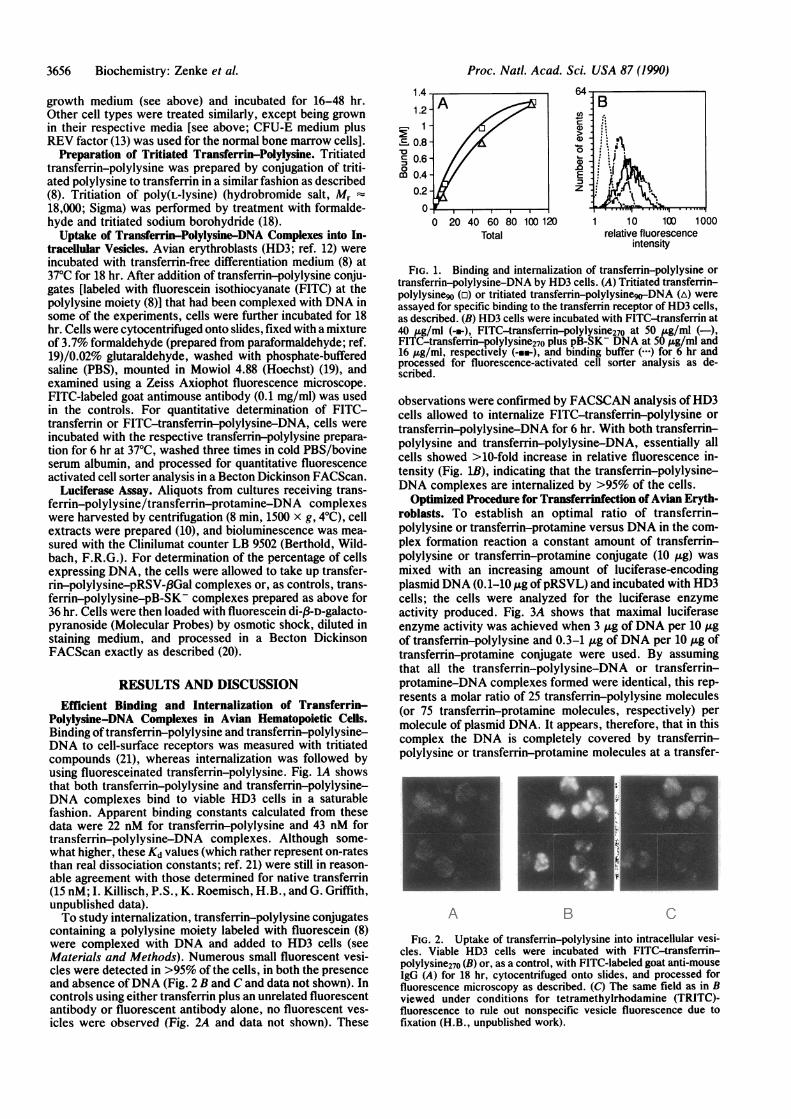
containing a polylysine moiety labeled with fluorescein (8)were complexed with DNA and added to HD3 cells (seeMaterials and Methods). Numerous small fluorescent vesi-cles were detected in >95% of the cells, in both the presenceand absence ofDNA (Fig. 2 B and C and data not shown). Incontrols using either transferrin plus an unrelated fluorescentantibody or fluorescent antibody alone, no fluorescent ves-icles were observed (Fig. 2A and data not shown). These
1.c
1.c
m 0.0.
4
.2 -A /1,.8.6-.4-.20
0 20 40
0)
Ez
60 80 100 120Total
1 10 100 1000relative fluorescence
intensity
FIG. 1. Binding and internalization of transfemn-polylysine or
transferrin-polylysine-DNA by HD3 cells. (A) Tritiated transferrin-polylysinego (r) or tritiated transferrin-polylysinego-DNA (A) wereassayed for specific binding to the transferrin receptor of HD3 cells,as described. (B) HD3 cells were incubated with FITC-transferrin at40 ,ug/ml (-e-), FITC-transferrin-polylysine270 at 50 ,Lg/ml (-),FITC-transferrin-polylysine270 plus pB-SK- DNA at 50 ,ug/ml and16 jig/ml, respectively (->e), and binding buffer (.) for 6 hr andprocessed for fluorescence-activated cell sorter analysis as de-scribed.
observations were confirmed by FACSCAN analysis ofHD3cells allowed to internalize FITC-transferrin-polylysine or
transferrin-polylysine-DNA for 6 hr. With both transferrin-polylysine and transferrin-polylysine-DNA, essentially allcells showed >10-fold increase in relative fluorescence in-tensity (Fig. 1B), indicating that the transferrin-polylysine-DNA complexes are internalized by >95% of the cells.
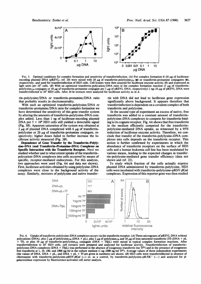
Optimized Procedure for Transferrinfection of Avian Eryth-roblasts. To establish an optimal ratio of transferrin-polylysine or transferrin-protamine versus DNA in the com-plex formation reaction a constant amount of transferrin-polylysine or transferrin-protamine conjugate (10 pug) was
mixed with an increasing amount of luciferase-encodingplasmid DNA (0.1-10 ,ug ofpRSVL) and incubated with HD3cells; the cells were analyzed for the luciferase enzymeactivity produced. Fig. 3A shows that maximal luciferaseenzyme activity was achieved when 3 ,ug of DNA per 10 ,ugof transferrin-polylysine and 0.3-1 ug of DNA per 10 ,ug oftransferrin-protamine conjugate were used. By assumingthat all the transferrin-polylysine-DNA or transferrin-protamine-DNA complexes formed were identical, this rep-resents a molar ratio of 25 transferrin-polylysine molecules(or 75 transferrin-protamine molecules, respectively) permolecule of plasmid DNA. It appears, therefore, that in thiscomplex the DNA is completely covered by transferrin-polylysine or transferrin-protamine molecules at a transfer-
A B
FIG. 2. Uptake of transferrin-polylysine into intracellular vesi-cles. Viable HD3 cells were incubated with FITC-transferrin-polylysine270 (B) or, as a control, with FITC-labeled goat anti-mouseIgG (A) for 18 hr, cytocentrifuged onto slides, and processed forfluorescence microscopy as described. (C) The same field as in Bviewed under conditions for tetramethylrhodamine (TRITC)-fluorescence to rule out nonspecific vesicle fluorescence due tofixation (H.B., unpublished work).
B
P.
F,
3656 Biochemistry: Zenke et al.
Proc. Natl. Acad. Sci. USA 87 (1990) 3657
(j)
.q
-C
0)
0 0.1 1 10,ug DNA 0 0.001 0.01 0.1 1 10
gg DNA
FIG. 3. Optimal conditions for complex formation and sensitivity of transferrinfection. (A) For complex formation 0-10 gg of luciferase-encoding plasmid DNA (pRSVL; ref. 10) were mixed with 10 ,Ag of transferrin-polylysine270 (x) or transferrin-protamine conjugates (0),respectively, and used for transferrinfection of HD3 cells. Cell lysates were then assayed for luciferase enzyme activity (8) and expressed aslight units per 107 cells. (B) With an optimized transferrin-polycation/DNA ratio in the complex formation reaction (3 ,ug of transferrin-polylysine270 conjugate or 10 Ag of transferrin-protamine conjugate per 1 Ag of pRSVL DNA, respectively) 1 ng-10 /Ig of pRSVL DNA weretransferrinfected to 107 HD3 cells. After 24 hr extracts were analyzed for luciferase activity as in A.
rin-polylysine/DNA or transferrin-protamine/DNA ratiothat probably results in electroneutrality.With such an optimized transferrin-polylysine/DNA or
transferrin-protamine/DNA ratio for complex formation wehave determined the sensitivity of this gene transfer systemby altering the amounts of transferrin-polylysine-DNA com-plex added. Less than 1 ng of luciferase-encoding plasmidDNA per 5 x 10⁶ HD3 cells still yielded a detectable signal(Fig. 3B). Apparent saturation of the system was obtained at2 ,ug of plasmid DNA complexed with 6 ,g of transferrin-polylysine or 20 ,ug of transferrin-protamine conjugate, re-spectively; higher doses failed to further increase the lu-ciferase activity measured (Fig. 3B).Dependence of Gene Transfer by the Transferrin-Polyly-
sine-DNA (and Transferrin-Protamine-DNA) Complexes onSpecific Interaction with the Transferrin Receptor. Next westudied whether and to what extent uptake of the transferrin-polycation-DNA complexes into cells occurred by means ofspecific, receptor-mediated endocytosis. For this analysis,two approaches were used (Fig. 4A and data not shown).First, luciferase activities obtained by using polylysine-DNAcomplexes were close to the background activity of theassay. Similarly, mixtures of polylysine and native transfer-
DNA
DNA+pL
DNA+pL+Tf
230
1777863
rin with DNA did not lead to luciferase gene expressionsignificantly above background. It appears therefore thattransferrinfection is dependent on a covalent complex ofbothtransferrin and polylysine.
In the second type of experiment an excess of native, freetransferrin was added to a constant amount of transferrin-polylysine-DNA complexes to compete for transferrin bind-ing to its cognate receptor. Fig. 4A shows that free transferrinin the medium efficiently competed for the transferrin-polylysine-mediated DNA uptake, as witnessed by a 95%reduction of luciferase enzyme activity. Therefore, we con-clude that transfer of the transferrin-polylysine-DNA com-plexes into cells depends on the transferrin receptor. Thisnotion is further confirmed by experiments in which theabundance of transferrin receptors on the surface of HD3cells and a human leukemia cell line has been modulated byvarious means, leading to the expected changes in transfer-rin-polylysine-mediated gene transfer efficiency (data notshown and ref. 22).To study which fraction of the cells actually express
plasmid DNA administered by transferrin-polylysine, HD3cells were incubated with transferrin-polylysine-pRSV-/3Galcomplexes. Expression of this reporter gene was then studied
A
no Tf* 1,1 ng/ml Tf*
DNA+TfpL 1 Ong/ml Tf-i1 OOng/mI Tf*m
1l OO0ng/mI Tf* 0 *-
0 2x10⁵ 4x10⁵lights units
1 10 50relative fluorescence
intensity
FIG. 4. Uptake oftransferrin-polylysine-DNA complexes occurs via the transferrin receptor. (A) Three micrograms ofpRSVL DNA withoutpolycations (DNA), plus 2 ;tg of polylysine270 (DNA + pL), plus 2 ;tg of polylysine270 and 10 j&g of iron-saturated transferrin (Tf) (DNA + pL+ Tf), or plus 10 ,ug of transferrin-polylysine270 conjugate (DNA + TfpL) were mixed in typical complex formation reactions. Aftertransferrinfection to 107 HD3 cells, cell extracts were prepared and analyzed for luciferase activity. Transferrinfection of transferrin-polylysine-DNA complexes (DNA + TfpL) was performed in the absence of exogenous transferrin (no Tf*) and in the presence of exogenousfree transferrin at 1, 10, 100, and 1000 ng/ml in the culture medium (1 ng-1000 ng/ml Tf*). Average values of three independent experiments(the values of DNA, DNA + pL, and DNA + pL + Tf are given in numbers) are shown. (B) HD3 cells were transferrinfected in absence ofchloroquine with transferrin-polylysine-pRSV-I3Gal (-) or, as a control, by transferrin-polylysine-pB-SK (---) and analyzed forBgalactosidase expression by fluorescence-activated cell sorter analysis.
Biochemistry: Zenke et al.
cn4x1 05
g-
- 2x1 05-
Proc. Natl. Acad. Sci. USA 87 (1990)
A
A-f,.. ...I"
0 10 100gM chloroquine
107
cn._
-.-_-C
1061 _
105 -]:
104I t. ...
0 2 4 10 40time of chloroquine pulse (hours)
FIG. 5. Chloroquine enhances the efficiency of transferrinfection. (A) Three micrograms of pRSVL DNA was transfected to 2 x 10⁷ HD3cells without and with 20, 60, and 200 ,uM chloroquine using a DEAE-dextran protocol optimized for chicken hematopoietic cells (ref. 6; o).Similarly, 3 ,g of pRSVL DNA complexed with 10 jig of transferrin-polylysine270 conjugate were transferrinfected to the same number ofHD3cells in the absence and presence of chloroquine (e). Twenty-four hours later cell extracts were analyzed for luciferase enzyme activity (thevalues for 106 cells are shown). (B) Transferrinfection of 3 ,g of pRSVL DNA (complexed with 10 ,g of transferrin-polylysine270 conjugate)to 2 x 10⁷ HD3 cells was performed both in the absence (0 hr) and presence of chloroquine (200 ,uM). After 2, 4, 6, and 8 hr of transferrinfectionin the presence of chloroquine, cells were extensively washed, seeded in fresh growth medium and incubated for a total of 24 hr. In addition,cells were treated with chloroquine (200 AiM) for the entire transferrinfection period (24 hr). Luciferase activity was determined as describedfor A.
at the single-cell level by FACS analysis (20) after introducingthe f-galactosidase substrate fluorescein di-p3-D-galactopy-ranoside. The unimodal distribution of cells containing fluo-rescein derived from fluorescein di-f3-D-galactopyranosidedue to ,B-galactosidase enzyme activity suggests that a largefraction of the cells exhibited expression of the P-galacto-sidase reporter gene (Fig. 4B).Chloroquine Augments the Efficiency of Transferrin-
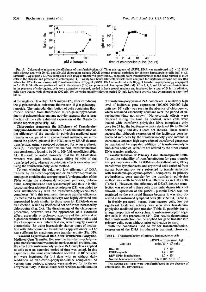
Polylysine-Mediated Gene Transfer. To obtain information onthe efficiency of the transferrin-polylysine-mediated genetransfer as compared with conventional methods, we intro-duced the pRSVL plasmid into HD3 cells by DEAE-dextrantransfection, using a protocol optimized for avian erythroidcells (6). In comparison with this method, transferrinfectionwas consistently found to be 50- to 100-fold less efficient (Fig.5A). It should be noted, however, that the DEAE-dextranprotocol was quite toxic, always killing 30-40% of thetransfected cells, whereas no cytotoxic effects were observedusing the transferrin-polylysine method.To test whether the relatively low efficiency of DNA
transfer by transferrin-polylysine or transferrin-protamineconjugates could be due to trapping and/or degradation oftheDNA within the endosomal/lysosomal pathway, chloro-quine, a drug known to alter lysosomal pH and thus to inhibitlysosomal degradation of macromolecules (23), was added tocells simultaneously with the transferrin-polylysine-DNAcomplexes. With this treatment, the gene transfer efficiency(as measured by luciferase activity) was highly elevated andapproached levels similar to those seen for DEAE-dextrantransfection, which by itself could not be further increased bychloroquine (Fig. SA). The disadvantage of the chloroquineprocedure, however, was the appearance of a cytotoxiceffect, especially at prolonged exposure of the cells and athigh concentrations ofchloroquine. We therefore tried to addthe chloroquine in a pulsed fashion, which largely avoidedcytotoxicity. By using increasingly shorter times of incuba-tion with chloroquine we found that its application for 3-4 hrwas sufficient for maximum gene transfer activity (Fig. SB).
Transient Expression ofDNA After Transferrin-Polylysine-Mediated Gene Transfer. Because the transferrin-polylysinegene transfer method was not deleterious to cell proliferation,the effect of transferrin-polylysine-DNA complexes appliedto cells over an extended period of time was tested. In thisexperiment, the same concentration of cells (1 x 10⁶ cells perml) were incubated for 1-4 days with or without dailyreaddition of transferrin-polylysine-DNA complexes. Atvarious time periods, aliquots were analyzed for luciferaseenzyme activity. In the cultures with repeated administration
of transferrin-polylysine-DNA complexes, a relatively highlevel of luciferase gene expression (100,000-200,000 lightunits per 107 cells) was seen in the absence of chloroquine,which remained essentially constant over the period of in-vestigation (data not shown). No cytotoxic effects wereobserved during this time. In contrast, when cells wereloaded with transferrin-polylysine-DNA complexes onlyonce for 24 hr, the luciferase activity declined 10- to 20-foldbetween day 2 and day 4 (data not shown). These resultssuggest that although expression of the luciferase gene in-troduced into cells by the transferrin-polylysine method istransient, a constant high expression of transferred genes canbe maintained by repeated addition of transferrin-polyly-sine-DNA complex, a feature not offered by the other knownDNA-transfer methods.
Transferrinfection of Primary Avian Hematopoietic Cells.To test the suitability of transferrinfection for gene transferinto primary avian cells, EGFR-ts-myb erythroblasts, REVTtransformed lymphoblasts, and erythroid cells enriched fromnormal bone marrow were subjected to transferrinfectionwith transferrin-polylysine-pRSVL complexes. In primaryerythroblasts, gene transfer by the transfemn-polylysinemethod was -30- to 50-fold less effective as in HD3 cells(Table 1). However, the efficiency of DEAE-dextran trans-fection was reduced in these cells to a similar degree (data notshown). Expression of the pRSVL plasmid DNA was notrestricted to the erythroid lineage because it was also ob-served in transformed lymphoid cells (REV NPB4, Table 1).
In freshly prepared, normal bone-marrow cells, low butsignificant luciferase activity was seen after transferrin-polylysine-mediated gene transfer (Table 1), possibly due toa large proportion of noncycling, transferrin-receptor nega-tive cells in this preparation (24). Our results demonstratethat transferrinfection can be applied for gene transfer intoprimary cells, even without prior cultivation in vitro.With the conditions used so far for transferrinfection,
expression of the DNA introduced is transient. However,
Table 1. Transferrinfection of primary hematopoietic cells
pRSVLuc expression, lightCell type units/10 x 106 cells
HD3 ebl 5 x 10⁵EGFR-myb-ebl 1.4 x 10⁴REV-NPB4 lymphoblasts 1.7 x 10⁵Normal bone-marrow cells 6.6 x 10³, 7.8 x l03The various cell types were transferrinfected in the absence of
chloroquine. ebl, Erythroblast.
108
V,, 107
2 1064-0)'- 105
104
Bwmm--.
3658 Biochemistry: Zenke et al.
Proc. Natl. Acad. Sci. USA 87 (1990) 3659
even when using a vast excess of transferrin-polylysine-DNA or transferrin-protamine-DNA complexes (10-foldabove the physiological concentration of native transferrin),no cytotoxic effects of the compounds have been observed,thereby enabling their repeated administration both at highconcentration and over an extended period of time.
We thank Gabi Stengl for excellent technical assistance, IngebrogHausmann for photography, and Marianne Vertes for typing themanuscript.
1. Graham, F. L. & Van der Eb, A. J. (1973) Virology 52, 456-467.
2. Feigner, P. L., Gadek, T. R., Holm, M., Roman, R., Chan,H. W., Wenz, M., Northrop, J. P., Ringold, G. M. &Danielsen, M. (1987) Proc. Nail. Acad. Sci. USA 84, 7413-7417.
3. Schaffner, W. (1980) Proc. Natl. Acad. Sci. USA 77, 2163-2167.
4. Nicolaou, C., Legrand, A. & Grosse, G. E. (1987) MethodsEnzymol. 149, 157-176.
5. Joyner, A. L., Skarnes, W. C. & Rossaut, J. (1989) Nature(London) 338, 153-155.
6. Choi, O.-R. B. & Engel, J. D. (1988) Cell 55, 17-26.7. Wu, G. Y. & Wu, C. H. (1987) J. Biol. Chem. 262, 4429-4432.8. Wagner, E., Zenke, M., Cotten, M., Beug, H. & Birnstiel,
M. L. (1990) Proc. Natl. Acad. Sci. USA 87, 3410-3414.9. Huebers, H. A. & Finch, C. A. (1987) Physiol. Rev. 67, 520-
582.
10. De Wet, J. R., Wood, K. V., DeLuca, M., Helinski, D. R. &Subramani, S. (1987) Mol. Cell. Biol. 7, 725-737.
11. Maniatis, T., Fritsch, E. & Sambrook, J. (1982) MolecularCloning:A Laboratory Manual (Cold Spring Harbor Lab., ColdSpring Harbor, NY).
12. Beug, H., Dbderlein, G., Freudenstein, C. & Graf, T. (1982) J.Cell Physiol. Suppl. 1, 195-207.
13. Zenke, M., Kahn, P., Disela, Ch.- Vennstrom, B., Leutz, A.,Keegan, K., Hayman, M., Choi, H. R., Yew, N., Engel, J. D.& Beug, H. (1988) Cell 52, 107-119.
14. Khazaie, K., Dull, T. J., Graf, T., Schlessinger, J., Ullrich, A.,Beug, H. & Vennstrom, B. (1988) EMBO J. 7, 3061-3071.
15. Radke, K., Beug, H., Kornfeld, S. & Graf, T. (1982) Cell 31,643-653.
16. Graf, T. (1973) Virology 54, 398-413.17. Adkins, B., Leutz, A. & Graf, T, (1984) Cell 39, 439-445.18. Ascoli, M. & Puett, D. (1974) Biochim. Biophys. Acta 371,
203-210.19. Royer-Pokora, B., Beug, H., Claviez, M., Winkhardt, H.-J.,
Friis, R. R. & Graf, T (1978) Cell 13, 751-750.20. Nolan, G. P., Fiering, S., Nicolas, J. F. & Herzenberg, L. A.
(1988) Proc. Nati. Acad. Sci. USA 85, 2603-2607.21. Stein, B. S., Bensch, K. G. & Sussman, H. H. (1984) J. Biol.
Chem. 259, 14762-14772.22. Cotten, M., Lingle-Rauoult, F., Kirlappos, H., Wagner, E.,
Mechtler, K., Zenke, M., Beug, H. & Birnstiel, M. L. (1990)Proc. Natl. Acad. Sci. USA 87, in press.
23. Dean, R. T., Jessup, W. & Roberts, C. R. (1984) Biochem. J.217, 27-40.
24. Schmidt, J. A., Marshall, J., Hayman, M. J., Doderlein, G. &Beug, H. (1986) Leuk. Res. 10, 257-272.
Biochemistry: Zenke et al.