Embed Size (px)
Citation preview
THE .]OL RNA I. or ( "' ESTIGATIVF. DER~IATOLOG\ , 6:2: 217-:22:J, 1971 C'"P'Ti)(ht @ I'll l hv The William• & Wilktn• ru.
\'nl. 6~ . Nn. :1 PrmtPd in l'.S.A .
RECEPTOR PROTEII\S FOR ANDROGEN lN HAMSTER SEBACEOUS GLANDS"
KE:\.Jl ADACHI. M.D .. P11.D.t
The coSto\·ertebral gland of the hamster is an androgen-::;ensitive tissue grossly discernible as a pair of pigmented nodules consisting of clumps of sebaceous glands. Ten to 14 day!' after castration. atrophy can be seen grossly and microscopically. but the atrophied gland can be react i\'ated by androgen (Hamilton and Montagna, 19-'lOl.
:-.lot withstanding our knowledge of androgen meta bolism and action in skin !Straus:- and Pochi. 1969). the specific mechanism of its action in sebaceous glands has not been reported in detail. Therefore. a few years ago we began to study the molecular mechanisms of androgen action in the hamster system and arrived a t a tentat ive conclus ion which we su mma rize here !Adachi a nd Takavasu. 197:2). Wh en testosterone carried in plasma reaches the sebaceous glands. it is converted l<> dihyd rotes tosteronet by !) a-reductase with the cofactor TPNH (Takayasu and Adachi. 1972}. Dihydrotestosterone. now generally believed to be a tissue-acti\·e a ndrogen. is transferred to the nuclei of the l-iebaceou~ cel ls where it initiate~ new protein synthesis. This synthesis is inhibited hy actinomycin 0 and t.hus involves new mRNA synthesis. The processes are selective and specific; not all. proteins (or enzymes) a re activated in the same way. Generally, certain key enzymes and regulatory proteins are sens it ive to the activation (Takayasu and Adachi, 1970}, which results in active metabolism through \'arious pos it ive feedback mechanisms.
In this scheme, the initial step in a ndrogenic action in t he sebaceous glands is the associat ion of an and rogen with receptors. Thu~ I he androgen receptors play a s ignificant rol e in the whole process of the molecular sequence'-. T he present paper !!ummarizes our studies on these receptors
Thi~ work was supported in part by Public Health Service. :\ational Instit ut es of Hea lth Gram A:\-1 -0;)I'il:?: Grant RR-00163 ot 1 he Animal Resources Branch , Di\ 1-sion of Research Resources; and b\ fund~ from Hofl-man- La Rnche, NE'w .Jer.;e\'. ·
• F'rom the Oregon Re~tional Primate Re~l'nrrh Center. Beaverton , Oregon 9iOO:i .
t Present address: Dermatulol(y Service. \'eterans Administration Hnspital , and linhersity nt Miami. Miam1. Florida :3:1 12!;.
:j: The followinl( 1 ri\·ial names and abhreviattuns are used in this paper: and rostenedi(lne ~·-andrn>tene-3, 17-dione; 5 a -dihydrotestosterone (DHT) = 5 trandrostan-17,8-ol-3 one; epiandrosterone <DHA) = 5 aandrostan-3 fj-ol- 17-one; testosterone 6 •-androsten-17 ,B-ol-3-one; estradiol-17 .8 = L, 3, 5(10)-estratrien-3. 17 ,B-d iol; estrone l, 3, 5! l0)-esn alrien-3-ol- l7-one; progesterone = 4-pregnen-3, 20-dione: ATP adenosine triphosphate; TPN - triphosphopyridine nucleotide: TPNH = reduced TPN; DPN = diphosphopyridine nucleotide: DPN H ~ reduced DPN: EDTA ethylene diamine tetra-acetate; TCA cycle = tricarboxylic acid cycle; GGPDH = glucose-6-phosphate dehydrogenase.
during the intervening years: a prel iminary report ha" already hecn pub! ished ! Adachi and Kano. 1972).
DETE('TJO!\ O F RECEfYI'OR PROTEINS
Pre parol ion of Sebaceous Glands
The experimental animals used in this series of experiments were male Syrian hamsters weighing 110 ± 10 gm . Generally they were castrated via the scrotum 16 hr before sacrifice . Costovertebral gla nds. seen as pigmented nodules on the back. were removed a nd dissected quil'kly at :1 o (' t o obtain clumps of sebaceous glands free from attached epidermis. subcutaneous fat. and hairs. Thus prepared. the sample consistPd of' at leas t 80 per cent pure sebaceous glands.
Detection of the Andro~en Receptors
Since the absolute amounts of' recept ors present in sebaceous gland:s are extremely minute. it is not practical to isolate them in pure form . At present, they a re demonstra ble only by indirect methods. The receptors are tagged wi th a radioacti\'e androgen such as 3 H-dihydrotestoslerone. a nd then the complex ( 3H -d ihyd rotestosterone- receptor complex ) is isolated by gel filtration. density gradient ultracen trifugation , or a combination of both.
The standard procedure fo r labe ling the receptor proteins is as follows. One ml of2oc;; homogenate of sebaceous glands in 0.32 M sucrose containing I mM M gCI2 and 20 mM Tris- HCI. pH 7.Ft. is incubated at :~o (' for 40 min with :l ~-tC'i nf dihydrotestosteron e. The mixture is centrifuged to obtain cytosol a nd nuclear fral·tions. The nuclear fraction is extracted with 0.4 M KCI !Fang et a l .. 19691 or 0...1 M \iaCI ( Bruchovskv and Wil:;on . 1968b.c) to obtain the nuclear rec~ptor complex. The nuclear extract is then subjected to gel filtration or density gradient centrifugation.
Gel filtration . Either the cytosol or nuclear frac tion is passed through a Sephadex G-2!1 column !1.5 :30 cml at :lo C' , and l..'l ml of each frac tion is collected and its radioactidty counted . Three reproducible peak:; are obtained !Pig. U. The fir~t peak to come out immediately after the \'oid volume i:; the 3H -androgen receptor <·omplex, a nd the second a nd t hird large peaks are 1 he conjugate a.nd free form of 3 H-dihydroteslosterune respectively. ln control experiments. the homogenate or sebaceous gla nds incubated with both 3H-androgen and 1.000 times exces, of cold dihy drotestosterone was run in an identical manner to rule out nonspecific binding.
S ucrose density wadient ultrac:entri{uf!ation. Alternatively, the cytosol or nuclear f'ract ion can be analyzed by the ultracentrifugation met hod. The
217
218 THE JOURNAL OF INVESTIGATIVE DERMATOLOGY
7
6
! • i? .... li ~ " 2
2
>< I
I 1 0 10 20 30 40 50 EO 70
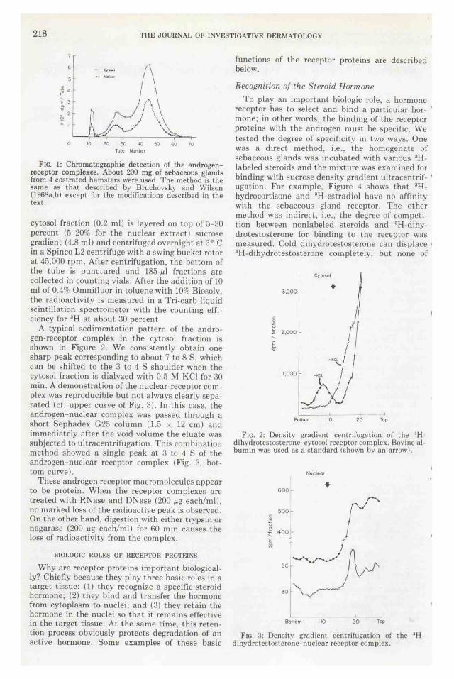
F1c. 1: Chromatographic detection of the androgenreceptor complexes. About 200 mg of sebaceous glands from 4 castrated hamsters were used. The method is the same as that described by Bruchovsky and Wilson (1968a,b) except for the modifications described in the text.
cytosol fraction (0.2 ml) is layered on top of 5-:~0 percent (5-200f for the nuclear extract) sucrose gradient (4.8 ml} and centrifuged overnight at 3° C in a Spinco L2 centrifuge with a swing bucket rotor at 45,000 rpm. After centrifugation, the bottom of the tube is punctured and 185-~1 fractions are collected in counting vials. After the addition of 10 ml of 0.4% Omnif1uor in toluene with lOOf Biosolv, the radioactivity is measured in a Tri-carb liquid scintillation spectrometer with the counting efficiency for "H at about 30 percent
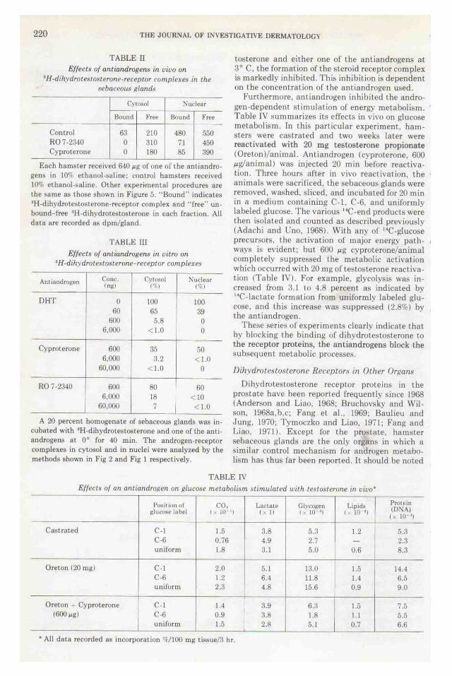
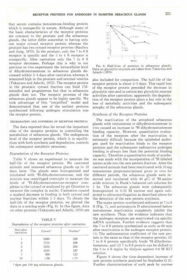
A typical sedimentation pattern of the androgen-receptor complex in the cytosol fraction is shown in Figure 2. We consistently obtain one sharp peak corresponding to about 7 to 8 S, which can be shifted to the 3 to 4 S shoulder when the cytosol fraction is dialyzed with 0.5 M KCl for :10 min. A demonstration of the nuclear-receptor complex was reproducible but not always clearly separated (cf. upper curve of Fig. ;~). In this case. the androgen- nuclear complex was passed through a short Sephadex G 25 column ( 1.5 ' 12 em) and immediately after the void volume the eluate was subjected to ultracentrifugation. This combination method showed a single peak at 3 to 4 S of the androgen- nuclear receptor complex (Fig. 3, bottom curve).
These androgen receptor macromolecules appear to be protein. When the receptor complexes are treated with RNase and 0:-.Jase (200 ~g each/ml). no marked loss of the radioactive peak is observed. On the other hand, digestion with either trypsin or nagarase (200 ~g each/ ml) for 60 min causes the loss of radioactivity from the complex.
BIOLOGIC ROLES OF RECEPTOR PROTEINS
Why are receptor proteins important biologically'? Chiefly because they play three basic roles in a target tissue: (l) they recognize a specific steroid hormone; (2) they bind and transfer the hormone from cytoplasm to nuclei; and (3) they retain the hormone in the nuclei so that it remains effective in the target tissue. At the same time, this retention process obviously protects degradation of an active hormone. Some example~ of these basic
functions of the receptor proteins are described below.
Recof(nition of the Steroid Hormone
To play an important biologic role, a hormone receptor has to select and bind a particular hor- · mone; in other words, the binding of the receptor proteins with the androgen must be specific. We tested the degree of specificity in two ways. One was a direct method. i.e ., the homogenate of sebaceous glands was incubated with various 3Hlabeled steroids and the mixture was examined for binding with sucrose density gradient ultracentrif- ' ugation . Por example, Figure 4 shows that 3Hhydrocortisone and 3H-estradiol have no affinity with the sebaceous gland receptor. The other method was indirect, i.e .. the degree of competition between nonlabeled steroids and 3H-dihydrotestosterone for binding to the receptor was measured. Cold dihydrotestosterone can displace • 3H-dihydrotestosterone completely, but none of
Cyiosol
J,OOO .. 2 u ! 2.000 .... E c>
"
1,0001
l=:: - .J Bo11om 10 20 Top
FIC. 2: Density gradient centrifugation ol the "Hdihydrolestosterone-cytO>OI receptor complex. Bovine albumin was used as a standard (shown by an arrow).
I soo ...
Bonom
Nuclear
I
10 ~
20 I
Top
F1c. 3: Density gradient centrifugation of the •Hdihydrotestosterone- nuclear receptor complex.

RECEPTOR PROTEINS FOR ANDROGEN IN HAMSTER SEBACEOUS GLANDS 219
1 \000
c
~ ,g ' E
<> .., 500
45.000 rpm
17 hri
j j
OL----------.----------.------Boftom 10 20 Top
FIG. 4: Specificity of the androgen receptors-bindings with 'H-Iabeled steroids. Twenty percent homogenate of sebaceous glands was incubated at o• for 40 min with 'H-estradiol, -cortisone, and -dihydrotesLosterone. The cytosol fraction shown in this figure was analyzed by density gradient cenLrifugation.
the other s teroids thus far tested can com pete with dihydrotestosterone (cf. Table 1 for summary). By comparison with d ihydrotestosterone binding. only one-fifth the amount of testosterone is bound to the receptor protein.
Transfer and Retention of the Andropen
According to the two-step mechanism for hormone action , the hormone must be incorporated and then retained in the cell (.Jensen et al.. 1968) . A specific receptor is also responsible for these processes. In Figure 5. the results of an in vivo experiment demonstrate the specific transfer and retention of dihvdrotestosterone in sebaceous glands. We injected 100 J.LCi of 3H-lestosterone intraperitoneally and remo\'ed the sebaceous glands at 10. 20, 30. 45, and 60 min . To detect the steroid-receptor complexes. they were homogenized and the homogenate was separated into cytosol and nuclear fractions and passed through a 025 column to obtain bound steroids as desc ribed in the previous section. The bound steroids were further extracted and analyzed by thin-layer chromatography . Within 10 min after the intraperitoneal injection of 3H-testosterone. the cytosol-androgen level reached saturation and decreased gradually. Ten minutes after the s teroid injection, testosterone was a major bound steroid only in the cytosol fraction. Otherwise the predominant androgen in both cytosol and the nuclear macromolecular fraction waR dihydrotestoRterone. The experiment clearly showed that dihydroteslosterone accumulates steadily in the nuclei with time. These data indicate that testosterone taken up by the sebaceous glands is converted to dihydrotestosterone in the cytoplasm. and the latter is subsequently incorporated and retained by the nuclei of the sebaceous glands.
Effects of Antiandrof!ens
The receptor proteins play a key role in androgen action and since thig step is an initial one, any interruption would result in incomplete androgen action . Such interruption can be achieved by the adm inistra tion of the antiandrogens. cyproterone acetate (Berlin Laboratories) or Ro 7-:23-10 (Hoffmann La Roche). When 3H-testosterone and one of the antiandrogens are given in vi\·o at the dosages shown in Table ll. 3H-dihydrotestoste rone binding to nuclei is decreased to 15 percent of control values. Since the free dihydrotestosterone level in nuclei is high (400- 500 dpm/gland). this indicates that these antiandrogens do not act direct ly on 5 a-reductase. which converts testosterone to d ihvdroteslosterone.
An in ~itro binding experiment subsequently showed that the antiandrogen com peted with dihydrotestosterone for the receptor binding sites (Table Ill). That is. when homogenates of sebaceous glands are incubated with 3 H-dihydrotes-
TABLE I
Competition between nonlabeled steroids and •H-dihvdrotestosterone for the androJ?en receptors
Sternids
DHT
T estosterone DHA Androstenedione Estradiol-17 {t Estrone Progesterone
. ~
Rind in~ percentag~ of dihvdrut eRtt>ht None
Cone. Cvtosol :-Juclenr (ngl I'll ('f I
0 100 100 60 65 39
600 5.8 0
600 9i) 98 600 100 99 600 103 95 600 98 104 600 LOO 100 600 105 110
FIG. 5: Transfer and retention of dihvdrotestosterone in sebaceous glands in an in vivo experiment. T -testosterone; DHT dihyd rotesLosterone.
220 THE JOURNAL OF INVESTIGATIVE DERMATOLOG"'
TABLE rJ Effects of antiandrogens in uiuo on
•H-dihydrotestosterone-receptor romplexe.~ in the sebaceous ~tland.,
Cyt~ol :"\udear
Bound Free Bound Free
Control 63 210 480 550 R07-2340 0 310 71 450 Cyproterone 0 180 85 390
Each hamster received 6-10 ~g of one of the anti androgens in 10c-; ethanol -saline: control hamsters recetved 10% ethanol-saline. Other exper~mental procedures are the same as those shown in Figure 5. ''Bound" indicates •H-dihydrolestosterone-receptor complex and "free" unbound free •H-dihydrotestosterone in each fraction. All data are recorded a~ dpm/gland.
TABLE ll1
Effects of antiandrogens in vitro on •H -dih) drote~tosterone-receptor complexes
Ant ia ndrogcn Cone Cywsol :-.ludear ln~tl ('~) t<; I
DHT 0 I(}() 100 60 65 39
600 5.8 0 6,000 < 1.0 0
Cyproterone 600 35 50 6,000 3.2 < 1.0
60,000 <l.O 0
RO 7-23-10 600 80 60 6,(}(10 18 <10
60,000 7 < 1.0
A 20 percent homogenate of sebaceous glands was incubated with •H-dihydrotestosterone and one of the antiandrogens at oo for -10 min. The androgen-receptor complexes in cytosol and in nuclei were analyzed by the methods shown in Fig 2 and Fig 1 respectively.
toslerone and either one of the antiandrogens at 3° C. the formaLion of the steroid receptor complex is markedly inhibited. This inhibition is dependent on the concentration of the amiandrogen used.
Furthermore, antiandrogen inhibited the androgen-dependent stimulation of energy metabolism. Table IV summarizes its effects in vivo on glucose metabolism. In this particular experiment, hamsters were castrated and lwo weeks later were reactivated with 20 mg testosterone propionate (Ore ton )/animal. Antiandrogen (cyproterone, 600 ,ug/animal) was injected 20 min before reactivation. Three hours after in vivo reactivation, the • animals were sacrificed, the sebaceous glands were removed, washed. sliced, and incubated for 20 min in a medium containing C-1, C-6. and uniformly labeled gluco~e. The various 14C-end products were then isolated and counted as described previously (Adachi and Uno. 1968). With any of 14C-glucose precursors, the activation of major energy path- , ways is evident; but 600 ,ug cyproterone/animal completely suppressed the metabolic activation which occurred with 20 mg of testosterone reactivation (Table lV). For example, glycolysis was increased from 3.1 to 4.8 percent as indicated by 14C-Iactate formation from uniformly labeled glucose. and this increase was suppressed (2.8%) by 1 the antiandrogen.
These series of experiments clearly indicate that by blocking the binding of dihydrotestosterone to the receptor proteins, the antiandrogens block the subsequent metabolic processes.
Dihvdrotesto.~terone Receptors in Other Orpans
Dihydrotestosterone receptor proteins in the prostate have been reported frequently since 1968 (Anderson and Liao, 1968: Bruchovsky and Wilson, 1968a.b.c; Fang et al.. 1969; Baulieu and .lung, 1970; Tymoczko and Liao, 1971; Fang and Liao, 1971) . Except for the prostate, hamster sebaceous glands are the only organs in which a similar control mechanism for androgen metabolism has thus far been reported. It should be noted
TABLE IV Effects of an antiandropen on pluco.~e metabolism ~tLmulated with testosterone in uiuo•
Posiuun of co, Lanete GI\'COI(en Liptds Protein {D'\>\) gluc<>'e label I >: tO 'l ('II r • tO '1 ! · 10 'I ( 10 1
)
Castrated C-1 1.5 3.8 5.3 1.2 5.3 C-6 0.76 4.9 2.7 - 2.:1 uniform 1.8 3.1 5.0 0.6 8.3
Oreton (20 mgl C-1 2.0 5.1 13.0 1.5 14.-t C-6 1.2 6.1 11.8 1.4 6.5 uniform 2.:3 4.8 15.6 0.9 9.0
Oreton ~ Cyproterone C-1 1.4 3.9 6.:l 1.5 7.5 (600~g) C-6 0.9 3.8 1.8 1.1 5.5
uniform l.5 2.8 5.1 0.7 6.6
• All data recorded as incorporation %/100 mg tissue/3 hr.
RECEPTOR PROTEINS FOR ANDROGEN JN HAMSTER SEBACEOUS GLANDS 221
that serum contaim testosterone-binding protein which is nonspecific in nature. Although many of the basic characteristics of the receptor proteins are common to the prostate and the sebaceous glands. the latter differ somewhat in having only one major cytosol receptor protein whereas the prostate has two cytosol receptor proteins (Baulieu and Jung, 1970). In the prostate, only the 7 to 8 S receptor is specific and the ~ to 4 S receptor is nonspeci fic. After cast rat ion only the 7 to 8 S receptor decreases. Perhaps this is why in our previous in vivo experiment the intracellular level of dihydrotestosterone in sebaceous glands decreased within :3- 4 days after castration whereas it remained high in the prostate and seminal vesicles (Takayasu and Adachi. 1972). The receptor protein in the prostatic cytosol fraction can bind 17{3-estradiol and progesterone but that in sebaceous glands does not. T he receptor proteins of the sebaceous glands appear to be highly specific. We took advantage of this ··simplified'' model and demonstrated that one of the earliest proteins synthesized following androgen stimulation was the receptor protein.
DEGRADATION AND SYNTHESIS OF RECEPTOR PROTEINS
The data reported thus far reveal the impor tant roles of the receptor proteins in controlling the metabolism of sebaceous glands. The endogenous level of the receptor protein, which is in equi librium with both synthesis and degradation, controls the subsequent metabolic processes.
Dewadation of the Receptor Proteins
Table V shows an experiment to measure the half-life of the receptor protein. We castrated animals and removed sebaceous glands up to I 0 days later. The glands were homogenized and incubated with GH-d ihydrotestosterone, and the mixture was centrifuged overnight to measure the leYels of GH-dihvdrotestosterone-receptor complexes in the ('ytosol or analyzed by gel filtration to measure the complex in nuclei. Castration caused a marked decrease in binding in both cytosol and nuclear fractions within 2-3 days. To obtain the half-l ife of the receptor proteins, we plotted the data in a semilog scale !Fig. 6). In this figure, data on other proteins (Takayasu and Adachi. 1970) are
TABLE V
Degradation of the receptor protein.~ after castration
Days after Cytosol' 1'\uclear• castration
1 2490 328 2 1710 306 3 1035 255 5 10:35 187 7 396 125
10 < 58 < 75
• dpm per 100 mg sebaceous glands.
:..:.:.· ... :.:.::·.::::,:·;;: ....... :::- ----..! __ .. __ _ \.""\ ·· .. .,· ................... \·-···-..
~ = "'' \ \
'·
0
' . ' .. 10 Drto!
•Tt RNA
' 2 Weeks
F tc. 6: Half-lives of proteins in sebaceous glancb. Data on glycolytic enzymes are taken f rom Takayasu and Adachi (1970).
a lso included for comparison. The half-life of the receptor protein is about 2-3 days. This rapid loss of the receptor protein preceded the decrease in glycolytic rate and in certain key glycolytic enzyme activities after cast ration: apparently the degradation of the receptor protein plays a key role in the loss of metabolic ac t ivities and the subsequent atrophy of the sebaceous glands.
Synthesis of the Receptor Proteins
The reactivation of the atrophied sebaceous glands with testosterone or dihydrotestosterone in vivo caused an increase in 3H-dihydrotestosterone bind ing capacity. However, quantitative evaluation of the receptors after the reactivation is extremely difficult because the unlabeled androgen used for react ivation binds to the receptor proteins and the subsequent radioactive androgen binding is always less than the potential binding capacity. Therefore a quantitat ive study of synthesis was made with the incorporation of 3H-labeled amino acids into the new protein fraction . After lhe castrated animals had been reactivated with 10 mg testosterone propionate/animal given in vivo for different periods. the sebaceous glands were removed and incubated with 40 J.LCi of 3H -amino acids mixture in Hank's balanced salt solution for 1 hr. The sebaceous glands were subsequently homogenized in 0.:~2 M sucrose and again subjected to ultracentrifugation as described above for the detection of the new protein synthesis.
The major protein-synthesized sediment at 'ito 8 S (Fig. 7). and actinomycin D administered before testosterone reactivation completely inhibit the new synt hesis. Thus, the evidence indicates that the androgen receptors are reactivated via specific mRNA synthesis. Further evidence suggests that the 7 to 8 S protein synthesized as early as 60 min after reactivation is the androgen receptor protein: ( 1) The sedimentation coeiTicient of the new protein is the same as that of the receptor protein: (21 7 to 8 S protein specifically binds 3H-dihydrolestosterone; and (3) 7 to 8 S protein can be shifted to the 3 to 4 S region by dialysis against 0.6 M KCl solution.
Figure 8 shows the time-dependent increase ol new protein synthesis analyzed by Sepbadex G 25. Further characterization of each peak by sucrose
222 THE JOURNAL OF INVESTIGATIVE DERMATOLOGY
density ultracentrifugation shows that the major peak is a 7 to 8 S receptor protein (Pig. 9).
In reviewing- the data in Figure 7. we note that the sebaceous glands of a 5-day castrate can s till synthesize 7 to 8 S protein without testosterone reactivation. This 7 to 8 S peak does. in fact, indicate the new receptor protein transcribed by endogenous mR:"JA. On the basis of this assumption, we can also measure a half-life of mR:-.JA specific for the receptor protein. We measured I he rate of the decrease in I he 7 to 8 S peak after 3H-arnino acid incorporation during various periods after cast ration . The data again showed a short half-life of the specific mRNA of 2-3 days similar to that of the cytosol receptor proteins (Fig. 6).
c 0
JOO
,,50 E ... ...
!/·· ... -·~ r
20 loll om Top
FtG. 7: Density ~radient centrifugation (at 45,000 rpm for 15 hrl of the cvtobol fraction obtained from the sebaceous ~lands incubated with labeled amino acids for l hr. The control animals received saline inJeCtions instead of Oreton, and 1 he actinomycin D group (---.11.---l received 1 mg actinomycin 0 /animal 30 min before the Orelon reactivation. Test : testosterone react ivation. (From Adachi and Kano. 197:2)
x 103 S E P H A D E X
c 0 -v D
10
G 25
6 7 8 9 10 '' 12 13 ,. 15
l U IE S
Ftc. 8: New protein synthesis after reactivation of the castrated sebaceous glands by Oreton .
c 0
200
::::_,oo E ... ...
10 loll om
b-o--o.q '•.. ,A
b o·C' O.•o,,
'b·C•<O./-q,
-vr-...1 ··,;·
20 30 lop
FtG. 9: Ultracentritugation analysis of each macromolecular peak obtained in the experiment shown in Figure 8. The major peak is about 7 to 8 S .
General stimulation of mRNA synthesis by steroid hormones has been reported (DeAngelo and Gorski, 1970), but the specific nature of the newly synthesized RNA and protein has not been defined. As far as we are aware, our study is the first to suggest androg-en st imulation of the new mRNA that specifically synthesizes the androgen receptor protein.
SUMMAR\
Chrom a tographic and ultracentr ifugal tech niques demonstrate that receptor proteins in hamster sebaceous glands play importan t biologic roles such as recognition, transfer. retent ion, and protection of the tissue-active androgen, dihydrotestosterone. Since this is the first step of androgen action, any interference ot the binding {for example, with an antiandrogen) causes a loss of subsequent biologic sequellae, such as the stimulation of energy metabolism. Both the synthesis and degradation nf the receptor proteins are highly dependent on dihydrotestosterone levels. The sh~Jrt halflives of the receptor proteins and of the messenger RNA for the receptors further contribute to the essential role of androgen receptors in controlling the dynamic actions of androgen in hamster sebaceous glands.
1 am greatly indebted to Dr. S. Takayasu and Mr. M. Kano for their collabora t ion.
REFERENCES
Adachi, K .. and Kano, M. (1972). The role of receptor proteins in controll ing androgen action in the sebaceous gla nds of hamstel'll. Steroids, 19: 567-574.
Adachi, K ., and Takayasu, S. ( 1972). The mechanisms of testosterone act ion on the sebaceous glands of the Syrian hamster. ln: Pharmacology and the Skin (ed.
RECEPTOR PROTE.INS FOR ANDROGEN lN HAMSTER SEBACEOUS GLANDS 223
by Montagna, W.. toughton. R. B .. and Van Scoll. E. J.). Appleton-Century Crofts. :-Jew York. pp. 381-401.
Adachi , K., and Uno, H . ( 1968). Glucose meta holism of growing and resting human hair follicles. Am .. J. Physiol. , 21.5: 1234-1239.
Anderson , K. M., and Liao. S . ( 1968). Selective retention of dihydrotestosterone hy prostatic nuclei. Nature (Lond .), 219: 277-279.
Baulieu. E. E., and Jung, I. ( 1970). A prostatic cytosol receptor. Biochem. Biophys. Res. Commun .. 38: 599--606.
Bruchovsky, N .. and Wilson , .J . D. ( 1968a). Evidence that dihydrotestosterone is the acti' e form of te,tosterone. Clin . Res., /4: 74.
Bruchovsky, N .. and Wilson, J.D. (1968b) . The com•ers ion of testosterone to 5a-andro~tan - 17P-ol-:3-ooe by rat prostate in vivo and in vitro. J. Bioi. Chern ., 243: 2012-2021.
Bruchovsky. N., and Wilson, J. D. ( L968c). The intranuclear binding of testosterone and 5a-androstan-17fJ-ol-3-onc by rat prostate. J. Bioi. Chern .. 243: 5953 5960.
DeAngelo, A. B .. and Gorski. J . ( 1970). Role of R:-<A synthesis in the estrogen induction of a specific uterine protein. Proc. Nat!. Acad. Sci. U.S.A., 66: 693- 700.
Fang. S., Anderson, K. M., and Liao, S. (1969). Receptor proteins for androgens. On the role of specific pro-
Leins in selective retention ol 17fJ-hydroxy-5aandrostan-3-one by rat ventral prostate in t•ivo and in t•itro. J . Bio!. Chern .. 244: 6584 6595.
Fan!(, S ., and Liau, S. ( 1971) Andro{(en receptors .. 1. Bioi. Chern., 246: 16 24.
Hamilton. J . B .. and Montagna. W. (1950). The sebaceous glands or the hamsters . 1. Morphological effects of androgens on integurnemary structures. Am. J. Anat., 86: 191 234.
.Jensen. E. V., Suzuki, T ., Kawashima, T .. Stumpf, W. E ... Jungblut, P. W., and DeSombre. E. R. (1968). A twv-step mechanism for the interaction of estradiol with rat uterus. Proc. :-.Inti. Acad. Sci. U.S.A., .59: 6:32-638.
StrauSl>, J. S., and Pochi. P. E. (1969). Recent advances in androgen metabolism and their relation to the skin. Arch. Dermatol., 100: 621- 636.
Takayasu, S., and Adachi, K. (1970) . Hormonal control or metabolism in hamster co~tovertebral glands . ,J. Invest. Dermatol., 55 · 13 19.
Takayasu. S .. and Adachi. K. Cl97:lJ. The tn ~:wu and 111 t•itro com·ersion of testosterone to 1 'i ,6-hydroxy-fie>androstan-:3-one (dihydrotestosteroneJ by the sebaceous gland of hamsters. Endocrinology, 90.· 73-80
Tymoczko, J . L .. and L1ao. S. (1971). Retention of an androgen- protein complex by nuclear chromatin aggregates: Heat -labi le factors. Biochim. Biophy:.. Acta , 252: 607- 611.


![SEBACEOUS NAEVUS LOCATED IN NASAL CAVITY – A … naevus.pdfcomplication of sebaceous naevi, however the life time risk is estimated to be less than 5% [3,4]. The majority of sebaceous](https://img.pdfslide.net/doc/110x75/5e67469b5680ed3ae440e92a/sebaceous-naevus-located-in-nasal-cavity-a-a-naevuspdf-complication-of-sebaceous.jpg)


![Suppression of Sebaceous Gland Development in Laboratory ... · of Win 17665 were the hamster flank organ [3] and, to a lesser extent, the supracaudal gland of the guinea pig [4,5]](https://img.pdfslide.net/doc/110x75/5faf66f08a49cb0f2d52940c/suppression-of-sebaceous-gland-development-in-laboratory-of-win-17665-were-the.jpg)