Embed Size (px)
Citation preview

Copy
right
© A
BE&
M to
dos o
s dire
itos r
eser
vado
s.
411Arq Bras Endocrinol Metab. 2014;58/5
Copy
right
© A
BE&
M to
dos o
s dire
itos r
eser
vado
s.
411
consensus
Recommendations of the Brazilian Society of Endocrinology and Metabology (SBEM) for the diagnosis and treatment of hypovitaminosis DRecomendações da Sociedade Brasileira de Endocrinologia e Metabologia (SBEM) para o diagnóstico e tratamento da hipovitaminose D
Sergio Setsuo Maeda1, Victoria Z. C. Borba2, Marília Brasilio Rodrigues Camargo1, Dalisbor Marcelo Weber Silva3, João Lindolfo Cunha Borges4, Francisco Bandeira5, Marise Lazaretti-Castro1
ABSTRACTObjective: The objective is to present an update on the diagnosis and treatment of hypovitami-nosis D, based on the most recent scientific evidence. Materials and methods: The Department of Bone and Mineral Metabolism of the Brazilian Society of Endocrinology and Metabology (SBEM) was invited to generate a document following the rules of the Brazilian Medical As-sociation (AMB) Guidelines Program. Data search was performed using PubMed, Lilacs and SciELO and the evidence was classified in recommendation levels, according to the scientific strength and study type. Conclusion: A scientific update regarding hypovitaminosis D was pre-sented to serve as the basis for the diagnosis and treatment of this condition in Brazil. Arq Bras
Endocrinol Metab. 2014;58(5):411-33
KeywordsVitamin D; cholecalciferol; PTH; osteoporosis; deficiency; insufficiency; diagnosis; treatment
RESUMOObjetivo: Apresentar uma atualização sobre o diagnóstico e tratamento da hipovitaminose D baseada nas mais recentes evidências científicas. Materiais e métodos: O Departamento de Metabolismo Ósseo e Mineral da Sociedade Brasileira de Endocrinologia e Metabologia (SBEM) foi convidado a conceber um documento seguindo as normas do Programa Diretrizes da Associação Médica Brasileira (AMB). A busca dos dados foi realizada por meio do PubMed, Lilacs e SciELO e foi feita uma classificação das evidências em níveis de recomendação, de acordo com a força científica por tipo de estudo. Conclusão: Foi apresentada uma atualização científica a respeito da hipovitaminose D que servirá de base para o diagnóstico e tratamento dessa condição no Brasil. Arq Bras Endocrinol Metab. 2014;58(5):411-33
DescritoresVitamina D; colecalciferol; PTH; osteoporose; deficiência; insuficiência; diagnóstico; tratamento
1 Endocrinology Division, Federal University of São Paulo (Unifesp), Escola Paulista de Medicina, São Paulo, SP, Brazil2 Department of Medicine, Paraná Federal University (UFPR), Curitiba, PR, Brazil3 Department of Medicine, Univille Medical School, Joinville, SC, Brazil4 Endocrinology Division, Brasília Catholic University (UCB), Brasília, DF, Brazil 5 Endocrinology Division, Agamenon Magalhães Hospital, University of Pernambuco (UPE), Escola de Medicina, Recife, PE, Brazil
Correspondence to:Sergio Setsuo MaedaRua Conselheiro Furtado, 847, ap. 9301511-001 – São Paulo, SP, [email protected]
Recebido em Mar /31/2014Aceito em Jun/18/2014
DOI: 10.1590/0004-2730000003388
INTRODUCTION
H ypovitaminosis D is highly prevalent and represents a public health problem in the entire
world. Studies show an elevated prevalence of this disease in several geographic regions, including Brazil. It can affect more than 90% of individuals, depending on the population studied (1).
Vitamin D is essential in functions related to bone metabolism, but it seems to be related in the patho
physiology of many diseases. In children, vitamin D deficiency leads to growth retardation and rickets. In adults, hypovitaminosis D leads to osteomalacia, to secondary hyperparathyroidism and consequently, to an increase in bone resorption, favoring bone mass loss and the development of osteopenia and osteoporosis. Muscle weakness can also happen, which further contributes to elevating the risk of fall and bone fractures among patients with low bone mass (2,3).

Copy
right
© A
BE&
M to
dos o
s dire
itos r
eser
vado
s.
412 Arq Bras Endocrinol Metab. 2014;58/5
The correct diagnosis of this condition and the identification of improvement or worsening factors can help the elaboration of more efficient strategies for the treatment of risk populations, such as the elderly and postmenopausal women.
This document represents the efforts of the Department of Bone Metabolism of the Brazilian Society of Endocrinology and Metabology (SBEM) for the development of recommendations based on evidence available in the scientific literature regarding the diagnosis and treatment of this condition. The objective of this document is to respond daily questions and to be a guideline for endocrinologists and clinicians in the Brazilian context.
MATERIALS AND METHODS
The elaboration of this guideline was motivated by the SBEM within its Practical Guidelines program. The model applied followed the Guidelines Program of the Brazilian Medical Association (AMB) and the Federal Council of Medicine (CFM). After the selection of collaborators, with a significant role and relevant publications in the area, clinical questions to be approached were elaborated.
The publication search was performed using MedLinePubMed and SciELOLilacs. We used the Oxford Classification, which evaluates the study design and considers the best available evidence for each question, to categorize the recommendation level or evidence strength of each article (4,5).
The levels of recommendation and evidence strength were reported as:
A: experimental or observational studies with better consistency.
B: experimental or observational studies with less consistency.
C: case reports (noncontroled studies).D: opinion lacking critical evaluation, based on
guidelines, physiological studies or animal models.
DEFINITION AND PHYSIOLOGY1. What is vitamin D: a nutrient or a prohormone?
Although it is defined as a vitamin, conceptually it is a prohormone. In conjunction with the parathyroid hormone (PTH), it acts as an important regulator of calcium homeostasis and bone metabolism.
It can be obtained from food sources, such as cod liver oil and from other fatrich fish (salmon, tuna, mackerel), or from endogenous cutaneous synthesis, which represents the most important source of this “vitamin” for the majority of human beings (2,3,6,7) (A). Table 1 shows some food sources of vitamin D (3).
Vitamin D can be found in the form of ergocalciferol or vitamin D2 and cholecalciferol or vitamin D3 (8). Vitamin D2 can be obtained from some yeast and plants, being produced for commercial use, through irradiation of the ergosterol present in some mushrooms (8) (D).
Figure 1. Photobiosynthesis of vitamin D.
7-dehidrocholesterolCholecalciferol or vitamin D or D
3
Ergocalciferol or vitamin D
2 25 hydroxilase
25(OH) vitamin D
1 a hydroxilase
1,25 (OH)2D
3
24, 25(OH)2 D
3Receptor
Skin
Ultra-Violet Rays
In the skin, the precursor is the 7dehydrocholesterol (7DHC) (8,9). During sun exposure, UVB photons (ultraviolet B, 290315 nm) penetrate the epidermis and produce a photochemical fragmentation to originate precholecalciferol. This intermediate is converted to vitamin D (or cholecalciferol) through a temperaturedependent isomerization (Figure 1).
Table 1. Vitamin D food sources
Food Portion Vitamin D content per portion
Wild salmon 100 g ~ 600-1,000 IU vitamin D3
Fish farming salmon 100 g ~ 100-250 IU vitamin D3
Canned sardine 100 g ~ 300 IU vitamin D3
Canned mackerel 100 g ~ 250 IU vitamin D3
Canned tuna 100 g ~ 230 IU vitamin D3
Cod liver oil 5 mL ~ 400-1,000 UI vitamin D3
Egg yolk 1 unit ~ 20 IU vitamin D3
Fresh mushroom 100 g ~ 100 IU vitamin D2
Sun dried mushroom
100 g ~ 1,600 IU vitamin D2
Adapted from ref. 3.
Recommendations for the diagnosis and treatment of hypovitaminosis D

Copy
right
© A
BE&
M to
dos o
s dire
itos r
eser
vado
s.
413Arq Bras Endocrinol Metab. 2014;58/5
Cholecalciferol is transported to the liver by DBP (vitamin D binding protein). In the liver, there is the hydroxylation of carbon 25 (CYP27B1), forming the 25hydroxyvitamin D (25(OH)D), through a process which is not strictly regulated, since it happens without control, and depends on the combination of cutaneous and diet stocks of vitamin D (8).
After the liver step, 25(OH)D is transported to the kidneys by DBP, where it is converted to calcitriol or 1,25dihydroxyvitamin D [1,25(OH)2D] (Figure 1). This is the most active metabolite and it is responsible for stimulating intestinal calcium and phosphate absorption. The kidney hydroxylation is stimulated by PTH and suppressed by phosphate and FGF23. Calcitriol production is strictly controlled by negative feedback, in a way to influence its own synthesis by the decrease of 1ahydroxylase. It is responsible for accelerating its inactivation through the conversion of 25(OH)D to 24,25(OH)2D. This mechanism reflects a direct action of 1,25(OH)2D in the kidneys, however there is still an inhibitory action on PTH production in the parathyroids (8,9). The 1ahydroxylase can also be found in other cells and tissues, such as the skin, prostate, breast, intestine, lungs, pancreatic β cells, monocytes and parathyroid cells. The 1,25(OH)2D molecule can also be locally synthesized by these cells and tissues (8,9) (D).
The vitamin D receptor (VDR) belongs to the superfamily of nuclear receptors regulating the transcription factors of the steroid hormones, retinoic acid, thyroid hormones and vitamin D. After binding 1,25(OH)2D to VDR, it interacts with the retinoic acid receptor, forming a heterodimeric complex (RXRVDR), which then binds specific sequences of DNA, known as Vitamin D Responsive Elements (VDRE) (10,11). The main target organs for 1,25(OH)2D are the intestine, bones, parathyroid glands and kidneys. However, its receptors have been found in several other tissues (10,11) (D).
SBEM recommendation: food sources are scarce in vitamin D and humans depend mainly on the skin production catalysed by UVB sunlight exposure (Evidence A).
2. What are the effects on bone metabolism?
SBEM recommendation: active vitamin D modulates PTH synthesis, increases intestinal calcium absorption, and improves bone mass and muscular function (Evidence A).
The best known and studied actions of vitamin D are related to bone metabolism, where it plays a crucial role. It participates in intestinal calcium absorption, muscle function, modulation of PTH secretion and bone cell function.
Parathyroid cells express the 1ahydroxylase enzyme, and can synthetize the active form of vitamin D (1,25(OH)2D) inside the cells using the 25(OH)D serum pool (12) (B). In hypovitaminosis D, due to a minor intracellular synthesis, there is a secondary hyperparathyroidism, which is associated to an increase in bone resorption (2,1316) (B), besides the fact that the circulating levels of 1,25(OH)2D are, generally, normal. There is an inverse correlation between PTH and 25(OH)D, described in children (17) and the elderly (2). Several cut off values for 25(OH)D for the PTH normalization have been published, the majority, being around 28 and 40 ng/mL (70 to 100 nmol/L) (2,1823) (C). Other causes of secondary hyperparathyroidism also have to be investigated, such as chronic kidney disease (creatinine clearance below 60 mL/min), Paget’s disease, hungry bone syndrome and the calcium and vitamin D malabsorption syndromes (24).
Intestinal calcium absorption depends on the active vitamin D action in the duodenum, through a transcellular saturable process, whose stimulus leads to the synthesis of proteins such as calbindinD9K (CaBP9k) and the epithelial apical channel TRPV6 (13,14) (D). However, there is evidence that the nonsaturable transport, which happens with part of calcium absorption in the human ileum is also vitamin D sensitive (25). According to Heaney and cols., individuals with 35 ng/mL of 25(OH)D presented higher absorption than those with 25 ng/mL (26) (B). Increase of calcium absorption with increasing dose of vitamin D3 or serum 25(OH)D was recently observed, but there is no evidence of what the minimum value of 25(OH)D to ensure calcium absorption from the intestine in the range of 1652 ng/mL evaluated in the study (27).
Population studies correlated positively vitamin D concentration with bone mass, mainly of the hip, but with 25(OH)D cut off points varying from 12 to 36 ng/mL (3090 nmol/L) (2830) (C).
The muscle tissue expresses vitamin D receptors (13) and, clinically, muscle weakness and myopathy are observed in patients presenting severe vitamin D deficiency. Dhesi and cols., observed that the number of falls is higher among the elderly when they present the vitamin D deficiency (31) (C). The administration
Recommendations for the diagnosis and treatment of hypovitaminosis D

Copy
right
© A
BE&
M to
dos o
s dire
itos r
eser
vado
s.
414 Arq Bras Endocrinol Metab. 2014;58/5
of 800 IU of cholecalciferol for 12 weeks decreased in 49% the number of falls (32) (B). Cholecalciferol use is associated with the prevention of falls among the elderly with hypovitaminosis D, but not among those presenting normal serum values (33) (B).
In a metaanalysis of the main osteoporosis intervention studies; BischoffFerrari and cols. indicated again 25(OH)D serum concentration above 30 ng/mL (75 nmol/L) to be the most beneficial for health in general (A). Bone health, here represented by a better bone mineral density (BMD), decreased risk of fall and femural and nonvertebral osteoporotic fractures, seems to be benefited by 25(OH)D concentrations equal to or higher than 30 ng/mL (75 nmol/L), concentrations around 36 ng/mL (90 nmol/L) being suggested as the most advantageous (3436). The same 25(OH)D values seem to benefit the muscle strength of lower limbs, which was evaluated by the TUG (Time Up and Go) test, where the individual is evaluated based on the time he needs to walk a distance equivalent to eight steps. Indivi duals presenting 25(OH)D in the range of 36 to 40 ng/mL (90 and 100 nmol/L) seem to perform with higher speed. Evidence also suggests that higher 25(OH)D values are associated with a lower risk for colorectal cancer and periodontal disease (36) (A).
Regarding the bone tissue, evidence suggests that 1,25(OH)2D stimulates mineralization, through an indirect process that happens with the increase in intestinal absorption of the minerals which are incorporated into the bone matrix. Physiological concentrations of calcitriol promote calcium mobilization to the bones, while the administration of large doses promotes excessive bone remodeling. Osteoblasts present 1,25(OH)2D receptor. This hormone modulates the gene expression of alkaline phosphatase and osteocalcin. Therefore, in the process of bone remodeling, 1,25(OH)2D is important for bone formation and reabsorption (37).
Priemel and cols. evaluated 675 bone biopsies and correlated the histomorphometry findings with serum 25(OH)D concentration. The presence of bone mineralization defects was only found in individuals with concentrations below 30 ng/mL (75 nmol/L) (38) (B).
The role of vitamin D in nonbone related endpoints, such as mortality, cardiovascular risk, cancer and autoimmune diseases is still controversial (39,40).
DIAGNOSIS
3. How to define hypovitaminosis D?
SBEM recommendation: the analysis of the 25 hydroxyvitamin D metabolite (25(OH)D) should be used for the evaluation of the vitamin D status of an individual (Evidence A).
There is a consensus that 25(OH)D (calcidiol) is the most abundant metabolite and the best indicator for the evaluation of vitamin D status (A), the individuals being classified as: deficient, insufficient of sufficient in vitamin D (3,6,41,42). On the other hand, there is no consensus regarding the cut off value for the definition of “vitamin D sufficiency” (6,43,44).
The values discussed in the medical literature, based on populational studies, with emphasis on calcium homeostasis and bone health, vary from 20 to 32 ng/mL (50 to 80 nmol/L) (26,4347). Several specialists agree that for correction of secondary hyperparathyroidism, reduction of the risk of fall and fractures and maximum calcium reabsorption, the best 25(OH)D cut off value is 30 ng/mL (75 nmol/L) (6,41,46). Thus, serum concentrations below 20 ng/mL (50 nmol/L) are classified as deficiency; those ranging from 20 to 29 ng/mL (50 to 74 nmol/L) as insufficiency and between 30 and 100 ng/mL (75 and 250 nmol/L) as sufficiency. Therefore, many consider 25(OH)D serum concentrations below 30 ng/mL as hypovitaminosis D (3,4143,48,49). These values were recognized by the Endocrine Society guideline, although they differ from the ones accepted (20 ng/mL) by the Institute of Medicine (IOM) (50) (B). In general population, there is no evidence of benefit in the measurement of 25(OH)D due to the high cost, but according to the Endocrine Society, to maximize bone health; supplementation is recommended for children up to 1 year with at least 400 IU/day; between 1 and 70 years, at least 600 IU/day while over 70 years old, 800 IU/day (41).
SBEM recommendation: 25(OH)D concentrations above 30 ng/mL are desirable and should be the target for higher risk populations, because above this concentration, vitamin D benefits are more evident, especially regarding osteometabolic diseases and fall risk reduction (Evidence B).
Recommendations for the diagnosis and treatment of hypovitaminosis D

Copy
right
© A
BE&
M to
dos o
s dire
itos r
eser
vado
s.
415Arq Bras Endocrinol Metab. 2014;58/5
4. What are the methodological implications for the plasma determinations of 25(OH)D?
SBEM recommendation: the methods based on chromatography are considered gold standard for the laboratory evaluation of 25(OH)D, although, automated immunometric assays can be used in the clinical practice taking into account the good correlation with the method of excellence, besides the practicality and lower cost. However, the clinician should be aware of the possible mistakes caused by several interfering conditions, possibly leading to diagnostic classification errors (Evidence B).
Circulating 25(OH)D level is the best method to evaluate the individual vitamin D status. Nevertheless, there are controversies regarding the best method for 25(OH)D determination. Some factors should be considered when the levels of this vitamin are evaluated, such as the lack of a precise physiological regulatory control (feedback), the variability of methods and standards, the inclusion of contaminant metabolites in the analysis, among others. Radioimmunoassays (RIA) used in the past underestimated the levels of 25(OH)D when the dominant levels were 25(OH)D2. RIA have been replaced by automated chemiluminescent immunoassays, resulting in higher concentrations and by immunoenzymatic assays which measure total 25(OH)D, a combination of vitamin D2 (25(OH)D2) and vitamin D3 (25(OH)D3 (51) (B).
The methods that do not employ direct immune detection are high performance liquid chromatography (HPLC) coupled to mass spectrometry (LCMS), which can distinguish individual levels of 25(OH)D2 and 25(OH)D3. These are considered the gold standard for analysis and currently used as reference (52) (B). Both 1,25(OH)2D and 25(OH)D circulate predominantly bound to proteins and their concentration can be determined. However, to evaluate the vitamin D status, 25(OH)D total serum level is used, including both D3 and D2 forms. The results can be reported in nanograms per milliliter (ng/mL) or nanomol per liter (nmol/L). For conversion, you just need to multiply the value obtained in ng/mL by 2.5, to obtain the value in nmol/L. Automated methods allow the use in clinical routines, they are fast and report 25(OH)D2 and 25(OH)D3 together, while LCMS methods can distinguish between 25(OH)D2 and 25(OH)D3, being useful, then, for the evaluation
of the effectiveness of D2 supplementation, versus endogenous D3 production. These chromatographic methods, although more precise, are more laborious and expensive (53) (B).
SBEM recommendation: for conversion of 25(OH)D concentration from ng/mL to nmol/L, the multiplication factor 2.5 is applied.
The accuracy of the measurements varies widely between laboratories and between different assays, and even when testing identical samples, this variation can achieve 17 ng/mL (53). The immunoassay requires the development of selective antibodies for 25(OH)D2 and 25(OH)D3, which preferentially do not show cross reaction with any other metabolite. Matrix effects can still occur, caused by endogenous components that modify the binding of the antibody to the material to be analyzed. Metabolites with lower physio logical potential end up being included in the quantification, such as the 3epimer of the 25(OH)D, which can correspond to up to 5% of the total 25(OH)D. As its molecular weight is identical to 25(OH)D, these are not separated by LCMS. Finally, 24,25 dihydroxyvitamin D (24,25(OH)2D), considered an inactive metabolite, can correspond to up to 20% of the 25(OH)D measured, whereas some assays show 100% crossreaction (51,54).
The use of a standard cut off value to evaluate vitamin D status is problematic if applied to all laboratories and all methods, considering there are still differences on vitamin D extraction from its binding protein, cross reaction between 25(OH)D2, 25(OH)D3 and other metabolites, besides the lack of standardization (52,53) and for this reason, quality control tools, such as DEQAS (International Vitamin D External Quality Assessment Scheme) were created, as an attempt to decrease the variation in data analysis (55).
The most used methods nowadays are competitive assays, based on specific antibodies and nonradioactive markers, the improvement in the comparison between results obtained from different methodologies being necessary. Whatever the method employed is a precise definition of the normality range is fundamental (56). It is also important to highlight that the intraindividual variability can vary from 12.1 to 40.3% (57).
The clinical conditions that interfere with 25(OH)D serum concentrations are highly dependent on environmental factors and lifestyle, particularly UVB sun
Recommendations for the diagnosis and treatment of hypovitaminosis D

Copy
right
© A
BE&
M to
dos o
s dire
itos r
eser
vado
s.
416 Arq Bras Endocrinol Metab. 2014;58/5
light exposure. Polymorphisms in CYP27B1, which codes for 1ahydroxylase, showed strong correlation with variations in 25(OH)D level. The vitamin D binding protein (DBP) is the main transporter for vitamin D metabolites, its phenotype helping predict 25(OH)D serum concentrations. Certain polymorphic forms can be more efficient for vitamin D binding, activation and metabolism, interfering with circulating levels. Genetic polymorphisms greatly contribute to the heterogeneity of clinical manifestations of hypovitaminosis D, especially among ethnic groups (51,58) (B).
EPIDEMIOLOGY5. Which are the risk populations for hypovitaminosis D? What is the prevalence in Brazil?
SBEM recommendation: the analysis of 25(OH)D concentration is not recommended for the general population. It is recommended for specific groups belonging to populations at risk or for those with relevant clinical condition in which deficiency is suspected (Evidence A).
The Department of Bone and Mineral Metabolism from SBEM agrees with the guidelines published by the Endocrine Society, which does not recommend the 25(OH)D test for the general population considering the cost of this evaluation. The laboratory test is recommended for individuals under risk for hypovitaminosis D or for those with a relevant clinical condition. The candidates to be tested are the ones presenting the following conditions: rickets or osteomalacia, osteoporosis, history of falls and fractures in the elderly, obesity, pregnant and lactating women,
patients with malabsorption syndromes (cystic fibrosis, inflammatory bowel disease, Chron’s disease, bariatric surgery), renal or liver insufficiency, hyperparathyroidism, medications interfering in vitamin D metabolism (anticonvulsants, glucocorticoids, antifungal, antiretroviral, cholestyramine, orlistat), granulomatous diseases and lymphomas (41,59) (A). It is also useful for the evaluation of hypothesis of vitamin D intoxication.
Besides that, it is important to highlight that all the conditions that limit sunlight exposure can potentially cause hypovitaminosis D and can be added to the list of individuals in photoprotection regimen (60) (D) and religious garment users (veil, burqa, cassock and others) (6163) (C).
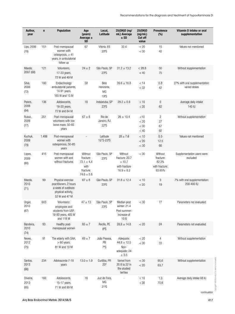
Hypovitaminosis D is a world health problem and Brazil is part of this scenario, also presenting an elevated prevalence of hypovitaminosis D in the population (B). Table 2 presents some of the main Brazilian and international studies that included Brazil, published in the last decade. In general, in several regions of the country, the values indicate suboptimum vitamin D concentrations, verifying high prevalence of hypovitaminosis D in several age groups. The majority of the studies included mainly the elderly and postmenopausal women which are the populations at risk for osteoporosis (C). However, three studies involving adolescents showed high prevalence of hypovitaminosis D in this age group of the Brazilian population (6466). The factors that seem to favor the presence of higher serum concentrations in our population are: younger age (64,6769), community life (70), the practice of outdoors physical activity (64,71), vitamin D oral supplementation (72), season of the year (spring, summer) (6873), residence in sunny beach areas (74,75) and in lower latitudes (76).
Table 2. Prevalence of hypovitaminosis D in Brazil
Author, year
n Population Age (years)
Average ± SD
Local, Latitude
25(OH)D (ng/mL) Average
± SD
25(OH)D (ng/mL) Cut off value
Prevalence(%)
Vitamin D intake or oral supplementation
Saraiva,
2005
(73)
and
2007 (70)
420
177
243
Elderly, > 65 years
Institutionalized
125 W and 52 M
From the community
168 W and 75 M
76 ± 9
79 ± 6
São Paulo, SP
23ºS
14.4 ± 9.2
19.6 ± 11.2
< 10
< 20
< 40
< 10
< 20
< 40
41
71
99
16
42
96
7% with oral supplementation: daily dose
125-1.000 IU
4% with supplementation
10% with supplementation
continuation
Recommendations for the diagnosis and treatment of hypovitaminosis D
Copy
right
© A
BE&
M to
dos o
s dire
itos r
eser
vado
s.
417Arq Bras Endocrinol Metab. 2014;58/5
Lips, 2006
(78)
151 Post-menopausal women with
osteoprosis, > 41 years, in ambulatorial
follow up
67 Vitória, ES
20ºS
32.4 < 20
< 30
15
42
Values not mentioned
Maeda, 2007 (68)
121 Volunteers,
17-33 years,
72 W and 49 M
24 ± 2 São Paulo, SP
23ºS
31.2 ± 13.2 < 28.8
< 40
50
75
Without supplementation
Silva, 2008
(72)
180 Endocrinology ambulatorial patients,
14-91 years,
165 W and 15 M
58 Belo Horizonte,
MG
19ºS
39.6 ± 16.8 < 14
< 32
0.8
42
27% with oral supplementation: varied doses
Peters, 2008
(64)
136 Adolescents,
16-20 years,
72 W and 64 M
18 Indaiatuba, SP
23ºS
29.2 ± 0.8 ≤ 10
≤ 30
0
62
Average daily intake
140 IU
Russo, 2009
(77)
251 Post-menopausal volunteers with low bone mass, 50-85
years
67 ± 6 Rio de Janeiro, RJ
22ºS
26 ± 10.4 <10
< 20
< 30
< 40
2
27
67
92
Without supplementation
Kuchuk, 2009
(79)
1.486 Post-menopausal women with
osteoporosis, 50-85 years
- Latitude 15°S-23ºS
28 ± 7.6 < 10
< 20
< 30
0.5
12.5
66
Values not mentioned
Lopes,
2009
(80)
415 Post-menopausal women with and without fractures
Without fracture:
72.1 ± 4.4
with fracture:
74.6 ± 5.8
São Paulo, SP
23ºS
Without fracture: 20.7
± 10.7
with fracture 16.9 ± 8.2
< 30 Without fracture: 82.3%
with fracture: 93.65%
Supplementation users were excluded
Maeda, 2010
(71)
99 Physical exercise practitioners, 2 hours a week of outdoors
physical activity,
52 W and 47 M
67 ± 6 São Paulo, SP
23ºS
31.6 ± 12.4 < 10
< 20
3
19
7% with oral supplementation: 200-400 IU
Unger, 2010
(67)
603 Volunteers:
employees and students from USP,
18-80 years, 485 W and 118 M
47 ± 13 São Paulo, SP
23ºS
Median post winter: 21.4
Post summer: increase of
10.6
< 30 77 Parameters not evaluated
Bandeira, 2010
(74)
93 Healthy post-menopausal women
65 ± 7 Recife, PE
8ºS
28.8 ± 14.8 < 20 24 Parameters not evaluated
Neves, 2012
(75)
91 The elderly with SAH, > 60 years,
81 W and 10 M
69 ± 7 João Pessoa, PB
7ºS
Adequate: 44.8 ± 12.5
Non-adequate: 24
± 3.5
< 20
< 30
4
33
Without supplementation
Santos, 2013
(66)
234 Adolescents 7-18 years
13.0 ± 1.9 Curitiba, PR 25º
Varied from 20.8 to 22 in the studied
tertiles
< 30
< 20
90,6
63,7
Without supplementation
Oliveira,
2013
(65)
160 Adolescents,
15-17 years,
71 W and 89 M
16 Juiz de Fora, MG
21ºS
- ≤ 10
≤ 30
1,3
70,6
Average daily intake 88 IU
Author, year
n Population Age (years)
Average ± SD
Local, Latitude
25(OH)D (ng/mL) Average
± SD
25(OH)D (ng/mL) Cut off value
Prevalence(%)
Vitamin D intake or oral supplementation
continuation
Recommendations for the diagnosis and treatment of hypovitaminosis D

Copy
right
© A
BE&
M to
dos o
s dire
itos r
eser
vado
s.
418 Arq Bras Endocrinol Metab. 2014;58/5
Maeda, 2013
(69)
591 Volunteers,
17-100 years,
388 W and 203 M
Nursing homes:
76.2 ± 9.0
Community: 79.6 ± 5.3
Exercise: 67.6 ± 5.4
young: 23.9 ± 2.8
São Paulo, SP
23ºS
Nursing homes: 15.0
± 11.9
community: 19.8 ± 11.0
Exercise: 31.5 ± 12.4
young: 34.5 ± 14.0
< 10
< 20
< 30
19
47
73
6% with oral supplementation: 200-400 IU
Arantes,
2013
(76)
1.933 Post-menopausal women with low bone
mass, 60-85 years
67 ± 5 Latitude
8°S-33°S
27.2 ± 8.4 ≤ 30 68,3 Parameters not evaluated
Martini, 2013
(81)
636 Adolescents, adults and the elderly
- São Paulo, SP
23ºS
M: 16.7
W: 19.2
< 20 - Intake varied from 108 to 140 IU/d
Cabral,
2013
(82)
284 Men, skin phototype evaluated
69.4 ± 6.5 Recife, PE
8ºS
28.0 ± 13.6 < 20
< 30
31,5
66,7
2,5% took supplements
M: men; W: women; IU: international units.
TREATMENT
6. How to treat hypovitaminosis D in patients who are at high risk for the deficiency?
SBEM recommendation: generalized vitamin D supplementation is not indicated for the entire population. The benefits of the vitamin D treatment are more evident in populations presenting high risk for the deficiency (Evidence A).
Current evidence does not support the concept of general population supplementation (41) (A). As the adequacy of vitamin D concentration in our population is closely related to the cutaneous production, secon dary to sunlight exposure, individuals with low exposure represent the main population of deficient indivi duals. Therefore, a simple interview can bring important information on the probability of vitamin D deficiency in a specific individual.
The complementation of the daily needs, as well as the treatment of the deficiency should be performed for individuals with hypovitaminosis D risk (see Epidemiology section) or those to whom sunlight exposure is prohibited, due to skin cancer, transplants or systemic lupus erythematosus (A).
The most available vitamin D form for treatment and supplementation is cholecalciferol or vitamin D3 and this is the metabolite that has been shown to be the most effective one. Ergocalciferol or vitamin D2 can also be used as a supplement, however the studies show that, as its halflife is a little shorter than the one of D3, it should be used preferentially daily (83). Besides that, some laboratory methods that test 25(OH)D recog nize only 25(OH)D3, what can bring problems for the control of plasma levels when vitamin D2 is used for supplementation. Therefore, although supplementation and treatment can be done with both vitamin D metabolites, preference should be given to vitamin D3, due to the advantages on the maintenance of more stable concentrations.
The treatment doses vary according to the degree of deficiency and the target to be achieved. Apparently, 25(OH)D concentrations higher than 12 ng/mL would be sufficient to avoid rickets and osteomalacia, as well as to normalize intestinal absorption of calcium (84,85). However, to reduce fractures, concentrations above 24 ng/mL are necessary (86), while to avoid the development of secondary hyperparathyroidism, concentrations above 30 ng/mL are desirable (69). Therefore, especially during osteoporosis treatment, it
Author, year
n Population Age (years)
Average ± SD
Local, Latitude
25(OH)D (ng/mL) Average
± SD
25(OH)D (ng/mL) Cut off value
Prevalence(%)
Vitamin D intake or oral supplementation
Recommendations for the diagnosis and treatment of hypovitaminosis D

Copy
right
© A
BE&
M to
dos o
s dire
itos r
eser
vado
s.
419Arq Bras Endocrinol Metab. 2014;58/5
is recommended that plasma 25(OH)D is kept above 30 ng/mL.
As a practical rule, one can predict that, for every 100 IU supplemented, an increase of 0.7 to 1.0 ng/mL is gained in the concentration of 25(OH)D (41). However, other studies show that this doseresponse curve is not linear.
In a study developed in Brazil, with an institutionalized population, showing high prevalence of hypovitaminosis D (40.4% with 25(OH)D < 20 ng/mL), supplementation with 7,000 IU/day, produced an average elevation of 7.5 ng/mL in 25(OH)D concentration after three months (87) and this elevation achieved a plateau around six weeks. However, as it has already been recognized by other authors, this increase was more evident among those individuals with lower initial values (< 20 ng/mL), in whom the average increase was 10.3 ng/mL after three months of treatment, while those showing 25(OH)D concentration above 20 ng/mL, increased on average only 5.18 ng/mL. Besides that, 45% of the individuals still kept insufficient (30 ng/mL) and 10% still kept deficient (< 20 ng/mL) at the end of three months of supplementation. This demonstrates that, for values lower than 20 ng/mL, doses higher than 1,000 IU/day will be necessary if the target to be achieved is 30 ng/mL (B).
In a similar institutionalized population, MoreiraPfrimer and cols. demonstrated, in a randomized, double blind prospective placebo controlled study, that an average dose of 3,700 IU/day of vitamin D3 for six months was able to take the treated group to average concentrations of 34.6 (variation from 20.9 to 48.4) ng/mL, while the placebo group kept in 20.7 (variation from 9.4 to 41.2) ng/mL (p < 0.0001). There was a significant increase in calcemia for the treated group, but no patient developed hypercalcemia (88) (A).
Those institutionalized and bedridden are a population with elevated risk for deficiency. Mocanu and cols. evaluated the effect of the fortification of the bread roll with 320 mg of calcium and 5,000 IU of vitamin D on an institutionalized population for 12 months. It was possible to verify an effective increase in 25(OH)D (initial average 11.4 ng/mL and final average 50.0 ng/mL), with 92% of the individuals achieving concentrations higher than 30 ng/mL. No individual developed hypercalcemia ou hypercalciuria. PTH concentrations were reduced during treatment and there was a significant increase in BMD of the lumbar spine and proximal femur (89). However, when reevaluated, this same
population, three years after removal of supplementation, it was verified that the benefits gained with vitamin D supplementation had been lost (90).
In a population of postmenopausal women undergoing treatment for osteoporosis, followed in a specific outpatient clinic, Camargo (91) demonstrated that weekly doses higher than 7,000 IU (> 1,000 IU/day) are necessary to achieve vitamin D sufficiency (25(OH)D > 30 ng/mL), which is in accordance with the propo sition of the Endocrine Society for the elderly at risk (Table 3). According to this Brazilian study, 73% of the patients, followed for at least three months in ambulatory directed to the treatment of osteoporosis were below the desired target concentration (> 30 ng/mL) (Figure 2). In the same study, a positive correlation was found between 25(OH)D concentrations and the femur BMD and a negative correlation with PTH (91).
Recommendations for the diagnosis and treatment of hypovitaminosis D
Table 3. Vitamin D daily maintenance doses recommended for the general population and the population at risk for the deficiency
Age groups General population (IU)
Population at risk (IU)
0 – 12 months 400 400 – 1,000
1 – 8 years 400 600 – 1,000
9 – 18 years 600 600 – 1,000
19 – 70 years 600 1,500 – 2,000
> 70 years 800 1,500 – 2,000
Pregnant women 14 – 18 years
600 600 – 1,000
Pregnant women > 18 years
600 1,500 – 2,000
Lactating 14 – 18 years 600 600 – 1,000
Lactating > 18 years 600 1,500 – 2,000
Adapted from the nutritional tables of the Institute of Medicine and the Endocrine Society.
Figure 2. Percentage of non-adequacy of vitamin D according to the plasma concentrations of 25-hydroxyvitamin D (25(OH)D) in a population of individuals presenting osteoporosis at a medical school teaching ward (91).
100908070
605040
302010
0ng/mL (n) < 10 (19)
5,2
36,4
73,3
< 20 (132) < 30 (266)
25(OH) D
%

Copy
right
© A
BE&
M to
dos o
s dire
itos r
eser
vado
s.
420 Arq Bras Endocrinol Metab. 2014;58/5
In children and adolescents, the doses, apparently, do not vary much from the ones in the adults, with the exception of the first year of life (Table 3). Winzenberg and cols., in a metaanalysis involving six studies that evaluated vitamin D supplementation in healthy children, were able to group 343 participants that received placebo and 541 participants that received vitamin D and the analysis suggested a benefit of the supplementation on the in lumbar spine bone mineral density and total body bone mineral content of those children that were previously deficient (92). Therefore, as already observed in other studies, the positive endpoints of the supplementation are always much more evident when the populations studied were initially deficient (A).
Vieth in a study done in Canada, tested two vitamin D3 doses, compared to placebo. The first dose was 1,400 IU/week (or 200 IU/day), the same quantity recommended at current nutritional tables from the Ministry of Health in Brazil. The second dose was 14,000 IU/week, both groups being followed for 12 months. The group that received 1,400 IU/week did not present significant increase in 25(OH)D concentration, while the group receiving 14,000 IU obtained an increment of 15 to 30 ng/mL at the end of 12 months (93). Taking from that and similar studies, an alteration on the vitamin D daily recommendation tables was proposed in several countries.
In 2011, the Institute of Medicine, in the USA, an organ that regulates the reference tables for daily recom mended intake (DRI) for the general population, increased the daily recommendation to 600 IU for individuals between the ages of 1 and 70, and to 800 IU for those older than 70 (Table 3) (50,94). However, the Brazilian nutritional table remains with the daily recommended intake of 200 IU, although several national studies have demonstrated that the typical meals of the Brazilian population are not a relevant vitamin D source (64,95), that we depend on cutaneous synthesis to obtain sufficiency and that the deficiency is present in all age and populational groups, especially among the elderly (70,74,76,80) (C).
Generally speaking, when 25(OH)D is lower than the target concentration (below 20 ng/mL), an attack dose is necessary to replenish the body stocks. The most used scheme currently is to administer 50,000 IU/week (or 7,000 IU/day) of vitamin D for six to eight weeks (41). In case the desired concentration of 25(OH)D is not achieved, a new cycle can be proposed. As there can be some individual variation in the
response to treatment, the reevaluation of the plasma values after each cycle is ideal, especially in the cases of more serious deficiencies, up to achieving the desired concentration. After this period, the maintenance dose should be defined and it varies according to the age group and concurrent conditions (Table 3). For the adults, maintenance doses vary between 400 and 2,000 IU, depending on the sunlight exposure and skin tone. For the elderly, the recommended doses vary from 1,000 to 2,000 IU/day or 7,000 to 14,000 IU/week. Obese individuals, presenting malabsorption conditions or in use of anticonvulsants might need doses that are twice or three times higer (41) (A).
SBEM recommendation: doses recommended for the maintenance of vitamin D sufficiency are indicated in table 3, varying for the different age groups. Howe ver, for deficiency correction, higher doses (attack doses) are necessary (Evidence A).
Due to the fact that it is a liposoluble substance, vitamin D is absorbed with fats and follows the enterohepatic circulation, meaning it is normally secreted through the gallbladder and reabsorbed in the small intestine. Thus, especially in cases of malabsorption, doses which are much higher than usual can be necessary in order to normalize 25(OH)D concentrations. Besides that, for supplementation studies and in the daily practice it is possible to notice some individual variation in blood concentrations of 25(OH)D reached in response to a same dose of vitamin D, suggesting individuals might present different competencies in intestinal absorption or its metabolization (96). It seems there is no difference considering vitamin D absorption in relation to fasting or meal type (97).
SBEM recommendation: the risk of intoxication with the defined doses in this document is almost null (Evidence A). Special care must be taken when pharmaceutical compounding is used (Evidence D) and with patients that have diseases with anomalous production of 1,25 dihydroxyvitamin D by the tissues, such as in granulomatous diseases and some lymphomas (Evidence B).
Vitamin D3, when administered as described above is very safe. Doses of up to 10,000 IU per day for five months did not induce signals of toxicity, which can be translated as hypercalcemia and hypercalciuria (98) (A). Toxic concentrations of 25(OH)D (> 90 ng/mL) are
Recommendations for the diagnosis and treatment of hypovitaminosis D

Copy
right
© A
BE&
M to
dos o
s dire
itos r
eser
vado
s.
421Arq Bras Endocrinol Metab. 2014;58/5
difficult to achieve with these routine doses (99). In rare clinical situations, such as in some cases of granulomatous diseases (sarcoidosis, tuberculosis and chronic fungal infections) and some lymphoma, activated macrophages can locally produce 1,25(OH)2D in excess and induce hypercalcemia and hypercalciuria (B). Children with Williams syndrome are more predisposed to hypercalcemia. Therefore, under these conditions, supplementation should be more criterious and follow frequent monitoring of plasmatic and urinary calcium (41).
7. What are the differences between vitamin D2 and D3?
SBEM recommendation: vitamin D deficiency treatment and supplementation can be done either with vitamin D3 (cholecalciferol) or with vitamin D2 (ergocalciferol), although the first metabolite presents some advantages over the second, due its commercial avaiability, for being more evaluated in clinical studies, for allowing more posologic formulations, promoting more effective increases and for being identified by all laboratory methods (Evidence B).
Vitamin D sources are: sunlight exposure, diet and supplementation. The main difference between vitamin D2 (ergocalciferol) and vitamin D3 (cholecalciferol) is the source. In summary, vitamin D2 is the vitamin D from the plant sources, while the one from animal sources is in the form of vitamin D3. The D2 and D3 sources differ only due to the presence of an additional double bond and a methyl group incorporated to the long side chain of the biological form called D2 (100). The two forms present equivalent biological power and are activated in equally efficient ways by hydroxylases in humans. However, there is controversy on the bioequivalence of these formulations for supplementation. A metaanalysis evaluating only controlled and randomized studies that use vitamin D2 and D3 showed that vitamin D3 increased 25(OH)D levels more significantly when compared to vitamin D2 (p = 0.001), the single or in bolus dose of vitamin D3 being better than D2 (p = 0.0002). However, this advantage was lost in daily supplementation (101) (A). Previous studies had already shown a small superiority of vitamin D3 when administered in single dose, to maintain 25(OH)D le vels for longer time (102).
When the two formulations were compared in daily use for 25 weeks, it was observed that those u sing vitamin D2 presented 25(OH)D average concentrations lower when compared to those that received D3, although, without altering PTH levels (103) (A).
A recent study did not demonstrate difference in effectiveness when higher 25(OH)D levels were found and also in sustained serum concentration of 1,25(OH)2D3, after 11 weeks of supplementation with 1,000 IU of vitamin D2 or D3 per day (104) (A).
The same was observed in the treatment of children with rickets and controls, where there was a similar increase in 25(OH)D and 1,25(OH)D levels with both formulations (105) (A). It is possible to conclude that both forms are equivalent in relation to daily supplementation and that vitamin D3 presents superiority in relation to the maintenance of the 25(OH)D levels for single dose supplementation.
8. What is the difference between vitamin D and calcitriol?
SBEM recommendation: vitamin D active forms, such as calcitriol or alfacalcidol, should not be used when the objective is supplementation, or in the treatment of vitamin D deficiency, because of their higher risk of side effects (Evidence A).
Calcitriol or 1,25(OH)2D is an active hormone, a final product of two vitamin D hydroxylations. Its endocrine action starts with renal production, finely controlled by the activity of the 1ahydroxylase enzyme. This enzyme, present in the epithelial cells or the proximal convoluted tubules is stimulated mainly by the PTH and is inhibited by FGF23, among other less important regulators. Circulating calcitriol itself also deviates its synthesis to an inactive product, the 24,25(OH)2D and, this way, protects the organism from its excess. The 1ahydroxylase enzyme was identified in different tissues, what makes us believe there is some local production of calcitriol, with autocrine and paracrine actions. Opposite to renal cells, where calcitriol production is rigorously controlled, in these other tissues it is believed that production only depends on the presence of substrate (14,41). While all the systems of the organism are fully functioning, the recommendation is for the supplementation and treatment of the deficiency to be done with vitamin D itself, allowing tissues to produce their necessities, controlled by local or hormonal mechanisms, in the case of renal tubular cells. The use of calcitriol should be reserved for special situations, such as in chronic kidney insufficiency, in type 1 and type 2 vitamin D dependent rickets and in hypophosphatemic rickets, or in cases of extreme malabsorption. The use of calcitriol presupposes a much more rigorous
Recommendations for the diagnosis and treatment of hypovitaminosis D

Copy
right
© A
BE&
M to
dos o
s dire
itos r
eser
vado
s.
422 Arq Bras Endocrinol Metab. 2014;58/5
control of calcemia and calciuria, because hypercalcemia can frequently occur (14,41) (A).
9. How to do supplementation in special cases?
a) In chronic kidney disease
The patient with chronic renal disease presents higher risk for vitamin D deficiency. In renal disease, PTH concentrations also correlate with circulating 25(OH)D levels (A). Therefore, it is believed that vitamin D deficiency contributes to the development of secondary hyperparathyroidism in chronic renal patients, independent on the renal calcitriol production. Nowadays it is known that several tissues, such as macrophages and osteoblasts have the capacity of producing active vitamin D (calcitriol) and that this synthesis depends on the subs trate, therefore, it is not strictly regulated as the renal synthesis. Because of that, the treatment of the deficiency and the adequacy of the circulating levels of 25(OH)D is always recommended each time plasma concentrations are lower than 30 ng/mL (106109). According to the opinion of the committee responsible for writing the treatment guidelines for osteomineral disease in chronic renal patients in Brazil, 25(OH)D quantification is recommended at the end of each cycle of attack doses, until the target concentration is achieved, and from then on, every six months (110) (D).
b) In the treatment of osteoporosis
A good part of vitamin D benefits on the risk of fractures observed in the literature has been associated with the concomitant use of calcium. Therefore, the adequacy of calcium intake, either through diet, or through the use of calcium salts, is part of any protocol for osteoporosis treatment. Recommended vitamin D doses are those capable of taking and maintaining plasma concentration to 30 ng/mL or above, avoiding, this way, the secondary hyperparathyroidism and the increase in bone resorption (111113) (A). The nonadequacy of vitamin D concentration is considered one of the potential failures in medicinal treatment of osteoporosis (significant BMD loss and fractures).
SBEM recommendation: for patients with osteoporosis and increased risk of fractures, it is recommended that the 25(OH)D concentrations are maintained above 30 ng/mL for full benefits, for the prevention of secondary hyperparathyroidism, decreased risk of fall and improvement of BMD. To this end, daily doses between 1,000 and 2,000 IU are necessary (Evidence A).
c) In obesity and post-bariatric surgery
Obese patients present lower vitamin D concentration when compared to nonobese and are considered a population at risk for the deficiency (41) (A). Nowadays, bariatric surgery is a very used alternative to induce weight loss in these individuals, possibly further aggravating this deficiency. Therefore, it is advisable to correct this condition prior to surgery. Santos and cols. demonstrated that women submitted to bariatric surgery at least three years before, presented lower 25(OH)D values when compared to normal paired controls, 77.1% of them presenting vitamin D insufficiency/deficiency and 41.7% presenting secondary hyperparathyroidism (Figure 3) (114). 25(OH)D concentrations correlated inversely with PTH (r = 0,57, p < 0,05) and directly with bone remodeling markers (CTX and osteocalcin) which, together, can justify the increased risk for fracture observed in this population by other researchers (C). Depending on the surgical technique used and the degree of disabsorption promoted, some individuals might have a lot of difficulty normalizing 25(OH)D and PTH concentrations, being necessary to follow these parameters and to consider a new attack dose or even higher maintenance doses that should be individually adjusted (D). In some cases, the orientation for frequent sunlight exposure and use of parenteral vitamin D are resources that can be necessary (115117).
Recommendations for the diagnosis and treatment of hypovitaminosis D
Figure 3. Prevalence of secondary hyperparathyroidism in a population of women submitted to bariatric surgery at least three years prior to the study enrollment. Different ranges of circulating 25(OH)D (114).
100%
< 10 ng/mL(n = 2)
> 30 ng/mL(n = 11)
25(OH)D
25(OH)D: 25-hydroxyvitamin D. Signi�cance level of the chi-square test (p < 0.025).
10 ¬ 20 ng/mL(n = 25)
20 ¬ 30 ng/mL(n = 10)
52%
30%
9,1%
90,9%70%
48%
PTH > 65 ng/L PTH < 65 ng/L
%

Copy
right
© A
BE&
M to
dos o
s dire
itos r
eser
vado
s.
423Arq Bras Endocrinol Metab. 2014;58/5
SBEM recommendation: obese people are a population at risk for deficiency and after bariatric surgery it is further aggravated, leading to a secondary hyperparathyroidism and an increased risk for fractures (Evidence A). 25(OH)D test in this situation is very useful for the titration of the daily vitamin D doses, which can be as high as ten times the routine ones (Evidence D).
d) Pregnancy
It is a critical period, because women are oriented to avoid sunlight exposure. Vitamin D deficiency in pregnant women was associated to low birth weight of the newborn, besides some late endpoints, such as low bone mass and cardiovascular risk markers in school age children. In a recent metaanalysis, Aghajafari and cols. analyzed 31 studies, including 18,869 individuals and they concluded that the serum le vels of 25(OH)D are related to gestational diabetes, preeclampsia, low birth weight newborn and bacterial vaginosis (118). In another metaanalysis, vitamin D supplementation showed positive effect on the low birth weight reduction (119) (A). The doses recommended for supplementation in this period of life can be found in table 3.
When vitamin D deficiency is suspected, the treatment with higher doses is still indicated, but daily doses are preferable. The 25(OH)D concentration in the newborn shows high correlation with the one found in the mother. The placenta presents the 1ahydroxylase enzyme and therefore, has the capacity of converting 25(OH)D to calcitriol. Apparently, this production is not strict controlled as that what happens in renal tubules, and depends only on the amount of substrate (41) (C). For this reason, the recommendation during pregnancy is to avoid weekly or monthly doses.
SBEM recommendation: for pregnant women presenting deficiency risk, treatment brings benefits for the mother (Evidence B) and the newborn (Evidence A). Daily doses of vitamin D are recommended during pregnancy and weekly or monthly higher doses should be avoided, because the placental production of calcitriol is substratedependant (Evidence C).
10. When should active vitamin D analogues be used?
SBEM recommendation: vitamin D analogues can be used to suppress PTH concentrations in patients with hyperparathyroidism secondary to chronic kidney disea se (CKD), the indication and the dose depending on the CKD stage and the PTH level (Evidence A). There is no superiority of the analogues currently available in Brazil in comparison with cholecalciferol and calcitriol for the prevention of fractures (Evidence B). The analogues are not recommended for the prevention of cancer (Evidence B).
Active vitamin D analogues are synthetic substances that directly bind the vitamin D receptor (VDR). They present different selectivity to the parathyroid cells, depending on its chemical structure, calcitriol (1,25(OH)2D3) shows smaller selectivity, promoting more side effects such as hypercalcemia, hyperphosphatemia and vascular calcifications. More selective compounds such as paricalcitol (19nor1a,25(OH)2D2), maxacalcitol (22oxa1a,25(OH)2D3) and doxercalciferol (1a(OH)D2) and eldecalcitol (1a,25(OH)22β(3hydroxypropyloxy)D3) and doxercalciferol (1a(OH)D2) and eldecalcitol (1a,25(OH)22β(3hydroxypropyloxy)D3) promote less adverse effects. Doxercalciferol and the alfacalcidol require liver 25hydroxylation to become active (120) (A).
a) Use in secondary hyperparathyroidism
The analogues are classically used to suppress PTH levels in patients with secondary hyperparathyroidism (SHPT) and chronic kidney disease (CKD). In CKD there is an increase in PTH levels, secondary to an alteration of the regulation of the fibroblast growth factor (FGF23) in the PTHvitamin D axis and the decrease of calcitriol production due to CKD itself. The suppression of PTH levels in patients in stages 34 of CKD is more than 40% in 90% of the patients (121,122) (A).
The dose used is variable depending on the CKD stage, if the patient is undergoing dialysis or not and also on the serum PTH concentration. In CKD stages 35, the ideal PTH levels are not defined yet, other PTH increasing factors having to be discarded. Hypocalcemia, vitamin D deficiency and hyperphosphatemia should be corrected initially. If PTH levels remain elevated and progressively increasing, the use of analogues such as calcitriol, should be considered. In CKD stage 5D with elevated and sustained PTH levels, the recommendation is to maintain PTH levels between two and
Recommendations for the diagnosis and treatment of hypovitaminosis D

Copy
right
© A
BE&
M to
dos o
s dire
itos r
eser
vado
s.
424 Arq Bras Endocrinol Metab. 2014;58/5
nine times the upper limit of normality. There is no consensus on the doses of doxercalciferol and paricalcitol, some studies calculated the dose in relation to the initial PTH value dividing it by 80 to 120, to minimize the excessive suppression of PTH or hypercalcemia and hyperphosphatemia (106) (B).
The use of vitamin D analogues minimizes bone loss in CKD due to the suppression of PTH levels and prevents bone remodeling reduction, due to the effect on the differentiation of normal osteoblasts and inhibition of osteoclastogenesis. However, the concern with the excessive suppression, which would lead to adynamic bone disease persists (122) (B).
Several studies have demonstrated benefit on the survival of patients undergoing dialysis treated with calcitriol or vitamin D analogues. Besides that, there is a smaller risk of progression for terminal renal disease and an increase in the survival in patients with CKD stages 34 (123,124) (B). There is doubt in relation to the benefit of vitamin D active analogues, compared to placebo, in relation to fractures, quality of life, hospitalizations, muscle function and fall in these patients (125,126) (A).
The most selective analogues, compared to calcitriol, demonstrate lower mortality, lower number of hospitalizations and lower duration of each hospitalization per year (124) (B). Doxercalciferol, similar to paracalcitol, presents higher benefit in relation to survival, when compared to calcitriol. A doseresponse was observed in the benefit when the levels of PTH were adjusted according to vitamin D (127) (B).
The use of analogues to avoid the evolution of nephropaty in diabetic patients is discussed, although the results are still conflicting, using microalbuminuria and albuminuria as markers. Results from VITAL study are awaited for a more definitive observation (120,128,129) (B).
b) Fracture prevention
The relationship between vitamin D levels with falls and fractures has been described, also as a significant linear predictor of major osteoporotic fractures in ten years (130) (B). A recent metaanalysis demonstrated a modest decrease in the prevention of fractures with the use of 1,25(OH)2D3 (calcitriol) and 1ahydroxyvitamin D3 (alfacalcidol), similar to that obtained using doses higher than 700 IU/day of vitamin D3 (35) (A).
Eldecalcitol (1a,25dihydroxy2β[3hydroxypropiloxy] vitamin D3 is a new analogue of vitamin D
active form, recently approved for the treatment of osteoporosis in Japan. Eldecalcitol presents a strong inhibitory effect on bone resorption and significantly increases bone mineral density. This drug showed a 26% decrease in the incidence of new vertebral fractures in three years and 71% decrease in the risk of wrist fracture, in comparison to alfacalcidol, but showed no bene fit against hip fracture. An increase in serum calcium was observed, although hypercalcemia was only seen in 0.4% of patients (131) (A). Edelcalcitol compared to alfacalcidol promoted better quality of life and decreased severity of vertebral fractures (125,126) (A).
NON-BONE ACTIONS OF VITAMIN D11. What is the evidence for extra-skeletal effects of vitamin D?
SBEM recommendation: although observational studies show an association between low vitamin D concentration and alterations in several extraskeletal systems, in the moment it is still not possible to prove a causeeffect relationship (Evidence B).
Traditionally, vitamin D was associated only with calcium metabolism functions. The possibility of existence of extraskeletal effects occurred after the disco very of vitamin D receptor (VDR) in tissues not involved with calcium metabolism (e. g. skin, placenta, breast, prostate, and colon cancer cells) and the identification of the enzyme 1ahydroxylase in extrarenal tissues. The question to be discussed is the real biological meaning of the presence of VDR and 1ahydroxylase in different tissues (132).
Nagpal and cols. (133) reported that 1,25(OH)2D3
through its transcriptional activity was able to directly or indirectly regulate at least 200 genes. These genes are involved in the control of proliferation, apoptosis and angiogenesis in several tissues. The etiological connection between vitamin D deficiency and specific extraskeletal diseases still needs to be identified in humans. Findings using animal models, regarding the beneficial effects of 1,25(OH)2D3 suggest mechanisms that involve similar human signaling pathways (134,135) (B). The main nonskeletal effects studied in the literature will be described here.
a) Vitamin D and cardiovascular disease
Vitamin D deficiency was included as a new risk factor for cardiovascular diseases (CVD) based on obser
Recommendations for the diagnosis and treatment of hypovitaminosis D

Copy
right
© A
BE&
M to
dos o
s dire
itos r
eser
vado
s.
425Arq Bras Endocrinol Metab. 2014;58/5
vational studies that demonstrate a strong association between vitamin D deficiency and mortality due to cardiovascular disease, the mechanism being unclear in the literature (136) (B). Potential hypothesis include the action in the regulation of genes involving renin production, proliferation of cardiac and vascular muscle cells, negative regulation of reactive C protein and other involvement in other proinflammatory processes. Fiscella and Franker (137) showed that black individuals, presenting calcitriol levels in the lower quartile presented 40% increase in the risk of death from coronary arterial disease (CAD) or stroke (B).
Serious vitamin D deficiency in patients with stabilized CVD is related to 50% more deaths from stroke and three to five times more sudden death (138). On the other hand, another study, demonstrated that very high levels are associated with an increase in the risk of ischemic myocardial disease (139). An increased risk of systemic arterial hypertension and metabolic syndrome has been demonstrated when suboptimum vitamin D levels are detected (140). Observational and cohort studies led to the potential vitamin D use as an antihypertensive agent. Some studies demonstrated decreased levels of systolic blood pressure upon supplementation. However, larger studies were not able to prove these positive effects. Two prospective studies did not show reduction in cardiovascular mortality upon vitamin D supplementation (131). A metaanalysis presented 8% reduction in the mortality from all causes with mo dest doses (141). Nonetheless, a recent metaanalysis including 51 studies, concluded that supplementation did not have a significant effect in the mortality (RR 0.96), CAD incidence (RR 1.02) and stroke (RR 1.05) (136). So far, there is no strong evidence for the screening of vitamin D deficiency in patients under risk for CVD, as well as patients with previous CVD. Good prospective studies are necessary for a better understanding of the efficacy of supplementation in the risk reduction of cardiovascular disease (132134) (B).
b) Vitamin D and diabetes
Epidemiological and observational studies demonstrate a potential involvement of vitamin D in the pathogenesis of the inflammatory process and in the prevention and control of both diabetes mellitus type 1 and type 2 (DM1 and DM2). Studies performed in animals and humans suggest that vitamin D can be a potential modifier of these diseases (142) (A).
Animal studies demonstrate that immunomodulatory and antiinflammatory actions of vitamin D reduce autoimmune insulinitis in DM1. It seems to suppress the antigen capacity of macrophages, inhibit the maturation of dendritic cells, modulate the development of CD4 lymphocytes and inhibit the production of cytokines such as interferon (IFN) and interleukin2 (IL2). These cytokines are known for activate macrophages and cytotoxic T cells, that lead to the destruction of pancreatic cells (143).
In DM2, vitamin D acts reducing insulin resistance and increasing its secretion, through the modulation of the immune and inflammatory process. DM2 is associated to an increase in the levels of tumor necrosis factors a and β, Creactive protein (CRP), plasminogen activating factor and interleukin6 (142).
Epidemiological studies demonstrate that children with vitamin D deficiency present 2.4 times higher risk of developing DM1. In the EURODIAB study there was as a reduction in the risk of developing DM1 in 33% among supplemented children (144) (B). The same way the maternal supplementation demonstrates a protective effect to the newborn. A metaanalysis concluded that, childhood supplementation seems to be protective against the development of DM1 (130). In adults with the disease, a reduction in the insulin dose was seen with the calcitriol supplementation (145) (B).
In rats with vitamin D deficiency, after supplementation there was improved insulin secretion (146). Two large studies evidenced that the combined use of calcium and vitamin D reduced the risk of DM2. In a doseresponse analysis, DM2 risk was reduced in 4% at each increment of 4 ng/mL in the concentration of 25(OH)D (147) (B). In a metaanalysis, it was concluded that the insufficiency of calcium and vitamin D can negatively influence glycemia and that supplementation of both can be beneficial for the optimization of glucose metabolism (146147) (B).
There is evidence suggesting that vitamin D has a role in the prevention and treatment of DM1 and DM2, through its action on the immune system, insulin secretion and resistance. However, further studies, using larger populations, are necessary to better elucidate the mechanisms of action and the doses necessary to present the best benefits (132134,148) (A).
c) Vitamin D and cancer
Epidemiological studies demonstrated a correlation between sunlight exposure and mortality due to some
Recommendations for the diagnosis and treatment of hypovitaminosis D

Copy
right
© A
BE&
M to
dos o
s dire
itos r
eser
vado
s.
426 Arq Bras Endocrinol Metab. 2014;58/5
types of cancer, as well as skin color seems to be related to an increase in the prevalence of colorectal, breast and prostate cancer (149) (B). The risk of development and death due to neoplasia is more elevated in places of higher latitude and this can be related to lower sunligh exposure. Women presenting vitamin D insufficiency show a higher risk of developing colorectal cancer when compared to women presenting sufficient vitamin D levels, although no benefit was observed in the use of vitamin D for prevention (150,151).
In tissues where 25(OH)D is available, there is paracrine production of 1,25(OH)2D3, which through binding to its receptor, VDR, regulates transcription of target genes, that act in the differentiation of normal and tumor cells. Epidemiological and preclinical studies suggest the action of vitamin D in cancer prevention and treatment. Polymorphisms in the VDR gene are associated with an increased risk for the development of neoplasias (150). Local production of 1,25(OH)2D3 does not have any function in the control of calcium metabolism, but it presents autocrine and paracrine effects. In vitro, it is possible to observe a decrease in 1ahydroxylase (CYP27B1) and in vitamin D receptor (VDR) as the tumor progresses, associated to an increase in 24hydroxylase (CYP24A1), which is inactivating. In vitro and in vivo studies demonstrate the direct or indirect effect of 1,25(OH)2D3 and its analogues on proliferation, differentiation, apoptosis, angiogenesis, invasion and inflammation of malignant cells. Microarray studies show that 1,25(OH)2D3 influences the transcription of a great number of genes, mainly related to apoptosis control (149,152) (B).
Low vitamin D levels make the tissues more sensitive to procarcinogenic events. Vitamin D analogues are not able to eradicate tumor cells, however they can be used as adjuvants in cancer treatment. It is believed that high doses of these substances are necessary for a real benefit to be observed, although it increases adverse effects (153) (B).
A doubleblind placebo controlled study determined that the use of 1,25(OH)2D3 in preleukemia showed promising results at the beginning, although it determined an increase in calcemia during the blastic crisis (154). In prostate cancer, the administration of 2,000 IU/day resulted in PSA levels reduction, though the presence of severe hypercalcemia determined the end of the study (134).
Some nonhypercalcemic calcitriol analogues are associated with good prognosis in patients that present
elevated VDR expression. However, the use of calcitriol and analogues for the treatment of cancer patients is so far uncertain. Most of the clinical studies were conducted in patients with prostate cancer and patients with advanced cancer that do not respond to traditional therapies. Laboratory evidence indicates that calcitriol generates a biological response that results in the inhibition of the neoplastic progress. However, large scale clinical studies are necessary to confirm the benefits of vitamin D use in neoplasias (155,156) (B).
d) Vitamin D and autoimmune disease
Vitamin D action on the immune system seems to be mediated by B and T lymphocytes. VDR is present in these cells. The molecule 1,25(OH)2D3 inhibits the proliferation of T cells, suppresses the synthesis and proliferation of immunoglobulins, prevents the formation of IFNγ (interferonγ) and IL2 (interleukin2); besides increasing the activity of suppressor T cells (TH2). In humans, there is epidemiological evidence of the importance of vitamin D in the immune system (157) (C).
Sunlight or vitamin D are environmental factors in the etiology of multiple sclerosis and can have a role together with class II MHC inherited factors. Epidemiological studies suggest that adults with elevated vitamin D levels present lower risk of developing multiple sclerosis. Women with high vitamin D intake have 42% less chance to develop this disease (134,158) (B).
Preliminary studies suggest that activated vitamin D can be an efficacious treatment for rheumatoid arthritis. Treated mice showed decreased activity of the cell responsible for this disease. In humans, epidemiological studies also confirmed a negative association between the levels of vitamin D and the prevalence of the disease. Other autoimmune diseases that are being associated with vitamin D are: autoimmune encephalitis, systemic lupus erythematosus, intestinal inflammatory disease and autoimmune thyroiditis. Additional studies are necessary for the confirmation of data, and the use of vitamin D for the prevention and treatment of autoimmune diseases (155,156).
e) Vitamin D and innate immunity
Recent studies suggest that vitamin D can modulate innate immunity. Hypovitaminosis D can present a negative impact in infectious diseases. It has been observed that 1,25(OH)2D3 has an antimicrobial activity, including Mycobacterium tuberculosis, through the stimula
Recommendations for the diagnosis and treatment of hypovitaminosis D

Copy
right
© A
BE&
M to
dos o
s dire
itos r
eser
vado
s.
427Arq Bras Endocrinol Metab. 2014;58/5
tion of cathelicidin production (protein that acts on the destruction of pathological agents) (159).
A study that used elevated vitamin D doses, 600,000 IU, in tuberculosis patients, demonstrated higher body weight increase and less residual disease in those who received the vitamin, in comparison to the controls. The patients that were deficient when they were enrolled in the study (25hydroxyvitaminD < 20 ng/mL) showed a more significant increase in Mycobacterium tuberculosis induced IFNγ (160) (A).
A research with postmenopausal women who received 2,000 IU of vitamin D per day, showed a 90% reduction in upper respiratory tract infections, when compared to those who received 400 IU per day (B). Some studies also demonstrate that lower vitamin D levels can be a risk factor for sepsis. Studies for bacterial vaginosis, skin infection and of the oral cavity are being developed (155).
f) Vitamin D and psoriasis
The active form of vitamin D is a powerful inhibitor of keratynocytes proliferation and can be safely used in nonmalignant hyperproliferative diseases of the skin, such as psoriasis. Data from controlled randomized studies demonstrated that the active form is an effective and welltolerated treatment in patients with chronic initial or moderate psoriasis plaques. The topic application of 1,25(OH)2D3 or its analogue calcipotriol can be used as a first line treatment against psoriasis (161).
g) Vitamin D and respiratory diseases
In children with asthma, the level of 25(OH)D seems to positively correlate with the control of the disease and the pulmonary function; and negatively with the use of corticoids. Few intervention studies, evaluating vitamin D supplementation with asthma exists in the literature (155,156,162). One of them demonstrated that 1,200 IU per day in children was associated to 83% reduction in the risk of disease exacerbation. It is believed that the immunomodulating effects of vitamin D and the effects on the pulmonary function can be useful for the treatment of respiratory diseases (163).
h) Vitamin D and physical and cognitive function of the elderly
In large populational studies, low vitamin D levels are associated with mobility reduction, worsening in muscular function and this way, an increase in the risk of
fall (132,156,164). Vitamin D receptors present high concentrations in several areas of the central nervous system. Epidemiological studies demonstrated that low ingestion of vitamin D is associated with a cognitive decline, an increased risk for Alzheimer disease and depression. The mechanism suggested for this association includes the formation and aggregation of βamiloid fibers, a deregulation of the gabaergic system and an increase in the calcium influx in the neurons (165).
Vitamin D seems to be involved in physiological and possibly pathologic changes that follow aging. If the supplementation can have a positive impact in the aging process is still uncertain and longterm intervention studies are necessary (133,134).
i) Vitamin D and obesityObesity is associated with a higher prevalence of vi
tamin D deficiency, interpreted as a sequestration by the adipose tissue. In fact, when compared with nonobese individuals, the necessary dose for the reposition of vitamin D is higher among the obese. Recent data suggest that low concentrations of 25(OH)D could predict an acceleration in the increase of fat mass and this way would be involved with an increase of obesity incidence (135137). Sergeev and cols., are investigating the mechanism through which 1,25(OH)2D3 regulates the apoptosis of adipocytes. Preliminary studies in rats suggest that the supplementation with elevated doses of calcium and vitamin D reduce the weight and fat mass in obese rats. Studies in humans are necessary for the evaluation of the efficiency of vitamin D in the treatment of obesity (166).
In recent metaanalyzes and systematic reviews, it was observed an association between 25hydroxyvitamin D and several nonskeletal outcomes in observational studies, but that was not seen in randomized controlled trials (RCTs) (40,151). The effects of vitamin D in other tissues is still controversial.
CONCLUSIONS
Vitamin D food sources are scarce and humans depend mainly on cutaneous synthesis. Hypovitaminosis D is very frequent in our country. Laboratorial evaluation should be performed measuring 25(OH)D and the following should be considered individuals at risk for vitamin D deficiency: the elderly, osteoporosis patients, patients with history of falls and fractures, obese peo
ple, pregnant and lactating women, patients making use of medications that interfere with vitamin D me
Recommendations for the diagnosis and treatment of hypovitaminosis D

Copy
right
© A
BE&
M to
dos o
s dire
itos r
eser
vado
s.
428 Arq Bras Endocrinol Metab. 2014;58/5
tabolism (glucocorticoids, anticonvulsants, antifungal drugs), patients with malabsorption syndromes, primary hyperparathyroidism, renal or liver insufficiency, granulomatous diseases and lymphomas.
The most adequate normality value taking bone benefits into consideration is 30 ng/mL. The factors that seem to favor the presence of higher serum concentrations in our population are: younger age, community life, practice of outdoor physical exercises, oral vitamin D supplementation, season of the year (spring, summer), residence in sunny beach areas and in lower latitudes.
The most available vitamin D form for treatment and supplementation is cholecalciferol or vitamin D3. For patients with osteoporosis and increased risk of fractures it is recommended that 25(OH)D concentrations are kept above 30 ng/mL for full benefits on the prevention of secondary hyperparathyroidism, decreased risk of fall and better impact on BMD. For this purpose, maintenance doses between 1,000 and 2,000 IU are necessary. Vitamin D active forms, such as calcitriol or alfacalcidol should not be used when the objective is supplementation or in the treatment of vitamin D deficiency, because of their higher risk of adverse effects. Special considerations regarding pregnant and lactating women, patients with chronic kidney disease, obese people and those submitted to bariatric surgery.
Nowadays there is special interest in the research of extraskeleton effects of vitamin D, due to observational studies that demonstrated association between low vitamin D concentration and several endpoints with mortality, cardiovascular complications, diabetes, cancer, autoimmune diseases, cognitive function, among others. However, in the moment it is still not possible to prove a causeeffect relationship.
Disclosures: Sergio Setsuo Maeda has received grants and is speak er from Sanofi. Marília Brasilio Rodrigues Camargo has nothing to disclosure. Victória Zeghbi Cochenski Borba has received speaker fees from MantecorpFarmasa and Sanofi. Dalisbor Marcelo Weber Silva has received speaker fees from MantecorpFarmasa. Francisco Bandeira is consultant for Sanofi. João Lindolfo Cunha Borges has nothing to disclosure. Marise LazarettiCastro has received speaker and consultant fees from Sanofi and MantecorpFarmasa.
REFERENCES1. Mithal A, Wahl DA, Bonjour JP, Burckhardt P, Dawson-Hughes B,
Eisman JA, et al.; IOF Committee of Scientific Advisors (CSA) Nu-
trition Working Group. Global vitamin D status and determinants
of hypovitaminosis D. Osteoporos Int. 2009;20(11):1807-20.
2. Lips P. Vitamin D deficiency and secondary hyperparathyroidism
in the elderly: consequences for bone loss and fractures and the-
rapeutic implications. Endocr Rev. 2001;22(4):477-501.
3. Holick MF. Vitamin D deficiency. N Engl J Med. 2007;357(3):266-
281.
4. Levels of evidence and Grades of Recommendations – Oxford
Centre for Evidence-Based Medicine. Available at: http://www.
cebm.net/index.aspx?o=1025.
5. Programa Diretrizes. Associação Médica Brasileira. Available at:
http://www.projetodiretrizes.amb.org.br.
6. Norman AW, Bouillon R. Vitamin D nutritional policy needs a vi-
sion for the future. Exp Biol Med (Maywood). 2010;235(9):1034-45.
7. Wacker M, Holick MF. Vitamin D – Effects on skeletal and ex-
traskeletal health and the need for supplementation. Nutrients.
2013;5(1):111-48.
8. Holick MF. Vitamin D: evolutionary, physiological and health pers-
pectives. Curr Drug Targets. 2011;12(1):4-18.
9. Holick MF, Chen TC. Vitamin D deficiency: a worldwide problem
with health consequences. Am J Clin Nutr. 2008;87:1080S-6S.
10. McDonnell DP, Pike JW, O’Malley BW. The vitamin D receptor: a
primitive steroid receptor related to thyroid hormone receptor. J
Steroid Biochem. 1988;30(1-6):41-6.
11. Walters MR. Newly identified actions of the vitamin D endocrine
system. Endocr Rev. 1992;13(4):719-64.
12. Vieth R, Ladak Y, Walfish PG. Age-related changes in the 25-hydro-
xyvitamin D versus parathyroid hormone relationship suggest a
different reason why older adults require more vitamin D. J Clin
Endocrinol Metab. 2003;88(1):185-91.
13. Bouillon R, Carmeliet G, Verlinden L, van Etten E, Verstuyf A, Lu-
derer HF, et al. Vitamin D and human health: lessons from vitamin
D receptor null mice. Endocr Rev. 2008;29(6):726-76.
14. Dusso AS, Brown AJ, Slatopolsky E. Vitamin D. Am J Physiol Re-
nal Physiol. 2005;289(1):F8-28.
15. Prestwood KM, Pannullo AM, Kenny AM, Pilbeam CC, Raisz LG.
The effect of a short course of calcium and vitamin D on bone
turnover in older women. Osteoporos Int. 1996;6:314-9.
16. Kamel S, Brazier M, Rogez JC, Vincent O, Maamer M, Desmet G,
et al. Different responses of free and peptide-bound cross-links to
vitamin D and calcium supplementation in elderly women with
vitamin D insufficiency J Clin Endocrinol Metab. 1996;81(10):3717-
21.
17. Cranney A, Weiler HA, O’Donnell S, Puil L. Summary of evidence-
-based review on vitamin D efficacy and safety in relation to bone
health. Am J Clin Nutr. 2008;88(2):513S-9S.
18. McKenna MJ. Differences in vitamin D status between countries
in young adults and the elderly. Am J Med. 1992;93:69-77.
19. van der Wielen RP, Löwik MR, van den Berg H, de Groot LC, Haller
J, Moreiras O, et al. Serum vitamin D concentration among elder-
ly people in Europe. Lancet. 1995;346(8969):207-10.
20. Chapuy MC, Pamphile R, Paris E, Kempf C, Schlichting M, Arnaud
S, et al. Combined calcium and vitamin D3 supplementation in
elderly women: confirmation of reversal of secondary hyperpara-
thyroidism and hip fracture risk: The Decalyos II Study. Osteropo-
ros Int. 2002;13:257-64.
Recommendations for the diagnosis and treatment of hypovitaminosis D

Copy
right
© A
BE&
M to
dos o
s dire
itos r
eser
vado
s.
429Arq Bras Endocrinol Metab. 2014;58/5
21. Freaney R, McBrinn Y, McKenna MJ. Secondary hyperparathyroi-dism in elderly people: combined effect of renal insufficiency and vitamin D deficiency. Am J Clin Nutr. 1993;58:187-91.
22. Souberbielle JC, Cormier C, Kindermans C, Gao P, Cantor T, Fo-rette F, et al. Vitamin D status and redefining serum parathyoid hormone reference range in the elderly. J Clin Endocrinol Metab. 2001;86(7):3086-90.
23. McKenna MJ, Freaney R. Secondary hyperparathyroidism in the elderly: means to defining hypovitaminosis D. Osteoporos Int. 1998;8 Suppl 2:S3-6.
24. Cunningham J, Locatelli F, Rodriguez M. Secondary hyperpara-thyroidism: pathogenesis, disease progression, and therapeutic options. Clin J Am Soc Nephrol. 2011;6(4):913-21.
25. Fleet JC, Schoch RD. Molecular mechanisms for regulation of in-testinal calcium absorption by vitamin D and other factors. Crit Rev Clin Lab Sci. 2010;47(4):181-95.
26. Heaney RP, Dowell MS, Hale CA, Bendich A. Calcium absorption varies within the reference range for serum 25-hydroxyvitamin D. J Am Coll Nutr. 2003;22(2):142-6.
27. Aloia JF, Dhaliwal R, Shieh A, Mikhail M, Fazzari M, Ragolia L, et al. Vitamin D supplementation increases calcium absorption wi-thout a threshold effect. Am J Clin Nutr. 2014;99(3):624-31.
28. Bischoff-Ferrari HA, Dietrich T, Orav EJ, Dawson-Hughes B. Posi-tive association between 25-hydroxy vitamin D levels and bone mineral density: a population-based study of younger and older adults. Am J Med. 2004;116(9):634-9.
29. Kuchuk NO, Pluijm SM, van Schoor NM, Looman CW, Smit JH, Lips P. Relationships of serum 25-hydroxyvitamin D to bone mineral density and serum parathyroid hormone and markers of bone turnover in older persons. J Clin Endocrinol Metab. 2009;94(4):1244-50.
30. Kuchuk NO, van Schoor NM, Pluijm SM, Chines A, Lips P. Vita-min D status, parathyroid function, bone turnover, and BMD in postmenopausal women with osteoporosis: global perspective. J Bone Miner Res. 2009;24(4):693-701.
31. Dhesi JK, Bearne LM, Moniz C, Hurley MV, Jackson SH, Swift CG, et al. Neuromuscular and psychomotor function in elderly sub-jects who fall and the relationship with vitamin D status. J Bone Miner Res. 2002;17(5):891-7.
32. Bischoff HA, Stähelin HB, Dick W, Akos R, Knecht M, Salis C, et al. Effects of vitamin D and calcium supplementation on falls: a randomized controlled trial. J Bone Miner Res. 2003;18(2):343-51.
33. Janssen HC, Samson MM, Verhaar HJ. Vitamin D deficiency, muscle function, and falls in elderly people. Am J Clin Nutr. 2002;75(4):611-5.
34. Bischoff-Ferrari HA, Willett WC, Wong JB, Giovannucci E, Dietri-ch T, Dawson-Hughes B. Fracture prevention with vitamin D su-pplementation: a meta-analysis of randomized controlled trials. JAMA. 2005;293(18):2257-64.
35. Bischoff-Ferrari HA, Dawson-Hughes B, Staehelin HB, Orav JE, Stuck AE, Theiler R, et al. Fall prevention with supplemental and active forms of vitamin D: a meta-analysis of randomised control-led trials. BMJ. 2009;339:b3692.
36. Bischoff-Ferrari HA, Giovannucci E, Willett WC, Dietrich T, Daw-son-Hughes B. Estimation of optimal serum concentrations of 25-hydroxyvitamin D for multiple health outcomes. Am J Clin Nutr. 2006;84(1):18-28.
37. Bikle DD. Vitamin D and bone. Curr Osteoporos Rep. 2012;10(2):151-9.
38. Priemel M, von Domarus C, Klatte TO, Kessler S, Schlie J, Meier
S, et al. Bone mineralization defects and vitamin D deficiency:
histomorphometric analysis of iliac crest bone biopsies and cir-
culating 25-hydroxyvitamin D in 675 patients. J Bone Miner Res.
2010;25(2):305-12.
39. de Paula FJA, Rosen CJ. Vitamin D safety and requirements. Arch
Biochem Biophys. 2012;523(1):64-72.
40. Bolland MJ, Grey A, Gamble GD, Reid IR. The effect of vitamin
D supplementation on skeletal, vascular, or cancer outcomes:
a trial sequential meta-analysis. Lancet Diabetes Endocrinol.
2014;2(4):307-20.
41. Holick MF, Binkley NC, Bischoff-Ferrari HA, Gordon CM, Hanley DA,
Heaney RP, et al.; Endocrine Society. Evaluation, treatment, and
prevention of vitamin D deficiency: an Endocrine Society clinical
practice guideline. J Clin Endocrinol Metab. 2011;96(7):1911-30.
42. Heaney RP. What is vitamin D insufficiency? And does it matter?
Calcif Tissue Int. 2013;92(2):177-83.
43. Heaney RP, Holick MF. Why the IOM recommendations for vitamin
D are deficient. J Bone Miner Res. 2011;26(3):455-7.
44. Chapuy M-C, Preziosi P, Maamer M, Arnaud S, Galan P, Hercberg
S, et al. Prevalence of vitamin D insufficiency in an adult normal
population. Osteoporos Int. 1997;7(5):439-43.
45. Hollis BW. Circulating 25-hydroxyvitamin D levels indicative
of vitamin D sufficiency: implications for establishing a new
effective dietary intake recommendation for vitamin D. J Nutr.
2005;135(2):317-22.
46. Dawson-Hughes B, Heaney RP, Holick MF, Lips P, Meunier PJ,
Vieth R. Estimates of optimal vitamin D status. Osteoporos Int.
2005;16(7):713-6.
47. Silva BCC, Camargos BM, Fujii JB, Dias EP, Soares MMS. Pre-
valence of vitamin D deficiency and its correlation with PTH,
biochemical bone turnover markers and bone mineral density,
among patients from ambulatories. Arq Bras Endocrinol Metabol.
2008;52(3):482-8.
48. Vieth R, Bischoff-Ferrari H, Boucher BJ, Dawson-Hughes B, Gar-
land CF, Heaney RP, et al. The urgent need to recommend an intake
of vitamin D that is effective. Am J Clin Nutr. 2007;85(3):649-50.
49. Holick MF, Binkley NC, Bischoff-Ferrari HA, Gordon CM, Hanley
DA, Heaney RP, et al. Guidelines for preventing and treating vi-
tamin D deficiency and insufficiency revisited. J Clin Endocrinol
Metab. 2012;97(4):1153-8.
50. Institute of Medicine (IOM). Dietary Reference Intakes (DRIs)
for calcium and vitamin D. Report at a glance 2011. Available at:
http://www.iom.edu/Reports/2010/Dietary-Reference-Intakes-for-
-Calcium-and-Vitamin-D/DRI-Values.aspx.
51. Lai JK, Lucas RM, Clements MS, Harrison SL, Banks E. Asses-
sing vitamin D status: pitfalls for the unwary. Mol Nutr Food Res.
2010;54(8):1062-71.
52. El-Khoury JM, Reineks EZ, Wang S. Progress of liquid chromato-
graphy-mass spectrometry in measurement of vitamin D meta-
bolites and analogues. Clin Biochem. 2011;44(1):66-76.
53. Ong L, Saw S, Sahabdeen NB, Tey KT, Ho CS, Sethi SK. Current 25
hydroxyvitamin D assays: do they pass the test? Clin Chim Acta.
2012;413(13-14):1127-34.
54. Binkley N, Wiebe D. Clinical controversies in vitamin D: 25(OH)D
measurement, target concentration, and supplementation. J Clin
Densitom. 2013;16(4):402-8.
Recommendations for the diagnosis and treatment of hypovitaminosis D

Copy
right
© A
BE&
M to
dos o
s dire
itos r
eser
vado
s.
430 Arq Bras Endocrinol Metab. 2014;58/5
55. Carter GD, Berry JL, Gunter E, Jones G, Jones JC, Makin HL, et al. Proficiency testing of 25-hydroxyvitamin D (25-OHD) assays. J Steroid Biochem Mol Biol. 2010;121(1-2):176-9.
56. Barake M, Daher RT, Salti I, Cortas NK, Al-Shaar L, Habib RH, et al. 25-hydroxyvitamin D assay variations and impact on clinical decision making. J Clin Endocrinol Metab. 2012;97(3):835-43.
57. Singh DK, Farrington K, Twomey PJ. Analytical quality goals for 25-vitamin D based on biological variation. J Clin Lab Anal. 2011;25(2):130-3.
58. Powe CE, Karumanchi SA, Thadhani R. Vitamin D-binding protein and vitamin D in blacks and whites. N Engl J Med. 2014;370(9):880-1.
59. McDuffie JR, Calis KA, Booth SL, Uwaifo GI, Yanovski JA. Effects of orlistat on fat-soluble vitamins in obese adolescents. Pharma-cotherapy. 2002;22(7):814-22.
60. Reichrath J, Nürnberg B. Cutaneous vitamin D synthesis versus skin cancer development: The Janus faces of solar UV-radiation. Dermatoendocrinol. 2009;1(5):253-61.
61. Jamali Z, Asadikaram G, Mahmoodi M, Sayadi A, Jamalizadeh A, Saleh-Moghadam M, et al. Vitamin D status in female students and its relation to calcium metabolism markers, lifestyles, and poly-morphism in vitamin D receptor. Clin Lab. 2013;59(3-4):407-13.
62. Allali F, El Aichaoui S, Khazani H, Benyahia B, Saoud B, El Kabbaj S, et al. High prevalence of hypovitaminosis D in Morocco: re-lationship to lifestyle, physical performance, bone markers, and bone mineral density. Semin Arthritis Rheum. 2009;38(6):444-51.
63. Gannagé-Yared MH, Chemali R, Yaacoub N, Halaby G. Hypovita-minosis D in a sunny country: relation to lifestyle and bone ma-rkers. J Bone Miner Res. 2000;15(9):1856-62.
64. Peters BSE, dos Santos LC, Fisberg M, Wood RJ, Martini LA. Pre-valence of vitamin D insufficiency in Brazilian adolescents. Ann Nutr Metab. 2009;54(1):15-21.
65. Oliveira RM, Novaes JF, Azeredo LM, Cândido AP, Leite IC. Asso-ciation of vitamin D insufficiency with adiposity and metabolic disorders in Brazilian adolescents. Public Health Nutr. 2013;9:1-8.
66. Santos BR, Mascarenhas LP, Satler F, Boguszewski MC, Spritzer PM. Vitamin D deficiency in girls from South Brazil: a cross-sec-tional study on prevalence and association with vitamin D recep-tor gene variants. BMC Pediatr. 2012;12:62.
67. Unger MD, Cuppari L, Titan SM, Magalhães MC, Sassaki AL, dos Reis LM, et al. Vitamin D status in a sunny country: where has the sun gone? Clin Nutr. 2010;29(6):784-8.
68. Maeda SS, Kunii IS, Hayashi L, Lazaretti-Castro M. The effect of sun exposure on 25-hydroxyvitamin D concentrations in young healthy subjects living in the city of São Paulo, Brazil. Braz J Med Biol Res. 2007;40(12):1653-9.
69. Maeda SS, Saraiva GL, Kunii IS, Hayashi LF, Cendoroglo MS, Ramos LR, et al. Factors affecting vitamin D status in different populations in the city of São Paulo, Brazil: the São PAulo vitamin D Evaluation Study (SPADES). BMC Endocr Disord. 2013;13(1):14.
70. Saraiva GL, Cendoroglo MS, Ramos LR, Araújo LM, Vieira JG, Maeda SS, et al. Prevalence of vitamin D deficiency, insufficiency and secondary hyperparathyroidism in the elderly inpatients and living in the community of the city of São Paulo, Brazil. Arq Bras Endocrinol Metabol. 2007;51(3):437-42.
71. Maeda SS, Kunii IS, Hayashi LF, Lazaretti-Castro M. Increases in summer serum 25-hydroxyvitamin D (25OHD) concentrations in elderly subjects in São Paulo, Brazil vary with age, gender and ethnicity. BMC Endocr Disord. 2010;10:12.
72. Silva BCC, Camargos BM, Fujii JB, Dias EP, Soares MMS. Pre-
valence of vitamin D deficiency and its correlation with PTH,
biochemical bone turnover markers and bone mineral density,
among patients from ambulatories. Arq Bras Endocrinol Metabol.
2008;52(3):482-8.
73. Saraiva GL, Cendoroglo MS, Ramos LR, Araújo LMQ, Vieira JGH,
Kunii I, et al. Influence of ultraviolet radiation on the production
of 25 hydroxyvitamin D in the elderly population in the city of
São Paulo (23° 34’S), Brazil. Osteoporos Int. 2005;16(12):1649-54.
74. Bandeira F, Griz L, Freese E, Lima DC, Thé AC, Diniz ET, et al. Vita-
min D deficiency and its relationship with bone mineral density
among postmenopausal women living in the tropics. Arq Bras
Endocrinol Metabol. 2010;54(2):227-32.
75. Neves JP, Silva AS, Morais LC, Diniz Ada S, Costa MJ, Asciutti
LS, et al. 25-hydroxyvitamin D concentrations and blood pressure
levels in hypertensive elderly patients. Arq Bras Endocrinol Meta-
bol. 2012;56(7):415-22.
76. Arantes HP, Kulak CA, Fernandes CE, Zerbini C, Bandeira F, Barbo-
sa IC, et al. Correlation between 25-hydroxyvitamin D levels and
latitude in Brazilian postmenopausal women: from the Arzoxife-
ne Generations Trial. Osteoporos Int. 2013;24(10):2707-12.
77. Russo LA, Gregório LH, Lacativa PG, Marinheiro LP. Concen-
tration of 25-hydroxyvitamin D in postmenopausal women
with low bone mineral density. Arq Bras Endocrinol Metabol.
2009;53(9):1079-87.
78. Lips P, Hosking D, Lippuner K, Norquist JM, Wehren L, Maalouf G,
et al. The prevalence of vitamin D inadequacy amongst women
with osteoporosis: an international epidemiological investiga-
tion. J Int Med. 2006;260(3):245-54
79. Kuchuk NO, van Schoor NM, Pluijm SM, Chines A, Lips P. Vita-
min D status, parathyroid function, bone turnover, and BMD in
postmenopausal women with osteoporosis: global perspective.
J Intern Med. 2006;260(3):245-54.
80. Lopes JB, Danilevicius CF, Takayama L, Caparbo VF, Scazufca M,
Bonfá E, et al. Vitamin D insufficiency: a risk factor to vertebral
fractures in community-dwelling elderly women. Maturitas.
2009;64(4):218-22.
81. Martini LA, Verly E Jr, Marchioni DM, Fisberg RM. Prevalence and
correlates of calcium and vitamin D status adequacy in adoles-
cents, adults, and elderly from the Health Survey-São Paulo. Nu-
trition. 2013;29(6):845-50.
82. Cabral MA, Borges CN, Maia JM, Aires CA, Bandeira F. Prevalence
of vitamin D deficiency during the summer and its relationship
with sun exposure and skin phototype in elderly men living in the
tropics. Clin Interv Aging. 2013;8:1347-51.
83. Binkley N, Gemar D, Engelke J, Gangnon R, Ramamurthy R, Krue-
ger D, et al. Evaluation of ergocalciferol or cholecalciferol dosing,
1,600 IU daily or 50,000 IU monthly in older adults. J Clin Endo-
crinol Metab. 2011;96(4):981-8.
84. Aloia JF, Chen DG, Yeh JK, Chen H. Serum vitamin D metabolites
and intestinal calcium absorption efficiency in women. Am Am J
Clin Nutr. 2010;92(4):835-40.
85. Nordin BE. Evolution of the calcium paradigm: the relation be-
tween vitamin D, serum calcium and calcium absorption. Nu-
trients. 2010;2(9):997-1004.
86. Bischoff-Ferrari HA, Willett WC, Orav EJ, Lips P, Meunier PJ, Lyons
RA, et al. A pooled analysis of vitamin D dose requirements for
fracture prevention. N Engl J Med. 2012;367(1):40-9.
Recommendations for the diagnosis and treatment of hypovitaminosis D

Copy
right
© A
BE&
M to
dos o
s dire
itos r
eser
vado
s.
431Arq Bras Endocrinol Metab. 2014;58/5
87. Canto-Costa MH, Kunii I, Hauache OM. Body fat and cholecalci-ferol supplementation in elderly homebound individuals. Braz J Med Biol Res. 2006;39(1):91-8.
88. Moreira-Pfrimer LD, Pedrosa MA, Teixeira L, Lazaretti-Castro M. Treatment of vitamin D deficiency increases lower limb muscle strength in institutionalized older people independently of regu-lar physical activity: a randomized double-blind controlled trial. Ann Nutr Metab. 2009;54(4):291-300.
89. Mocanu V, Stitt PA, Costan AR, Voroniuc O, Zbranca E, Luca V, et al. Long-term effects of giving nursing home residents bread for-tified with 125 microg (5000 IU) vitamin D(3) per daily serving. Am J Clin Nutr. 2009;89(4):1132-7.
90. Mocanu V, Vieth R. Three-year follow-up of serum 25-hydroxyvi-tamin D, parathyroid hormone, and bone mineral density in nur-sing home residents who had received 12 months of daily bread fortification with 125 μg of vitamin D3. Nutr J. 2013;12:137.
91. Camargo MBR. Fatores determinantes do status de vitamina D em pacientes de um ambulatório especializado em osteoporose e sua interferência sobre a absorção de cálcio [tese]. São Paulo: Universidade Federal de São Paulo; 2013.
92. Winzenberg T, Powell S, Shaw KA, Jones G. Effects of vitamin D supplementation on bone density in healthy children: systematic review and meta-analysis. BMJ. 2011;342:c7254.
93. Vieth R. Implications for 25-hydroxyvitamin D testing of public health policies about the benefits and risks of vitamin D for-tification and supplementation. Scand J Clin Lab Invest Suppl. 2012;243:144-53.
94. Ross AC, Manson JE, Abrams SA, Aloia JF, Brannon PM, Clinton SK, et al. The 2011 report on dietary reference intakes for calcium and vitamin D from the Institute of Medicine: what clinicians need to know. J Clin Endocrinol Metab. 2011;96(1):53-8.
95. Pinheiro MM, Schuch NJ, Genaro PS, Ciconelli RM, Ferraz MB, Martini LA. Nutrient intakes related to osteoporotic fractures in men and women--the Brazilian Osteoporosis Study (BRAZOS). Nutr J. 2009;8:6.
96. Autier P, Gandini S, Mullie P. A systematic review: influence of vitamin D supplementation on serum 25-hydroxyvitamin D con-centration. J Clin Endocrinol Metab. 2012;97(8):2606-13.
97. Dawson-Hughes B, Harris SS, Palermo NJ, Ceglia L, Rasmussen H. Meal conditions affect the absorption of supplemental vitamin D3 but not the plasma 25-hydroxyvitamin D response to supple-mentation. J Bone Miner Res. 2013;28(8):1778-83.
98. Heaney RP, Davies KM, Chen TC, Holick MF, Barger-Lux MJ. Hu-man serum 25-hydroxycholecalciferol response to extended oral dosing with cholecalciferol. Am J Clin Nutr. 2003;77(1):204-10. Erratum in: Am J Clin Nutr. 2003;78(5):1047.
99. Vieth R. Vitamin D supplementation, 25-hydroxyvitamin D con-centrations, and safety. Am J Clin Nutr. 1999;69(5):842-56.
100. Holick MF, Wacker M. Vitamin-D effects on skeletal and ex-traskeletal health and the need of supplementation. Nutrients. 2013;5(1):111-48.
101. Tripkovic L, Lambert H, Hart K, Smith CP, Bucca G, Penson S, et al. Comparison of vitamin D2 and vitamin D3 supplementation in raising serum 25-hydroxyvitamin D status: a systematic review and meta-analysis. Am J Clin Nutr. 2012;95(6):1357-64.
102. Houghton LA, Vieth R. The case against ergocalciferol (vitamin D2) as a vitamin supplement. Am J Clin Nutr. 2006;84(4):694-7.
103. Logan VF, Gray AR, Peddie MC, Harper MJ, Houghton LA. Long--term vitamin D3 supplementation is more effective than vitamin
D2 in maintaining serum 25-hydroxyvitamin D status over the winter months. Br J Nutr. 2013;109(6):1082-8.
104. Biancuzzo RM, Clarke N, Reitz RE, Travison TG, Holick MF. Serum concentrations of 1,25-dihydroxyvitamin D2 and 1,25-dihydro-xyvitamin D3 in response to vitamin D2 and vitamin D3 supple-mentation. J Clin Endocrinol Metab. 2013;98(3):973-9.
105. Thacher TD, Fischer PR, Obadofin MO, Levine MA, Singh RJ, Petti-for JM. Comparison of metabolism of vitamins D2 and D3 in chil-dren with nutritional rickets. J Bone Miner Res. 2010;25(9):1988-95.
106. Kidney Disease: Improving Global Outcomes (KDIGO) CKD-MBD Work Group. KDIGO clinical practice guideline for the diagnosis, evaluation, prevention, and treatment of Chronic Kidney Dise-ase-Mineral and Bone Disorder (CKD-MBD). Kidney Int Suppl. 2009;(113):S1-130.
107. Metzger M, Houillier P, Gauci C, Haymann JP, Flamant M, Thervet E, et al.; NephroTest Study Group. Relation between circulating levels of 25(OH) vitamin D and parathyroid hormone in chronic kidney disease: quest for a threshold. J Clin Endocrinol Metab. 2013;98(7):2922-8.
108. London G, Coyne D, Hruska K, Malluche HH, Martin KJ. The new kidney disease: improving global outcomes (KDIGO) guidelines – expert clinical focus on bone and vascular calcification. Clin Ne-phrol. 2010;74(6):423-32.
109. Cuppari L, Garcia Lopes MG, Kamimura MA. Vitamin D biology: from the discovery to its significance in chronic kidney disease. J Ren Nutr. 2011;21(1):113-6.
110. Carvalho AB, Gueiros AP, Gueiros JE, Neves CL, Karohl C, Sam-paio E, et al. Guidelines on bone mineral disorder in chronic kid-ney disease--addendum chapter 2. J Bras Nefrol. 2012;34(2):199-205.
111. Lewiecki EM. Nonresponders to osteoporosis therapy. J Clin Den-sitom. 2003 Winter;6(4):307-14.
112. Adami S, Giannini S, Bianchi G, Sinigaglia L, Di Munno O, Fiore CE, et al. Vitamin D status and response to treatment in post-me-nopausal osteoporosis. Osteoporos Int. 2009;20(2):239-44.
113. Shab-Bidar S, Bours SP, Geusens PP, van der Velde RY, Janssen MJ, van den Bergh JP. Suboptimal effect of different vitamin D3 supplementations and doses adapted to baseline serum 25(OH)D on achieved 25(OH)D levels in patients with a recent fracture: a prospective observational study. Eur J Endocrinol. 2013;169(5):597-604.
114. Santos MT, Souza FI, Fonseca FL, Lazaretti-Castro M, Sarni RO. Changes in bone metabolism markers in women after Roux-en-Y gastric bypass. Arq Bras Endocrinol Metabol. 2012;56(6):376-82.
115. Viégas M, Vasconcelos RS, Neves AP, Diniz ET, Bandeira F. Baria-tric surgery and bone metabolism: a systematic review. Arq Bras Endocrinol Metabol. 2010;54(2):158-63.
116. Nakamura KM, Haglind EG, Clowes JA, Achenbach SJ, Atkinson EJ, Melton LJ 3rd, et al. Fracture risk following bariatric surgery: a population-based study. Osteoporos Int. 2014;25(1):151-8.
117. Censani M, Stein EM, Shane E, Oberfield SE, McMahon DJ, Ler-ner S, et al. Vitamin D Deficiency Is Prevalent in Morbidly Obese Adolescents Prior to Bariatric Surgery. ISRN Obes. 2013;2013. pii: 284516.
118. Aghajafari F, Nagulesapillai T, Ronksley PE, Tough SC, O’Beirne M, Rabi DM. Association between maternal serum 25-hydro-xyvitamin D level and pregnancy and neonatal outcomes: sys-
Recommendations for the diagnosis and treatment of hypovitaminosis D

Copy
right
© A
BE&
M to
dos o
s dire
itos r
eser
vado
s.
432 Arq Bras Endocrinol Metab. 2014;58/5
tematic review and meta-analysis of observational studies. BMJ. 2013;346:f1169.
119. Thorne-Lyman A, Fawzi WW. Vitamin D during pregnancy and maternal, neonatal and infant health outcomes: a systematic review and meta-analysis. Paediatr Perinat Epidemiol. 2012;26 Suppl 1:75-90.
120. Cunningham J, Zehnder D. New vitamin D analogs and changing therapeutic paradigms. Kidney Int. 2011;79(7):702-7.
121. Coyne D, Acharya M, Qiu P, Abboud H, Batlle D, Rosansky S, et al. Paricalcitol capsule for the treatment of secondary hyperparathyroidism in stages 3 and 4 CKD. Am J Kidney Dis. 2006;47(2):263-76.
122. Zangeneh F, Clarke BL, Hurley DL, Watts NB, Miller PD. Chronic Kidney Disease Mineral and Bone Disorders (CKD-MBD) What the Endocrinologist Needs to Know. Endocr Pract. 2013;10:1-46.
123. Xu L, Wan X, Huang Z, Zeng F, Wei G, Fang D, et al. Impact of vitamin D on chronic kidney diseases in non-dialysis patients: a meta-analysis of randomized controlled trials. PLoS One. 2013;8(4):e61387.
124. Duranton F, Rodriguez-Ortiz ME, Duny Y, Rodriguez M, Daurès JP, Argilés A. Vitamin D treatment and mortality in chronic kidney disease: a systematic review and meta-analysis. Am J Nephrol. 2013;37(3):239-48.
125. Hagino H, Takano T, Fukunaga M, Shiraki M, Nakamura T, Matsu-moto T. Eldecalcitol reduces the risk of severe vertebral fractures and improves the health-related quality of life in patients with osteoporosis. J Bone Miner Metab. 2013;31(2):183-9.
126. Nakamura T, Takano T, Fukunaga M, Shiraki M, Matsumoto T. Elde-calcitol is more effective for the prevention of osteoporotic frac-tures than alfacalcidol. J Bone Miner Metab. 2013;31(4):417-22.
127. Tentori F, Hunt WC, Stidley CA, Rohrscheib MR, Bedrick EJ, Meyer KB, et al.; Medical Directors of Dialysis Clinic Inc. Mortality risk among hemodialysis patients receiving different vitamin D ana-logs. Kidney Int. 2006;70(10):1858-65.
128. Bonakdaran S, Hami M, Hatefi A. The effects of calcitriol on albu-minuria in patients with type-2 diabetes mellitus. Saudi J Kidney Dis Transpl. 2012;23(6):1215-20.
129. Jørgensen HS, Winther S, Povlsen JV, Ivarsen P. Effect of vitamin--D analogue on albuminuria in patients with non-dialysed chro-nic kidney disease stage 4-5: a retrospective single center study. BMC Nephrol. 2012;13:102.
130. Looker AC. Serum 25-hydroxyvitamin D and risk of major os-teoporotic fractures in older U.S. adults. J Bone Miner Res. 2013;28(5):997-1006.
131. Noguchi Y, Kawate H, Nomura M, Takayanagi R. Eldecalcitol for the treatment of osteoporosis. Clin Interv Aging. 2013;8:1313-21.
132. Christakos S, DeLuca H. Minireview: vitamin D: is there a role in extraskeletal health? Endocrinology. 2011;152(8):2930-6.
133. Nagpal S, Na S, Rathachalam R. Non calcemic actions of vitamina D receptor ligands. Endocr Rev. 2005;26(5):662-87.
134. Visweswaran R, Lekha H. Extraskeletal effects and manifes-tations of Vitamin D deficiency. Indian J Endocrinol Metab. 2013;17(4):602-10.
135. Holick MF. Sunlight and vitamin D for bone health and prevention of autoimmune diseases, cancers and cardiovascular disease. Am J Clin Nutr. 2004;80(6 Suppl):1678S-88S.
136. Elamin MB, Abu Elnour NO, Elamin KB, Fatourechi MM, Alkatib AA, Almandoz JP, et al. Vitamin D and cardiovascular outcomes:
a systematic review and meta-analysis. J Clin Endocrinol Metab. 2011;96(7):1931-42.
137. Fiscella K, Franker P. Vitamin D, race and cardiovascular mor-tality with findings from a national US sample. Ann Fam Med. 2010;8(1):11-8.
138. Pilz S, März W, Wellnitz B, Seelhorst U, Fahrleitner-Pammer A, Dimai HP, et al. Association of vitamin D deficiency with heart failure and sudden cardiac death in a large cross-sectional study of patients referred for coronary angiography. J Clin Endocrinol Metab. 2008;93(10):3927-35.
139. Rajasree S, Rajpal K, Kartha CC, Sarma PS, Kutty VR, Iyer CS, et al. Serum 25-hydroxivitamin D3 levels are elevated in South Indian patients with ischemic heart disease. Eur J Epidemiol. 2001;17(6):567-71.
140. Christakos S, Hewison M, Gardner DG, Wagner CL, Sergeev IN, Rutten E, et al. Vitamin D: beyond bone. Ann N Y Acad Sci. 2013;1287:45-58.
141. Motiwala SR, Wang TJ. Vitamin D and cardiovascular disease. Curr Opin Nephrol Hypertens. 2011;20(4):345-53.
142. Pittas AG, Harris SS, Stark PC, Dawson Hughes B. The effects of calcium and vitamin D supplementation on blood glucose and markers of inflammation in nondiabetic adults. Diabetes Care. 2007;30(4):980-6.
143. Danescu LG, Levy S, Levy J. Vitamin D and diabetes mellitus. En-docrine. 2009;35(1):11-7.
144. Vitamin D supplement in early childhood and risk for Type 1 (in-sulin-dependent) diabetes mellitus. The EURODIAB Substudy 2 Study Group. Diabetologia. 1999;42(1):51-4.
145. Zipitis CS, Akobeng Ak. Vitamin D supplementation in early chil-dhood and risk of type 1 diabetes: a systematic review and meta--analysis. Arch Dis Child. 2008;93(6):512-7.
146. Pittas AG, Lau J, Hu B, Dawson Hughes B. The role of vitamin D and calcium in type 2 diabetes. A systematic review and meta--analysis. J Clin Endocrinol Metab. 2007;92(6):2017-29.
147. Song Y, Wang L, Pittas AG, Del Gobbo LC, Zhang C, Manson JE, et al. Blood 25-hydroxy vitamin D levels and incident type 2 dia-betes: a meta-analysis of prospective studies. Diabetes Care. 2013;36(5):1422-8.
148. de Boer IH, Tinker LF, Connelly S, Curb JD, Howard BV, Kesten-baum B, et al.; Women’s Health Initiative Investigators. Calcium plus vitamin D supplementation and the risk of incident diabetes in the Women’s Health Initiative. Diabetes Care. 2008;31(4):701-7.
149. Swami S, Krishnan AV, Wang JY, Jensen K, Horst R, Albertelli MA, et al. Dietary vitamin D3 and 1,25-dihydroxyvitamin D3 (calcitriol) exhibit equivalent anticancer activity in mouse xenograft models of breast and prostate cancer. Endocrinology. 2012;153(6):2576-87.
150. Krishnan AV, Feldman D. Mechanisms of the anti-cancer and anti--inflammatory actions of vitamin D. Annu Rev Pharmacol Toxicol. 2011;51:311-36.
151. Autier P, Boniol M, Pizot C, Mullie P. Vitamin D status and ill heal-th: a systematic review. Lancet Diabetes Endocrinol. 2014;2:76-89.
152. Pike JW. Genome-wide principles of gene regulation by the vi-tamin D receptor and its activating ligand Mol Cell Endocrinol. 2011;347(1-2):3-10.
153. Leyssens C, Verlinden L, Verstuyf A. Antineoplastic effects of 1,25(OH)2D3 and its analogs in breast, prostate and colorectal cancer. Endocr Relat Cancer. 2013;20(2):R31-47.
Recommendations for the diagnosis and treatment of hypovitaminosis D

Copy
right
© A
BE&
M to
dos o
s dire
itos r
eser
vado
s.
433Arq Bras Endocrinol Metab. 2014;58/5
154. Tanaka H, Abe E, Miyaura C, Kuribayashi T, Konno K, Nishii Y, et al. 1 alpha,25-dihydroxycholecalciferol and a human myeloid leuka-emia cell line (HL-60). Biochem J. 1982;204(3):713-9.
155. Holick MF. Vitamin D: A millennium perspective. J Cell Biochem. 2003;88(2):296-307.
156. Holick MF. Vitamin D: extraskeletal health. Endocrinol Metab Clin North Am. 2010;39(2):381-400, table of contents.
157. Hewison M, Freeman L, Hughes SV, Evans KN, Bland R, Eliopoulos AG, et al. Differential regulation of vitamin D receptor and its ligand in human monocyte-derived dendritic cells. J Immunol. 2003;170(11):5382-90.
158. Jeffery LE, Wood AM, Qureshi OS, Hou TZ, Gardner D, Briggs Z, et al. Availability of 25-hydroxyvitamin D(3) to APCs controls the balance between regulatory and inflammatory T cell responses. J Immunol. 2012;189(11):5155-64.
159. Hewison M. Antibacterial effects of vitamin D. Nat Rev Endocri-nol. 2011;7(6):337-45.
160. Salahuddin N, Ali F, Hasan Z, Rao N, Aqeel M, Mahmood F. Vita-min D accelerates clinical recovery from tuberculosis: results of the SUCCINCT Study [Supplementary Cholecalciferol in recovery from tuberculosis]. A randomized, placebo-controlled, clinical
trial of vitamin D supplementation in patients with pulmonary tuberculosis’. BMC Infect Dis. 2013;13:22.
161. Holick MF, Chen ML, Kong XF, Sanan DK. Clinical uses for cal-ciotropic hormones 1,25-dihydroxyvitamin D3 and parathyroid hormone-related peptide in dermatology: a new perspective. J Investig Dermatol Symp Proc. 1996;1(1):1-9.
162. Brown SD, Calvert HH, Fitzpatrick AM. Vitamin D and asthma. Dermatoendocrinol. 2012;4(2):137-45.
163. Searing DA, Zhang Y, Murphy JR, Hauk PJ, Goleva E, Leung DY. Decreased serum vitamin D levels in children with asthma are associated with increased corticosteroid use. J Allergy Clin Im-munol. 2010;125(5):995-1000.
164. Gallagher JC. The effects of calcitriol on falls and fractures and physical performance tests. J Steroid Biochem Mol Biol. 2004;89-90(1-5):497-501.
165. Llewellyn DJ, Lang IA, Langa KM, Muniz-Terrera G, Phillips CL, Cherubini A, et al. Vitamin D and risk of cognitive decline in elder-ly persons. Arch Intern Med. 2010;170(13):1135-41.
166. Song Q, Sergeev IN. Calcium and vitamin D in obesity. Nutr Res Rev. 2012;25(1):130-41.
Recommendations for the diagnosis and treatment of hypovitaminosis D