Embed Size (px)
Citation preview
GASTRIC CANCER
College voor Oncologie Nationale Richtlijnen
V1.2008 © 2008 College of Oncology
College voor Oncologie Nationale Richtlijnen
College voor Oncologie Nationale Richtlijnen
College voor Oncologie Nationale Richtlijnen
RReeccttuumm
CCOOLLLLEEGGEE OOFF OONNCCOOLLOOGGYY
NNaattiioonnaall CClliinniiccaall PPrraaccttiiccee GGuuiiddeelliinneess
CCaanncceerr
VVeerrssiioonn 11..22000044
GGaassttrriicc CCaanncceerr
VVeerrssiioonn 11..22000088
Continue
GASTRIC CANCER
College voor Oncologie Nationale Richtlijnen
V1.2008 © 2008 College of Oncology
College voor Oncologie Nationale Richtlijnen
College voor Oncologie Nationale Richtlijnen
College voor Oncologie Nationale Richtlijnen College of Oncology National Guidelines
Expert panel
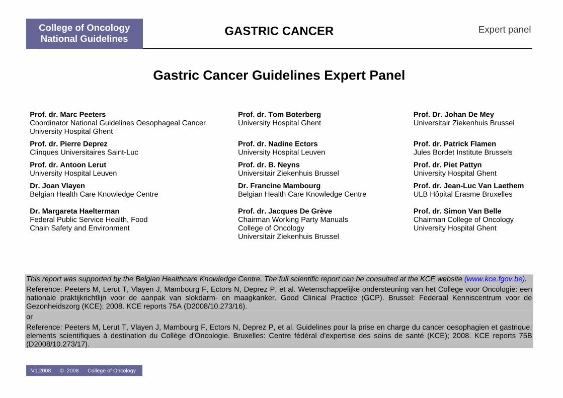
Gastric Cancer Guidelines Expert Panel Prof. dr. Marc Peeters Coordinator National Guidelines Oesophageal Cancer University Hospital Ghent
Prof. dr. Tom Boterberg University Hospital Ghent
Prof. Dr. Johan De Mey Universitair Ziekenhuis Brussel
Prof. dr. Pierre Deprez Clinques Universitaires Saint-Luc
Prof. dr. Nadine Ectors University Hospital Leuven
Prof. dr. Patrick Flamen Jules Bordet Institute Brussels
Prof. dr. Antoon Lerut University Hospital Leuven
Prof. dr. B. Neyns Universitair Ziekenhuis Brussel
Prof. dr. Piet Pattyn University Hospital Ghent
Dr. Joan Vlayen Belgian Health Care Knowledge Centre
Dr. Francine Mambourg Belgian Health Care Knowledge Centre
Prof. dr. Jean-Luc Van Laethem ULB Hôpital Erasme Bruxelles
Dr. Margareta Haelterman Federal Public Service Health, Food Chain Safety and Environment
Prof. dr. Jacques De Grève Chairman Working Party Manuals College of Oncology Universitair Ziekenhuis Brussel
Prof. dr. Simon Van Belle Chairman College of Oncology University Hospital Ghent
This report was supported by the Belgian Healthcare Knowledge Centre. The full scientific report can be consulted at the KCE website (www.kce.fgov.be). Reference: Peeters M, Lerut T, Vlayen J, Mambourg F, Ectors N, Deprez P, et al. Wetenschappelijke ondersteuning van het College voor Oncologie: een nationale praktijkrichtlijn voor de aanpak van slokdarm- en maagkanker. Good Clinical Practice (GCP). Brussel: Federaal Kenniscentrum voor de Gezonheidszorg (KCE); 2008. KCE reports 75A (D2008/10.273/16). or Reference: Peeters M, Lerut T, Vlayen J, Mambourg F, Ectors N, Deprez P, et al. Guidelines pour la prise en charge du cancer oesophagien et gastrique: elements scientifiques à destination du Collège d'Oncologie. Bruxelles: Centre fédéral d'expertise des soins de santé (KCE); 2008. KCE reports 75B (D2008/10.273/17).
GASTRIC CANCER
College voor Oncologie Nationale Richtlijnen
V1.2008 © 2008 College of Oncology
College voor Oncologie Nationale Richtlijnen
College voor Oncologie Nationale Richtlijnen
College voor Oncologie Nationale Richtlijnen
College of Oncology National Guidelines
External reviewers
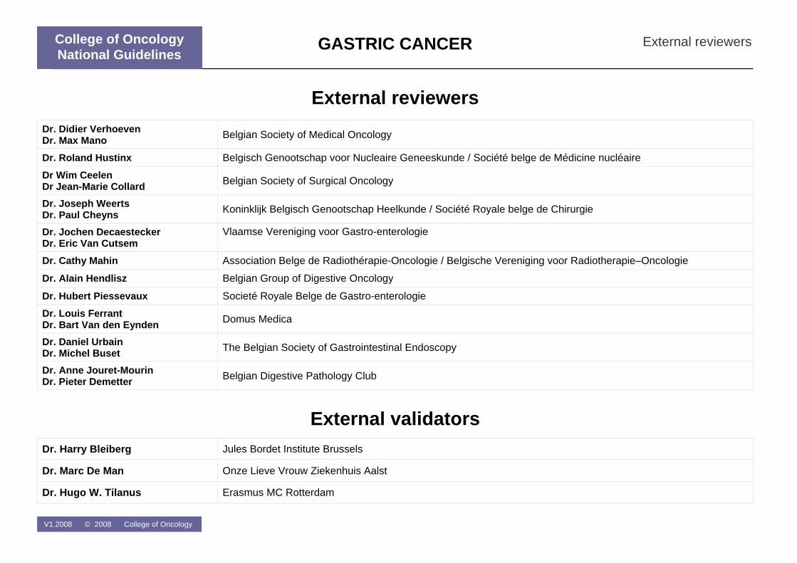
External reviewers Dr. Didier Verhoeven Dr. Max Mano Belgian Society of Medical Oncology
Dr. Roland Hustinx Belgisch Genootschap voor Nucleaire Geneeskunde / Société belge de Médicine nucléaire
Dr Wim Ceelen Dr Jean-Marie Collard Belgian Society of Surgical Oncology
Dr. Joseph Weerts Dr. Paul Cheyns Koninklijk Belgisch Genootschap Heelkunde / Société Royale belge de Chirurgie
Dr. Jochen Decaestecker Dr. Eric Van Cutsem
Vlaamse Vereniging voor Gastro-enterologie
Dr. Cathy Mahin Association Belge de Radiothérapie-Oncologie / Belgische Vereniging voor Radiotherapie–Oncologie
Dr. Alain Hendlisz Belgian Group of Digestive Oncology
Dr. Hubert Piessevaux Societé Royale Belge de Gastro-enterologie
Dr. Louis Ferrant Dr. Bart Van den Eynden Domus Medica
Dr. Daniel Urbain Dr. Michel Buset The Belgian Society of Gastrointestinal Endoscopy
Dr. Anne Jouret-Mourin Dr. Pieter Demetter Belgian Digestive Pathology Club
External validators
Dr. Harry Bleiberg Jules Bordet Institute Brussels
Dr. Marc De Man Onze Lieve Vrouw Ziekenhuis Aalst
Dr. Hugo W. Tilanus Erasmus MC Rotterdam
GASTRIC CANCER College voor Oncologie Nationale Richtlijnen
College voor Oncologie Nationale Richtlijnen
College voor Oncologie Nationale Richtlijnen
College voor Oncologie Nationale Richtlijnen
College of Oncology National Guidelines
Table of contents
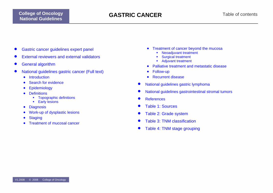
• Gastric cancer guidelines expert panel
• nd external validators External reviewers a
• General algorithm
• Na ional guidelit nes gastric cancer (Full text)
ence gy
• Ddefinitions
lesions
of dysplastic lesions
• Treatment of mucosal cancer
• Table 3: TNM classification
• Table 4: TNM stage grouping
• Introduction • Search for evid• Epidemiolo
efinitions Topographic Early
• Diagnosis • Work-up• Staging
• Treatment of cancer beyond the mucosa Neoadjuvant treatment Surgical treatment Adjuvant treatment
• Palliative treatment and metastatic disease • Follow-up • Recurrent disease
• National guidelines gastric lymphoma
• ines gastrointestinal stromal tumors National guidel
• References
• Table 1: Sources
• Table 2: Grade system
V1.2008 © 2008 College of Oncology
GASTRIC CANCER College voor Oncologie Nationale Richtlijnen
College voor Oncologie Nationale Richtlijnen
College voor Oncologie Nationale Richtlijnen
College voor Oncologie Nationale Richtlijnen
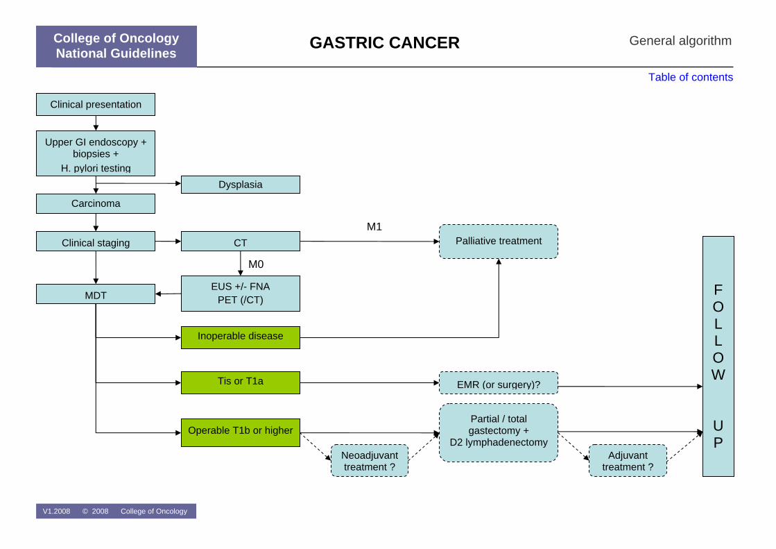
Carcinoma
Tis or T1a
Inoperable disease
Operable T1b or higher
F O L L O W
U P
EUS +/- FNA PET (/CT)
Dysplasia
Clinical staging
MDT
Upper GI endoscopy + biopsies +
H. pylori testing
Clinical presentation
EMR (or surgery)?
Partial / total gastectomy +
D2 lymphadenectomy Adjuvant
treatment ?Neoadjuvant treatment ?
Palliative treatment CTM1
M0
General algorithm
Table of contents
College of Oncology National Guidelines
V1.2008 © 2008 College of Oncology
GASTRIC CANCER College voor Oncologie
Nationale Richtlijnen College voor Oncologie
Nationale Richtlijnen College voor Oncologie
Nationale Richtlijnen College voor Oncologie
Nationale Richtlijnen
National Guidelines Gastric Cancer
INTRODUCTION This document provides an overview of the clinical practice guidelines for gastric cancer. For more in-depth information and the scientific background, we would like to ask the readers to consult the full scientific report at www.kce.fgov.be. The guidelines are developed by a panel of experts (see 'expert panel') comprising clinicians of different specialties and were reviewed by relevant professional associations (see 'external reviewers') The guidelines are based on the best evidence available at the time they are derived (date restriction 2001-2007). The aim of these guidelines is to assist all care providers involved in the care of patients with gastric ancer. c
SEARCH FOR EVIDENCE Clinical practice guidelines
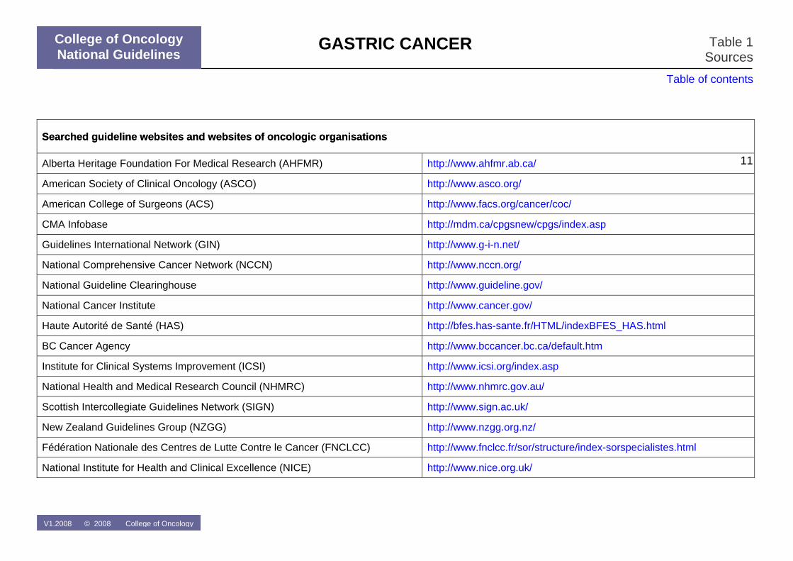
Sources A broad search of electronic databases (Medline, EMBASE), specific guideline websites and websites of oncologic organisations (Table 1) was conducted in July 2007.
In- and exclusion criteria Both national and international clinical practice guidelines (CPGs) on oesophageal cancer were searched. A language (English, Dutch, French)
nd date restriction (2001 – 2007) were used. CPGs without referencea s s without clear recommendations.
Reviews from the search date of the CPG on (search date
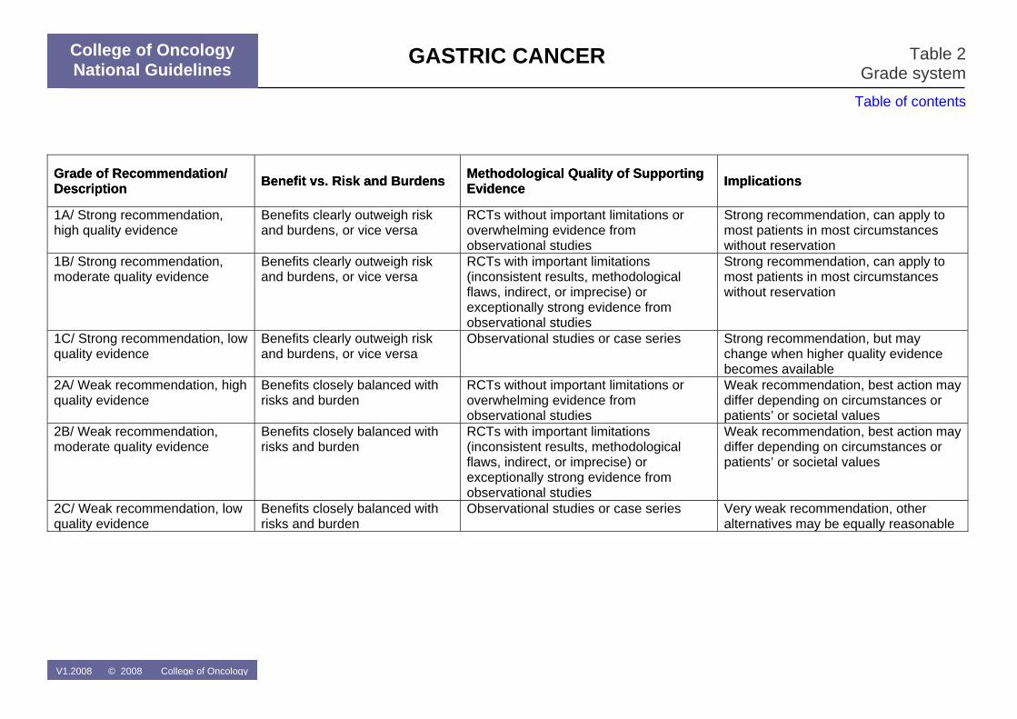
tion was assigned to each recommendation using he GRADE system (Table 2).
ies [1]. It is the second most commocause of death from cancer.
Table of contents
Full Text
were excluded, as were CPG
Additional evidence For each clinical question, the evidence – identified through the included CPGs – was updated by searching Medline and the Cochrane Database
f SystematicoAugust-September 2007).
Grade of recommendation A grade of recommendat
EPIDEMIOLOGY With an estimated 934.000 new cases per year in 2002 worldwide (8.6% of all new cancer cases), gastric cancer is in fourth place behind cancers of the lung, breast, and colon and rectum, with almost two-third of the cases occurring in developing countr
V1.2008 © 2008 College of Oncology
n
College of Oncology National Guidelines
1
GASTRIC CANCER
College of Oncology National Guidelines
Full Text
Table of contents
Gastric cancer incidence rates vary by up to ten-fold throughout the world. Japan and Korea have the highest gastric cancer incidence rates in the world. In Belgium, the crude incidence rate of gastric cancer rose from 12.9 per 100.000 males in 1997 to 14.9 per 100.000 males in 2003, and from 8.0 per 100.000 females in 1997 to 8.4 per 100.000 females in 2003 (Belgian Cancer Registry, personal communication). Age standardised incidence increased by 2.6% and 0.8% per year (1997 – 2003) for males and females respectively. However, in these rates tumours of the gastro-oesophageal junction (GOJ) are also included. While the incidence rates of these GOJ tumours recently increased, the incidence rates of ‘real’ gastric tumours declined [2].
DEFINITIONS Topographic definitions [3-8] • If more than 50% of the mass of the tumour is situated in the cardia, the
tumour should be considered to be of cardiac origin and classified as a gastric tumour
• If the mass of the tumour is predominantly found in the oesophagus, it should be classified as an oesophageal tumour.
• Tumours of the gastro-oesophageal junction should be classified and have the same concept of treatment as oesophageal tumours.
Early lesions [9-38] • There is no consensus about the definition of Barrett’s oesophagus.
• Several classifications are available for dysplasia. For the physician, the used classification should be clinically relevant.
DIAGNOSIS [39-45] • Patients presenting with any of the following alarm symptoms within the
clinical context of potential gastric pathology should be referred for early endoscopy and biopsies: dysphagia, recurrent vomiting, anorexia, weight loss, gastrointestinal blood loss (1C recommendation).
• Flexible upper gastrointestinal endoscopy with at least biopsies of all suspicious lesions is recommended as the diagnostic procedure of choice in patients with suspected gastric cancer (1C recommendation).
• High-resolution endoscopy (HRE) and chromoendoscopy is not routinely recommended, but may be of value in screening and follow-up of high-risk patients (2C recommendation).
• H. pylori testing should be systematically done on histology and ideally with a second test. Serology should be considered if gastric sampling remains negative (2C recommendation).
WORK-UP DYSPLASTIC LESIONS [39] • Patients confirmed with high-grade dysplasia should have subsequent
careful endoscopic and pathological assessment (1C recommendation).
V1.2008 © 2008 College of Oncology 2
GASTRIC CANCER
College of Oncology National Guidelines
Full Text
Table of contents
• Pathologists should follow a classification for reporting dysplasia that the multidisciplinary team is familiar with (1C recommendation).
• Where therapeutic intervention is contemplated on the basis of high-grade dysplasia, the diagnosis should be validated by a second pathologist experienced in this area. Further biopsies should be done if there is uncertainty (1C recommendation).
• Biopsies should be reviewed at a multidisciplinary meeting with access to the clinical information (expert opinion).
• Patients with high-grade dysplasia should be referred to centres or network reference centres with the appropriate endoscopic and surgical expertise and facilities (1C recommendation).
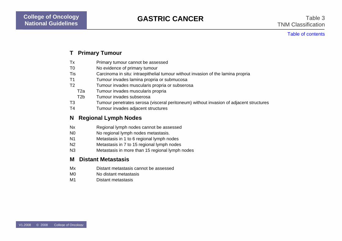
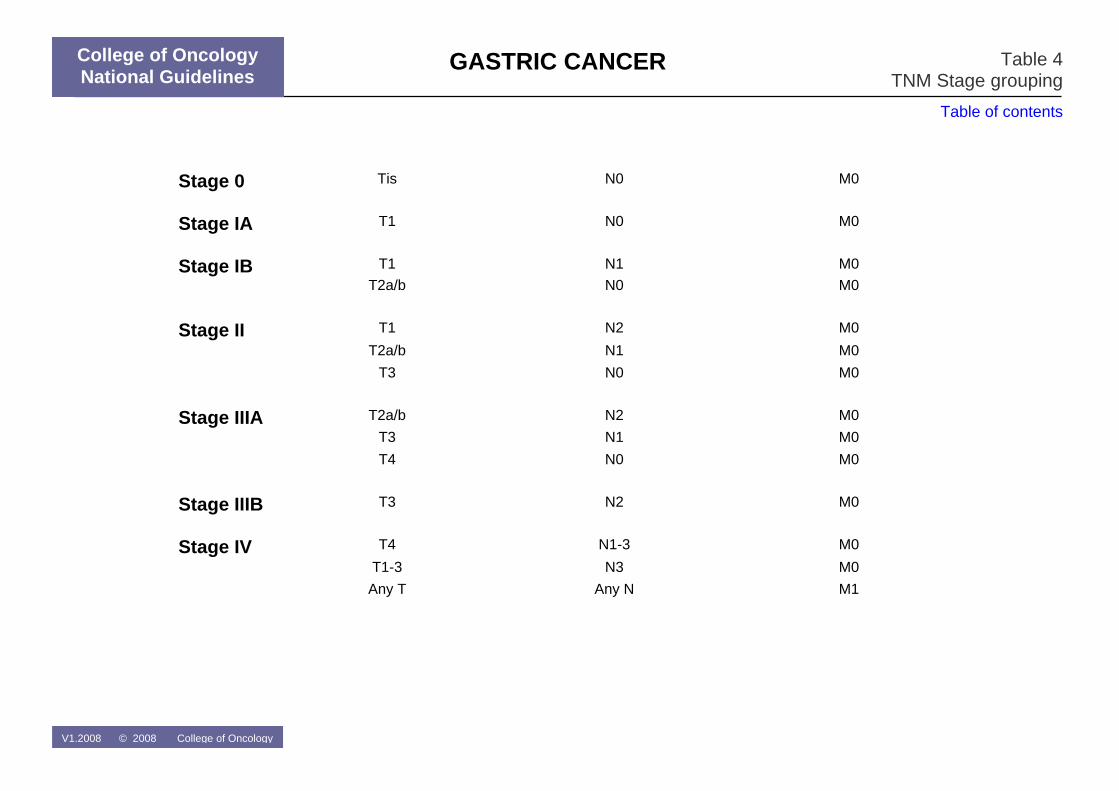
STAGING [39,46-57] TNM classifcation and TNM stage grouping are presented in table 3 and table 4.
• In patients with gastric cancer, CT scan of the chest and abdomen with IV contrast and gastric distension with oral contrast or water should be performed routinely. The liver should at least be imaged in the arterial and portal venous phase (1C recommendation).
• Endoscopic ultrasonography with or without fine-needle aspiration cytology can be considered in patients to be treated with curative intent based on clinical presentation and/or CT (1C recommendation).
• The following examinations can be considered for specific indications (as explained in the text above): PET scan, Magnetic Resonance Imaging, laparoscopy (1C recommendation).
TREATMENT OF MUCOSAL CANCER [39,58-64] • Biopsies should be reviewed by an experienced pathologist in this area
and discussed at a multidisciplinary meeting with access to the clinical information (expert opinion).
• Superficial gastric cancer limited to the mucosa can be treated with endoscopic mucosal resection (EMR), taking into account the stage, size, histological type and differentiation grade (2C recommendation).
• Mucosal ablative techniques, such as photodynamic therapy (PDT), laser or argon plasma coagulation (APC), cannot be recommended as a curative option (expert opinion).
TREATMENT OF CANCER BEYOND THE MUCOSA Neoadjuvant treatment [65-70] • Neoadjuvant treatment is not routinely indicated for patients with gastric
cancer, but is an option to be discussed during a multidisciplinary meeting (2A recommendation).
• Prospective registration of clinical outcomes and adverse events of combined treatment is recommended (expert opinion).
Surgical treatment [39,46,71-88] • Surgical resection should be considered standard treatment for patients
with resectable gastric cancer (1A recommendation).
V1.2008 © 2008 College of Oncology 3
GASTRIC CANCER
College of Oncology National Guidelines
Full Text
Table of contents
• Surgery for gastric cancer should aim at achieving an R0 resection (1A recommendation).
• D2 lymphadenectomy (with a minimum of 15 lymph nodes removed and examined) should be standard during gastrectomy to improve staging and local disease control (1B recommendation).
• Gastric cancer surgery should be carried out in high volume specialist surgical units by surgeons with experience and/or specialist training in this area (1C recommendation).
Adjuvant treatment [39,68,89-98] • Postoperative adjuvant chemotherapy is not recommended for patients
with gastric cancer (2A recommendation). • Postoperative adjuvant radiotherapy is not recommended for patients
with gastric cancer (2A recommendation). • Postoperative adjuvant chemoradiotherapy is not routinely
recommended for patients with gastric cancer, but can be considered after discussion in the multidisciplinary team (2A recommendation).
PALLIATIVE TREATMENT AND METASTATIC DISEASE [39,46,99-103] • Palliative gastric surgery is limited to symptomatic stenoses, bleeding
tumours and perforation (2C recommendation). • For patients with malignant gastric outlet obstruction, treatment options
include endoscopic stenting or surgical gastroenterostomy (2C recommendation).
• In patients with locally advanced or metastatic cancer of the stomach with good performance status combination chemotherapy should be considered (1A recommendation).
• Patients with gastric cancer should have access to a specialist palliative care team, in particular in relation to comfort and symptom control, and quality of life (1C recommendation).
FOLLOW-UP [62,104-106] • It is recommended that the follow-up of patients treated for gastric
cancer includes a physical examination and blood analysis every three months, and a CT scan every six months in the first year and afterwards annually until the fifth year (expert opinion).
• Patients treated with endoscopic mucosal resection (EMR) should have a follow-up endoscopy after three months, then every six months in the first two years, and then annually (expert opinion).
RECURRENT DISEASE [63,107-111] • In patients with recurrent gastric cancer, treatment options should be
discussed in the multidisciplinary team (expert opinion). • In patients with a local recurrence or new tumour after endoscopic
mucosal resection (EMR), treatment options, including local treatment, should be discussed in the multidisciplinary team (expert opinion).
V1.2008 © 2008 College of Oncology 4
GASTRIC CANCER
College of Oncology National Guidelines
Full Text
Table of contents
National Guidelines Gastric lymphoma
INTRODUCTION [112-116] Primary gastric lymphoma is a rare tumour, accounting for less than 5% of primary gastric neoplasms. However, it is the most common extranodal lymphoma, representing 4-20% of all extranodal lymphomas [190]. Helicobacter pylori infection, immunosuppression after solid-organ transplantation, celiac disease, inflammatory bowel disease, and human immunodeficiency virus (HIV) infection are known risk factors for GI lymphoma. A significant proportion of gastric lymphomas is of low-grade histology and arises from mucosa-associated lymphoid tissue (MALT) [191]. According to the most recent WHO classification, the term ‘MALT lymphoma’ should only be applied to tumours previously defined as low-grade MALT lymphomas composed mostly by small cells. High-grade lymphomas are known as large B-cell lymphoma [192]. Patients with low-grade B-cell lymphoma or MALT lymphoma have a better prognosis than patients with diffuse large B-cell lymphoma (DLBCL) [193]. Tumours of T-cell origin are rare [190]. Patients with gastric lymphomas are currently staged using the Ann Arbor staging system with the Cotswold modification. This has largely replaced the older International Workshop staging system [194].
DIAGNOSIS AND STAGING [117-125] • In patients with suspected gastric lymphoma, subtle endoscopic-bioptic
techniques are needed, including a minimum of 8–12 biopsies from visible lesions, mapping of macroscopically normal-appearing areas, and repeated examinations in the individual case. Biopsies should be preserved in such a way to allow molecular diagnostic investigation (expert opinion).
• Lymphomas should be diagnosed and classified according to the most recent appropriate classification (expert opinion).
• In patients with histologically confirmed gastric lymphoma, endoscopic ultrasonography is indicated. Endoscopic ultrasound-guided fine needle aspiration of suspicious lymph nodes is not recommended (1C recommendation).
• For patients with low-grade MALT lymphoma no further staging procedures are recommended, unless otherwise required for differential diagnostic reasons (expert opinion).
V1.2008 © 2008 College of Oncology 5
GASTRIC CANCER
College of Oncology National Guidelines
Full Text
Table of contents
TREATMENT [45,116,126-131] • H. pylori eradication is the treatment of first choice for H. pylori infected
patients with stage I low grade gastric MALT lymphoma (1C recommendation).
• In patients with low-grade MALT lymphoma treated with antibiotic eradication, close follow-up with upper GI endoscopy and biopsies is required, including evaluation of H. pylori eradication within 3 months (expert opinion).
• Patients with successful H. pylori eradication but without tumour regression after 1 year or with tumour progression should be referred to a specialized haematology centre (expert opinion).
V1.2008 © 2008 College of Oncology 6
GASTRIC CANCER
College of Oncology National Guidelines
Full Text
Table of contents
National Guidelines Gastrointestinal Stromal Tumors
INTRODUCTION [132,133] Gastrointestinal stromal tumours (GIST) are relatively rare tumours, representing less than 1% of all tumours of the gastrointestinal tract. They are most common in the stomach (39% to 70%) and the small intestine (20% to 32%), whereas the colon, rectum and oesophagus are affected in less than 15% of cases [210]. GISTs predominantly occur in individuals over 40 years of age, with the majority occurring between the ages of 55 to 65. Estimates of incidence vary widely from 4 to 14 cases per million populations [211]. Presenting features of these tumours include abdominal discomfort or pain, a feeling of abdominal fullness and the presence of a palpable mass. However, many people with GISTs are asymptomatic during early stages of the disease until tumours reach a large size, at which time the tumours rupture and bleed or obstruct the gastrointestinal tract [211]. Overall, literature on GIST is relatively scarce and of low quality. Most studies are observational studies or case series without an adequate control. Therefore, the recommendations presented below often have a low level of evidence or are based on expert opinion.
DIAGNOSIS AND STAGING [134-139] • In patients with clinical suspicion of GIST, endoscopic ultrasonography
and endoscopic ultrasound-guided fine needle aspiration can be recommended for differential diagnostic reasons and to confirm the presence of positive lymph nodes or malignancy in adjacent organs (2C recommendation).
• In patients with a (suspected) GIST, immunohistochemical testing of CD117 is recommended (1C recommendation).
• In patients with a (suspected) GIST tumour, a CT abdomen is recommended if treatment is considered (expert opinion).
TREATMENT Non-metastatic resectable GIST [132-134,140-142] • In patients with a histologically confirmed non-metastatic GIST and who
are fit for surgery, resectional surgery is indicated (expert opinion). • In patients with a gastric tumour of >5 cm that is highly suspicious of a
GIST, without evidence for metastatic disease, and who are fit for surgery, resectional surgery is indicated (expert opinion).
V1.2008 © 2008 College of Oncology 7
GASTRIC CANCER
College of Oncology National Guidelines
Full Text
Table of contents
• In patients with a gastric tumour of 2-5 cm that is highly suspicious of a GIST, without evidence for metastatic disease, and who are fit for surgery, the choice between surveillance and resectional surgery should be discussed at the multidisciplinary team (expert opinion).
• In patients with a gastric tumour of <2 cm that is highly suspicious of a GIST and without evidence for metastatic disease, surveillance is indicated (expert opinion).
• The use of imatinib as adjuvant treatment is investigational (expert opinion).
Metastatic or unresectable GIST [132,133,141,143-147] • In patients with inoperable or metastatic (suspected) GIST imatinib is
recommended (1C recommendation). • PET/CT is indicated to evaluate treatment response to imatinib (expert
opinion). • In patients with imatinib resistance or intolerance sunitinib can be
considered as second-line treatment (2A recommendation).
V1.2008 © 2008 College of Oncology 8
GASTRIC CANCER
College of Oncology National Guidelines
References
Table of contents
References 1 Parkin, D.M., et al., Global cancer statistics, 2002. CA Cancer J Clin, 2005.
55(2): p. 74-108. 2 Crane, S.J., et al., The changing incidence of oesophageal and gastric
adenocarcinoma by anatomic sub-site. Aliment Pharmacol Ther, 2007. 25(4): p. 447-53.
3 Siewert, J.R. and H.J. Stein, Classification of adenocarcinoma of the oesophagogastric junction. Br J Surg, 1998. 85(11): p. 1457-9.
4 Spechler, S.J., et al., Adenocarcinoma of the esophago-gastric junction., in Pathology and Genetics of Tumours of the Digestive System., S.R. Hamilton and L.A. Aaltonen, Editors. 2000, IARC Press: Lyon, France. p. 31-36.
5 (UICC), I.U.A.C., TNM classification of malignant tumours. 6th ed. ed. 2002, Berlin: Springer-Verlag.
6 Corley, D.A. and A. Kubo, Influence of site classification on cancer incidence rates: an analysis of gastric cardia carcinomas. J Natl Cancer Inst, 2004. 96(18): p. 1383-7.
7 Driessen, A., et al., Identical cytokeratin expression pattern CK7+/CK20- in esophageal and cardiac cancer: etiopathological and clinical implications. Mod Pathol, 2004. 17(1): p. 49-55.
8 Driessen, A., et al., Are carcinomas of the cardia oesophageal or gastric adenocarcinomas? Eur J Cancer, 2003. 39(17): p. 2487-94.
9 Skinner, D.B., et al., Barrett's esophagus. Comparison of benign and malignant cases. Ann Surg, 1983. 198(4): p. 554-65.
10 Paull, A., et al., The histologic spectrum of Barrett's esophagus. N Engl J Med, 1976. 295(9): p. 476-80.
11 Naef, A.P., M. Savary, and L. Ozzello, Columnar-lined lower esophagus: an acquired lesion with malignant predisposition. Report on 140 cases of Barrett's esophagus with 12 adenocarcinomas. J Thorac Cardiovasc Surg, 1975. 70(5): p. 826-35.
12 Haggitt, R.C., et al., Adenocarcinoma complicating columnar epithelium-lined (Barrett's) esophagus. Am J Clin Pathol, 1978. 70(1): p. 1-5.
13 Reid, B.J., et al., Barrett's esophagus. Correlation between flow cytometry and histology in detection of patients at risk for adenocarcinoma. Gastroenterology, 1987. 93(1): p. 1-11.
14 Spechler, S.J. and R.K. Goyal, The columnar-lined esophagus, intestinal metaplasia, and Norman Barrett. Gastroenterology, 1996. 110(2): p. 614-21.
15 Sampliner, R.E., Practice guidelines on the diagnosis, surveillance, and therapy of Barrett's esophagus. The Practice Parameters Committee of the American College of Gastroenterology. Am J Gastroenterol, 1998. 93(7): p. 1028-32.
16 Sharma, P., et al., A critical review of the diagnosis and management of Barrett's esophagus: the AGA Chicago Workshop. Gastroenterology, 2004. 127(1): p. 310-30.
17 British Society of Gastroenterology, Guidelines for the diagnosis and management of Barrett’s columnar-lined oesophagus., B.S.o. Gastroenterology, Editor. 2005, British Society of Gastroenterology: London.
18 Flejou, J.F. and M. Svrcek, Barrett's oesophagus--a pathologist's view. Histopathology, 2007. 50(1): p. 3-14.
19 Takubo, K., et al., Double muscularis mucosae in Barrett's esophagus. Hum Pathol, 1991. 22(11): p. 1158-61.
20 Nigro, J.J., et al., Prevalence and location of nodal metastases in distal esophageal adenocarcinoma confined to the wall: implications for therapy. J Thorac Cardiovasc Surg, 1999. 117(1): p. 16-23; discussion 23-5.
21 Westerterp, M., et al., Outcome of surgical treatment for early adenocarcinoma of the esophagus or gastro-esophageal junction. Virchows Arch, 2005. 446(5): p. 497-504.
22 Watanabe, H., J.R. Jass, and L.H. Sobin, Histological typing of oesophageal and gastric tumours. 2nd ed. ed. 1990, Berlin: Springer-Verlag.
23 Rubio, C.A., F.S. Liu, and H.Z. Zhao, Histological classification of intraepithelial neoplasias and microinvasive squamous carcinoma of the esophagus. Am J Surg Pathol, 1989. 13(8): p. 685-90.
V1.2008 © 2008 College of Oncology
GASTRIC CANCER
College of Oncology National Guidelines
References
Table of contents
24 Riddell, R.H., et al., Dysplasia in inflammatory bowel disease: standardized classification with provisional clinical applications. Hum Pathol, 1983. 14(11): p. 931-68.
25 Montgomery, E., Is there a way for pathologists to decrease interobserver variability in the diagnosis of dysplasia? Arch Pathol Lab Med, 2005. 129(2): p. 174-6.
26 Montgomery, E., et al., Reproducibility of the diagnosis of dysplasia in Barrett esophagus: a reaffirmation. Hum Pathol, 2001. 32(4): p. 368-78.
27 Reid, B.J., et al., Observer variation in the diagnosis of dysplasia in Barrett's esophagus. Hum Pathol, 1988. 19(2): p. 166-78.
28 Schlemper, R.J., et al., International comparability of the pathological diagnosis for early cancer of the digestive tract: Munich meeting. J Gastroenterol, 2000. 35 Suppl 12: p. 102-10.
29 Schlemper, R.J., et al., Differences in diagnostic criteria for esophageal squamous cell carcinoma between Japanese and Western pathologists. Cancer, 2000. 88(5): p. 996-1006.
30 Schlemper, R.J., et al., Differences in diagnostic criteria for gastric carcinoma between Japanese and western pathologists. Lancet, 1997. 349(9067): p. 1725-9.
31 Antonioli, D.A. and H.H. Wang, Morphology of Barrett's esophagus and Barrett's-associated dysplasia and adenocarcinoma. Gastroenterol Clin North Am, 1997. 26(3): p. 495-506.
32 Haggitt, R.C., Barrett's esophagus, dysplasia, and adenocarcinoma. Hum Pathol, 1994. 25(10): p. 982-93.
33 Rugge, M., et al., Gastric dysplasia: the Padova international classification. Am J Surg Pathol, 2000. 24(2): p. 167-76.
34 Schlemper, R.J., et al., The Vienna classification of gastrointestinal epithelial neoplasia. Gut, 2000. 47(2): p. 251-5.
35 Schlemper, R.J., Y. Kato, and M. Stolte, Diagnostic criteria for gastrointestinal carcinomas in Japan and Western countries: proposal for a new classification system of gastrointestinal epithelial neoplasia. J Gastroenterol Hepatol, 2000. 15 Suppl: p. G49-57.
36 Schlemper, R.J., Y. Kato, and M. Stolte, Review of histological classifications of gastrointestinal epithelial neoplasia: differences in
diagnosis of early carcinomas between Japanese and Western pathologists. J Gastroenterol, 2001. 36(7): p. 445-56.
37 Hamilton, S.R. and L.A. Aaltonen, Pathology and genetics of tumours of the digestive system., ed. S.R. Hamilton and L.A. Aaltonen. 2000, Lyon, France: IARC Press.
38 Odze, R.D., Diagnosis and grading of dysplasia in Barrett's oesophagus. J Clin Pathol, 2006. 59(10): p. 1029-38.
39 Scottish Intercollegiate Guidelines Network, Management of oesophageal and gastric cancer. A national clinical guideline., SIGN, Editor. 2006, SIGN: Edinburgh.
40 Bowrey, D.J., et al., Use of alarm symptoms to select dyspeptics for endoscopy causes patients with curable esophagogastric cancer to be overlooked. Surgical Endoscopy, 2006. 20(11): p. 1725-8.
41 Graham, D.Y., et al., Prospective evaluation of biopsy number in the diagnosis of esophageal and gastric carcinoma. Gastroenterology, 1982. 82(2): p. 228-31.
42 Sugano, K., K. Sato, and K. Yao, New diagnostic approaches for early detection of gastric cancer. Dig Dis, 2004. 22(4): p. 327-33.
43 Wang, C., Y. Yuan, and R.H. Hunt, The association between Helicobacter pylori infection and early gastric cancer: a meta-analysis. Am J Gastroenterol, 2007. 102(8): p. 1789-98.
44 Fuccio, L., et al., Systematic review: Helicobacter pylori eradication for the prevention of gastric cancer. Aliment Pharmacol Ther, 2007. 25(2): p. 133-41.
45 Malfertheiner, P., et al., Current concepts in the management of Helicobacter pylori infection: the Maastricht III Consensus Report. Gut, 2007. 56(6): p. 772-81.
46 Fédération Nationale des Centres de Lutte Contre le Cancer (FNCLCC), Recommandations pour la pratique clinique: Standards, Options et Recommandations 2003 pour la prise en charge des patients atteints d’adénocarcinomes de l’estomac (cancers du cardia, autres types histologiques exclus) (rapport intégral), in Standards, Options et Recommandations. 2004, FNCLCC: Paris.
V1.2008 © 2008 College of Oncology
GASTRIC CANCER
College of Oncology National Guidelines
References
Table of contents
47 Kwee, R.M. and T.C. Kwee, Imaging in local staging of gastric cancer: a systematic review. Journal of Clinical Oncology, 2007. 25(15): p. 2107-16.
48 Chen, C.-Y., et al., Gastric cancer: preoperative local staging with 3D multi-detector row CT--correlation with surgical and histopathologic results. Radiology, 2007. 242(2): p. 472-82.
49 Lauren, P., The Two Histological Main Types of Gastric Carcinoma: Diffuse and So-Called Intestinal-Type Carcinoma. an Attempt at a Histo-Clinical Classification. Acta Pathol Microbiol Scand, 1965. 64: p. 31-49.
50 Goseki, N., T. Takizawa, and M. Koike, Differences in the mode of the extension of gastric cancer classified by histological type: new histological classification of gastric carcinoma. Gut, 1992. 33(5): p. 606-12.
51 Mukai, K., et al., Usefulness of preoperative FDG-PET for detection of gastric cancer. Gastric Cancer, 2006. 9(3): p. 192-6.
52 Mortensen, M.B., et al., Combined preoperative endoscopic and laparoscopic ultrasonography for prediction of R0 resection in upper gastrointestinal tract cancer. British Journal of Surgery, 2006. 93(6): p. 720-5.
53 Gretschel, S., et al., Prediction of gastric cancer lymph node status by sentinel lymph node biopsy and the Maruyama computer model. European Journal of Surgical Oncology, 2005. 31(4): p. 393-400.
54 Lee, J.H., et al., Sentinel node biopsy using dye and isotope double tracers in early gastric cancer. Annals of Surgical Oncology, 2006. 13(9): p. 1168-74.
55 Miwa, K., et al., Mapping sentinel nodes in patients with early-stage gastric carcinoma. British Journal of Surgery, 2003. 90(2): p. 178-82.
56 Mochiki, E., et al., Sentinel lymph node mapping with technetium-99m colloidal rhenium sulfide in patients with gastric carcinoma. American Journal of Surgery, 2006. 191(4): p. 465-9.
57 Simsa, J., et al., Sentinel node biopsy in gastric cancer: preliminary results. Acta Chirurgica Belgica, 2003. 103(3): p. 270-3.
58 Ono, H., et al., Endoscopic mucosal resection for treatment of early gastric cancer. Gut, 2001. 48(2): p. 225-9.
59 Tajima, Y., et al., Histopathologic findings predicting lymph node metastasis and prognosis of patients with superficial esophageal carcinoma: analysis of 240 surgically resected tumors. Cancer, 2000. 88(6): p. 1285-93.
60 Gotoda, T., et al., Incidence of lymph node metastasis from early gastric cancer: estimation with a large number of cases at two large centers. Gastric Cancer, 2000. 3(4): p. 219-225.
61 Kitajima, K., et al., Correlations between lymph node metastasis and depth of submucosal invasion in submucosal invasive colorectal carcinoma: a Japanese collaborative study. J Gastroenterol, 2004. 39(6): p. 534-43.
62 Wang, Y.P., C. Bennett, and T. Pan, Endoscopic mucosal resection for early gastric cancer. Cochrane Database of Systematic Reviews, 2006(1): p. CD004276.
63 Oka, S., et al., Endoscopic submucosal dissection for residual/local recurrence of early gastric cancer after endoscopic mucosal resection. Endoscopy, 2006. 38(10): p. 996-1000.
64 Japanese Gastric Cancer, A., Japanese Classification of Gastric Carcinoma - 2nd English Edition. Gastric Cancer, 1998. 1(1): p. 10-24.
65 Wu, A.W., et al., Neoadjuvant chemotherapy versus none for resectable gastric cancer. Cochrane Database of Systematic Reviews, 2007(2): p. CD005047.
66 Cunningham, D., et al., Perioperative chemotherapy versus surgery alone for resectable gastroesophageal cancer.[see comment]. New England Journal of Medicine, 2006. 355(1): p. 11-20.
67 Boige, V., et al., Final results of a randomized trial comparing preoperative 5-fluorouracil (F)/cisplatin (P) to surgery alone in adenocarcinoma of stomach and lower esophagus (ASLE): FNLCC ACCORD07-FFCD 9703 trial. J Clin Oncol (Meeting Abstracts), 2007. 25(18_suppl): p. 4510-.
68 Earle, C.C., et al., Neoadjuvant or Adjuvant Therapy for Resectable Gastric Cancer. Practice Guideline Report #2-14., CCO, Editor. 2003, CCO: Ottawa.
69 Romano, F., et al., Phase-II randomized study of preoperative IL-2 administration in radically operable gastric cancer patients. Hepato-Gastroenterology, 2004. 51(60): p. 1872-6.
V1.2008 © 2008 College of Oncology
GASTRIC CANCER
College of Oncology National Guidelines
References
Table of contents
70 FOD Volksgezondheid Veiligheid van de voedselketen en Leefmilieu, K.B. 21 maart 2003. Koninklijk besluit houdende vaststelling van de normen waaraan het zorgprogramma voor oncologische basiszorg en het zorgprogramma voor oncologie moeten voldoen om te worden erkend. 2003: B.S. 25-04-2003.
71 McCulloch, P., et al., Extended versus limited lymph nodes dissection technique for adenocarcinoma of the stomach.[update of Cochrane Database Syst Rev. 2003;(4):CD001964; PMID: 14583942]. Cochrane Database of Systematic Reviews, 2004(4): p. CD001964.
72 Roukos, D.H. and A.M. Kappas, Perspectives in the treatment of gastric cancer. Nat Clin Pract Oncol, 2005. 2(2): p. 98-107.
73 Degiuli, M., et al., Morbidity and mortality after D1 and D2 gastrectomy for cancer: interim analysis of the Italian Gastric Cancer Study Group (IGCSG) randomised surgical trial. European Journal of Surgical Oncology, 2004. 30(3): p. 303-8.
74 Degiuli, M., et al., Survival results of a multicentre phase II study to evaluate D2 gastrectomy for gastric cancer. Br J Cancer, 2004. 90(9): p. 1727-32.
75 Hartgrink, H.H., et al., Extended lymph node dissection for gastric cancer: who may benefit? Final results of the randomized Dutch gastric cancer group trial.[see comment]. Journal of Clinical Oncology, 2004. 22(11): p. 2069-77.
76 Kulig, J., et al., Standard D2 versus extended D2 (D2+) lymphadenectomy for gastric cancer: an interim safety analysis of a multicenter, randomized, clinical trial. American Journal of Surgery, 2007. 193(1): p. 10-5.
77 Sano, T., et al., Gastric cancer surgery: morbidity and mortality results from a prospective randomized controlled trial comparing D2 and extended para-aortic lymphadenectomy--Japan Clinical Oncology Group study 9501.[see comment]. Journal of Clinical Oncology, 2004. 22(14): p. 2767-73.
78 Yonemura, Y., et al., Operative morbidity and mortality after D2 and D4 extended dissection for advanced gastric cancer: a prospective randomized trial conducted by Asian surgeons. Hepato-Gastroenterology, 2006. 53(69): p. 389-94.
79 Iwata, T., et al., Evaluation of reconstruction after proximal gastrectomy: prospective comparative study of jejunal interposition and jejunal pouch interposition. Hepato-Gastroenterology, 2006. 53(68): p. 301-3.
80 Yoo, C.H., et al., Proximal gastrectomy reconstructed by jejunal pouch interposition for upper third gastric cancer: prospective randomized study. World Journal of Surgery, 2005. 29(12): p. 1592-9.
81 Kono, K., et al., Improved quality of life with jejunal pouch reconstruction after total gastrectomy. American Journal of Surgery, 2003. 185(2): p. 150-4.
82 Hayashi, H., et al., Prospective randomized study of open versus laparoscopy-assisted distal gastrectomy with extraperigastric lymph node dissection for early gastric cancer. Surgical Endoscopy, 2005. 19(9): p. 1172-6.
83 Hosono, S., et al., Meta-analysis of short-term outcomes after laparoscopy-assisted distal gastrectomy. World Journal of Gastroenterology, 2006. 12(47): p. 7676-83.
84 Huscher, C.G.S., et al., Laparoscopic versus open subtotal gastrectomy for distal gastric cancer: five-year results of a randomized prospective trial. Annals of Surgery, 2005. 241(2): p. 232-7.
85 Lee, J.H., H.S. Han, and J.H. Lee, A prospective randomized study comparing open vs laparoscopy-assisted distal gastrectomy in early gastric cancer: early results. Surgical Endoscopy, 2005. 19(2): p. 168-73.
86 Halm, E.A., C. Lee, and M.R. Chassin, Is volume related to outcome in health care? A systematic review and methodologic critique of the literature. Ann Intern Med, 2002. 137(6): p. 511-20.
87 Killeen, S.D., et al., Provider volume and outcomes for oncological procedures. Br J Surg, 2005. 92(4): p. 389-402.
88 Callahan, M.A., et al., Influence of surgical subspecialty training on in-hospital mortality for gastrectomy and colectomy patients. Ann Surg, 2003. 238(4): p. 629-36; discussion 636-9.
89 Oba, K., et al., Efficacy of adjuvant chemotherapy using oral fluorinated pyrimidines for curatively resected gastric cancer: a meta-analysis of centrally randomized controlled clinical trials in Japan. Journal of Chemotherapy, 2006. 18(3): p. 311-7.
V1.2008 © 2008 College of Oncology
GASTRIC CANCER
College of Oncology National Guidelines
References
Table of contents
90 Bouche, O., et al., Adjuvant chemotherapy with 5-fluorouracil and cisplatin compared with surgery alone for gastric cancer: 7-year results of the FFCD randomized phase III trial (8801). Annals of Oncology, 2005. 16(9): p. 1488-97.
91 Chipponi, J., et al., Randomized trial of adjuvant chemotherapy after curative resection for gastric cancer. American Journal of Surgery, 2004. 187(3): p. 440-5.
92 Nashimoto, A., et al., Randomized trial of adjuvant chemotherapy with mitomycin, Fluorouracil, and Cytosine arabinoside followed by oral Fluorouracil in serosa-negative gastric cancer: Japan Clinical Oncology Group 9206-1.[see comment]. Journal of Clinical Oncology, 2003. 21(12): p. 2282-7.
93 Nitti, D., et al., Randomized phase III trials of adjuvant FAMTX or FEMTX compared with surgery alone in resected gastric cancer. A combined analysis of the EORTC GI Group and the ICCG. Annals of Oncology, 2006. 17(2): p. 262-9.
94 Macdonald, J.S., et al., Chemoradiotherapy after surgery compared with surgery alone for adenocarcinoma of the stomach or gastroesophageal junction. N Engl J Med, 2001. 345(10): p. 725-30.
95 Xu, D.-Z., et al., Meta-analysis of intraperitoneal chemotherapy for gastric cancer. World Journal of Gastroenterology, 2004. 10(18): p. 2727-30.
96 Yan, T.D., et al., Hp40p a systematic review and meta-analysis of the randomised controlled trials on adjuvant intraperitoneal chemotherapy for advanced gastric cancer. ANZ Journal of Surgery, 2007. 77 Suppl 1: p. A49.
97 Oba, K., et al., Efficacy of adjuvant immunochemotherapy with polysaccharide K for patients with curative resections of gastric cancer. Cancer Immunology, Immunotherapy, 2007. 56(6): p. 905-11.
98 Popiela, T., et al., Efficiency of adjuvant immunochemotherapy following curative resection in patients with locally advanced gastric cancer. Gastric Cancer, 2004. 7(4): p. 240-5.
99 Hosono, S., et al., Endoscopic stenting versus surgical gastroenterostomy for palliation of malignant gastroduodenal obstruction: a meta-analysis. Journal of Gastroenterology, 2007. 42(4): p. 283-90.
100 Jeurnink, S.M., et al., Stent versus gastrojejunostomy for the palliation of gastric outlet obstruction: a systematic review. BMC Gastroenterology, 2007. 7: p. 18.
101 Casaretto, L., P.L.R. Sousa, and J.J. Mari, Chemotherapy versus support cancer treatment in advanced gastric cancer: a meta-analysis. Brazilian Journal of Medical & Biological Research, 2006. 39(4): p. 431-40.
102 Wagner, A.D., et al., Chemotherapy in advanced gastric cancer: a systematic review and meta-analysis based on aggregate data. Journal of Clinical Oncology, 2006. 24(18): p. 2903-9.
103 Kuchler, T., et al., Impact of psychotherapeutic support for patients with gastrointestinal cancer undergoing surgery: 10-year survival results of a randomized trial.[see comment]. Journal of Clinical Oncology, 2007. 25(19): p. 2702-8.
104 Whiting, J., et al., Follow-up of gastric cancer: a review. Gastric Cancer, 2006. 9(2): p. 74-81.
105 Tan, I.T. and B.Y. So, Value of intensive follow-up of patients after curative surgery for gastric carcinoma. J Surg Oncol, 2007. 96(6): p. 503-6.
106 Sinning, C., et al., Gastric stump carcinoma - epidemiology and current concepts in pathogenesis and treatment. Eur J Surg Oncol, 2007. 33(2): p. 133-9.
107 Ohashi, M., et al., Cancer of the gastric stump following distal gastrectomy for cancer. Br J Surg, 2007. 94(1): p. 92-5.
108 Chen, L., et al., Surgical management of gastric stump cancer: a report of 37 cases. J Zhejiang Univ Sci B, 2005. 6(1): p. 38-42.
109 Hirai, I., et al., Surgical management for metastatic liver tumors. Hepatogastroenterology, 2006. 53(71): p. 757-63.
110 Lehnert, T., et al., Surgical therapy for loco-regional recurrence and distant metastasis of gastric cancer. Eur J Surg Oncol, 2002. 28(4): p. 455-61.
111 Yokoi, C., et al., Endoscopic submucosal dissection allows curative resection of locally recurrent early gastric cancer after prior endoscopic mucosal resection. Gastrointest Endosc, 2006. 64(2): p. 212-8.
112 Al-Akwaa, A.M., N. Siddiqui, and I.A. Al-Mofleh, Primary gastric lymphoma. World J Gastroenterol, 2004. 10(1): p. 5-11.
V1.2008 © 2008 College of Oncology
GASTRIC CANCER
College of Oncology National Guidelines
References
Table of contents
113 Crump, M., M. Gospodarowicz, and F.A. Shepherd, Lymphoma of the gastrointestinal tract. Semin Oncol, 1999. 26(3): p. 324-37.
114 Zucca, E. and F. Cavalli, Are antibiotics the treatment of choice for gastric lymphoma? Curr Hematol Rep, 2004. 3(1): p. 11-6.
115 Binn, M., et al., Surgical resection plus chemotherapy versus chemotherapy alone: comparison of two strategies to treat diffuse large B-cell gastric lymphoma.[see comment]. Ann Oncol, 2003. 14(12): p. 1751-7.
116 Yoon, S.S. and E.P. Hochberg, Chemotherapy is an effective first line treatment for early stage gastric mucosa-associated lymphoid tissue lymphoma. Cancer Treat Rev, 2006. 32(2): p. 139-43.
117 Fischbach, W., et al., Primary gastric B-cell lymphoma: results of a prospective multicenter study. The German-Austrian Gastrointestinal Lymphoma Study Group.[see comment][erratum appears in Gastroenterology 2000 Dec;119(6):1809]. Gastroenterology, 2000. 119(5): p. 1191-202.
118 Strecker, P., et al., [Diagnostic value of stomach biopsy in comparison with surgical specimen in gastric B-cell lymphomas of the MALT type]. Pathologe, 1998. 19(3): p. 209-13.
119 Isaacson, P.G., J. Spencer, and D.H. Wright, Classifying primary gut lymphomas. Lancet, 1988. 2(8620): p. 1148-9.
120 Harris, N.L., et al., A revised European-American classification of lymphoid neoplasms: a proposal from the International Lymphoma Study Group. Blood, 1994. 84(5): p. 1361-92.
121 Caletti, G., et al., Accuracy of endoscopic ultrasonography in the diagnosis and staging of gastric cancer and lymphoma. Surgery, 1993. 113(1): p. 14-27.
122 Fischbach, W., M.-E. Goebeler-Kolve, and A. Greiner, Diagnostic accuracy of EUS in the local staging of primary gastric lymphoma: results of a prospective, multicenter study comparing EUS with histopathologic stage. Gastrointest Endosc, 2002. 56(5): p. 696-700.
123 Rodriguez, M., et al., [18F] FDG PET in gastric non-Hodgkin's lymphoma. Acta Oncol, 1997. 36(6): p. 577-84.
124 Vorbeck, F., et al., Comparison of spiral-computed tomography with water-filling of the stomach and endosonography for gastric lymphoma of mucosa-associated lymphoid tissue-type. Digestion, 2002. 65(4): p. 196-9.
125 Hoepffner, N., et al., [Value of endosonography in diagnostic staging of primary gastric lymphoma (MALT type)]. Med Klin (Munich), 2003. 98(6): p. 313-7.
126 Hong, S.S., et al., A prospective analysis of low-grade gastric malt lymphoma after Helicobacter pylori eradication. Helicobacter, 2006. 11(6): p. 569-73.
127 Nakamura, S., et al., Long-term clinical outcome of Helicobacter pylori eradication for gastric mucosa-associated lymphoid tissue lymphoma with a reference to second-line treatment. Cancer, 2005. 104(3): p. 532-40.
128 Wundisch, T., et al., Long-term follow-up of gastric MALT lymphoma after Helicobacter pylori eradication. J Clin Oncol, 2005. 23(31): p. 8018-24.
129 Fischbach, W., et al., Long term outcome of patients with gastric marginal zone B cell lymphoma of mucosa associated lymphoid tissue (MALT) following exclusive Helicobacter pylori eradication therapy: experience from a large prospective series. Gut, 2004. 53(1): p. 34-7.
130 Montalban, C., et al., Treatment of low grade gastric mucosa-associated lymphoid tissue lymphoma in stage I with Helicobacter pylori eradication. Long-term results after sequential histologic and molecular follow-up. Haematologica, 2001. 86(6): p. 609-17.
131 Aviles, A., et al., Mucosa-associated lymphoid tissue (MALT) lymphoma of the stomach: results of a controlled clinical trial. Medical Oncology, 2005. 22(1): p. 57-62.
132 Verma, S., et al., Imatinib Mesylate (Gleevec™) for the Treatment of Adult Patients with Unresectable or Metastatic Gastrointestinal Stromal Tumours:A Clinical Practice Guideline. 2006, CCO: Ottawa.
133 Wilson, J.S., et al., Imatinib mesylate for the treatment of patients with unresectable and/or metastatic gastro-intestinal stromal tumours (GIST). 2004, West Midlands Health Technology Assessment Collaboration, University of Birmingham: Birmingham.
V1.2008 © 2008 College of Oncology
GASTRIC CANCER
College of Oncology National Guidelines
References
Table of contents
145 Heinicke, T., et al., Very early detection of response to imatinib mesylate therapy of gastrointestinal stromal tumours using 18fluoro-deoxyglucose-positron emission tomography. Anticancer Res, 2005. 25(6C): p. 4591-4.
134 Hwang, J.H., S.D. Rulyak, and M.B. Kimmey, American Gastroenterological Association Institute technical review on the management of gastric subepithelial masses. Gastroenterology, 2006. 130(7): p. 2217-28.
146 Goldstein, D., et al., Gastrointestinal stromal tumours: correlation of F-FDG gamma camera-based coincidence positron emission tomography with CT for the assessment of treatment response--an AGITG study. Oncology, 2005. 69(4): p. 326-32.
135 Rimondini, A., et al., Contribution of CT to treatment planning in patients with GIST. Radiol Med (Torino), 2007. 112(5): p. 691-702.
136 Lupescu, I.G., et al., Computer tomographic evaluation of digestive tract non-Hodgkin lymphomas. J Gastrointestin Liver Dis, 2007. 16(3): p. 315-9.
147 Demetri, G.D., et al., Efficacy and safety of sunitinib in patients with advanced gastrointestinal stromal tumour after failure of imatinib: a randomised controlled trial. Lancet, 2006. 368(9544): p. 1329-38.
137 De Leo, C., et al., Gastrointestinal stromal tumours: experience with multislice CT. Radiol Med (Torino), 2006. 111(8): p. 1103-14.
138 Da Ronch, T., A. Modesto, and M. Bazzocchi, Gastrointestinal stromal tumour: spiral computed tomography features and pathologic correlation. Radiol Med (Torino), 2006. 111(5): p. 661-73.
139 Hong, X., et al., Gastrointestinal stromal tumor: role of CT in diagnosis and in response evaluation and surveillance after treatment with imatinib. Radiographics, 2006. 26(2): p. 481-95.
140 Fletcher, C.D.M., et al., Diagnosis of gastrointestinal stromal tumors: A
consensus approach. Hum Pathol, 2002. 33(5): p. 459-65. 141 Blay, J.-Y., et al., [Recommendations for the management of GIST patients].
Bull Cancer, 2005. 92(10): p. 907-18. 142 Dematteo, R.P., et al. Adjuvant imatinib mesylate increases recurrence free
survival (RFS) in patients with completely resected localized primary gastrointestinal stromal tumor (GIST): North American Intergroup Phase III trial ACOSOG Z9001. 2007 [cited 2008 23-01-2008]; Available from: http://www.asco.org/portal/site/ASCO/menuitem.34d60f5624ba07fd506fe310ee37a01d/?vgnextoid=76f8201eb61a7010VgnVCM100000ed730ad1RCRD&vmview=abst_detail_view&confID=47&abstractID=100001.
143 Zincirkeser, S., et al., Early detection of response to imatinib therapy for
gastrointestinal stromal tumor by using 18F-FDG-positron emission tomography and computed tomography imaging. World J Gastroenterol, 2007. 13(15): p. 2261-2.
144 Goh, B.K., et al., Pathologic, radiologic and PET scan response of gastrointestinal stromal tumors after neoadjuvant treatment with imatinib mesylate. Eur J Surg Oncol, 2006. 32(9): p. 961-3.
V1.2008 © 2008 College of Oncology
GASTRIC CANCER
College of Oncology National Guidelines
Table 1 Sources
Table of contents
Searched guideline websites and websites of oncologic organisations Searched guideline websites and websites of oncologic organisations
Alberta Heritage Foundation For Medical Research (AHFMR) http://www.ahfmr.ab.ca/
American Society of Clinical Oncology (ASCO) http://www.asco.org/
American College of Surgeons (ACS) http://www.facs.org/cancer/coc/
CMA Infobase http://mdm.ca/cpgsnew/cpgs/index.asp
Guidelines International Network (GIN) http://www.g-i-n.net/
National Comprehensive Cancer Network (NCCN) http://www.nccn.org/
National Guideline Clearinghouse http://www.guideline.gov/
National Cancer Institute http://www.cancer.gov/
Haute Autorité de Santé (HAS) http://bfes.has-sante.fr/HTML/indexBFES_HAS.html
BC Cancer Agency http://www.bccancer.bc.ca/default.htm
Institute for Clinical Systems Improvement (ICSI) http://www.icsi.org/index.asp
National Health and Medical Research Council (NHMRC) http://www.nhmrc.gov.au/
Scottish Intercollegiate Guidelines Network (SIGN) http://www.sign.ac.uk/
New Zealand Guidelines Group (NZGG) http://www.nzgg.org.nz/
Fédération Nationale des Centres de Lutte Contre le Cancer (FNCLCC) http://www.fnclcc.fr/sor/structure/index-sorspecialistes.html
National Institute for Health and Clinical Excellence (NICE) http://www.nice.org.uk/
11
V1.2008 © 2008 College of Oncology
GASTRIC CANCER
College of Oncology National Guidelines
Table 2 Grade system
Table of contents
Grade of Recommendation/ Description Grade of Recommendation/ Description Benefit vs. Risk and Burdens Benefit vs. Risk and Burdens Methodological Quality of Supporting
Evidence Methodological Quality of Supporting Evidence Implications Implications
1A/ Strong recommendation, high quality evidence
Benefits clearly outweigh risk and burdens, or vice versa
RCTs without important limitations or overwhelming evidence from observational studies
Strong recommendation, can apply to most patients in most circumstances without reservation
1B/ Strong recommendation, moderate quality evidence
Benefits clearly outweigh risk and burdens, or vice versa
RCTs with important limitations (inconsistent results, methodological flaws, indirect, or imprecise) or exceptionally strong evidence from observational studies
Strong recommendation, can apply to most patients in most circumstances without reservation
1C/ Strong recommendation, low quality evidence
Benefits clearly outweigh risk and burdens, or vice versa
Observational studies or case series Strong recommendation, but may change when higher quality evidence becomes available
2A/ Weak recommendation, high quality evidence
Benefits closely balanced with risks and burden
RCTs without important limitations or overwhelming evidence from observational studies
Weak recommendation, best action may differ depending on circumstances or patients’ or societal values
2B/ Weak recommendation, moderate quality evidence
Benefits closely balanced with risks and burden
RCTs with important limitations (inconsistent results, methodological flaws, indirect, or imprecise) or exceptionally strong evidence from observational studies
Weak recommendation, best action may differ depending on circumstances or patients’ or societal values
2C/ Weak recommendation, low quality evidence
Benefits closely balanced with risks and burden
Observational studies or case series Very weak recommendation, other alternatives may be equally reasonable
V1.2008 © 2008 College of Oncology
GASTRIC CANCER
College of Oncology National Guidelines
Table 3 TNM Classification
Table of contents
T Primary Tumour Tx Primary tumour cannot be assessed T0 No evidence of primary tumour Tis Carcinoma in situ: intraepithelial tumour without invasion of the lamina propria T1 Tumour invades lamina propria or submucosa T2 Tumour invades muscularis propria or subserosa T2a Tumour invades muscularis propria T2b Tumour invades subserosa T3 Tumour penetrates serosa (visceral peritoneum) without invasion of adjacent structures T4 Tumour invades adjacent structures
N Regional Lymph Nodes Nx Regional lymph nodes cannot be assessed N0 No regional lymph nodes metastasis. N1 Metastasis in 1 to 6 regional lymph nodes N2 Metastasis in 7 to 15 regional lymph nodes N3 Metastasis in more than 15 regional lymph nodes
M Distant Metastasis Mx Distant metastasis cannot be assessed
M0
No distant metastasisM1 Distant metastasis
V1.2008 © 2008 College of Oncology
GASTRIC CANCER
College of Oncology National Guidelines
Table 4 TNM Stage grouping
Table of contents
Stage 0 Tis
N0 M0
Stage IA T1 N0 M0
Stage IB T1 N1 M0
T2a/b N0 M0
Stage II T1 N2 M0
T2a/b N1 M0
T3 N0 M0
Stage IIIA T2a/b N2 M0
T3 N1 M0
T4 N0 M0
Stage IIIB T3 N2 M0
Stage IV T4 N1-3 M0
T1-3 N3 M0
Any T Any N M1
V1.2008 © 2008 College of Oncology