Embed Size (px)
Citation preview
Yonsei Medical Journal
Vol. 46, No. 5, pp. 715 - 718, 2005
Yonsei Med J Vol. 46, No. 5, 2005
Retroperitoneal cystic lymphangioma is a rare congenitalmalformation. The majority of lymphangiomas are present atbirth and nearly all present before the age of two years. Wereport a case of giant cystic retroperitoneal lymphangioma ina patient who first presented with symptoms at the age of 7,underwent surgery, and who then suffered a recurrent mass 11years later.
Key Words: Lymphangioma, retroperitoneal, ultrasonography,computed tomography, magnetic resonance imaging
INTRODUCTION
Abdominal cystic lymphangiomas are rare
benign tumors of the lymphatic vessels.1 They are
characterized by the appearance of uni- or multi-
septate cystic masses.2 The incidence of cystic
lymphangioma is approximately 1/6000 live born
and localization of the retroperitoneum is less
than 5%.3 More than half (50-65%) of neck and
axillar lymphangiomas are present at birth and
90% present symptoms before the age of two
years.4 Cystic lymphangiomas of the retroperi-
toneum are frequently found in older children or
adults.5 Although the lesion is benign in nature,
it causes pressure on vital organs by mass effect
and can therefore cause symptoms. Here, we pres-
ent a case of a giant retroperitoneal cystic lym-
phangioma which first presented with symptoms
in a 7-year-old who underwent surgery and who
returned with a recurrent mass 11 years later.
CASE REPORT
An 18-year-old male was admitted with epi-
gastric and left upper quadrant pain. Distention
due to an abdominal mass was detected on
physical examination. The patient had also suf-
fered from abdominal distension, abdominal pain,
nausea and vomiting 11 years previously and
sonographic findings revealed a hypoechoic mass
with smooth margins which lay between the liver
hilus and splenic hilus. Laparotomic measurement
of the mass, which contained multiple cysts, was
30×20 cm. It was localized between the stomach
and transverse colon and filled the bursa
omentalis. During laparotomy, the cysts and bursa
omentalis were drained, the vessels and capsule of
cysts were excised and capitonnage were per-
formed. The cysts contained 2.5 liters of yellow,
transudative fluid. Microscopically, the tumor
consisted of dilate, large, lymphatic channels of
different sizes growing in loose connective tissue.
These channels were lined by flattened endothe-
lium. A few bundles of smooth muscle were pres-
ent in the walls (pathologic number: B-1895/92).
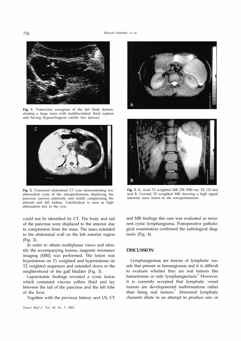
In the current hospitalization, ultrasound ex-
amination (US) showed an anechoic multicystic
mass of 14×15×20 cm with thick septations. There
were echogenic areas within the lesion consistent
with calcifications (Fig. 1).
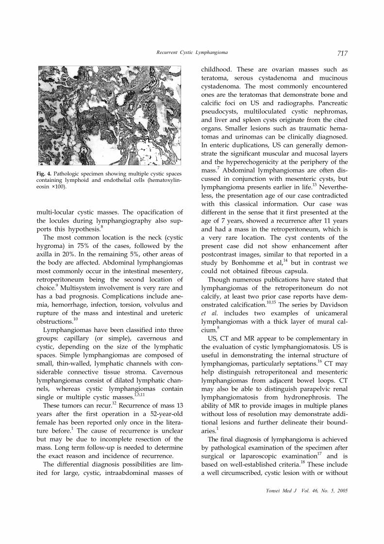
On CT following the administration of contrast
material, there was a non-enhancing mass lesion
having low attenuation values (+ 12 HU) while
including hyperdense calcific foci. The mass
surrounded the body and tail of the pancreas and
extended to the right side of the midline. It also
had extensions into left renal spaces on its pos-
terior aspect, and thereby created a mass effect.
The internal septations that were observed on US
Recurrent Retroperitoneal Cystic Lymphangioma
Huseyin Ozdemir,1 Ercan Kocakoc,1 Zulkif Bozgeyik,1 and Bengu Cobanoglu2
Departments of 1Radiology and 2Pathology, Firat University School of Medicine, Elazig, Turkey.
Received March 12, 2003Accepted October 4, 2003
Reprint address: requests to Dr. Huseyin Ozdemir, Departmentof Radiology, Firat University, Faculty of Medicine, 23119, Elazig,
Turkey. Tel: 90 424 23335, Fax: 90 424 2376773, E-mail:
ozdemir@ firat.edu.tr
Huseyin Ozdemir, et al.
Yonsei Med J Vol. 46, No. 5, 2005
could not be identified by CT. The body and tail
of the pancreas were displaced to the anterior due
to compression from the mass. The mass extended
to the abdominal wall on the left anterior region
(Fig. 2).
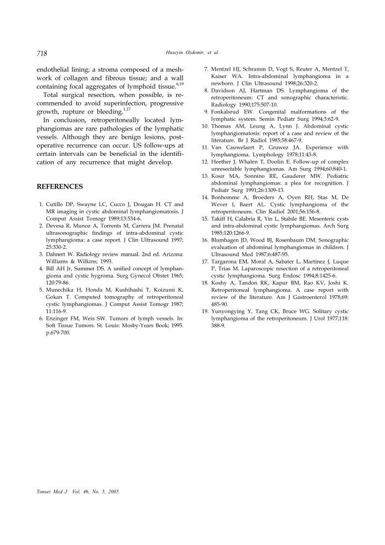
In order to obtain multiplanar views and iden-
tify the accompanying lesions, magnetic resonance
imaging (MRI) was performed. The lesion was
hypointense on T1 weighted and hyperintense on
T2 weighted sequences and extended down to the
neighborhood of the gall bladder (Fig. 3).
Laparotomic findings revealed a cystic lesion
which contained viscous yellow fluid and lay
between the tail of the pancreas and the left lobe
of the liver.
Together with the previous history and US, CT
and MR findings this case was evaluated as recur-
rent cystic lymphangioma. Postoperative patholo-
gical examination confirmed the radiological diag-
nosis (Fig. 4).
DISCUSSION
Lymphangiomas are lesions of lymphatic ves-
sels that present as hemangiomas and it is difficult
to evaluate whether they are real tumors like
hamartomas or only lymphangiectasis.6However,
it is currently accepted that lymphatic vessel
tumors are developmental malformations rather
than being real tumors.7Abnormal lymphatic
channels dilate in an attempt to produce uni- or
Fig. 1. Transverse sonogram of the left flank demon-strating a large mass with multiloculated, thick septumand having hyperechogenic calcific foci (arrow).
Fig. 2. Transaxial abdominal CT scan demonstrating lowattenuation cysts of the retroperitoneum displacing thepancreas (arrow) anteriorly and mildly compressing theadrenal and left kidney. Calcification is seen as highattenuation foci in the cyst.
Fig. 3. A. Axial T2 weighted MR (TR 3000 ms, TE 112 ms)and B. Coronal T2 weighted MR showing a high signalintensity mass lesion in the retroperitoneum.
A
B
Recurrent Cystic Lymphangioma
Yonsei Med J Vol. 46, No. 5, 2005
multi-locular cystic masses. The opacification of
the locules during lymphangiography also sup-
ports this hypothesis.8
The most common location is the neck (cystic
hygroma) in 75% of the cases, followed by the
axilla in 20%. In the remaining 5%, other areas of
the body are affected. Abdominal lymphangiomas
most commonly occur in the intestinal mesentery,
retroperitoneum being the second location of
choice.9 Multisystem involvement is very rare and
has a bad prognosis. Complications include ane-
mia, hemorrhage, infection, torsion, volvulus and
rupture of the mass and intestinal and ureteric
obstructions.10
Lymphangiomas have been classified into three
groups: capillary (or simple), cavernous and
cystic, depending on the size of the lymphatic
spaces. Simple lymphangiomas are composed of
small, thin-walled, lymphatic channels with con-
siderable connective tissue stroma. Cavernous
lymphangiomas consist of dilated lymphatic chan-
nels, whereas cystic lymphangiomas contain
single or multiple cystic masses.1,5,11
These tumors can recur.12 Recurrence of mass 13
years after the first operation in a 52-year-old
female has been reported only once in the litera-
ture before.1 The cause of recurrence is unclear
but may be due to incomplete resection of the
mass. Long term follow-up is needed to determine
the exact reason and incidence of recurrence.
The differential diagnosis possibilities are lim-
ited for large, cystic, intraabdominal masses of
childhood. These are ovarian masses such as
teratoma, serous cystadenoma and mucinous
cystadenoma. The most commonly encountered
ones are the teratomas that demonstrate bone and
calcific foci on US and radiographs. Pancreatic
pseudocysts, multiloculated cystic nephromas,
and liver and spleen cysts originate from the cited
organs. Smaller lesions such as traumatic hema-
tomas and urinomas can be clinically diagnosed.
In enteric duplications, US can generally demon-
strate the significant muscular and mucosal layers
and the hyperechogenicity at the periphery of the
mass.7 Abdominal lymphangiomas are often dis-
cussed in conjunction with mesenteric cysts, but
lymphangioma presents earlier in life.13 Neverthe-
less, the presentation age of our case contradicted
with this classical information. Our case was
different in the sense that it first presented at the
age of 7 years, showed a recurrence after 11 years
and had a mass in the retroperitoneum, which is
a very rare location. The cyst contents of the
present case did not show enhancement after
postcontrast images, similar to that reported in a
study by Bonhomme et al,14 but in contrast we
could not obtained fibrous capsula.
Though numerous publications have stated that
lymphangiomas of the retroperitoneum do not
calcify, at least two prior case reports have dem-
onstrated calcification.10,15 The series by Davidson
et al. includes two examples of unicameral
lymphangiomas with a thick layer of mural cal-
cium.8
US, CT and MR appear to be complementary in
the evaluation of cystic lymphangiomatosis. US is
useful in demonstrating the internal structure of
lymphangiomas, particularly septations.16 CT may
help distinguish retroperitoneal and mesenteric
lymphangiomas from adjacent bowel loops. CT
may also be able to distinguish parapelvic renal
lymphangiomatosis from hydronephrosis. The
ability of MR to provide images in multiple planes
without loss of resolution may demonstrate addi-
tional lesions and further delineate their bound-
aries.1
The final diagnosis of lymphangioma is achieved
by pathological examination of the specimen after
surgical or laparoscopic examination17 and is
based on well-established criteria.18These include
a well circumscribed, cystic lesion with or without
Fig. 4. Pathologic specimen showing multiple cystic spacescontaining lymphoid and endothelial cells (hematoxylin-eosin ×100).
Huseyin Ozdemir, et al.
Yonsei Med J Vol. 46, No. 5, 2005
endothelial lining; a stroma composed of a mesh-
work of collagen and fibrous tissue; and a wall
containing focal aggregates of lymphoid tissue.6,19
Total surgical resection, when possible, is re-
commended to avoid superinfection, progressive
growth, rupture or bleeding.1,17
In conclusion, retroperitoneally located lym-
phangiomas are rare pathologies of the lymphatic
vessels. Although they are benign lesions, post-
operative recurrence can occur. US follow-ups at
certain intervals can be beneficial in the identifi-
cation of any recurrence that might develop.
REFERENCES
1. Cutillo DP, Swayne LC, Cucco J, Dougan H. CT and
MR imaging in cystic abdominal lymphangiomatosis. J
Comput Assist Tomogr 1989;13:534-6.
2. Devesa R, Munoz A, Torrents M, Carrera JM. Prenatal
ultrasonographic findings of intra-abdominal cystic
lymphangioma: a case report. J Clin Ultrasound 1997;
25:330-2.
3. Dahnert W. Radiology review manual. 2nd ed. Arizona:
Williams & Wilkins; 1993.
4. Bill AH Jr, Summer DS. A unified concept of lymphan-
gioma and cystic hygroma. Surg Gynecol Obstet 1965;
120:79-86.
5. Munechika H, Honda M, Kushihashi T, Koizumi K,
Gokan T. Computed tomography of retroperitoneal
cystic lymphangiomas. J Comput Assist Tomogr 1987;
11:116-9.
6. Enzinger FM, Weis SW. Tumors of lymph vessels. In:
Soft Tissue Tumors. St. Louis: Mosby-Years Book; 1995.
p.679-700.
7. Mentzel HJ, Schramm D, Vogt S, Reuter A, Mentzel T,
Kaiser WA. Intra-abdominal lymphangioma in a
newborn. J Clin Ultrasound 1998;26:320-2.
8. Davidson AJ, Hartman DS. Lymphangioma of the
retroperitoneum: CT and sonographic characteristic.
Radiology 1990;175:507-10.
9. Fonkalsrud EW. Congenital malformations of the
lymphatic system. Semin Pediatr Surg 1994;3:62-9.
10. Thomas AM, Leung A, Lynn J. Abdominal cystic
lymphangiomatosis: report of a case and review of the
literature. Br J Radiol 1985;58:467-9.
11. Van Cauwelaert P, Gruwez JA. Experience with
lymphangioma. Lymphology 1978;11:43-8.
12. Heether J, Whalen T, Doolin E. Follow-up of complex
unresectable lymphangiomas. Am Surg 1994;60:840-1.
13. Kosir MA, Sonnino RE, Gauderer MW. Pediatric
abdominal lymphangiomas: a plea for recognition. J
Pediatr Surg 1991;26:1309-13.
14. Bonhomme A, Broeders A, Oyen RH, Stas M, De
Wever I, Baert AL. Cystic lymphangioma of the
retroperitoneum. Clin Radiol 2001;56:156-8.
15. Takiff H, Calabria R, Yin L, Stabile BE. Mesenteric cysts
and intra-abdominal cystic lymphangiomas. Arch Surg
1985;120:1266-9.
16. Blumhagen JD, Wood BJ, Rosenbaum DM. Sonographic
evaluation of abdominal lymphangiomas in children. J
Ultrasound Med 1987;6:487-95.
17. Targarona EM, Moral A, Sabater L, Martinez J, Luque
P, Trias M. Laparoscopic resection of a retroperitoneal
cystic lymphangioma. Surg Endosc 1994;8:1425-6.
18. Koshy A, Tandon RK, Kapur BM, Rao KV, Joshi K.
Retroperitoneal lymphangioma. A case report with
review of the literature. Am J Gastroenterol 1978;69:
485-90.
19. Yunyongying Y, Tang CK, Bruce WG. Solitary cystic
lymphangioma of the retroperitoneum. J Urol 1977;118:
388-9.