Embed Size (px)
Citation preview
Red Palm Weevil: Understanding the fungal disease mechanism and host defense
Abid Hussain, Muhammad Rizwan-ul-Haq and Ahmed M. Al-Jabr
Department of Arid Land Agriculture, College of Agricultural and Food Sciences, King Faisal University, Al-Hofuf 31982, The Kingdom of Saudi Arabia
E-mail: [email protected]; [email protected]
Synthetic pesticides remained the mainstay of Red Palm Weevil, Rhynchophorus ferrugineus (Olivier) (Coleoptera: Curculionidae) control over 50 years. However, insecticide resistance, pest resurgence and concerns over human health and environmental pollution by insecticides have encouraged researchers for the development of environmentally benign strategies for pest control including the use of entomopathogenic fungi. Entomopathogenic fungi form the largest single group of insect pathogens. Such insect killing fungi are fast growing microorganisms to be recognized as disease causing agents in insects. Recent developments have revealed that successful invasion of pathogens to cause infection among insect populations relied on many fitness factors. Their failure or attenuation led to the development of disease resistance. The main purpose of this chapter is to highlight the interaction between virulence factors responsible for pathogen invasion and host defense mechanism to eradicate pathogen.
Keywords Fungal pathogens; Red palm weevil; host defense; virulence factors; Host–pathogen interaction
1. Historic perspective of Red Palm Weevil (RPW)
Red Palm Weevil, Rhynchophorus ferrugineus (Olivier) (Coleoptera: Curculionidae), native to Indian sub-continent was unintentionally introduced to other parts of the world probably due to the import of infested palms. This carelessness allows the red palm weevils to flourish and now RPW has been reported to become a major pest in Far East (Cambodia, China, Hong Kong, Indonesia, Japan, Laos, Malaysia, Myanmar, Philippines, Singapore, Taiwan, Thailand, Vietnam), South Asia (Pakistan, India, Bangladesh, Sri Lanka), Arabian Peninsula (Bahrain, Cyprus, Egypt, Iran, Iraq, Israel, Jordan, Kingdom of Saudi Arabia, Kuwait, Lebanon, Palestine, Qatar, Sultanate of Oman, Syria, Turkey, United Arab Emirates, Yemen), Europe (Albania, France, Greece, Italy, Malta, Monaco, Netherlands Antilles, Portugal, Slovenia, Spain), Oceania (Australia, Papua New Guinea, Samoa and Solomon Islands), and United States of America [1].
2. Damage
RPW is among the most highly destructive pest of palms. It has been reported to infest ≥ 29 different palm species belonging to Agavaceae and Arecaceae [2]. The susceptibility of different palm species towards RPW varies with the geographical area. In Peoples Republic of China and India, RPW has been reported as primary pest against coconut palm (Cocos nucifera). In Spain, Canary Palm (Phoenix canariensis) is being reported as the most susceptible palm species. However, the infestation of RPW in the Arabian Peninsula is mainly responsible for the destruction of date palm plantations [3]. Their creamy white color larvae (grubs) are the most destructive stage. These legless larvae feed on the succulent plant tissues that create feeding galleries and move towards the center of the infested palms. Such feeding pattern disrupts the vascular system of the infested palm resulting toppling, collapse and death of the infested palm under severe attack [1].
3. Management of Red Palm Weevil
In the past, the control of RPW relies mainly on several approaches. Different strategies have been adopted against different life stages of RPW. The previous investigations have reported the control of adult RPW by adopting different tactics such as the use of Sterile Insect Technique (SIT), insect pheromones and insecticidal applications to prevent the adult entry into the tree trunk. The use of SIT to control RPW was considered for the first time during 1970s. The investigations carried out by Rahalkar et al., [4] suggested that the 1-2 d exposure of X-rays to the newly emerged male populations of RPW at a dose of 1.5 Krad greatly (~90) induced the sterility. In the meanwhile, when the exposed male RPWs were allowed to sex with unexposed females, they produced fertile eggs that might because of resistance in sperms against radiations. In another study, field trials were conducted to investigate the effect of radiations on the growth of RPWs and the viability of eggs laid by the females. They reported that the sterile R. ferrugineus males remained live till 100 days post-exposure. However, they observed significant difference in the viability of the eggs between trapped females from
Microbial pathogens and strategies for combating them: science, technology and education (A. Méndez-Vilas, Ed.)
© FORMATEX 2013
____________________________________________________________________________________________
1278
experimental areas (58.9%) and wild females (72.9%) [5]. Recently, in the Kingdom of Saudi Arabia a study was conducted in order to standardize the dose of gamma radiations for sterilization among the male RPWs. They successfully optimized the dose in the laboratory for the sterilization of male RPWs [6]. Despite successful dose optimization and eggs viability reduction under controlled conditions, the use of SIT could not be practiced successfully under natural field conditions because RPWs mate within the date palm tree (concealed environment). In addition, the previous investigations further reported that the females prefer to mate with normal males that might create hurdle not to become a sole management strategy for the control of RPWs in the field. The incorporation of pheromone usage into the management strategy of RPW started with the identification of aggregation pheromones (ferrugineol {4-Methyl-5-nonanol} and ferrugineone {4-methyl-5-nonanone} during 1993 [7]. Later on, the work on the use of pheromones to enhance their trapping potential started in different parts of the world. In Sri Lanka, different alcohols including n-propanol, n-butanol, n-pentanol, n-hexanol and n-nonanol were incorporated solely and in combination with ferrugineol to enhance the trapping potential of RPW populations in coconut plam plantations. Their results revealed that n-pentanol in combination with ferrugineol significantly enhance the trapping potential compared with all the treatments [8]. In Saudi Arabia, enhanced trapping potential (65 % increase) of the aggregation pheromone was reported during 1996. They obtained fruitful results by combining little fractions of 4-methyl-5-nonanone with 4-methyl-5-nonanol [9]. In another study, field trials were conducted at Qatif, Saudi Arabia by using 2252 pheromone traps. Their results revealed the significant reduction in RPW infestation [10]. In Egypt, it has been reported that ethyl-acetate greatly enhance the trapping potential of the aggregation pheromone [11]. In the Sultanate of Oman, field trials were conducted in the date palm plantations. Their main objective was to explore the trapping potential of food bait (fermented dates), pheromones lure (ferrugineol) and kairomone (ethyl-acetate). Their results reported interesting findings such as 1) among all the treatments, the combined effect of lure, kairomone and bait trapped the maximum number of RPWs, 2) among the trapped RPWs, majority of them were females, 3) colour of the trap might play an important role for trapping RPWs [12]. More recently, trials were conducted in order to observe the longevity of the pheromones. Their findings suggested that the pheromones used for RPWs rapidly declined in summer compared to winter [13]. Despite aggregation pheromones have multiple advantages including, easy handling, environmentally friendly, cheap, safe to humans and mammals, till now, the use of pheromones could not become a sole strategy to control the populations of RPWs. The failure might be because of high temperature prevailing especially in the Gulf. Much research needs to be done on different aspects in order to implement a successful Integrated RPW management strategy. Insecticides are being applied to control RPW populations in different ways including spraying, dipping the offshoots with insecticidal solutions, wound dressing, frond axil filling, trunk injection, fumigation and crown drenching. Historically, insecticidal application to control RPW populations started with the use of most hazardous insecticides. For instance during 1950s, benzene hexachloride (BHC) or Chlordane dust remained the major control measure by filling the frond axil [14]. Subsequently, laboratory bioassays calimed EndrinTM as the most potent insecticide [15]. In the meanwhile, awareness of the public health hazard concerns regarding the use of insecticides came on the scene. At that advent, voices are being heard to use alternate control methods to protect environment, humans and wildlife. As a result of that most of the insecticides belonged to cyclodienes were banned because of their ability to persist within the environment. The withdrawal of banned insecticides closed this chapter and led scientists to search for alternate insecticides. In the meanwhile, the infestation of RPW in different countries was reported for the first time during 1980s and 1990s. Therefore, the search for safe, environmentally less hazardous insecticides started in different parts of the world. In its native range India, RPW was successfully controlled by monocrotophos and dichlorvos, solely and in combination through trunk injection [16]. In UAE, carbosulfan, pirimiphos-ethyl and RogodialTM were used to explore their insecticidal potential against different larval instars of RPW. Their promising laboratory results enabled them to evaluate their potential under field conditions. They reported that the injection of these insecticides into the tree trunk successfully control RPWs [17]. Laboratory experiments conducted at Saudi Arabia reported the use of chlorpyriphos, endosulfan and pirimphos-methyl as successful preventive measure to control the attack of RPWs [18]. In another study, mixture of piperonyl butoxide and carbaryl was investigated against RPWs. They reported that the mixture is more toxic when incorporated into the diet [19]. More recently, insecticides belonging to different groups were evaluated against different life stages of RPWs. Their findings claimed pyrethroids as the most potent compared to other insecticides [20]. In Spain, different larval instars of RPWs were investigated by using imidacloprid and oxamyl. Among all the laboratory bioassays, imidacloprid effectively controlled the RPWs compared to oxamyl [21]. More recently, efforts are being done to reduce the dose of a previously effective insecticide, chlorpyriphos by introducing a micro-encapsulation formulation. This newly introduced formulation was found to be effective under laboratory and semi-field conditions [22]. Until now, multiple preventive and curative measures have been adopted to control the populations of RPWs. Because of concealed nature of grubs, the insecticides are being utilized to target adults that require frequent application. This drawback raises the concern over human health and environmental pollution that provides the impetus to look for alternative methods of RPW management.
Microbial pathogens and strategies for combating them: science, technology and education (A. Méndez-Vilas, Ed.)
© FORMATEX 2013
____________________________________________________________________________________________
1279
4. Potential of bio-control agents
Naturally occurring bio-control agents are alternative to reverse the use of hazardous synthetic insecticides. Among these microorganisms, the use of entomopathogenic fungi was found to be promising alternate for insects control. According to an estimate, more than 700 species of fungi belonging to different genera are known to infect insects. In the past, the potential of entomopathogenic fungi especially Beauveria bassiana, Metarhizium anisopliae and Isaria fumosorosea have been evaluated against different pests including Aphis craccivora [23], Aedes aegypti [24], Bemisia argentifolii [25], Coptotermes formosanus Shiraki [26], Melanoplus sanguinipes [27], Ocinara varians Walker [28], Odontotermes obesus [29], Periplaneta americana [30], Rhynchophorus ferrugineus [31], Scolytus scolytus [32], Thrips tabaci [33]. The success of these naturally occurring microorganisms mainly depends on the host pathogen interaction. The most important pathogen characteristics and host events that led to the success and failure of any fungal pathogen attack are explained below.
4.1. Pathogenicity related characteristics of Entomopathogenic fungi
The access of entomopathogenic fungi to invade the host is through the cuticle that involves complex biochemical interactions between the host and the pathogen (fungus) before germination, penetration, growth, and reproduction of the fungus. Prior to host invasion, there are certain characteristics of fungi that designate them virulent or avirulent strains.
4.1.1. Conidial attachment to the host cuticle
Conidial attachment of entomopathogenic fungi (EPF) corresponds to be the first step for the establishment of mycosis as shown in Figure 1. Generally, the conidia of the EPF applied on the host through 1) direct application on the substrate, 2) dipping the target host in conidial suspensions, 3) conidial dispersion from different parts of the host. After inoculation, the success and failure of fungal infection depends on the host pathogen characteristics. In case of compatible reaction, the application of conidia could lead to a successful infection that greatly depends on the adhesion of fungal spores to the host cuticle. The previous investigations have clearly elaborated that the adhesion of conidia on the host cuticle is mainly because of some mechanisms including non-specific (hydrophobic or electrostatic) or specific (glycoprotein) [34-36].
The outer wall of the conidia that mainly determines the conidial adhesion to the host cuticle varies with the type of conidia. Approximately four decades before, adhesion process was proposed [37-38]. This model illustrated that the infection could only proceed after a successful penetration has been achieved. Subsequently, Fargues proposed that the conidial adhesion involves three steps, 1) adsorption of the fungal propagules to the cuticular surface; 2) adhesion or consolidation of the interface between pre-germinant propagules and the epicuticle; 3) fungal germination and development at the insect cuticular surface, until appresorium is developed to start the penetration stage [39]. In addition, carbohydrate binding glycoproteins such as lectins, have also been detected on the conidial surface that involved in binding between spores and the insect cuticle [40]. On the whole, the successful attachment among susceptible hosts is due to the rodlet layer of the spores that facilitates contact with the host’s epicuticle, and the topography and chemical properties of the epicuticle that enhance adhesion of the spores and help to germinate the spores on the cuticle. However, non-compatible reaction might lead to the failure of infection. The fungal strains with high adhesion ability, an important trait related to pathogenicity should be considered for the development of bio-control agents.
4.1.2. Directly penetrating structures
The conidial invasion through the host depends on the penetration pattern of the conidia. Previously, it has been reported that virulence is directly related with the penetration. The conidia with early penetration are more virulent compared with late penetrating conidia [41]. Recent investigations greatly enhanced our understandings. The results obtained from these findings showed that each strain has its own penetrating potential. The strains having high potential to produce directly penetrating structures are advantageous to invade the host. Furthermore, they suggested that directly penetrating conidia greatly help to penetrate into the host’s cuticle. After penetration, conidia firmly attach with the host’s cuticle [42].
Microbial pathogens and strategies for combating them: science, technology and education (A. Méndez-Vilas, Ed.)
© FORMATEX 2013
____________________________________________________________________________________________
1280

Fig. 1 Sporulation of Beauveria bassiana on the cadaver of Rhynchophorus ferrugineus (Olivier) (Source: Abid Hussain).
4.1.3. Cuticle degrading enzymes
Conidial germination under terrestrial conditions lead with the formation of penetrating structures including germ tubes or appressoria [35, 37-38, 43]. These penetrating structures breach the cuticle of the host through mechanical or enzymatic means [37-38]. Among enzymes, a number of different extra-cellular enzymes including chitinases, esterases, lipases and proteases have been discovered in various fungi that synergistically degrade the cuticle of the host. However, a major virulent determinant spore bound protease Pr1 was found to play a pivotal role in host penetration. Previously, a number of investigations have been done to enhance the Pr1 expression that ultimately enhance the virulence of the EPF.
There are many reasons that might reduce the expression of this particular enzyme. Among them, continuous sub-culturing on the artificial nutrient growth medium lead to the reduction of Pr1. Recently, it has been reported that high levels of spore bound protease Pr1 in spores resulted in faster infection. The in vivo produced entomopathogenic spores from a lepidopterous host (Ocinara varians Walker) greatly enhanced the activity of Pr1 compared to the spores grown on artificial growth medium. Furthermore, they suggested that enhanced Pr1 activity along with high virulence showed that the host clearly provides the nutrition to the invading pathogenic fungal spores [44].
4.2. Host defense to combat invading pathogens
Pathogenesis among the infected insects is an important interaction between the host and invading pathogenic fungal conidia. Generally, most of the insects are resistant to microbial infections. Because they actively combat invading
Microbial pathogens and strategies for combating them: science, technology and education (A. Méndez-Vilas, Ed.)
© FORMATEX 2013
____________________________________________________________________________________________
1281
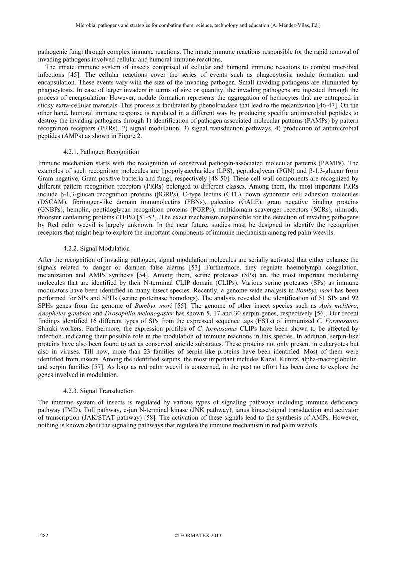
pathogenic fungi through complex immune reactions. The innate immune reactions responsible for the rapid removal of invading pathogens involved cellular and humoral immune reactions. The innate immune system of insects comprised of cellular and humoral immune reactions to combat microbial infections [45]. The cellular reactions cover the series of events such as phagocytosis, nodule formation and encapsulation. These events vary with the size of the invading pathogen. Small invading pathogens are eliminated by phagocytosis. In case of larger invaders in terms of size or quantity, the invading pathogens are ingested through the process of encapsulation. However, nodule formation represents the aggregation of hemocytes that are entrapped in sticky extra-cellular materials. This process is facilitated by phenoloxidase that lead to the melanization [46-47]. On the other hand, humoral immune response is regulated in a different way by producing specific antimicrobial peptides to destroy the invading pathogens through 1) identification of pathogen associated molecular patterns (PAMPs) by pattern recognition receptors (PRRs), 2) signal modulation, 3) signal transduction pathways, 4) production of antimicrobial peptides (AMPs) as shown in Figure 2.
4.2.1. Pathogen Recognition
Immune mechanism starts with the recognition of conserved pathogen-associated molecular patterns (PAMPs). The examples of such recognition molecules are lipopolysaccharides (LPS), peptidoglycan (PGN) and β-1,3-glucan from Gram-negative, Gram-positive bacteria and fungi, respectively [48-50]. These cell wall components are recognized by different pattern recognition receptors (PRRs) belonged to different classes. Among them, the most important PRRs include β-1,3-glucan recognition proteins (βGRPs), C-type lectins (CTL), down syndrome cell adhesion molecules (DSCAM), fibrinogen-like domain immunolectins (FBNs), galectins (GALE), gram negative binding proteins (GNBPs), hemolin, peptidoglycan recognition proteins (PGRPs), multidomain scavenger receptors (SCRs), nimrods, thioester containing proteins (TEPs) [51-52]. The exact mechanism responsible for the detection of invading pathogens by Red palm weevil is largely unknown. In the near future, studies must be designed to identify the recognition receptors that might help to explore the important components of immune mechanism among red palm weevils.
4.2.2. Signal Modulation
After the recognition of invading pathogen, signal modulation molecules are serially activated that either enhance the signals related to danger or dampen false alarms [53]. Furthermore, they regulate haemolymph coagulation, melanization and AMPs synthesis [54]. Among them, serine proteases (SPs) are the most important modulating molecules that are identified by their N-terminal CLIP domain (CLIPs). Various serine proteases (SPs) as immune modulators have been identified in many insect species. Recently, a genome-wide analysis in Bombyx mori has been performed for SPs and SPHs (serine proteinase homologs). The analysis revealed the identification of 51 SPs and 92 SPHs genes from the genome of Bombyx mori [55]. The genome of other insect species such as Apis melifera, Anopheles gambiae and Drosophila melanogaster has shown 5, 17 and 30 serpin genes, respectively [56]. Our recent findings identified 16 different types of SPs from the expressed sequence tags (ESTs) of immunized C. Formosanus Shiraki workers. Furthermore, the expression profiles of C. formosanus CLIPs have been shown to be affected by infection, indicating their possible role in the modulation of immune reactions in this species. In addition, serpin-like proteins have also been found to act as conserved suicide substrates. These proteins not only present in eukaryotes but also in viruses. Till now, more than 23 families of serpin-like proteins have been identified. Most of them were identified from insects. Among the identified serpins, the most important includes Kazal, Kunitz, alpha-macroglobulin, and serpin families [57]. As long as red palm weevil is concerned, in the past no effort has been done to explore the genes involved in modulation.
4.2.3. Signal Transduction
The immune system of insects is regulated by various types of signaling pathways including immune deficiency pathway (IMD), Toll pathway, c-jun N-terminal kinase (JNK pathway), janus kinase/signal transduction and activator of transcription (JAK/STAT pathway) [58]. The activation of these signals lead to the synthesis of AMPs. However, nothing is known about the signaling pathways that regulate the immune mechanism in red palm weevils.
Microbial pathogens and strategies for combating them: science, technology and education (A. Méndez-Vilas, Ed.)
© FORMATEX 2013
____________________________________________________________________________________________
1282
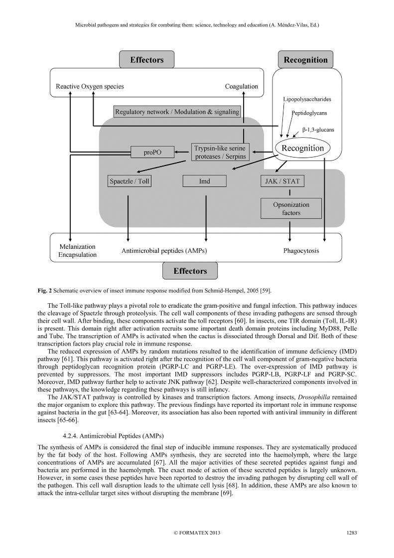
Fig. 2 Schematic overview of insect immune response modified from Schmid-Hempel, 2005 [59]. The Toll-like pathway plays a pivotal role to eradicate the gram-positive and fungal infection. This pathway induces
the cleavage of Spaetzle through proteolysis. The cell wall components of these invading pathogens are sensed through their cell wall. After binding, these components activate the toll receptors [60]. In insects, one TIR domain (Toll, IL-IR) is present. This domain right after activation recruits some important death domain proteins including MyD88, Pelle and Tube. The transcription of AMPs is activated when the cactus is dissociated through Dorsal and Dif. Both of these transcription factors play crucial role in immune response.
The reduced expression of AMPs by random mutations resulted to the identification of immune deficiency (IMD) pathway [61]. This pathway is activated right after the recognition of the cell wall component of gram-negative bacteria through peptidoglycan recognition protein (PGRP-LC and PGRP-LE). The over-expression of IMD pathway is prevented by suppressors. The most important IMD suppressors includes PGRP-LB, PGRP-LF and PGRP-SC. Moreover, IMD pathway further help to activate JNK pathway [62]. Despite well-characterized components involved in these pathways, the knowledge regarding these pathways is still infancy.
The JAK/STAT pathway is controlled by kinases and transcription factors. Among insects, Drosophilla remained the major organism to explore this pathway. The previous findings have reported its important role in immune response against bacteria in the gut [63-64]. Moreover, its association has also been reported with antiviral immunity in different insects [65-66].
4.2.4. Antimicrobial Peptides (AMPs)
The synthesis of AMPs is considered the final step of inducible immune responses. They are systematically produced by the fat body of the host. Following AMPs synthesis, they are secreted into the haemolymph, where the large concentrations of AMPs are accumulated [67]. All the major activities of these secreted peptides against fungi and bacteria are performed in the haemolymph. The exact mode of action of these secreted peptides is largely unknown. However, in some cases these peptides have been reported to destroy the invading pathogen by disrupting cell wall of the pathogen. This cell wall disruption leads to the ultimate cell lysis [68]. In addition, these AMPs are also known to attack the intra-cellular target sites without disrupting the membrane [69].
Microbial pathogens and strategies for combating them: science, technology and education (A. Méndez-Vilas, Ed.)
© FORMATEX 2013
____________________________________________________________________________________________
1283

These evolutionary conserved peptides are selectively toxic against invading pathogens. Till now, > 800 AMPs have been discovered from different organisms belonging to vertebrates, invertebrates, plants, protozoans and microbes. Because of the diversity of AMPs, it is very difficult to classify them except on the basis of secondary structure. The four major classes of AMPs based on their structure include β-sheet, amphipathic α-helical peptides, loop and extended peptides [70].
5. Conclusion and Future perspective
In summary, the successful control of red palm weevils mainly depends on the host pathogen interactions. So, there is a constant struggle between host and pathogen that ultimately lead to the success or failure of pathogens. In case of compatible interaction, the pathogen must have high number of conidia with strong adhesion that ultimately penetrate into the host through directly penetrating structures. Moreover, the invading pathogen must have the capacity to bypass or overcome the host immune system by producing toxins. In future, experiments must be conducted to explore in detail the immune mechanism of red palm weevil that might help to find out major genes involved in host defense. These findings might help to develop new products for the control of invasive populations of red palm weevils through gene silencing.
6. References
[1] Hussain A, Haq MRU, Al-Jabr AM, Al-Ayied HY. Managing Invasive Populations of Red Palm Weevil: A Worldwide Perspective. Journal of Food Agriculture and Environment. 2013; 11: 456-463.
[2] Malumphy C, Moran H. Red palm Weevil, Rhynchophorus ferrugineus. Plant Pest Fact sheet. 2009. [3] Dembilio Ó, Jacas JA. Bio-ecology and integrated management of the red palm weevil, Rhynchophorus ferrugineus
(Coleoptera: Curculionidae), in the region of Valencia (Spain). Hellenic Plant Protection Journal. 2012; 5: 1-12. [4] Rahalkar GW, Harwalkar MR, Rananavare HD, Shantaram K, Ayengar ARG. Laboratory studies on radiation sterilization of
the red palm weevil (Rhynchophorus ferrugineus Oliv.) males. Journal of Plantation Crops. 1973; 1: 141-145. [5] Krishnakumar R, Maheshwari P. Assessment of the sterile insect technique to manage red palm weevil Rhynchophorus
ferrugineus in coconut. In: Vreysen MJB, Robinson AS, Hendrichs J, eds. Area-Wide Control of Insect Pests from research to field implementation. Netherlands: Springer. 2007:475-485.
[6] Al-Ayedh HY, Rasool KG. Determination of the optimum sterilizing radiation dose for control of the red date palm weevil Rhynchophorus ferrugineus Oliv. (Coleoptera: Curculionidae). Crop Protection. 2010; 29: 1377-1380.
[7] Hallett RH, Gries G, Gries R, Borden JH, Czyzewska E, Oehlschlgar AC, Pierce HD, Angerilli NPD, Rauf A. Aggregation pheromone of two Asian palm weevils, Rhynchophorus ferrugineus and R. vulneratus. Naturwissenschaften. 1993; 80: 328-331.
[8] Gunawardena NE, Herath HMWKB. Enhancement of the activity of Ferrugineol by n-pentanol in an attractant baited trap for the coconut pest, Rhynchophorus ferrugineus F. (Coleoptera : Curculionidae). Journal of National Scientific Council of Sri Lanka. 1995; 23(2): 81-86.
[9] Abozuhairah RA, Vidyasagar PSPV, Abraham VA. Integrated management of red palm weevil, Rhynchophorus ferrugineus in date palm plantations of the Kingdom of Saudi Arabia. Proceedings of XX International Congress of Entomology. Firenze, Italy; 1996: 541.
[10] Vidyasagar PSPV, Hagi M, Abozuhairah RA, Al-Mohanna OE, Al-Saihati AA. Impact of mass pheromone trapping on red palm weevil adult population and infestation level in date palm gardens of Saudi Arabia. Planter. 2000; 76(891): 347-355.
[11] El-Sebay Y. Ecological studies on the red palm weevil Rhynchophorus ferrugineus Oliv., (Coleoptera: Curculionidae) in Egypt. Egyptian Journal of Agricultural Research. 2003; 81: 523-529.
[12] Abd-Allah FF, Al-Khatri SA. The effect of pheromone, kairomone and food bait on attracting males and females of red palm weevil. Egyptian Journal of Agricultural Research. 2005; 83: 169-177.
[13] Abdel-Moety EM, Lotfy HM, Rostom Y. Trace determination of Red Palm Weevil, Rhynchophorus ferrugineus, pheromone at trapping locations under Egyptian climate. International Journal of Agricultural and Food Sciences. 2012; 2(2): 44-50.
[14] Anon. Red palm weevil. The hidden enemy that works from within. Coconut Bulletin. 1956; 10: 77-81. [15] Mathen K, Kurian C. Comparative efficacy of different insecticides on Rhynchophorus ferrugineus F. Proceedings of 1st
Conference of Coconut Research Workers, Indian Central Coconut Committee, Ernakulam. 1962. [16] Muthuraman M. Trunk injection of undiluted insecticides – a method to control coconut red palm weevil, Rhynchophorus
ferrugineus Fab. Indian Coconut Journal. 1984; 15: 12-14. [17] El-Ezaby F. A biological in-vitro study on the red Indian date palm weevil. Arab Journal of Plant Protection. 1997; 15(2): 84-
87. [18] Abraham VA, Al-Shuaibi MA, Faleiro JR, Abozuhairah RA, Vidyasagar PSPV. 1998. An integrated approach for the
management of red palm weevil Rhynchophorus ferrugineus Oliv. A key pest of date palm in the Middle East. Sultan Qaboos University Journal for Scientific Research. 1998; 3: 77-83.
[19] Al-Rajhy DH, Hussein HI, Al-Shawaf AMA. Insecticidal activity of carbaryl and its mixture with piperonylbutoxide against red palm weevil Rhynchophorus ferrugineus (Olivier) (Curculionidae: Coleoptera) and their effects on Acetylcholinesterase activity. Pakistan Journal of Biological Sciences. 2005; 8: 679-682.
[20] Abo-El-Saad MM, Elshafie HA, Faleiro JR, Goa, Bou-Khowh IA. Toxicity evaluation of certain insecticides against the red palm weevil, Rhynchophorus ferrugineus (Olivier), under laboratory conditions. ESA Annual meeting, 2011.
[21] Cabello TP, de la Peña J, Barranco P, Belda J. Laboratory evaluation of imidacloprid and oxamyl against Rhynchophorus ferrugineus. Tests of Agrochemicals and Cultivars. 1997; 18: 6-7.
Microbial pathogens and strategies for combating them: science, technology and education (A. Méndez-Vilas, Ed.)
© FORMATEX 2013
____________________________________________________________________________________________
1284
[22] Jacas JA, Dembilio Ó, Llácer E. Research activities focused on management of red palm weevil at the UJI-IVIA associated unit (Region of Valencia, Spain). Bulletin of OEPP/EPPO Bulletin. 2011; 41: 122-127.
[23] Saranya S, Ushakumari R, Jacob S, Philip BM. Efficacy of different entomopathogenic fungi against cowpea aphid, Aphis craccivora (Koch). Journal of Biopesticides. 2010; 3(1 Special Issue): 138-142.
[24] García-Munguía AM, Garza-Hernández JA, Rebollar-Tellez EA, Rodríguez-Pérez MA, Reyes-Villanueva F. Transmission of Beauveria bassiana from male to female Aedes aegypti mosquitoes. Parasites and Vectors. 2011; 4: 24.
[25] James RR, Buckner JS, Freeman TP. Cuticular lipids and silverleaf whitefly stage affect conidial germination of Beauveria bassiana and Paecilomyces fumosoroseus. Journal of Invertebrate Pathology. 2003; 84(2): 67-74.
[26] Hussain A, Tian MY, He YR, Bland JM, Gu WX. Behavioral and electrophysiological responses of C. formosanus towards entomopathogenic fungal volatiles. Biological Control. 2010; 55: 166-173.
[27] Inglis GD, Johnson DL, Goettel MS. Effects of temperature and thermo-regulation on mycosis by Beauveria bassiana in Grasshoppers. Biological Control. 1996; 7: 131-139.
[28] Hussain A, Tian MY, He YR, Ahmed S. Entomopathogenic fungi disturbed the larval growth and feeding performance of Ocinara varians Walker (Lepidoptera: Bombycidae) larvae. Insect Science. 2009; 16: 511-517.
[29] Hussain A, Ahmed S, Shahid M. Laboratory and field evaluation of Metarhizium anisopliae var. anisopliae for controlling subterranean termites. Neotropical Entomology. 2011; 40(2): 244-50.
[30] Mohan CM, Lakshmi KA, Devi KU. Laboratory evaluation of the pathogenicity of three isolates of the entomopathogenic fungus Beauveria bassiana (Bals.) Vuillemin on the American cockroach (Periplaneta americana). Biocontrol Science and Technology. 1999; 9(1): 29-33.
[31] Dembilio O´, Quesada-Moraga E, Santiago-Alvarez C, Jacas JA. Potential of an indigenous strain of the entomopathogenic fungus Beauveria bassiana as a biological control agent against the Red Palm Weevil, Rhynchophorus ferrugineus. Journal of Invertebrate Pathology. 2010; 104(3): 214-221.
[32] Doberski JW. Comparative laboratory studies on three fungal pathogens of the elm bark beetle Scolytus scolytus: Effect of temperature and humidity on infection by Beauveria bassiana, Metarhizium anisopliae, and Paecilomyces farinosus. Journal of Invertebrate Pathology. 1981; 37(2): 195-200.
[33] Al-mazra'awi MS, Al-Abbadi A, Shatnawi MA, Ateyyat M. Effect of application method on the interaction between Beauveria bassiana and neem tree extract when combined for Thrips tabaci (Thysanoptera: Thripidae) control. Journal of Food Agriculture and Environment. 2009; 7(2): 869-873.
[34] Boucias DG, Pendland JC, Latge JP. Non-specific factors involved in attachment of entomopathogenic deuteromycetes to host insect cuticle. Applied and Environmental Microbiology. 1988 ; 54: 1795-1805.
[35] Boucias D, Pendland J. Attachment of mycopathogens to cuticle. In: Cole GT, Hoch HC, eds. The fungal spore and disease initiation in plants and animals. Plenum Press, New York, NY; 1991: 101-127.
[36] Doss RP, Potter SW, Chastagner GA, Christian JK. Adhesion of non-germinated Botrytis cinerea conidia to several substrata. Applied and Environmental Microbiology. 1993; 59: 1786-1791.
[37] Zacharuk RY. Fine structure of the fungus Metarhizium anisopliae infecting three species of larval Elateridae (Coleoptera). II. Conidial germ tubes and appressoria. Journal of Invertebrate Pathology. 1970; 15: 81-91.
[38] Zacharuk RY. Fine structure of the fungus Metarhizium anisopliae infecting three species of larval Elateridae (Coleoptera): III. Penetration of the host integument. Journal of Invertebrate Pathology. 1970; 15: 372-396.
[39] Fargues J. Adhesion of the fungal spore to the insect cuticle in relation to pathogenicity. In: Roberts DW, Aist JR, eds. Infection processes of fungi. The Rockefeller Foundation, New York; 1984: 90-110.
[40] Latge JP, Monsigny M, Prevost MC. Visualization of exo-cellular lectins in the entomopathogenic fungus Conidiobolus obscurus. Journal of Histochemistry and Cytochemistry. 1988; 36(11): 1419-1424.
[41] Neves PMOJ, Alves SB. External events related to the infection process of Cornitermes cumulans (Kollar) (Isoptera: Termitidae) by the entomopathogenic fungi Beauveria bassiana and Metarhizium anisopliae. Neotropical Entomology. 2004; 33: 51-56.
[42] Hussain A, Tian MY. Germination pattern and inoculum transfer of entomopathogenic fungi and their role in disease resistance among Coptotermes formosanus (Isoptera: Rhinotermitidae). International Journal of Agriculture and Biology. 2013; 15: 319-324.
[43] Madelin MF, Robinson RF, Williams RS. Appressorium like structures in insect parasitizing deuteromycetes. Journal of Invertebrate Pathology. 1967; 9: 404-412.
[44] Hussain A, Tian MY, He YR, Lin R. In vitro and in vivo culturing impacts on the virulence characteristics of serially passed entomopathogenic fungi. Journal of Food Agriculture and Environment. 2010; 8(3&4): 481-487.
[45] Khush RS, Lemaitre B. Genes that fight infection: what the Drosophila genome says about animal immunity? Trends in Genetics. 2000; 16: 442-449.
[46] Johansson MW. Cell adhesion molecules in invertebrate immunity. Developmental and Comparative Immunology. 1999; 23: 303-315.
[47] Gillespie JP, Bailey AM, Cobb B, Vilcinskas A. Fungi as elicitors of insect immune responses. Archives of Insect Biochemistry and Physiology. 2000; 44: 49-68.
[48] Yoshida H, Kinoshita K, Ashida M. Purification of a peptidoglycan recognition protein from hemolymph of the silkworm, Bombyx mori. The Journal of Biological Chemistry. 1996; 271: 13854-60.
[49] Koizumi N, Imamura M, Kadotani T, Yaoi K, Iwahana H, Sato R. The lipopolysaccharide-binding protein participating in hemocyte nodule formation in the silkworm Bombyx mori is a novel member of the C-type lectin superfamily with two different tandem carbohydrate-recognition domains. FEBS Letters. 1999; 443: 139-143.
[50] Yu XQ, Zhu YF, Ma C, Fabrick JA, Kanost MR. Pattern recognition proteins in Manduca sexta plasma. Insect Biochemistry and Molecular Biology. 2002; 32: 1287-1293.
[51] Kim YS, Ryu JH, Han SJ, Choi KH, Nam KB, Jang IH, Lemaitre B, Brey PT, Lee WJ. Gram-negative bacteria-binding protein, a pattern recognition receptor for lipopolysaccharide and beta-1,3-glucan that mediates the signaling for the
Microbial pathogens and strategies for combating them: science, technology and education (A. Méndez-Vilas, Ed.)
© FORMATEX 2013
____________________________________________________________________________________________
1285
induction of innate immune genes in Drosophila melanogaster cells. Journal of Biological Chemistry. 2000; 275: 32721-32727.
[52] Wang Y, Sumathipala N, Rayaprolu S, Jiang H. Recognition of microbial molecular patterns and stimulation of prophenoloxidase activation by a β-1,3-glucanase-related protein in Manduca sexta larval plasma. Insect Biochemistry and Molecular Biology. 2011; 41(5): 322-331.
[53] Christophides GK, Zdobnov E, Barillas-Mury C, Birney E, Blandin S, Blass C, Brey PT, Collins FH, Danielli A, Dimopoulos G, Hetru C, Hoa NT, Hoffmann JA, Kanzok SM, Letunic I, Levashina EA, Loukeris TG, Lycett G, Meister S, Michel K, Moita LF, Müller HM, Osta MA, Paskewitz SM, Reichhart JM, Rzhetsky A, Troxler L, Vernick KD, Vlachou D, Volz J, von Mering C, Xu J, Zheng L, Bork P, Kafatos FC. Immunity-related genes and gene families in Anopheles gambiae. Science. 2002; 298: 159-165.
[54] Gorman MJ, Paskewitz SM. Serine proteases as mediators of mosquito immune responses. Insect Biochemistry and Molecular Biology. 2001; 31: 257-262.
[55] Zhao P, Dong Z, Duan J, Wang G, Wang L, Li Y, Xiang Z, Xia Q. Genome-Wide identification and immune response analysis of serine protease Inhibitor genes in the Silkworm, Bombyx mori. PLoS ONE. 2012; 7(2); e31168.
[56] Tanaka H, Ishibashi J, Fujita K, Nakajima Y, Sagisaka A, Tomimoto K, Suzuki N, Yoshiyama M, Kaneko Y, Iwasaki T, Sunagawa T, Yamaji K, Asaoka A, Mita K, Yamakawa M. A genome-wide analysis of genes and gene families involved in innate immunity of Bombyx mori. Insect Biochemistry and Molecular Biology. 2008; 38: 1087-1110.
[57] Kanost MR. Serine proteinase inhibitors in arthropod immunity. Developmental and Comparative Immunology. 1999; 23: 291-301.
[58] Evans JD, Aronstein KA, Chen Y, Hetru C, Imler J, Jiang H, Zhou C, Kanost M, Thompson G, Hultmark D. Immune pathways and defence mechanisms in honey bees Apis mellifera. Insect Molecular Biology. 2006; 15(5); 645-656.
[59] Schmid-Hempel P. Natural insect host-parasite systems show immune priming and specificity: puzzles to be solved. BioEssays. 2005; 27(10): 1026-34.
[60] Weber AN, Tauszig-Delamasure S, Hoffmann JA, Lelievre E, Gascan H, Ray KP, Morse MA, Imler JL, Gay NJ. Binding of the Drosophila cytokine Spätzle to Toll is direct and establishes signalling. Nature Immunology. 2003; 4(8): 794-800.
[61] Lemaitre B, Kromer-Metzger E, Michaut L, Nicolas E, Meister M, Georgel P, Reichhart JM & Hoffmann JA. A recessive mutation, immune deficiency (imd) defines two distinct control pathways in the Drosophila host defence. Proceedings of National Academy of Sciences of the United States of America. 1995; 92: 9465-9469.
[62] Boutros M, Agaisse H, Perrimon N. Sequential activation of signaling pathways during innate immune responses in Drosophila. Developmental Cell. 2002; 3: 711-722.
[63] Buchon N, Broderick NA, Poidevin M, Pradervand S, Lemaitre B. Drosophila intestinal response to bacterial infection: activation of host defense and stem cell proliferation. Cell Host and Microbe. 2009; 5(2): 200-211.
[64] Cronin SJ, Nehme NT, Limmer S, Liegeois S, Pospisilik JA, Schramek D, Leibbrandt A, Simoes Rde M, Gruber S, Puc U, Ebersberger I, Zoranovic T, Neely GG, von Haeseler A, Ferrandon D, Penninger JM. Genome-wide RNAi screen identifies genes involved in intestinal pathogenic bacterial infection. Science (New York NY) 2009; 325(5938): 340-3.
[65] Dostert C, Jouanguy E, Irving P, Troxler L, Galiana-Arnoux D, Hetru C, Hoffmann JA, Imler JL. The Jak-STAT signaling pathway is required but not sufficient for the antiviral response of Drosophila. Nature Immunology. 2005; 6(9): 946-953.
[66] Souza-Neto JA, Sim S, Dimopoulos G. An evolutionary conserved function of the JAK-STAT pathway in anti-dengue defense. Proceedings of the National Academy of Sciences of the United States of America. 2009; doi:10.1073/pnas.0905006106.
[67] Meister M, Lemaitre B, Hoffmann JA. Antimicrobial peptide defense in Drosophila. BioEssays. 1997; 19: 1019-1026. [68] Hoffmann JA, Reichhart JM. Drosophila innate immunity: an evolutionary perspective. Nature Immunology. 2002; 3: 121-126. [69] Subbalakshimi C, Sitaram N. Mechanismm of antimicrobial action of indolicidin. FEMS Microbiol Letters. 1998; 160: 91-96. [70] Hancock REW, Lehrer R. Cationic peptides: a new source of antibiotics. Trends in Biotechnology. 1998; 16: 82-88.
Microbial pathogens and strategies for combating them: science, technology and education (A. Méndez-Vilas, Ed.)
© FORMATEX 2013
____________________________________________________________________________________________
1286