Embed Size (px)
Citation preview
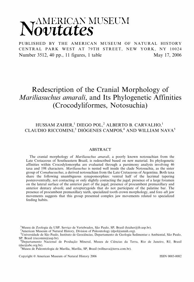
PUBLISHED BY THE AMERICAN MUSEUM OF NATURAL HISTORY
CENTRAL PARK WEST AT 79TH STREET, NEW YORK, NY 10024
Number 3512, 40 pp., 11 figures, 1 table May 17, 2006
Redescription of the Cranial Morphology ofMariliasuchus amarali, and Its Phylogenetic Affinities
(Crocodyliformes, Notosuchia)
HUSSAM ZAHER,1 DIEGO POL,2 ALBERTO B. CARVALHO,1
CLAUDIO RICCOMINI,3 DIOGENES CAMPOS,4 AND WILLIAM NAVA5
ABSTRACT
The cranial morphology of Mariliasuchus amarali, a poorly known notosuchian from theLate Cretaceous of Southeastern Brazil, is redescribed based on new material. Its phylogeneticaffinities within Crocodylomorpha are evaluated through a parsimony analysis involving 46taxa and 198 characters. Mariliasuchus is nested well inside the clade Notosuchia, as the sistergroup of Comahuesuchus, a derived notosuchian from the Late Cretaceous of Argentina. Both taxashare the following unambiguous synapomorphies: ventral half of the lacrimal taperingposteroventrally, not contacting or only slightly contacting the jugal; presence of a large foramenon the lateral surface of the anterior part of the jugal; presence of procumbent premaxillary andanterior dentary alveoli; and ectopterygoids that do not participate of the palatine bar. Thepresence of procumbent premaxillary teeth, specialized tooth crown morphology, and fore–aft jawmovements suggests that this group presented complex jaw movements related to specializedfeeding habits.
Copyright E American Museum of Natural History 2006 ISSN 0003-0082
1Museu de Zoologia da USP, Servico de Vertebrados, Sao Paulo, SP, Brasil ([email protected]).2American Museum of Natural History, Division of Paleontology ([email protected]).3Universidade de Sao Paulo, Instituto de Geociencias, Departamento de Geologia Sedimentar e Ambiental, Sao Paulo,
SP, Brasil ([email protected]).4Departamento Nacional de Producao Mineral, Museu de Ciencias da Terra, Rio de Janeiro, RJ, Brasil
([email protected]).5Museu de Paleontologia de Marılia, Marılia, SP, Brasil ([email protected]).
INTRODUCTION
Sediments of the Bauru Basin, located inSoutheastern Brazil, held one of the mostdiverse Upper Cretaceous Crocodyliform fau-na known so far (Kellner, 1998). This diversityis predominantly of Notosuchian taxa, withsome highly specialized forms such asSphagesaurus huenei (Price, 1950; Pol, 2003)and Mariliasuchus amarali (Carvalho andBertini, 1999). Despite the exquisite state ofconservation of several fossil crocodiliansunearthed from the Bauru Basin, few havebeen described in detail. Although the de-scription of Mariliasuchus amarali wasbased on a specimen with an almost com-plete skull and partially preserved postcra-nium, the authors provided little details ofits anatomy. Additionally, the originally de-scription is based on a juvenile specimen(Carvalho and Bertini, 1999). Carvalhoand Bertini (1999) concluded that M. amaraliis more closely related to Notosuchusterrestris than to any other notosuchian,allocating it in the family Notosuchidaealong with the genera Notosuchus andMalawisuchus. We present here a redescriptionof the species based on mostly complete skullsof a subadult and two adult specimens foundin the same locality from which the typespecimen was collected. The new materialdescribed here allows a more careful evalua-tion of the phylogenetic affinities ofMariliasuchus amarali.
GEOLOGICAL SETTING
The fossil remains of Mariliasuchus amaralireported here as well as the holotype describedby Carvalho and Bertini (1999) were collectedin a road cut at the right margin of the AguaFormosa creek (coordinates 22u209280S and49u569460W), 10 km south from the urbanarea of Marılia, about 500 m from thesecondary road between this city and thelocality of Ocaucu (Sao Paulo State), in thesoutheastern part of the Bauru Basin. Thisbasin is a large cratonic depression developedduring the Late Cretaceous in the central-southeastern portion of the South AmericanPlatform (Fernandes and Coimbra, 1996).
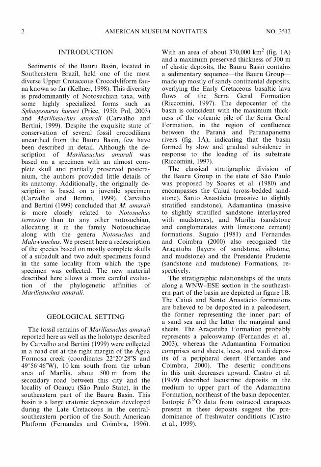
With an area of about 370,000 km2 (fig. 1A)and a maximum preserved thickness of 300 mof clastic deposits, the Bauru Basin containsa sedimentary sequence—the Bauru Group—made up mostly of sandy continental deposits,overlying the Early Cretaceous basaltic lavaflows of the Serra Geral Formation(Riccomini, 1997). The depocenter of thebasin is coincident with the maximum thick-ness of the volcanic pile of the Serra GeralFormation, in the region of confluencebetween the Parana and Paranapanemarivers (fig. 1A), indicating that the basinformed by slow and gradual subsidence inresponse to the loading of its substrate(Riccomini, 1997).
The classical stratigraphic division ofthe Bauru Group in the state of Sao Paulowas proposed by Soares et al. (1980) andencompasses the Caiua (cross-bedded sand-stone), Santo Anastacio (massive to slightlystratified sandstone), Adamantina (massiveto slightly stratified sandstone interlayeredwith mudstones), and Marılia (sandstoneand conglomerates with limestone cement)formations. Suguio (1981) and Fernandesand Coimbra (2000) also recognized theAracatuba (layers of sandstone, siltstone,and mudstone) and the Presidente Prudente(sandstone and mudstone) Formations, re-spectively.
The stratigraphic relationships of the unitsalong a WNW–ESE section in the southeast-ern part of the basin are depicted in figure 1B.The Caiua and Santo Anastacio formationsare believed to be deposited in a paleodesert,the former representing the inner part ofa sand sea and the latter the marginal sandsheets. The Aracatuba Formation probablyrepresents a paleoswamp (Fernandes et al.,2003), whereas the Adamantina Formationcomprises sand sheets, loess, and wadi depos-its of a peripheral desert (Fernandes andCoimbra, 2000). The desertic conditionsin this unit decreases upward. Castro et al.(1999) described lacustrine deposits in themedium to upper part of the AdamantinaFormation, northeast of the basin depocenter.Isotopic d18O data from ostracod carapacespresent in these deposits suggest the pre-dominance of freshwater conditions (Castroet al., 1999).
2 AMERICAN MUSEUM NOVITATES NO. 3512
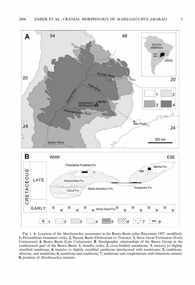
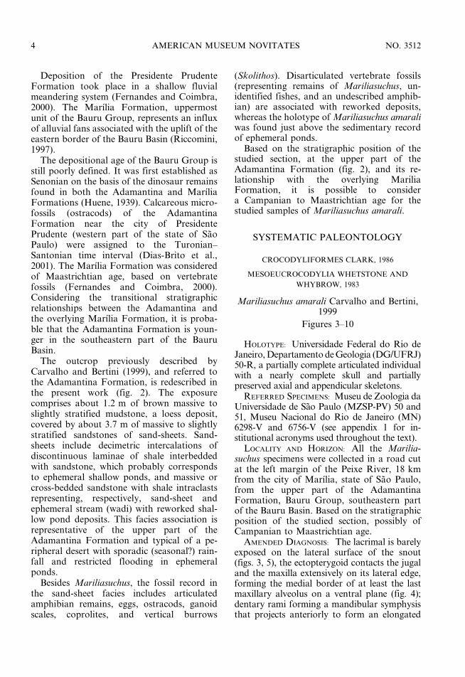
Fig. 1. A. Location of the Mariliasuchus occurrence in the Bauru Basin (after Riccomini 1997, modified):1, Precambrian basement rocks; 2, Parana Basin (Ordovician to Triassic); 3, Serra Geral Formation (EarlyCretaceous); 4, Bauru Basin (Late Cretaceous). B. Stratigraphic relationships of the Bauru Group in thesoutheastern part of the Bauru Basin: 1, basaltic rocks; 2, cross-bedded sansdstone; 3, massive to slightlystratified sandstone; 4, massive to slightly stratified sandstone interlayered with mudstones; 5, sandstone,siltstone, and mudstone; 6, sandstone and mudstone; 7, sandstone and conglomerate with limestone cement;8, position of Mariliasuchus remains.
2006 ZAHER ET AL.: CRANIAL MORPHOLOGY OF MARILIASUCHUS AMARALI 3
Deposition of the Presidente PrudenteFormation took place in a shallow fluvialmeandering system (Fernandes and Coimbra,2000). The Marılia Formation, uppermostunit of the Bauru Group, represents an influxof alluvial fans associated with the uplift of theeastern border of the Bauru Basin (Riccomini,1997).
The depositional age of the Bauru Group isstill poorly defined. It was first established asSenonian on the basis of the dinosaur remainsfound in both the Adamantina and MarıliaFormations (Huene, 1939). Calcareous micro-fossils (ostracods) of the AdamantinaFormation near the city of PresidentePrudente (western part of the state of SaoPaulo) were assigned to the Turonian–Santonian time interval (Dias-Brito et al.,2001). The Marılia Formation was consideredof Maastrichtian age, based on vertebratefossils (Fernandes and Coimbra, 2000).Considering the transitional stratigraphicrelationships between the Adamantina andthe overlying Marılia Formation, it is proba-ble that the Adamantina Formation is youn-ger in the southeastern part of the BauruBasin.
The outcrop previously described byCarvalho and Bertini (1999), and referred tothe Adamantina Formation, is redescribed inthe present work (fig. 2). The exposurecomprises about 1.2 m of brown massive toslightly stratified mudstone, a loess deposit,covered by about 3.7 m of massive to slightlystratified sandstones of sand-sheets. Sand-sheets include decimetric intercalations ofdiscontinuous laminae of shale interbeddedwith sandstone, which probably correspondsto ephemeral shallow ponds, and massive orcross-bedded sandstone with shale intraclastsrepresenting, respectively, sand-sheet andephemeral stream (wadi) with reworked shal-low pond deposits. This facies association isrepresentative of the upper part of theAdamantina Formation and typical of a pe-ripheral desert with sporadic (seasonal?) rain-fall and restricted flooding in ephemeralponds.
Besides Mariliasuchus, the fossil record inthe sand-sheet facies includes articulatedamphibian remains, eggs, ostracods, ganoidscales, coprolites, and vertical burrows
(Skolithos). Disarticulated vertebrate fossils(representing remains of Mariliasuchus, un-identified fishes, and an undescribed amphib-ian) are associated with reworked deposits,whereas the holotype of Mariliasuchus amaraliwas found just above the sedimentary recordof ephemeral ponds.
Based on the stratigraphic position of thestudied section, at the upper part of theAdamantina Formation (fig. 2), and its re-lationship with the overlying MariliaFormation, it is possible to considera Campanian to Maastrichtian age for thestudied samples of Mariliasuchus amarali.
SYSTEMATIC PALEONTOLOGY
CROCODYLIFORMES CLARK, 1986
MESOEUCROCODYLIA WHETSTONE AND
WHYBROW, 1983
Mariliasuchus amarali Carvalho and Bertini,1999
Figures 3–10
HOLOTYPE: Universidade Federal do Rio deJaneiro, Departamento de Geologia (DG/UFRJ)50-R, a partially complete articulated individualwith a nearly complete skull and partiallypreserved axial and appendicular skeletons.
REFERRED SPECIMENS: Museu de Zoologia daUniversidade de Sao Paulo (MZSP-PV) 50 and51, Museu Nacional do Rio de Janeiro (MN)6298-V and 6756-V (see appendix 1 for in-stitutional acronyms used throughout the text).
LOCALITY AND HORIZON: All the Marilia-suchus specimens were collected in a road cutat the left margin of the Peixe River, 18 kmfrom the city of Marılia, state of Sao Paulo,from the upper part of the AdamantinaFormation, Bauru Group, southeastern partof the Bauru Basin. Based on the stratigraphicposition of the studied section, possibly ofCampanian to Maastrichtian age.
AMENDED DIAGNOSIS: The lacrimal is barelyexposed on the lateral surface of the snout(figs. 3, 5), the ectopterygoid contacts the jugaland the maxilla extensively on its lateral edge,forming the medial border of at least the lastmaxillary alveolus on a ventral plane (fig. 4);dentary rami forming a mandibular symphysisthat projects anteriorly to form an elongated
4 AMERICAN MUSEUM NOVITATES NO. 3512

Fig
.2
.C
olu
mn
ar
sect
ion
of
the
stu
die
do
utc
rop
,A
raca
tub
aF
orm
ati
on
,1
8k
mso
uth
wes
tfr
om
Ma
rıli
a(c
oo
rdin
ate
s:4
9u5
694
50W
,2
2u2
093
20S
).
2006 ZAHER ET AL.: CRANIAL MORPHOLOGY OF MARILIASUCHUS AMARALI 5
6 AMERICAN MUSEUM NOVITATES NO. 3512

and spatulated process with parallel lateraledges (figs. 6, 7, 8, 9A); there are four pre-maxillary, five maxillary, and nine dentary teeththat are characterized by their heterodonty(fig. 9); most dentary and maxillary teeth withanastomosing longitudinal striations formed byenamel ridges (fig. 10); well-developed serrationof the mesial and distal margins of the crown(fig. 10A); and one to six longitudinally alignedtubercles at the base of each crow, which can bewell developed and ornament the whole surfaceof the crown base (fig. 10B).
TAXONOMIC COMMENTS: The name Mari-liasuchus amaralensis, published in a previouscontribution by Bertini and Carvalho (1999),is a nomen nudum according to Article 13 ofthe International Code of ZoologicalNomenclature (ICZN, 1999), and therefore isnot an available name. Several widelyused taxonomic names are followed through-out the text, such as Crocodyliformes, Meso-eucrocodylia, Neosuchia, and Notosuchia.Our usage of the first three of them followsthat of Clark (Benton and Clark, 1988; Clark,1994). Notosuchia was originally created byGasparini (1971) to cluster Araripesuchus,Uruguaysuchus, and Notosuchus. Later, sever-al authors pointed out that Sebecosuchia (Pol,2003; Sereno et al., 2003) was related to thisgroup, or at least to some of its members(Buckley et al., 2000; Ortega et al., 2000).Sereno et al. (2001) defined this taxon usinga stem-based definition of phylogenetic tax-onomy. Our results and usage of Notosuchiais consistent with these propositions.
DESCRIPTION
The material available is composed of fourspecimens. Specimen MZSP-PV 50 (figs. 3–8)consists of an almost complete individual withskull, mandibles, and most of the postcranialskeleton. The skull and mandibles are articulatedand crushed on the left side, which caused a slightdisplacement of elements on the parasagittal axisof the skull. The upper right temporal bar ispoorly preserved. Specimen MZSP-PV 51 (fig. 9)
consists of a complete skull and mandibles and theanterior part of the postcranial skeleton, includingthe cervical region, the anterior girdle andforelimbs, and part of the thoracic region. Thisskull is less distorted than MZSP-PV 50, beingslightly crushed dorsoventrally. The right post-orbital bar and upper temporal regions aremissing. Specimen MN 6298-V consists of analmost complete individual with skull, mandible,and most of the postcranial skeleton. Both skulland mandible are compressed laterally andslightly crushed on the sagittal axis. SpecimenMN 6756-V is composed of a partial skull with thenaris, the orbital, and palatal regions preserved,but lacking the braincase and temporal regions.
The postcranial material of specimensMZSP-PV 50, 51, and MN 6298-V is currentlyunder preparation and will be treated else-where.
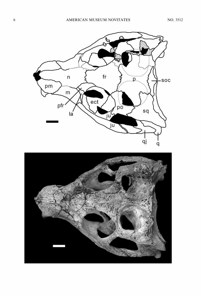
All four skulls analyzed here are oreini-rostral (sensu Busbey, 1994) with anteriorlyfacing external nares, characteristic of terres-trial forms. The snout is somewhat constrictedlaterally at its midpoint (at the level of thesecond maxillary tooth), being more conspic-uous in MZSP-PV 50 and MN 6298-V. Theskull widens significantly at the level of theorbital region. The orbits are large, rounded,and dorsolaterally exposed when the palpebralis removed. The temporal region is short andwide, with large, rounded supratemporalfossae (sensu Witmer, 1997) occupying mostof the flat skull table. The supratemporalfossae are separated by a wide flat surface ofthe parietal in MZSP-PV 51 and MN 6298-V(5.92 mm) whereas this separation is markedlynarrow in MZSP-PV 50 (3.33 mm). In MN6756-V, the parietal is not entirely preserved,precluding a precise measurement. Onto-genetic variation is visible on the shape ofthe supratemporal fossae, being more roundedand wider in the larger specimens whereas theyare more elongated and narrower in thesmaller specimen MN 6298-V. The infratem-poral fenestrae are large, triangular, and facelatero-dorsally, being almost completely ex-posed on dorsal view. The skull table and
r
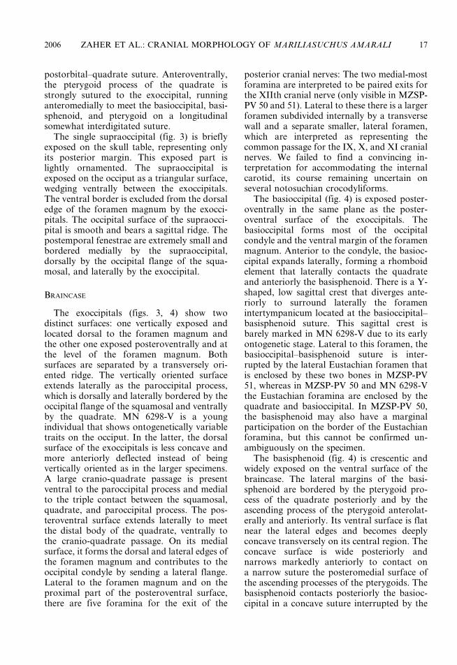
Fig. 3. Skull of MZSP-PV 50 in dorsal view. Scale bar 5 1 cm. Anatomical abbreviations are listedin appendix 1.
2006 ZAHER ET AL.: CRANIAL MORPHOLOGY OF MARILIASUCHUS AMARALI 7
8 AMERICAN MUSEUM NOVITATES NO. 3512
r
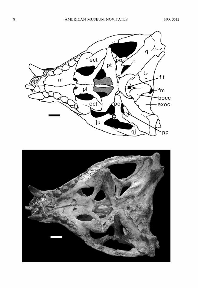
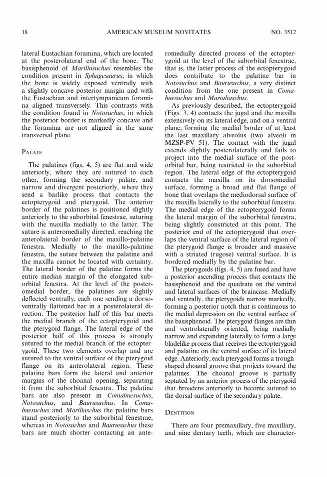
Fig. 4. Skull of MZSP-PV 50 in ventral view. Scale bar 5 1 cm. Anatomical abbreviations are listedin appendix 1.
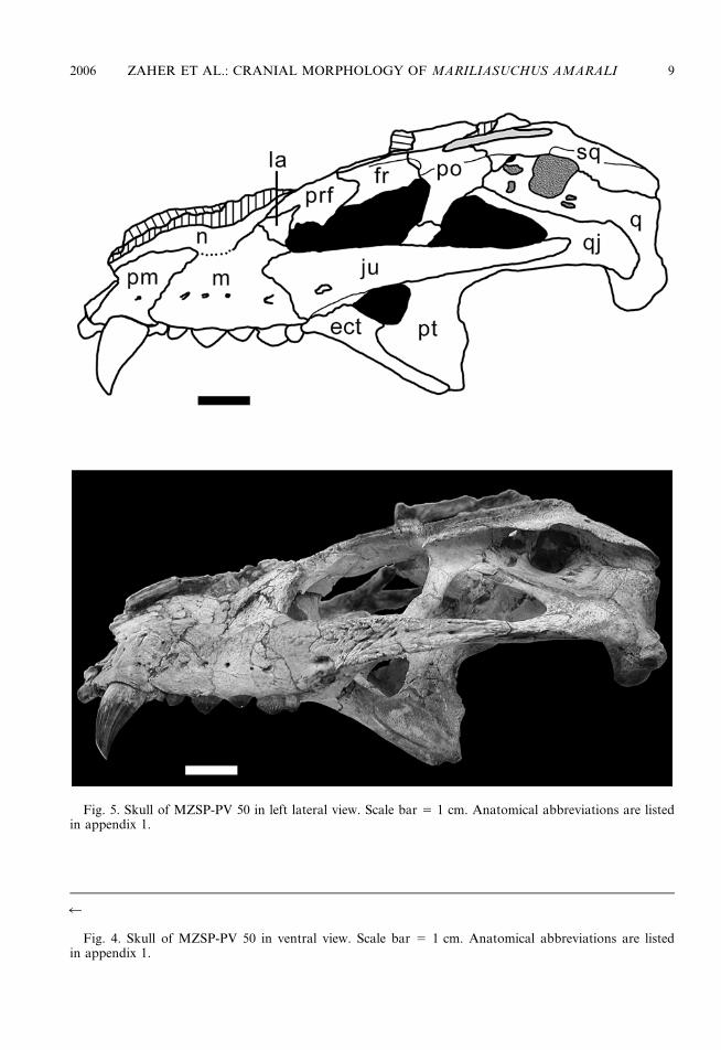
Fig. 5. Skull of MZSP-PV 50 in left lateral view. Scale bar 5 1 cm. Anatomical abbreviations are listedin appendix 1.
2006 ZAHER ET AL.: CRANIAL MORPHOLOGY OF MARILIASUCHUS AMARALI 9
10 AMERICAN MUSEUM NOVITATES NO. 3512
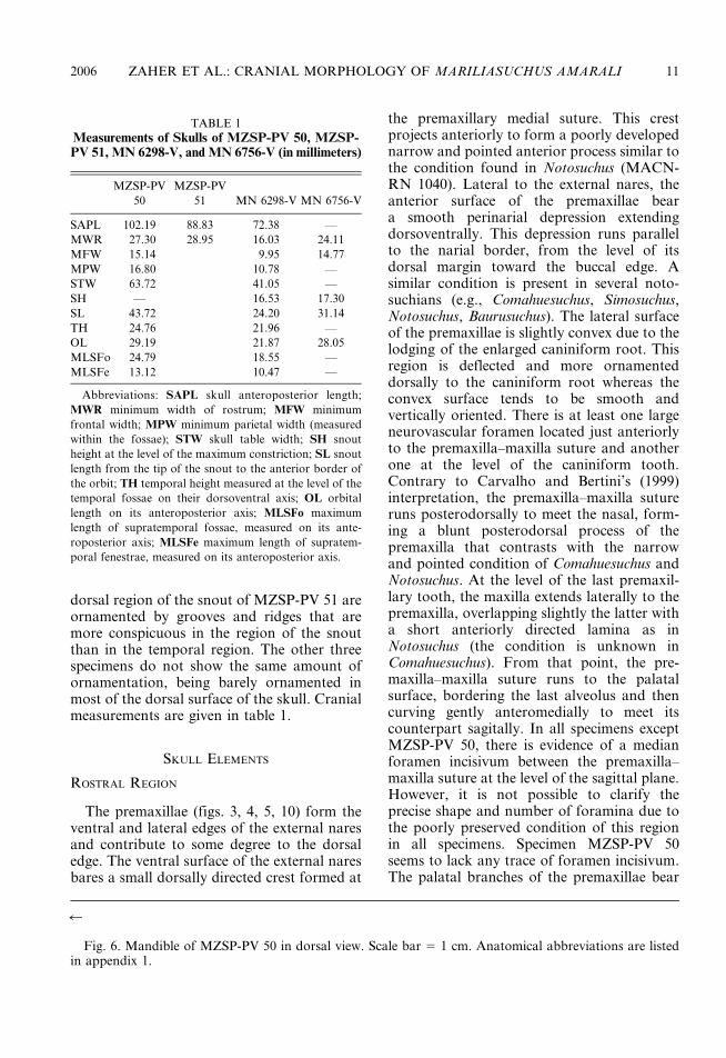
dorsal region of the snout of MZSP-PV 51 areornamented by grooves and ridges that aremore conspicuous in the region of the snoutthan in the temporal region. The other threespecimens do not show the same amount ofornamentation, being barely ornamented inmost of the dorsal surface of the skull. Cranialmeasurements are given in table 1.
SKULL ELEMENTS
ROSTRAL REGION
The premaxillae (figs. 3, 4, 5, 10) form theventral and lateral edges of the external naresand contribute to some degree to the dorsaledge. The ventral surface of the external naresbares a small dorsally directed crest formed at
the premaxillary medial suture. This crestprojects anteriorly to form a poorly developednarrow and pointed anterior process similar tothe condition found in Notosuchus (MACN-RN 1040). Lateral to the external nares, theanterior surface of the premaxillae beara smooth perinarial depression extendingdorsoventrally. This depression runs parallelto the narial border, from the level of itsdorsal margin toward the buccal edge. Asimilar condition is present in several noto-suchians (e.g., Comahuesuchus, Simosuchus,Notosuchus, Baurusuchus). The lateral surfaceof the premaxillae is slightly convex due to thelodging of the enlarged caniniform root. Thisregion is deflected and more ornamenteddorsally to the caniniform root whereas theconvex surface tends to be smooth andvertically oriented. There is at least one largeneurovascular foramen located just anteriorlyto the premaxilla–maxilla suture and anotherone at the level of the caniniform tooth.Contrary to Carvalho and Bertini’s (1999)interpretation, the premaxilla–maxilla sutureruns posterodorsally to meet the nasal, form-ing a blunt posterodorsal process of thepremaxilla that contrasts with the narrowand pointed condition of Comahuesuchus andNotosuchus. At the level of the last premaxil-lary tooth, the maxilla extends laterally to thepremaxilla, overlapping slightly the latter witha short anteriorly directed lamina as inNotosuchus (the condition is unknown inComahuesuchus). From that point, the pre-maxilla–maxilla suture runs to the palatalsurface, bordering the last alveolus and thencurving gently anteromedially to meet itscounterpart sagitally. In all specimens exceptMZSP-PV 50, there is evidence of a medianforamen incisivum between the premaxilla–maxilla suture at the level of the sagittal plane.However, it is not possible to clarify theprecise shape and number of foramina due tothe poorly preserved condition of this regionin all specimens. Specimen MZSP-PV 50seems to lack any trace of foramen incisivum.The palatal branches of the premaxillae bear
r
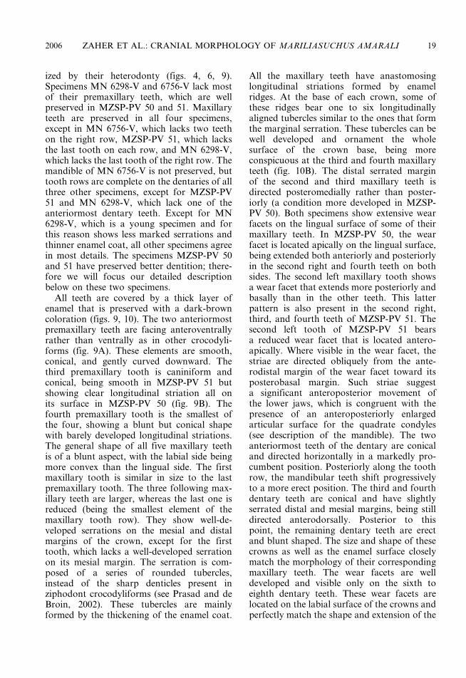
Fig. 6. Mandible of MZSP-PV 50 in dorsal view. Scale bar 5 1 cm. Anatomical abbreviations are listedin appendix 1.
TABLE 1
Measurements of Skulls of MZSP-PV 50, MZSP-PV 51, MN 6298-V, and MN 6756-V (in millimeters)
MZSP-PV
50
MZSP-PV
51 MN 6298-V MN 6756-V
SAPL 102.19 88.83 72.38 —
MWR 27.30 28.95 16.03 24.11
MFW 15.14 9.95 14.77
MPW 16.80 10.78 —
STW 63.72 41.05 —
SH — 16.53 17.30
SL 43.72 24.20 31.14
TH 24.76 21.96 —
OL 29.19 21.87 28.05
MLSFo 24.79 18.55 —
MLSFe 13.12 10.47 —
Abbreviations: SAPL skull anteroposterior length;
MWR minimum width of rostrum; MFW minimum
frontal width; MPW minimum parietal width (measured
within the fossae); STW skull table width; SH snout
height at the level of the maximum constriction; SL snout
length from the tip of the snout to the anterior border of
the orbit; TH temporal height measured at the level of the
temporal fossae on their dorsoventral axis; OL orbital
length on its anteroposterior axis; MLSFo maximum
length of supratemporal fossae, measured on its ante-
roposterior axis; MLSFe maximum length of supratem-
poral fenestrae, measured on its anteroposterior axis.
2006 ZAHER ET AL.: CRANIAL MORPHOLOGY OF MARILIASUCHUS AMARALI 11
12 AMERICAN MUSEUM NOVITATES NO. 3512
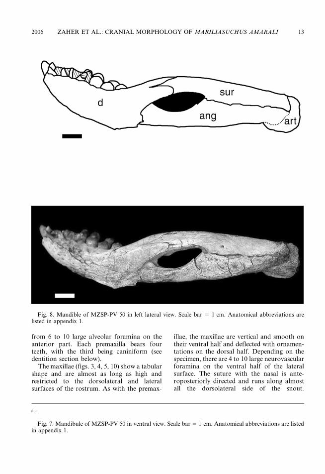
from 6 to 10 large alveolar foramina on theanterior part. Each premaxilla bears fourteeth, with the third being caniniform (seedentition section below).
The maxillae (figs. 3, 4, 5, 10) show a tabularshape and are almost as long as high andrestricted to the dorsolateral and lateralsurfaces of the rostrum. As with the premax-
illae, the maxillae are vertical and smooth ontheir ventral half and deflected with ornamen-tations on the dorsal half. Depending on thespecimen, there are 4 to 10 large neurovascularforamina on the ventral half of the lateralsurface. The suture with the nasal is ante-roposteriorly directed and runs along almostall the dorsolateral side of the snout.
r
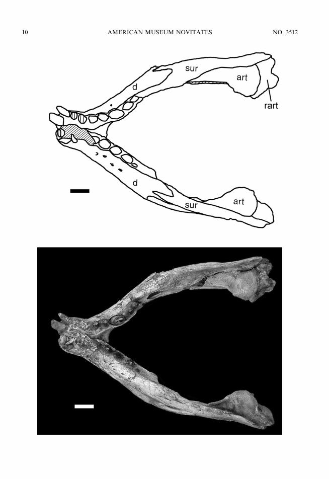
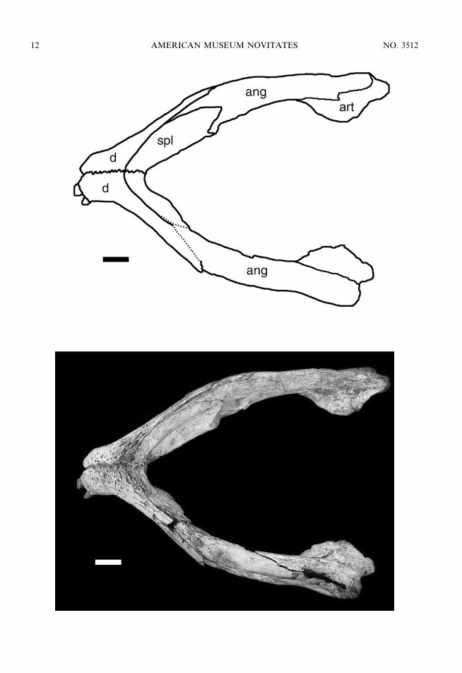
Fig. 7. Mandibule of MZSP-PV 50 in ventral view. Scale bar 5 1 cm. Anatomical abbreviations are listedin appendix 1.
Fig. 8. Mandible of MZSP-PV 50 in left lateral view. Scale bar 5 1 cm. Anatomical abbreviations arelisted in appendix 1.
2006 ZAHER ET AL.: CRANIAL MORPHOLOGY OF MARILIASUCHUS AMARALI 13

Posteriorly, the maxilla contacts the lacrimaland participates on the orbital margin, form-ing its anteroventral corner. There is novestige of an antorbital fenestra. The lateralcontact between the maxilla and the jugaloccurs just below the level of the anteriormargin of the orbit, with the latter sendinga short rounded anterior process laterally tothe maxilla on its dorsal half. The maxilla-jugal contact runs ventrally toward the buccalmargin where it meets the ectopterygoid. Thebuccal margin is straight, lacking the festoon-ing pattern present in derived neosuchians.The palatal branches of the maxillae meetalong their entire length in the sagittal line.The posterolateral border contacts the ectop-terygoid along the medial margin of the lastmaxillary tooth (in MZSP-PV 50 and MN6298-V) or the last two maxillary teeth(MZSP-PV 51 and MN 6756-V). Medially tothis point, the border of the maxillae runstransversally and barely enters the anterioredge of the suborbital fenestrae. The maxilla–palatine suture extends anteromedially fromthe level of the suborbital fenestrae to themaxillo-palatine foramina where the maxillacontributes to its anterior edge. The maxilla–palatine sutures seem to run transversely tomeet at the sagittal plane from the poster-omedial edge of the maxillo-palatine forami-na. There are five maxillary teeth on eachmaxilla that are located in individual alveolithat are only partially septate (i.e., the in-terdental plate is poorly developed). There are6 to 10 neurovascular foramina along thelingual edge of the alveoli. In all specimensboth labial and lingual margins are at thesame plane, except in MZSP-PV 51 where thelabial alveolar margin is much more developedventrally than the lingual margin.
The pair of nasals (figs. 3, 5) forms most ofthe posterodorsal border of the external nares.They slightly widen posteriorly along theircontact with the premaxillae and maxillaeuntil reaching the anterior tip of the lacrimal.The nasal then narrows posteriorly along thecontact with the anterodorsal surfaces of thelacrimal and prefrontal. The nasal–frontalsuture, positioned just behind the anteriormargin of the orbit, is transversally orientedand shows clear interdigitation in MZSP-PV50, MN 6756-V, and MN 6298-V. In MZSP-
PV 51, the precise position of the nasal–frontal suture cannot be determined due to theheavy ornamentation.
The lacrimal (figs. 3, 5) is barely exposed onthe lateral surface of the snout and is restrictedto the anterior border of the orbit. It showsa triangular shape being dorsoanteriorlyexpanded at the triple contact between lacri-mal, maxilla, and nasal. Ventrally, the lacri-mal tapers markedly along the anteroventralborder of the orbit, barely contacting theexpanded anterodorsal border of the jugalor not contacting at all, as in MN 6298-V.At that level the lacrimal is embraced mediallyby a dorsal flange of the maxilla. The lacrimalshows a posterior tuberosity exposed dorsallyon its posterodorsal surface that receives theanterior tip of the palpebral. Dorsally tothis tuberosity, the lacrimal contacts the pre-frontal along an anteroposteriorly orientedsuture. This suture continues ventromediallyon the inner surface of the orbit, borderingthe posterior opening of the lacrimal duct,which is completely included within thelacrimal.
Both anterior and posterior palpebrals werepreserved in position in specimen MN 6298-V.Specimens MZSP-PV 50 and 51 retained rightanterior palpebrals in position that wereremoved during preparation. A right posteriorpalpebral is also preserved in MZSP-PV 50,but dislocated and laying ventral to thesupratemporal fenestrae along the anterodor-sal process of the quadrate (fig. 4). In allspecimens, the anterior palpebrals are blade-like, somewhat triangular, elongated, andstrongly curved bones that are latero-poster-iorly and dorsally positioned to the orbit. Thedorsal surface of the anterior palpebrals isslightly ornamented. The posterior tip of theanterior palpebral lies close to the anterior tipof the posterior palpebral but does not contactthe latter. The posterior palpebrals are short,broad, and somewhat triangular bones thatlie along the anterior margin of the post-orbital.
The prefrontal (figs. 3, 5) is positionedmostly on the anterodorsal border of theorbit, running from the dorsal midpoint of theorbit to the anterodorsal corner, where itmeets the lacrimal. Anteriorly, the prefrontalexpands to meet the nasal on an oblique
14 AMERICAN MUSEUM NOVITATES NO. 3512
suture. Most of the dorsal surface is orna-mented by large rugosities where the palpebralarticulates. The anterodorsal region of theprefrontal is much wider than the posteriorregion, where the prefrontal forms the orbitalmargin. Posteromedially, on the inner marginof the orbit, the prefrontal bears a smallrounded process that overlaps the frontal. Theprefrontal has well-developed prefrontal pil-lars strongly sutured to the palate. These arethin laminae, entirely oriented in an obliqueposition. Each prefrontal pillar bears a medi-ally oriented process that meets it counterparton the sagittal plane, forming a closed bridgebelow the olfactory bulbs.
DORSAL AND TEMPORAL REGION
The frontals (figs. 3, 5) are completely fusedand contribute to most (two-thirds) of thedorsal margin of the orbit. In all specimensexcept MZSP-PV 51, the dorsal surface of thefrontals is flat and barely ornamented. In thelatter, this surface is rugose and bears a slightlymarked longitudinal ridge. A poorly definedlongitudinal ridge is also present inComahuesuchus and Notosuchus. It expandsslightly posterolaterally to contact the post-orbital in a strait longitudinal suture. Thefrontal meets the postorbital and the parietalat the anterior edge of the supratemporal fossain MZSP-PV 50 and MN 6298-V, whereas inMZSP-PV 51 it seems to expand into thesupratemporal fossa (not visible in MN 6756-V). The posterior margin of the frontal isstrongly sutured to the parietal via a slightlyconcave, transverse, interdigitated suture. Thelateral ventral flange of the frontals formsa concave and moderately developed dor-somedial inner orbital wall. Posteriorly, theflange is sutured to the postorbital and thedorsal projection of the laterosphenoid.Anteriorly, the flange is overlapped by a pos-terior process of the prefrontal.
The unpaired parietal (fig. 3) forms a dorsaltable that is constricted between the supra-temporal fossae, the constriction being widerin MZSP-PV 51 and MN 6298-V than in theother two specimens. In MZSP-PV 50, theparietal table represents only a thin string ofslightly ornamented bone running between thefossae, whereas in MZSP-PV 51 and MN
6298-V this surface is much broader andornamented, being almost as wide as thefrontal at its narrower orbital constriction.Anteriorly, the parietal contacts the frontal ona broad and slightly concave suture. Theparietal contacts the postorbital within thesupratemporal fossa. Posterior to this contact,the parietal forms the entire medial surface ofthe supratemporal fossa, being borderedventrally by the laterosphenoid and theascending anterodorsal process of the quad-rate. The laterosphenoid and the quadrateascending processes form the medial wall ofthe supratemporal fenestra. Lateroposteriorly,within the supratemporal fossa, the parietalcontacts the squamosal on an interdigitatedsuture that passes medially to the anteriortemporal orbital foramen, which is totallyenclosed within the squamosal. Posterior tothis point the parietal meets the supraoccipitalon the dorsal surface of the skull table.
The squamosal (figs. 3, 5) is triradiate, withthe anterior branch contacting the postorbital,the medial branch contacting the parietal, andthe posterolateral branch contacting the exoc-cipital and quadrate. In MZSP-PV 50 andMN 6298-V, the anterior branch is widelaterally and has a smooth dorsal surfacewhereas the medial branch is very narrow andornamented dorsally. In MZSP-PV 51, bothbranches are ornamented. The postorbital–squamosal contact is directed posteromediallyfrom the lateral margin of the skull table tothe lateral margin of the supratemporal fossa,meeting the anterodorsal process of thequadrate on a longitudinal suture positionedat the inner edge of the supratemporalfenestra. Then, the squamosal suture runsposteriorly along the inner posterolateralsurface of the supratemporal fossa to meetthe parietal in a triple contact with the latterand the anterodorsal process of the quadrate.The lateral margin of the postorbital andsquamosal overhangs the quadrate and quad-ratojugal, forming a deep otic recess. Thesquamosal is extensively sutured to the quad-rate within the otic recess, forming the dorsaland posterior margins of the otic aperture.The posterolateral branch is almost smooth onboth specimens, showing a median crest thatdivides the branch in two distinct lateralsurfaces: one dorsolaterally oriented and the
2006 ZAHER ET AL.: CRANIAL MORPHOLOGY OF MARILIASUCHUS AMARALI 15
other dorsoposteriorly oriented. The dorso-posterior surface meets the paroccipital pro-cess of the exoccipital on a broad contact thatruns mediolaterally along the occipital surfaceof the skull.
The postorbital (figs. 3, 4, 5) is alsotriradiate, with a narrow medial branchcontacting the frontal and parietal and sepa-rating the supratemporal fossa from the orbit,a posterior branch that contacts the squamo-sal, and a descending branch that forms thedorsal half of the postorbital bar separatingthe infratemporal fenestra from the orbit.Both medial and posterior branches constitutethe anterolateral border of the supratemporalfossa that is somewhat L-shaped. The ante-rolateral border of the postorbital showsa triangular peglike process forming a steppositioned just below the level of the skullroof. This process is also present inNotosuchus, Simosuchus, and Araripesuchus,where the posterior palpebral articulates. Theposterior branch articulates with the squamo-sal dorsally (see description of the squamosal)and ventrally, within the otic recess. Alsowithin the otic recess and anteriorly to the oticnotch, the postorbital articulates with thequadrate and sends a small posteroventralprocess that overlaps the ascending process ofthe quadratojugal. The ventral branch form-ing the dorsal half of the postorbital bar issmooth and somewhat cylindrical. It embracesthe ascending process of the jugal anteriorlyand contacts in a straight transversally orient-ed suture the same process of the jugalposteriorly.
The jugal (figs. 3, 4, 5) is triradiate, sendinga cylindrical ascending branch directed poster-odorsally and that meets the postorbital (seedescription of postorbital), a broad anteriorbranch that contacts the maxilla and ectopter-ygoid, and a posterior branch that contactsthe quadratojugal. The lateral surface of thejugal is densely ornamented (except forthe ascending branch) in all specimens. Theanterior branch reaches the level of theanterior margin of the orbit, where it isdorsoventrally tall. The anterior edge of thejugal contacts the maxilla in an extensive andsinuous suture, overlapping slightly the latter.The lateral surface of the anterior branchpresents a large foramen that is also present in
Comahuesuchus (as in MOZ P 6131). Theventral surface of the anterior branch of thejugal becomes sutured to the extensive pos-terolateral process of the ectopterygoid.Posteriorly, the jugal tapers gradually to forma thin posterior branch that overlaps laterallythe anterior branch of the quadratojugal. Bothbranches contribute to form a dorsoventrallyflattened infratemporal bar.
The quadratojugal (figs. 3, 4, 5) has ananterior process that forms the posterior halfof the infratemporal bar, lying medial to thejugal in an extensive longitudinally directedsuture. It expands posteriorly to form a broadcontact with the quadrate and sends a narrowanterodorsally ascending process that formsmost of the posterior border of the infra-temporal fenestra. The ascending processmeets dorsally with the postorbital in a narrowsuture. The posterior and posterodorsal bor-ders of the quadratojugal are solidly suturedto the anterior border of the quadrate. Theposteroventral tip of the quadratojugal doesnot reach the articular condyle of the quad-rate, thus not contributing to the cranio-mandibular articulation.
The quadrate (figs. 3, 4, 5) shares mostsynapomorphies of the notosuchian clade. Theanterodorsal branch is laterally exposed onthe otic recess whereas the distal body ofthe quadrate is projected ventrally at 90u inrespect to the longitudinal axis of the skull.The distal body of the quadrate is anteropos-teriorly thin and lateromedially wide andbears a well-developed ridge on its posteriorsurface running from the medial condyle tothe distal tip of the posterolateral process ofthe squamosal. A large foramen aereum islocated just medially to this ridge. The distalbody of the quadrate is projected ventrally inposterior view rather than ventrolaterally as inmost neosuchians. The anterior surface of thedistal body of the quadrate is concave andsmooth, lacking the ridges for the origin of theadductor bundles. The anterodorsal branch ofthe quadrate is broadly exposed laterally onthe otic recess and forms the ventral andanterior margin of the otic notch. Fouraccessory pneumatic foramina are present:Three are located posteroventrally and oneanteriorly to the otic notch. In addition, thereis a siphoneal foramen located near the
16 AMERICAN MUSEUM NOVITATES NO. 3512
postorbital–quadrate suture. Anteroventrally,the pterygoid process of the quadrate isstrongly sutured to the exoccipital, runninganteromedially to meet the basioccipital, basi-sphenoid, and pterygoid on a longitudinalsomewhat interdigitated suture.
The single supraoccipital (fig. 3) is brieflyexposed on the skull table, representing onlyits posterior margin. This exposed part islightly ornamented. The supraoccipital isexposed on the occiput as a triangular surface,wedging ventrally between the exoccipitals.The ventral border is excluded from the dorsaledge of the foramen magnum by the exocci-pitals. The occipital surface of the supraocci-pital is smooth and bears a sagittal ridge. Thepostemporal fenestrae are extremely small andbordered medially by the supraoccipital,dorsally by the occipital flange of the squa-mosal, and laterally by the exoccipital.
BRAINCASE
The exoccipitals (figs. 3, 4) show twodistinct surfaces: one vertically exposed andlocated dorsal to the foramen magnum andthe other one exposed posteroventrally and atthe level of the foramen magnum. Bothsurfaces are separated by a transversely ori-ented ridge. The vertically oriented surfaceextends laterally as the paroccipital process,which is dorsally and laterally bordered by theoccipital flange of the squamosal and ventrallyby the quadrate. MN 6298-V is a youngindividual that shows ontogenetically variabletraits on the occiput. In the latter, the dorsalsurface of the exoccipitals is less concave andmore anteriorly deflected instead of beingvertically oriented as in the larger specimens.A large cranio-quadrate passage is presentventral to the paroccipital process and medialto the triple contact between the squamosal,quadrate, and paroccipital process. The pos-teroventral surface extends laterally to meetthe distal body of the quadrate, ventrally tothe cranio-quadrate passage. On its medialsurface, it forms the dorsal and lateral edges ofthe foramen magnum and contributes to theoccipital condyle by sending a lateral flange.Lateral to the foramen magnum and on theproximal part of the posteroventral surface,there are five foramina for the exit of the
posterior cranial nerves: The two medial-mostforamina are interpreted to be paired exits forthe XIIth cranial nerve (only visible in MZSP-PV 50 and 51). Lateral to these there is a largerforamen subdivided internally by a transversewall and a separate smaller, lateral foramen,which are interpreted as representing thecommon passage for the IX, X, and XI cranialnerves. We failed to find a convincing in-terpretation for accommodating the internalcarotid, its course remaining uncertain onseveral notosuchian crocodyliforms.
The basioccipital (fig. 4) is exposed poster-oventrally in the same plane as the poster-oventral surface of the exoccipitals. Thebasioccipital forms most of the occipitalcondyle and the ventral margin of the foramenmagnum. Anterior to the condyle, the basioc-cipital expands laterally, forming a rhomboidelement that laterally contacts the quadrateand anteriorly the basisphenoid. There is a Y-shaped, low sagittal crest that diverges ante-riorly to surround laterally the foramenintertympanicum located at the basioccipital–basisphenoid suture. This sagittal crest isbarely marked in MN 6298-V due to its earlyontogenetic stage. Lateral to this foramen, thebasioccipital–basisphenoid suture is inter-rupted by the lateral Eustachian foramen thatis enclosed by these two bones in MZSP-PV51, whereas in MZSP-PV 50 and MN 6298-Vthe Eustachian foramina are enclosed by thequadrate and basioccipital. In MZSP-PV 50,the basisphenoid may also have a marginalparticipation on the border of the Eustachianforamina, but this cannot be confirmed un-ambiguously on the specimen.
The basisphenoid (fig. 4) is crescentic andwidely exposed on the ventral surface of thebraincase. The lateral margins of the basi-sphenoid are bordered by the pterygoid pro-cess of the quadrate posteriorly and by theascending process of the pterygoid anterolat-erally and anteriorly. Its ventral surface is flatnear the lateral edges and becomes deeplyconcave transversely on its central region. Theconcave surface is wide posteriorly andnarrows markedly anteriorly to contact ona narrow suture the posteromedial surface ofthe ascending processes of the pterygoids. Thebasisphenoid contacts posteriorly the basioc-cipital in a concave suture interrupted by the
2006 ZAHER ET AL.: CRANIAL MORPHOLOGY OF MARILIASUCHUS AMARALI 17
lateral Eustachian foramina, which are locatedat the posterolateral end of the bone. Thebasisphenoid of Mariliasuchus resembles thecondition present in Sphagesaurus, in whichthe bone is widely exposed ventrally witha slightly concave posterior margin and withthe Eustachian and intertympanicum forami-na aligned transversely. This contrasts withthe condition found in Notosuchus, in whichthe posterior border is markedly concave andthe foramina are not aligned in the sametransversal plane.
PALATE
The palatines (figs. 4, 5) are flat and wideanteriorly, where they are sutured to eachother, forming the secondary palate, andnarrow and divergent posteriorly, where theysend a barlike process that contacts theectopterygoid and pterygoid. The anteriorborder of the palatines is positioned slightlyanteriorly to the suborbital fenestrae, suturingwith the maxilla medially to the latter. Thesuture is anteromedially directed, reaching theanterolateral border of the maxillo-palatinefenestra. Medially to the maxillo-palatinefenestra, the suture between the palatine andthe maxilla cannot be located with certainty.The lateral border of the palatine forms theentire median margin of the elongated sub-orbital fenestra. At the level of the poster-omedial border, the palatines are slightlydeflected ventrally, each one sending a dorso-ventrally flattened bar in a posterolateral di-rection. The posterior half of this bar meetsthe medial branch of the ectopterygoid andthe pterygoid flange. The lateral edge of theposterior half of this process is stronglysutured to the medial branch of the ectopter-ygoid. These two elements overlap and aresutured to the ventral surface of the pterygoidflange on its anterolateral region. Thesepalatine bars form the lateral and anteriormargins of the choanal opening, separatingit from the suborbital fenestra. The palatinebars are also present in Comahuesuchus,Notosuchus, and Baurusuchus. In Coma-huesuchus and Mariliaschus the palatine barsstand posteriorly to the suborbital fenestrae,whereas in Notosuchus and Baurusuchus thesebars are much shorter contacting an ante-
romedially directed process of the ectopter-ygoid at the level of the suborbital fenestrae,that is, the latter process of the ectopterygoiddoes contribute to the palatine bar inNotosuchus and Baurusuchus, a very distinctcondition from the one present in Coma-huesuchus and Marialiaschus.
As previously described, the ectopterygoid(Figs. 3, 4) contacts the jugal and the maxillaextensively on its lateral edge, and on a ventralplane, forming the medial border of at leastthe last maxillary alveolus (two alveoli inMZSP-PV 51). The contact with the jugalextends slightly posterolaterally and fails toproject into the medial surface of the post-orbital bar, being restricted to the suborbitalregion. The lateral edge of the ectopterygoidcontacts the maxilla on its dorsomedialsurface, forming a broad and flat flange ofbone that overlaps the mediodorsal surface ofthe maxilla laterally to the suborbital fenestra.The medial edge of the ectopterygoid formsthe lateral margin of the suborbital fenestra,being slightly constricted at this point. Theposterior end of the ectopterygoid that over-laps the ventral surface of the lateral region ofthe pterygoid flange is broader and massivewith a striated (rugose) ventral surface. It isbordered medially by the palatine bar.
The pterygoids (figs. 4, 5) are fused and havea posterior ascending process that contacts thebasisphenoid and the quadrate on the ventraland lateral surfaces of the braincase. Mediallyand ventrally, the pterygoids narrow markedly,forming a posterior notch that is continuous tothe medial depression on the ventral surface ofthe basisphenoid. The pterygoid flanges are thinand ventrolaterally oriented, being mediallynarrow and expanding laterally to form a largebladelike process that receives the ectopterygoidand palatine on the ventral surface of its lateraledge. Anteriorly, each pterygoid forms a trough-shaped choanal groove that projects toward thepalatines. The choanal groove is partiallyseptated by an anterior process of the pterygoidthat broadens anteriorly to become sutured tothe dorsal surface of the secondary palate.
DENTITION
There are four premaxillary, five maxillary,and nine dentary teeth, which are character-
18 AMERICAN MUSEUM NOVITATES NO. 3512
ized by their heterodonty (figs. 4, 6, 9).Specimens MN 6298-V and 6756-V lack mostof their premaxillary teeth, which are wellpreserved in MZSP-PV 50 and 51. Maxillaryteeth are preserved in all four specimens,except in MN 6756-V, which lacks two teethon the right row, MZSP-PV 51, which lacksthe last tooth on each row, and MN 6298-V,which lacks the last tooth of the right row. Themandible of MN 6756-V is not preserved, buttooth rows are complete on the dentaries of allthree other specimens, except for MZSP-PV51 and MN 6298-V, which lack one of theanteriormost dentary teeth. Except for MN6298-V, which is a young specimen and forthis reason shows less marked serrations andthinner enamel coat, all other specimens agreein most details. The specimens MZSP-PV 50and 51 have preserved better dentition; there-fore we will focus our detailed descriptionbelow on these two specimens.
All teeth are covered by a thick layer ofenamel that is preserved with a dark-browncoloration (figs. 9, 10). The two anteriormostpremaxillary teeth are facing anteroventrallyrather than ventrally as in other crocodyli-forms (fig. 9A). These elements are smooth,conical, and gently curved downward. Thethird premaxillary tooth is caniniform andconical, being smooth in MZSP-PV 51 butshowing clear longitudinal striation all onits surface in MZSP-PV 50 (fig. 9B). Thefourth premaxillary tooth is the smallest ofthe four, showing a blunt but conical shapewith barely developed longitudinal striations.The general shape of all five maxillary teethis of a blunt aspect, with the labial side beingmore convex than the lingual side. The firstmaxillary tooth is similar in size to the lastpremaxillary tooth. The three following max-illary teeth are larger, whereas the last one isreduced (being the smallest element of themaxillary tooth row). They show well-de-veloped serrations on the mesial and distalmargins of the crown, except for the firsttooth, which lacks a well-developed serrationon its mesial margin. The serration is com-posed of a series of rounded tubercles,instead of the sharp denticles present inziphodont crocodyliforms (see Prasad and deBroin, 2002). These tubercles are mainlyformed by the thickening of the enamel coat.
All the maxillary teeth have anastomosinglongitudinal striations formed by enamelridges. At the base of each crown, some ofthese ridges bear one to six longitudinallyaligned tubercles similar to the ones that formthe marginal serration. These tubercles can bewell developed and ornament the wholesurface of the crown base, being moreconspicuous at the third and fourth maxillaryteeth (fig. 10B). The distal serrated marginof the second and third maxillary teeth isdirected posteromedially rather than poster-iorly (a condition more developed in MZSP-PV 50). Both specimens show extensive wearfacets on the lingual surface of some of theirmaxillary teeth. In MZSP-PV 50, the wearfacet is located apically on the lingual surface,being extended both anteriorly and posteriorlyin the second right and fourth teeth on bothsides. The second left maxillary tooth showsa wear facet that extends more posteriorly andbasally than in the other teeth. This latterpattern is also present in the second right,third, and fourth teeth of MZSP-PV 51. Thesecond left tooth of MZSP-PV 51 bearsa reduced wear facet that is located antero-apically. Where visible in the wear facet, thestriae are directed obliquely from the ante-rodistal margin of the wear facet toward itsposterobasal margin. Such striae suggesta significant anteroposterior movement ofthe lower jaws, which is congruent with thepresence of an anteroposteriorly enlargedarticular surface for the quadrate condyles(see description of the mandible). The twoanteriormost teeth of the dentary are conicaland directed horizontally in a markedly pro-cumbent position. Posteriorly along the toothrow, the mandibular teeth shift progressivelyto a more erect position. The third and fourthdentary teeth are conical and have slightlyserrated distal and mesial margins, being stilldirected anterodorsally. Posterior to thispoint, the remaining dentary teeth are erectand blunt shaped. The size and shape of thesecrowns as well as the enamel surface closelymatch the morphology of their correspondingmaxillary teeth. The wear facets are welldeveloped and visible only on the sixth toeighth dentary teeth. These wear facets arelocated on the labial surface of the crowns andperfectly match the shape and extension of the
2006 ZAHER ET AL.: CRANIAL MORPHOLOGY OF MARILIASUCHUS AMARALI 19
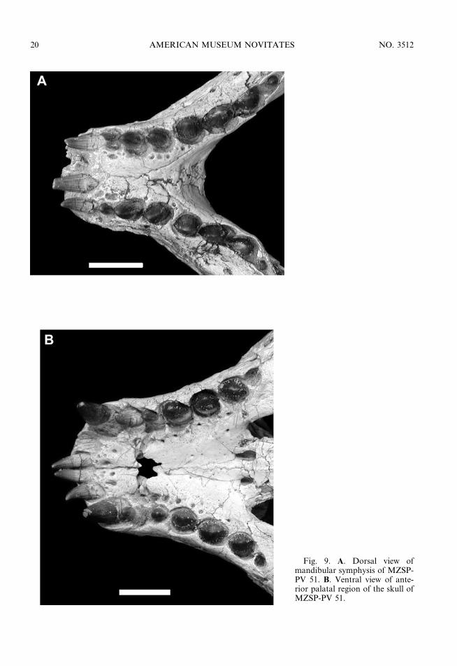
Fig. 9. A. Dorsal view ofmandibular symphysis of MZSP-PV 51. B. Ventral view of ante-rior palatal region of the skull ofMZSP-PV 51.
20 AMERICAN MUSEUM NOVITATES NO. 3512

2006 ZAHER ET AL.: CRANIAL MORPHOLOGY OF MARILIASUCHUS AMARALI 21

wear facets present in the correspondingmaxillary teeth, implying that a pattern oftooth-to-tooth occlusion was present inMariliasuchus amarali. Such a pattern wasonly found previously in Sphagesaurus hueneiamong crocodyliforms (Pol, 2003).
To accommodate the highly autapomorphicposition of the two anteriormost teeth, thecorresponding alveoli of both premaxillae anddentaries are in an almost horizontal position,with an enlarged labial wall and a highlyreduced lingual wall.
MANDIBLE
The complete mandible is preserved inMZSP-PV 50 (figs. 6, 7, 8), MZSP-PV 51,and MN 6298-V. The dentaries are dorsoven-trally low and laterally convex at the mandib-ular symphysis. The dentary rami converge tomeet and form a mandibular symphysis thattapers anteriorly along its posterior half.Anteriorly, the symphyseal region is elongatedand spatulated, with parallel lateral edges. Atthis point, the symphyseal region has a pecu-liar, large, and flattened dorsal surface be-tween the two parallel tooth rows. In all threespecimens the ventral surface of the symphysisis ornamented, whereas the lateral surface issmooth and bears several large neurovascularforamina, varying from four to eight. Justposterior to the mandibular symphysis, thedentaries diverge and the tooth rows (last fourteeth) are medially inset on their ramus, beinglingually bordered by a thin lamina of thesplenial. The lateral surface of the posteriorhalf of the dentary is mostly smooth andlateromedially broad in the larger specimens.The dorsal posterior process of the dentary isforked, receiving an acute anterior process ofthe surangular in an interdigitated suture. Theventral branch of the dorsal posterior processforms the anterodorsal margin of the externalmandibular fenestra, and contacts the angularat the anterior edge of the mandibularfenestra. The ventral posterior process of the
dentary is extremely reduced and fails toextend underneath the mandibular fenestra,the angular–dentary suture being directeddorsoventrally.
The splenials (fig. 7) represent thin laminaethat form the posterior third of the dorsalsurface of the mandibular symphysis. Thesplenial participation on the ventral surface ofthe symphysis is much more restricted, con-stituting only the posterior tip of the symphy-sis ventrally, where it forms a peglike pro-tuberance on the sagittal midline. The poste-rior surface of the symphysis is notably highand is entirely formed by the splenials, whichcontact each other on a solid interdigitatedsuture. Posterior to the symphysis, the sple-nials contribute to the ventral surface of themandibular rami on their anterior half,curving gently dorsally and being restrictedto the medial surface of the rami on theirposterior half. The medial surface of themandibular rami is covered by a flat laminaof the splenial that bears a moderately largeforamen intramandibularis oralis posterior tothe mandibular symphysis, located at the levelof the seventh dentary tooth. The posterioredge of the splenials forms the anterior borderof the internal mandibular fenestra. Its poster-oventral margin contacts the anterior processof the angular that wedges between thedentary and splenial.
The surangular (figs. 6, 8) is an elongatedbone that forms the dorsal margin of theposterior half of the mandibular rami. Theanterior process is forked like the posteriordorsal process of the dentary with which itinterdigitates. The medial surface of theanterior branch extends anteriorly betweenthe dorsal margin of the splenial and thedentary, reaching the lateral border of the lastdentary alveolus. At the level of the ante-rodorsal border of the inner mandibularfenestra, the anterior process of the surangularhas a concave and spatulate rugose surfacethat projects medially and ventrally. Thissurface might represent the coronoid bone
r
Fig. 10. A. Left lateral view of the skull of MZSP-PV 50 showing the buccal side of the premaxillary andmaxillary tooth rows. B. Detail of the second maxillary tooth of MZSP-PV 51 in lingual view. Scale bar 51 cm.
22 AMERICAN MUSEUM NOVITATES NO. 3512
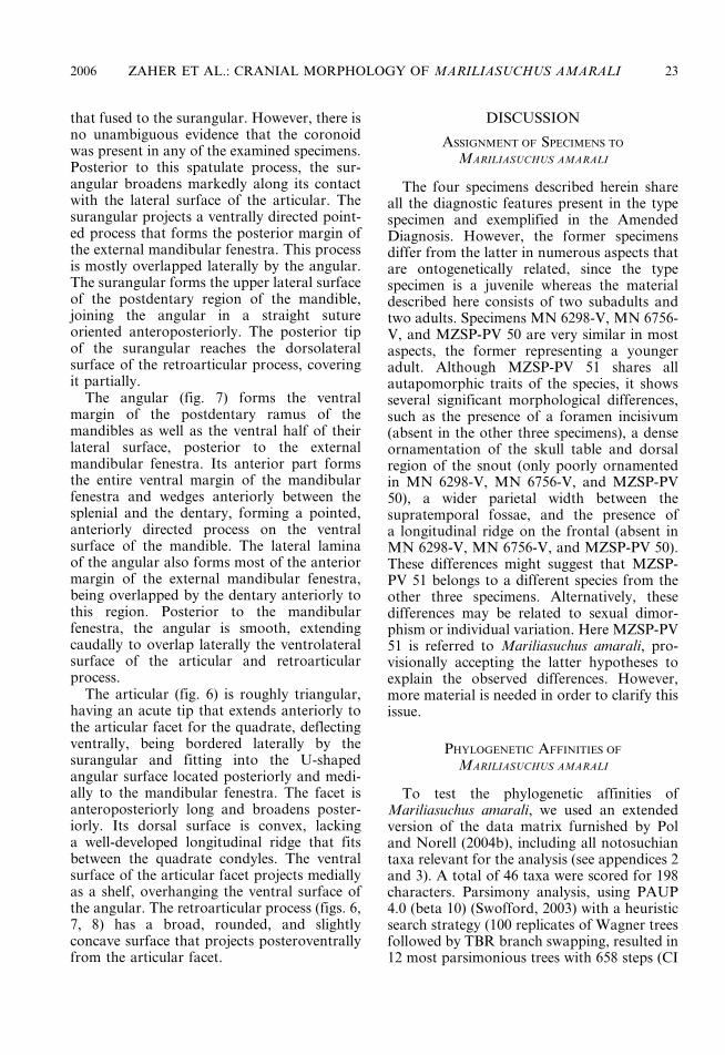
that fused to the surangular. However, there isno unambiguous evidence that the coronoidwas present in any of the examined specimens.Posterior to this spatulate process, the sur-angular broadens markedly along its contactwith the lateral surface of the articular. Thesurangular projects a ventrally directed point-ed process that forms the posterior margin ofthe external mandibular fenestra. This processis mostly overlapped laterally by the angular.The surangular forms the upper lateral surfaceof the postdentary region of the mandible,joining the angular in a straight sutureoriented anteroposteriorly. The posterior tipof the surangular reaches the dorsolateralsurface of the retroarticular process, coveringit partially.
The angular (fig. 7) forms the ventralmargin of the postdentary ramus of themandibles as well as the ventral half of theirlateral surface, posterior to the externalmandibular fenestra. Its anterior part formsthe entire ventral margin of the mandibularfenestra and wedges anteriorly between thesplenial and the dentary, forming a pointed,anteriorly directed process on the ventralsurface of the mandible. The lateral laminaof the angular also forms most of the anteriormargin of the external mandibular fenestra,being overlapped by the dentary anteriorly tothis region. Posterior to the mandibularfenestra, the angular is smooth, extendingcaudally to overlap laterally the ventrolateralsurface of the articular and retroarticularprocess.
The articular (fig. 6) is roughly triangular,having an acute tip that extends anteriorly tothe articular facet for the quadrate, deflectingventrally, being bordered laterally by thesurangular and fitting into the U-shapedangular surface located posteriorly and medi-ally to the mandibular fenestra. The facet isanteroposteriorly long and broadens poster-iorly. Its dorsal surface is convex, lackinga well-developed longitudinal ridge that fitsbetween the quadrate condyles. The ventralsurface of the articular facet projects mediallyas a shelf, overhanging the ventral surface ofthe angular. The retroarticular process (figs. 6,7, 8) has a broad, rounded, and slightlyconcave surface that projects posteroventrallyfrom the articular facet.
DISCUSSION
ASSIGNMENT OF SPECIMENS TO
MARILIASUCHUS AMARALI
The four specimens described herein shareall the diagnostic features present in the typespecimen and exemplified in the AmendedDiagnosis. However, the former specimensdiffer from the latter in numerous aspects thatare ontogenetically related, since the typespecimen is a juvenile whereas the materialdescribed here consists of two subadults andtwo adults. Specimens MN 6298-V, MN 6756-V, and MZSP-PV 50 are very similar in mostaspects, the former representing a youngeradult. Although MZSP-PV 51 shares allautapomorphic traits of the species, it showsseveral significant morphological differences,such as the presence of a foramen incisivum(absent in the other three specimens), a denseornamentation of the skull table and dorsalregion of the snout (only poorly ornamentedin MN 6298-V, MN 6756-V, and MZSP-PV50), a wider parietal width between thesupratemporal fossae, and the presence ofa longitudinal ridge on the frontal (absent inMN 6298-V, MN 6756-V, and MZSP-PV 50).These differences might suggest that MZSP-PV 51 belongs to a different species from theother three specimens. Alternatively, thesedifferences may be related to sexual dimor-phism or individual variation. Here MZSP-PV51 is referred to Mariliasuchus amarali, pro-visionally accepting the latter hypotheses toexplain the observed differences. However,more material is needed in order to clarify thisissue.
PHYLOGENETIC AFFINITIES OF
MARILIASUCHUS AMARALI
To test the phylogenetic affinities ofMariliasuchus amarali, we used an extendedversion of the data matrix furnished by Poland Norell (2004b), including all notosuchiantaxa relevant for the analysis (see appendices 2and 3). A total of 46 taxa were scored for 198characters. Parsimony analysis, using PAUP4.0 (beta 10) (Swofford, 2003) with a heuristicsearch strategy (100 replicates of Wagner treesfollowed by TBR branch swapping, resulted in12 most parsimonious trees with 658 steps (CI
2006 ZAHER ET AL.: CRANIAL MORPHOLOGY OF MARILIASUCHUS AMARALI 23
24 AMERICAN MUSEUM NOVITATES NO. 3512
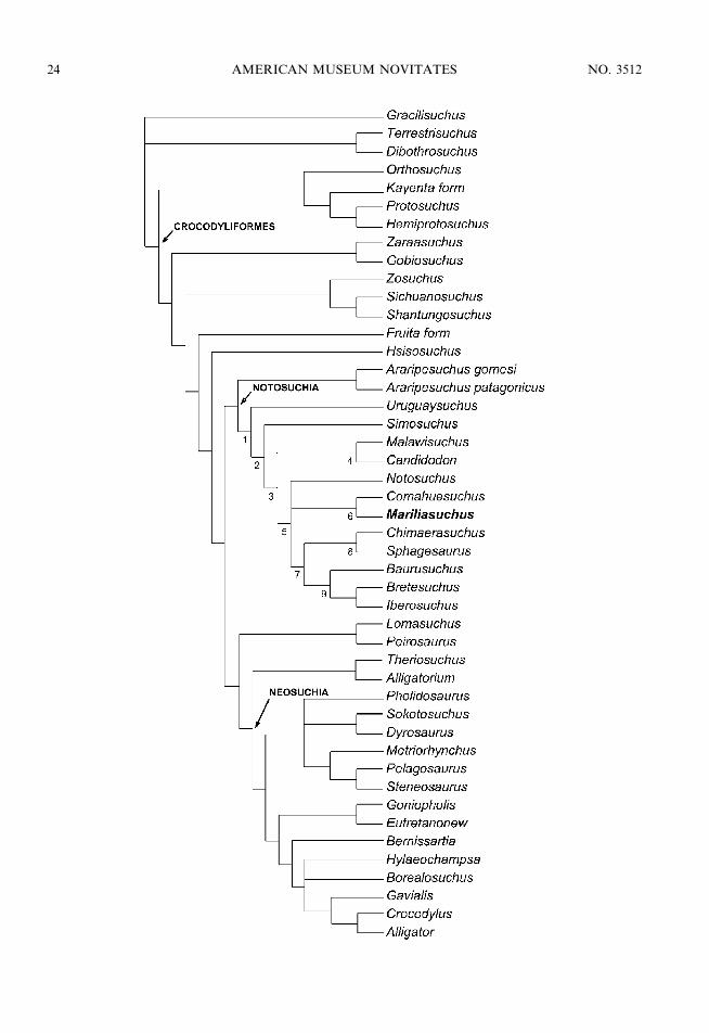
5 0.36; RI 5 0.67), the strict consensus ofwhich is presented in figure 11. The 12phylogenetic hypotheses differ in the relation-ships of some neosuchian crocodyliforms(e.g., Hylaeochampsa, Borealosuchus, andPholidosaurus) and the alternative position ofNotosuchus either as the sister group of clade 6or clade 7 within Notosuchia (or Ziposuchiasensu Ortega et al., 2000). In contrast to someprevious phylogenetic analyses (e.g., Clark,1994; Buckley et al., 2000; Ortega et al., 2000;Turner, 2004), the genus Araripesuchus ap-pears as the basalmost notosuchian instead ofbeing a basal member of Neosuchia, as ina recent analysis (Pol and Apesteguıa, 2005).
According to the present analysis,Mariliasuchus appears nested well insidethe clade Notosuchia, as the sister groupof Comahuesuchus, a derived and highlyautapomorphic notosuchian from the LateCretaceous of Argentina. These two taxashare four unambiguous synapomorphies:the ventral half of the lacrimal taperingposteroventrally, not contacting or only slight-ly contacting the jugal (character 192 [1]), thepresence of a large foramen on the lateralsurface of the anterior part of the jugal(character 193 [1]), the presence of procum-bent premaxillary and anterior dentary alveoli[character 194 (1)], and ectopterygoids that donot participate of the palatine bar (character196 [0]). Character 194 confers to bothMariliasuchus and Comahuesuchus an unusualcondition in which the anteriormost (pre-maxillary and dentary) teeth are set horizon-tally instead of vertically. These peculiarcharacteristics, related to specialized dentalcrown morphology and the presence in de-rived notosuchians of fore–aft jaw move-ments, suggests that this group presentedcomplex jaw movements related to specializedfeeding habits still poorly understood for themajority of the taxa.
Jacobs et al. (1990) and, more recently,Nobre and Carvalho (2002) suggested thatCandidodon itapecuruense from the LowerCretaceous Parnaıba basin is more closelyrelated to Malawisuchus than to any othernotosuchian, due to their similarities in dentalmorphology (i.e., lingual base of the crownornamented with a cuspidate cingulum; Clarket al., 1989; Gomani, 1997; Carvalho andBertini, 2000). Similarly, one could interpretthe complex lingual cuspidate ornamentationat the base of the crown of Mariliasuchus ashomologous to the lingual cuspidate cingula inthe teeth of Candidodon and Malawisuchus.However, such similarity may well be superfi-cial, as recent works documented a high di-versity of complex crown morphologies amongcrocodyliforms (Clark et al., 1989; Wu andSues, 1996; Gomani, 1997; Wu et al., 1997;Larsson and Sidor, 1999; Buckley et al., 2000;Pol, 2003). The present analysis supports thehypothesis previously proposed by Jacobs et al.(1990) and Nobre and Carvalho (2002), withCandidodon and Malawisuchus forming a clade(4) supported by five ambiguous synapomor-phies (9 [1], 122 [0], 140 [0], 149 [0], 161 [2];contra Carvalho et al., 2004).
Although the recently described notosu-chian Anatosuchus minor was hypothesized tobe the sister group of Comahuesuchus (Serenoet al., 2003), it was not included in thisanalysis, awaiting a more detailed description.However, Sereno et al.’s (2003) prelimi-nary description of the skull shows thatAnatosuchus may lack the synapomorphiessupporting clade 6 (fig. 11), suggestingthat it might not represent the sister groupof Comahuesuchus. A more detailed phylo-genetic analysis including Anatosuchus, aswell as Comahuesuchus, Mariliasuchus, andCandidodon, is needed before any moreaccurate taxonomic or biogeographic conclu-sions concerning these taxa can be drawn.
r
Fig. 11. Strict consensus of the 12 most parsimonious topologies that resulted from a strict parsimonyanalysis using PAUP 4.0 (beta 10). Unambiguous synapomorphies for the labeled nodes are: Node 1: 95 (0),104 (2), 151 (1). Node 2: 1 (1), 74 (1), 79 (0), 106 (1). Node 3: 78 (1), 107 (1), 141 (1). Node 4: 140 (0). Node 5:195 (1), 198 (1). Node 6: 192 (1), 193 (1), 194 (1), 196 (0). Node 7: 121 (1), 130 (1), 134 (1), 148 (1). Node 8:105 (3), 124 (1). Node 9: 3 (0), 9 (2), 79 (1), 80 (1), 106 (0), 118 (1), 120 (0), 128 (0), 155 (0), 158 (1).
2006 ZAHER ET AL.: CRANIAL MORPHOLOGY OF MARILIASUCHUS AMARALI 25
ACKNOWLEDGMENTS
The authors thank M. Norell (AmericanMuseum of Natural History, New York), M.Wilkinson (The Natural History Museum,London), F. de Broin, P. Taquet, C. de Muizon(Museum National d’Histoire Naturelle deParis), J. Bonaparte, A. Kramarz (MuseoArgentino de Ciencias Naturales), J. Clark(George Washington University), S.A.K. deAzevedo (Museu Nacional do Rio de Janeiro),and I.S. Carvalho (Universidade Federal doRio de Janeiro) for permission to analyze speci-mens under their care. Jim Clark and ChrisBrochu provided thoughtful comments thatimproved the quality of the manuscript. We alsodeeply thank Pablo Goloboff for his help, advice,and assistance in the computer-assisted phyloge-netic analysis using PAUP*. The present contri-bution benefited from grants of FAPESP (01/00162-3) and CNPq (303413/2002-6) to thesenior author.
REFERENCES
Benton, M.J., and J. Clark. 1988. Archosaurphylogeny and the relationships of theCrocodylia, In M.J. Benton (editor), Thephylogeny and classification of the tetrapods.Volume 1: amphibians, reptiles, birds. TheSystematics Association Special Volume 35A:295–338.
Bertini, R.J., and I.S. Carvalho. 1999. Distribuicaocronologica dos crocodilomorfos notossuquiose ocorrencias nas bacias cretacicas brasileiras.Boletim do 5u. Simposio sobre o Cretaceo doBrasil / 1er Simposio sobre el Cretacico deAmerica del Sur, Rio Claro, UniversidadeEstadual Paulista, 1999): 517–523.
Brochu, C.A. 1997. Fossils, morphology, diver-gence timing, and the phylogenetic relation-ships of Gavialis. Systematic Biology 46:479–522.
Buckley, G.A., and C.A. Brochu. 1999. Anenigmatic new crocodile from the UpperCretaceous of Madagascar. In D.M. Unwin(editor), Special Papers in Palaeontology 60:149–175.
Buckley, G.A., C.A. Brochu, D.W. Krause, andD. Pol. 2000. A pug-nosed crocodyliform fromthe Late Cretaceous of Madagascar. Nature405: 941–944.
Busbey, A.B. 1994. Structural consequences of skullflattening in crocodilians. In J. Thomason(editor), Functional morphology and verte-
brate paleontology: 173–192. Cambridge:Cambridge University Press.
Buscalioni, A.D., and J.L. Sanz. 1988. Phylogeneticrelationships of the Atoposauridae (Archo-sauria, Crocodylomorpha). Historical Biology1: 233–250.
Carvalho, I.S., and R.J. Bertini. 1999. Mariliasuchus:um novo Crocodylomorpha (Notosuchia) doCretaceo da Bacia Bauru. Geologıa Colombiana24: 83–105.
Carvalho, I.S., and R.J. Bertini. 2000. Contextogeologico dos notossuquios (Crocodylo-morpha) cretacicos do Brasil. GeologıaColombiana 25: 163–184.
Castro, J.C., D. Dias-Brito, E.A. Musacchio,J.M. Suarez, M.S.A.S. Maranhao, andR. Rodrigues. 1999. Arcabouco estratigraficodo Grupo Bauru no Oeste Paulista. Boletim do5u. Simposio sobre o Cretaceo do Brasil / 1erSimposio sobre el Cretacico de America delSur, Rio Claro, Universidade Estadual Paulista(1999): 509–515.
Clark, J.M. 1994. Patterns of evolution in Mesozoiccrocodyliformes. In N.C. Fraser and H.-D. Sues(editors), In the shadow of dinosaurs: 84–97.Cambridge: Cambridge University Press.
Clark, J.M., L.L. Jacobs, and W.R. Downs. 1989.Mammal-like dentition in a Mesozoic crocody-lian. Science 244: 1064–1066.
Dias-Brito, D., E.A. Musachchio, J.C. Castro,M.S.A.S. Maranhao, J.M. Suarez, and R.Rodrigues. 2001. Grupo Bauru: uma unidadecontinental do Cretaceo no Brasil—concepcoesbaseadas em dados micropaleontologicos,isotopicos e estratigraficos. Revue dePaleobiologie 20: 245–304.
Fernandes, L.A., and A.M. Coimbra. 1996. ABacia Bauru (Cretaceo Superior, Brasil). Anaisda Academia Brasileira de Ciencias 68:195–205.
Fernandes, L.A., and A.M. Coimbra. 2000.Revisao estratigrafica da parte oriental daBacia Bauru (Neocretaceo). Revista Brasileirade Geociencias 30: 717–728.
Fernandes, L.A., P.C.F. Giannini, and A.M. Goes.2003. Aracatuba Formation: palustrine depos-its from the initial sedimentation phase of theBauru Basin. Anais da Academia Brasileira deCiencias 75: 173–187.
Frey, E. 1988. Das Tragsystem der Krocodile—einebiomechanische und phylogenetische Analyse.Stuttgarter Beitrage zur NaturkundeSerie A (Biologie) 426: 1–60.
Gasparini, Z.B. 1971. Los Notosuchia del Cretacicode America del Sur como um nuevo infraordende los Mesosuchia (Crocodylia). Ameghiniana8: 83–103.
26 AMERICAN MUSEUM NOVITATES NO. 3512
Gasparini, Z.B., L.M. Chiappe, and M. Fernandez.1991. A new Senonian Peirosaurid (Crocody-lomorpha) from Argentina and a synopsis ofthe South American Cretaceous crocodilians.Journal of Vertebrate Paleontology 11: 316–333.
Gomani, E. 1997. A crocodyliform from the EarlyCretaceous dinosaur beds, northern Malawi.Journal of Vertebrate Paleontology 17:280–294.
Huene, F. 1939. Carta de F. von Huene ao Dr.Euzebio de Oliveira. Mineracao e Metalurgia 4:190.
ICZN. 1999. International Code of ZoologicalNomenclature, 4th ed. London: InternationalTrust of Zoological Nomenclature.
Jacobs, L.L., D.A. Winkler, Z.M. Kaufulu, andW.R. Downs. 1990. The dinosaur beds ofnorthern Malawi, Africa. National GeographicResearch 6(2): 162–204.
Kellner, A.W.A. 1998. Panorama e perspectiva doestudo de repteis fosseis no Brasil. Anais daAcademia Brasileira de Ciencias 70(3):647–676.
Larsson, H.C.E., and C.A. Sidor. 1999. Unusualcrocodyliform teeth from the Late Cretaceous(Cenomanian) of southeastern Morocco.Journal of Vertebrate Paleontology 19:398–401.
Nobre, P.E., and I.S. Carvalho. 2002. Osteologiado cranio de Candidodon itapecuruense(Crocodylomorpha, Mesoeucrocodylia) doCretaceo do Brasil. Boletim do 6u. Simposiosobre o Cretaceo do Brasil / 2nd. Simposiosobre el Cretacico de America del Sur (2002):77–82.
Ortega, F., A.D. Buscalioni, and Z.B. Gasparini.1996. Reinterpretation and new denominationof Atacisaurus crassiproratus (Middle Eocene;Issel, France) as cf. Iberosuchus (Croco-dylomorpha: Metasuchia). Geobios 29:353–364.
Ortega, F., Z.B. Gasparini, A.D. Buscalioni, andJ.O. Calvo. 2000. A new species ofAraripesuchus (Crocodylomorpha, Mesoeu-crocodylia) from the lower Cretaceous ofPatagonia (Argentina). Journal of VertebratePaleontology 20: 57–76.
Osmolska, H., S. Hua, and E. Buffetaut. 1997.Gobiosuchus kielanae (Protosuchia) from theLate Cretaceous of Mongolia: anatomy andrelationships. Acta Paleontologica Polonica 42:257–289.
Pol, D. 1999a. El esqueleto postcraneano deNotosuchus terrestris (Archosauria: Croco-dyliformes) del Cretacico Superior de laCuenca Neuquina y su informacion filogeneti-ca. Tesis de Licenciatura, Facultad de Ciencias
Exactas y Naturales, Universidad de BuenosAires, Argentina, 158 pp.
Pol, D. 1999b. Basal mesoeucrocodylian relation-ships: new clues to old conflicts. Journal ofVertebrate Paleontology 19(suppl. to no. 3):69A.
Pol, D. 2003. New remains of Sphagesaurus huenei(Crocodylomorpha: Mesoeucrocodylia) fromthe Late Cretaceous of Brazil. Journal ofVertebrate Paleontology 23: 817–831.
Pol, D., and S. Apesteguıa. 2005. NewAraripesuchus remains from the Early LateCretaceous (Cenomanian-Turonian) of Patagonia.American Museum Novitates 3490: 1–38.
Pol, D., and M.A. Norell. 2004a. A new crocodyli-form from Zos Canyon Mongolia. AmericanMuseum Novitates 3445: 1–36.
Pol, D., and M.A. Norell. 2004b. A new gobio-suchid crocodyliform taxon from theCretaceous of Mongolia. American MuseumNovitates 3458: 1–31.
Prasad, G.V.R., and L. de Broin. 2002. LateCretaceous crocodile remains from Naskal(India): comparisons and biogeographic affin-ities. Annales de Paleontologie 88(1): 19–71.
Price, L.I. 1950a. On a new crocodilian,Sphagesaurus, from the Cretaceous of theState of Sao Paulo, Brazil. Anais daAcademia Brassileira de Ciencias 22(1): 77–85.
Riccomini, C. 1997. Arcabouco estrutural e aspec-tos do tectonismo gerador e deformador daBacia Bauru no Estado de Sao Paulo. RevistaBrasileira de Geociencias 27: 153–162.
Sereno, P.C., H.C.E. Larsson, C.A. Sidor, andB. Gado. 2001. The giant crocodyliformSarcosuchus from the Cretaceous of Africa.Science 294: 1516–1519.
Sereno, P.C., C.A. Sidor, H.C.E. Larsson, andB. Gado. 2003. A new notosuchian from theEarly Cretaceous of Niger. Journal ofVertebrate Paleontology 23: 477–482.
Soares, P.C., P.M.B. Landim, V.J. Fulfaro, andA.F. Sobreiro Neto. 1980. Ensaio de caracter-izacao estratigrafica do Cretaceo no Estado deSao Paulo: Grupo Bauru. Revista Brasileira deGeociencias 10: 177–185.
Suguio, K. 1981. Fatores paleoambientais e paleo-climaticos e subdivisao estratigrafica do GrupoBauru. In A Formacao Bauru no Estado de SaoPaulo e regioes adjacentes: 15–26. Sao Paulo:Sociedade Brasileira de Geologia.
Swofford, D.L. PAUP*. Phylogenetic analysis usingparsimony (*and other methods). Version 4.Sunderland, MA: Sinauer Associates.
Turner, A.H.T. 2004. Crocodyliform biogeographyduring the Cretaceous: evidence of Gondwananvicariance from biogeographical analysis.
2006 ZAHER ET AL.: CRANIAL MORPHOLOGY OF MARILIASUCHUS AMARALI 27
Proceedings of the Royal Society London B271: 2003–2009.
Witmer, L.M. 1997. The evolution of the antorbitalcavity of archosaurs: a study in soft-tissuereconstruction in the fossil record with ananalysis of the function of pneumaticity.Journal of Vertebrate Paleontology, Memoir3: 1–73.
Wu, X.-C., and H.-D. Sues. 1996. Anatomy andphylogenetic relationships of Chimaeresuchus
paradoxus, an unusual crocodyliform reptilefrom the Lower Cretaceous of Hubei, China.Journal of Vertebrate Paleontology 16: 688–702.
Wu, X.-C., H.-D. Sues, and Z.-M. Dong. 1997.Sichuanosuchus shuhanensis: a new ?EarlyCretaceous protosuchian (Archosauria:Crocodyliformes) from Sichuan (China), andthe monophyly of Protosuchia. Journal ofVertebrate Paleontology 17: 89–103.
APPENDIX 1
INSTITUTIONAL ACRONYMS AND
ANATOMICAL ABBREVIATIONS
Institutional
DG/UFRJ Departamento de Geologia,Universidade Federal do Rio deJaneiro, Brazil.
MZSP-PV Museu de Zoologia, Universidadede Sao Paulo, Brazil.
MN Museu Nacional, UniversidadeFederal do Rio de Janeiro, Brazil.
MACN Museo Argentino de Ciencias Na-turales, Buenos Aires, Argentina.
Anatomical
ang angularart articularbocc basioccipitald dentaryect ectopterygoidexoc exoccipitalfit foramen intertympanicumfm foramen magnumfr frontalju jugalla lacrymalm maxillan nasalp parietalpm premaxillapl palatinepo postorbitalpp posterior palpebralprf prefrontalpt pterygoidq quadrateqj quadratojugalrart retroarticular processsoc supraoccipitalspl splenialsq squamosalsur surangular
APPENDIX 2
CHARACTER LIST CORRESPONDING TO DATA
MATRIX USED IN PHYLOGENETIC ANALYSIS
Character definitions 1–101 were takenfrom Clark (1994) and have the same numer-ation as in the original publication. Character5 was excluded from the analysis (because itdepends on the modified definition of charac-ter 6); however, its inclusion does not affectthe outcome of the analysis (except for the treelength). The additional characters are alsolisted here and their respective source is citedalong with the character number of theoriginal publication. Characters 1, 3, 6, 23,37, 45, 49, 65, 67, 69, 73, 77, 79, 90, 91, 96, 97,103, 104, 105, 107, 126, 143, 149, and 165 wereset as ordered characters (marked ‘‘+’’ in thislist).
Character 1 (modified from Clark, 1994;character 1). + External surface of dorsalcranial bones: smooth (0), slightly grooved (1),and heavily ornamented with deep pits andgrooves (2).
Character 2 (modified from Clark, 1994;character 2). Skull expansion at orbits: grad-ual (0) or abrupt (1).
Character 3 (modified from Clark, 1994;character 3). + Rostrum proportions: narroworeinirostral (0) or broad oreinirostral (1) ornearly tubular (2) or platyrostral (3).
Character 4 (Clark, 1994; character 4).Premaxilla participation in internarial bar:forming at least the ventral half (0) or withlittle participation (1).
Character 5 (Clark, 1994; character 5).Premaxilla anterior to nares: narrow (0) orbroad (1).
Character 6 (modified from Clark, 1994;character 6). + External nares facing: ante-rolaterally or anteriorly (0), dorsally notseparated by premaxillary bar from anterior
28 AMERICAN MUSEUM NOVITATES NO. 3512

edge of rostrum (1), or dorsally separated bypremaxillary bar (2).
Character 7 (Clark, 1994; character 7).Palatal parts of premaxillae: do not meetposterior to incisive foramen (0) or meetposteriorly along contact with maxillae (1).
Character 8 (Clark, 1994; character 8).Premaxilla–maxilla contact: premaxilla loose-ly overlies maxilla (0) or sutured togetheralong a butt joint (1).
Character 9 (modified from Clark, 1994;character 9). Ventrally opened notch onventral edge of rostrum at premaxilla–maxillacontact: absent (0) or present as a notch (1) orpresent as a large fenestra (2).
Character 10 (Clark, 1994; character 10).Posterior ends of palatal branches of maxillaeanterior to palatines: do not meet (0) or meet(1).
Character 11 (Clark, 1994; character 11).Nasal–lacrimal contact: (0) or do not contact(1).
Character 12 (Clark, 1994; character 12).Lacrimal contacts nasal along: medial edgeonly (0) or medial and anterior edges (1).
Character 13 (Clark, 1994; character 13).Nasal contribution to narial border: yes (0) orno (1).
Character 14 (Clark, 1994; character 14).Nasal–premaxilla contact: present (0) or ab-sent (1).
Character 15 (modified from Clark, 1994;character 15). Descending process of prefron-tal: does not contact palate (0) or contactspalate (1).
Character 16 (Clark, 1994; character 16).Postorbital–jugal contact: postorbital anteriorto jugal or postorbital medial to jugal (1) orpostorbital lateral to jugal (2).
Character 17 (Clark, 1994; character 17).Anterior part of the jugal with respect toposterior part: as broad (0) or twice as broad (1).
Character 18 (Clark, 1994; character 18).Jugal bar beneath infratemporal fenestra:flattened (0) or rod-shaped (1).
Character 19 (Clark, 1994; character 19).Quadratojugal dorsal process: narrow, con-tacting only a small part of postorbital (0) orbroad, extensively contacting the postorbital(1).
Character 20 (Clark, 1994; character 20).Frontal width between orbits: narrow, as
broad as nasals (0) or broad, twice as broadas nasals (1).
Character 21 (Clark, 1994; character 21).Frontals: paired (0), unpaired (1).
Character 22 (Clark, 1994; character 22).Dorsal surface of frontal and parietal: flat (0)or with midline ridge (1).
Character 23 (modified from Clark, 1994;character 23; by Buckley and Brochu, 1999;character 81). + Parieto-postorbital suture:absent from dorsal surface of skull roof andsupratemporal fossa (0) or absent from dorsalsurface of skull roof but broadly presentwithin supratemporal fossa (1) or presentwithin supratemporal fossa and on dorsalsurface of skull roof (2).
Character 24 (Clark, 1994; character 24).Supratemporal roof dorsal surface: complex(0) or dorsally flat ‘‘skull table’’ developed,with postorbital and squamosal with flatshelves extending laterally beyond quadratecontact (1).
Character 25 (modified from Clark, 1994;character 25). Postorbital bar: sculpted (ifskull sculpted) (0) or unsculpted (1).
Character 26 (modified from Clark, 1994;character 26). Postorbital bar: transverselyflattened (0) or cylindrical (1).
Character 27 (Clark, 1994; character 27).Vascular opening in dorsal surface of post-orbital bar: absent (0), present (1).
Character 28 (modified from Clark, 1994;character 28). Postorbital anterolateral pro-cess: absent or poorly developed (0) or welldeveloped, long, and acute (1).
Character 29 (Clark, 1994; character 29).Dorsal part of the postorbital: with anteriorand lateral edges only (0) or with anterolat-erally facing edge (1).
Character 30 (Clark, 1994; character 30).Dorsal end of the postorbital bar broadensdorsally, continuous with dorsal part of post-orbital (0) or dorsal part of the postorbital barconstricted, distinct from the dorsal part of thepostorbital (1).
Character 31 (Clark, 1994; character 31).Bar between orbit and supratemporal fossabroad and solid, with broadly sculpted dorsalsurface (0) or bar narrow, sculpting restrictedto anterior surface (1).
Character 32 (modified from Clark, 1994;character 32). Parietal: with broad occipital
2006 ZAHER ET AL.: CRANIAL MORPHOLOGY OF MARILIASUCHUS AMARALI 29
portion (0) or without broad occipital portion(1).
Character 33 (Clark, 1994; character 33).Parietal: with broad sculpted region separat-ing fossae (0) or with sagittal crest betweensupratemporal fossae (1).
Character 34 (Clark, 1994; character 34).Postparietal (dermosupraoccipital): a distinctelement (0) or not distinct (fused with parie-tal?) (1).
Character 35 (Clark, 1994; character 35).Posterodorsal corner of the squamosal:squared off, lacking extra ‘‘lobe’’ (0) or withunsculptured ‘‘lobe’’ (1).
Character 36 (modified from Clark, 1994;character 36). Posterolateral process of squa-mosal: poorly developed and projected hor-izontally at the same level of the skull (0) orelongated, thin, and posteriorly directed, notventrally deflected (1) or elongated, poster-olaterally directed, and ventrally deflected(2).
Character 37. (Clark, 1994; character 37). +Palatines: do not meet on palate below thenarial passage (0) or form palatal shelves thatdo not meet (1) or meet ventrally to the narialpassage, forming part of secondary palate (2).
Character 38 (Clark, 1994; character 38).Pterygoid: restricted to palate and suspensor-ium, joints with quadrate and basisphenoidoverlapping (0) or pterygoid extends dorsallyto contact laterosphenoid and form ventrolat-eral edge of the trigeminal foramen, stronglysutured to quadrate and laterosphenoid (1).
Character 39 (modified from Clark, 1994;character 39). Choanal opening: continuouswith pterygoid ventral surface except foranterior and anterolateral borders (0) or opensinto palate through a deep midline depression(choanal groove) (1).
Character 40 (Clark, 1994; character 40).Palatal surface of pterygoids: smooth (0) orsculpted (1).
Character 41 (Clark, 1994; character 41).Pterygoids posterior to choanae: separated (0)or fused (1).
Character 42 (modified from Clark, 1994;character 42; by Ortega et al., 2000; character139). Depression on primary pterygoideanpalate posterior to choana: absent or moder-ate in size being narrower than palatine bar (0)or wider than palatine bar (1).
Character 43 (Clark, 1994; character 43).Pterygoids: do not enclose choana (0) orenclose choana (1).
Character 44 (modified from Clark, 1994;character 44). Anterior edge of choanaesituated near posterior edge of suborbitalfenestra (or anteriorly) (0) or near posterioredge of pterygoid flanges (1).
Character 45 (Clark, 1994; character 45). +Quadrate: without fenestrae (0) or with singlefenestrae (1) or with three or more fenestraeon dorsal and posteromedial surfaces (2).
Character 46 (Clark, 1994; character 46).Posterior edge of quadrate: broad medial totympanum, gently concave (0) or posterioredge narrow dorsal to otoccipital contact,strongly concave (1).
Character 47 (Clark, 1994; character 47).Dorsal, primary head of quadrate articulateswith: squamosal, otoccipital and prootic (0) orwith prootic and laterosphenoid (1).
Character 48 (Clark, 1994; character 48).Ventrolateral contact of otoccipital withquadrate: very narrow (0) or broad (1).
Character 49 (Clark, 1994; character 49). +Quadrate, squamosal, and otoccipital: do notmeet to enclose cranioquadrate passage (0) orenclose passage near lateral edge of skull (1) ormeet broadly lateral to the passage (2).
Character 50 (Clark, 1994; character 50).Pterygoid ramus of quadrate: with flat ventraledge (0) or with deep groove along ventraledge (1).
Character 51 (Clark, 1994; character 51).Ventromedial part of quadrate: does notcontact otoccipital (0) or contacts otoccipitalto enclose carotid artery and form passage forcranial nerves IX–XI (1).
Character 52 (Clark, 1994; character 52).Eustachian tubes: not enclosed between ba-sioccipital and basisphenoid (0) or entirelyenclosed (1).
Character 53 (Clark, 1994; character 53).Basisphenoid rostrum (cultriform process):slender (0) or dorsoventrally expanded (1).
Character 54 (Clark, 1994; character 54).Basipterygoid process: prominent, formingmovable joint with pterygoid (0) or basipter-ygoid process small or absent, with basi-sphenoid joint suturally closed (1).
Character 55 (modified from Clark, 1994;character 55; by Ortega et al., 2000; character
30 AMERICAN MUSEUM NOVITATES NO. 3512

68). Basisphenoid ventral surface: shorter thanthe basioccipital (0) or wide and similar to orlonger in length than basioccipital (1).
Character 56 (Clark, 1994; character 56).Basisphenoid: exposed on ventral surface ofbraincase (0) or virtually excluded fromventral surface by pterygoid and basioccipital(1).
Character 57 (Clark, 1994; character 57).Basioccipital: without well-developed biltaeraltuberosities (0) or with large pendulous tubera(1).
Character 58 (Clark, 1994; character 58).Otoccipital: without laterally concave des-cending flange ventral to subcapsular process(0) or with flange (1).
Character 59 (Clark, 1994; character 59).Cranial nerves IX–XI: pass through commonlarge foramen vagi in otoccipital (0) or cranialnerve IX pass medial to nerves X and XI inseparate passage (1).
Character 60 (Clark, 1994; character 60).Otoccipital: without large ventrolateral partventral to paroccipital process (0) or withlarge ventrolateral part (1).
Character 61 (Clark, 1994; character 61).Crista interfenestralis between fenestrae pseu-dorotunda and ovalis nearly vertical (0) orhorizontal (1).
Character 62 (Clark, 1994; character 62).Supraoccipital: forms dorsal edge of theforamen magnum (0) or otoccipitals broadlymeet dorsal to the foramen magnum, separat-ing supraoccipital from foramen (1).
Character 63 (Clark, 1994; character 63).Mastoid antrum: does not extend into su-praoccipital (0) or extends through transversecanal in supraoccipital to connect middle earregions (1).
Character 64 (Clark, 1994; character 64).Posterior surface of supraoccipital: nearly flat(0), or with bilateral posterior prominences(1).
Character 65 (modified from Clark, 1994;character 65). + One small palpebral present inorbit (0) or one large palpebral (1) or two largepalpebrals (2).
Character 66 (Clark, 1994; character 66).External nares: divided by a septum (0) orconfluent (1).
Character 67 (Clark, 1994; character 67). +Antorbital fenestra: as large as orbit (0) or
about half the diameter of the orbit (1) ormuch smaller than the orbit (2) or absent (3).
Character 68 (modified from Clark, 1994;character 68; by Ortega et al., 2000; character41). Supratemporal fenestrae extension: rela-tively large, covering most of surface ofskull roof (0) or relatively short, fenestraesurrounded by a flat and extended skull roof(1).
Character 69 (modified from Clark, 1994;character 69). + Choanal groove: undivided(0) or partially septated (1) or completelyseptated (2).
Character 70 (Clark, 1994; character 70).Dentary: extends posteriorly beneath mandib-ular fenestra (0) or does not extend beneathfenestra (1).
Character 71 (modified from Clark, 1994;character 71). Retroarticular process: absentor extremely reduced (0) or very short, broad,and robust (1) or with an extensive rounded,wide, and flat (or slightly concave) surfaceprojected posteroventrally and facing dor-somedially (2) or posteriorly elongated, tri-angular shaped and facing dorsally (3) orposteroventrally projecting and paddle shaped(4).
Character 72 (Clark, 1994; character 72).Prearticular: present (0) or absent (1).
Character 73 (modified from Clark, 1994;character 73). + Articular without medialprocess (0) or with short process not contact-ing braincase (1) or with process articulatingwith otoccipital and basisphenoid (2).
Character 74 (Clark, 1994; character 74).Dorsal edge of surangular: flat (0) or archeddorsally (1).
Character 75 (Clark, 1994; character 75).Mandibular fenestra: present (0) or absent (1).
Character 76 (Clark, 1994; character 76).Insertion area for M. pterygoideous posterior:does not extend onto lateral surface of angular(0) or extends onto lateral surface of angular(1).
Character 77 (modified from Clark, 1994;character 77). + Splenial involvement insymphysis in ventral view: not involved (0)or involved slightly in symphysis (1) orextensively involved (2).
Character 78 (Clark, 1994; character 78).Posterior premaxillary teeth: similar in size toanterior teeth (0) or much longer (1).
2006 ZAHER ET AL.: CRANIAL MORPHOLOGY OF MARILIASUCHUS AMARALI 31
Character 79 (modified from Clark, 1994;character 79). + Maxillary teeth waves: absent,no tooth size variation (0) or one wave of teethenlarged (1) or enlarged maxillary teeth curvedin two waves (‘‘festooned’’) (2).
Character 80 (Clark, 1994; character 80).Anterior dentary teeth opposite premaxilla–maxilla contact: no more than twice the lengthof other dentary teeth (0) or more than twicethe length (1).
Character 81 (modified from Clark, 1994;character 81). Dentary teeth posterior to toothopposite premaxilla–maxilla contact: equal insize (0) or enlarged dentary teeth opposite tosmaller teeth in maxillary tooth row (1).
Character 82 (modified from Clark, 1994;character 82; by Ortega et al., 2000; character120). Anterior and posterior scapular edges:symmetrical in lateral view (0) or anterioredge more strongly concave than posterioredge (1) or dorsally narrow with straight edges(2).
Character 83 (modified from Clark, 1994;character 83; by Ortega et al., 2000; character121). Coracoid length: up to two-thirds of thescapular length (0) or subequal in length toscapula (1).
Character 84 (Clark, 1994; character 84).Anterior process of ilium: similar in length toposterior process (0) or one-quarter or less ofthe length of the posterior process (1).
Character 85 (Clark, 1994; character 85).Pubis: rodlike without expanded distal end (0)or with expanded distal end (1).
Character 86 (Clark, 1994; character 86).Pubis: forms anterior half of ventral edge ofacetabulum (0) or pubis at least partiallyexcluded from the acetabulum by the anteriorprocess of the ischium (1).
Character 87 (Clark, 1994; character 87).Distal end of femur: with large lateral facet forthe fibula (0) or with very small facet (1).
Character 88 (Clark, 1994; character 88).Fifth pedal digit: with phalanges (0) orwithout phalanges (1).
Character 89 (Clark, 1994; character 89).Atlas intercentrum: broader than long (0) oras long as broad (1).
Character 90 (modified from Clark, 1994;character 90). + Cervical neural spines: allanteroposteriorly large (0), only posterior onesrodlike (1), or all spines rodlike (2).
Character 91 (modified from Clark, 1994;character 91; by Buscalioni and Sanz, 1988;and by Brochu, 1997; character 37 andcharacter 7, respectively). + Hypapophyses incervicodorsal vertebrae: absent (0) or presentonly in cervical vertebrae (1) or present incervical and the first two dorsal vertebrae (2)or present up to the third dorsal vertebra (3)or up to the fourth dorsal vertebrae (4).
Character 92 (Clark, 1994; character 92).Cervical vertebrae: amphicoelous or amphy-platian (0) or procoelous (1).
Character 93 (Clark, 1994; character 93).Trunk vertebrae: amphicoelous or amphypla-tian (0) or procoelous (1).
Character 94 (Clark, 1994; character 94).All caudal vertebrae: amphicoelous or am-phyplatian (0) or first caudal biconvex withother procoelous (1) or procoelous (2).
Character 95 (Clark, 1994; character 95).Dorsal osteoderms: rounded or ovate (0) orrectangular, broader than long (1) or square (2).
Character 96 (modified from Clark, 1994;character 96; and Brochu, 1997; character 40).+ Dorsal osteoderms without articular anteri-or process (0) or with a discrete convexity onanterior margin (1) or with a well-developedprocess located anterolaterally in dorsal para-sagittal osteoderms (2).
Character 97 (modified from Clark, 1994;character 97; by Ortega et al., 2000; characters107 and 108). + Rows of dorsal osteoderms:two parallel rows (0) or more than two (1) ormore than four with ‘‘accessory ranges ofosteoderms’’ (sensu Frey, 1988) (2).
Character 98 (Clark, 1994; character 98).Osteoderms: Some or all imbricated (0) orsutured to one another (1).
Character 99 (Clark, 1994; character 99).Tail osteoderms: dorsal only (0) or completelysurrounded by osteoderms (1).
Character 100 (Clark, 1994; character 100).Trunk osteoderms: absent from ventral part ofthe trunk (0) or present (1).
Character 101 (Clark, 1994; character 101).Osteoderms: with longitudinal keels on dorsalsurfaces (0) or without longitudinal keels (1).
Character 102 (Wu and Sues, 1996; charac-ter 14). Jugal: participating in margin ofantorbital fossa (0) or separated from it (1).
Character 103 (modified from Wu and Sues,1996; character 23). + Articular facet for
32 AMERICAN MUSEUM NOVITATES NO. 3512

quadrate condyle: equal in length to thequadrate condyles (0) or slightly longer (1)or close to three times the length of thequadrate condyles (2).
Character 104 (modified from Wu and Sues,1996, and Wu et al., 1997; character 24 andcharacter 124, respectively). + Jaw joint:placed at level with basioccipital condyle (0)or below basioccipital condyle about abovelevel of lower tooth row (1) or below level oftooth row (2).
Character 105 (modified from Wu and Sues,1996, and Ortega et al., 2000; character 27 andcharacter 133, respectively). + Premaxillaryteeth: five (0), four (1), three (2), or two (3).
Character 106 (modified from Wu and Sues,1996; character 29). Unsculptured regionalong alveolar margin on lateral surface ofmaxilla: absent (0) or present (1).
Character 107 (modified from Wu and Sues,1996; character 30). + Maxilla: with eight ormore teeth (0) or seven (1) or six (2) or five (3)or four (4) teeth.
Character 108 (Wu and Sues, 1996; charac-ter 33). Coracoid: without posteromedial orventromedial process (0) or with elongateposteromedial process (1) or distally expandedventromedial process (2).
Character 109 (Wu and Sues, 1996; charac-ter 40). Radiale and ulnare: short and massive(0) or elongate (1).
Character 110 (Wu and Sues, 1996; charac-ter 41). Postacetabular process: directed pos-teroventrally or posteriorly (0) or directedposterodorsally and much higher in positionthan preacetabular process (1).
Character 111 (modified from Gomani,1997; character 4). Prefrontals anterior toorbits: elongated, oriented parallel to ante-roposterior axis of the skull (0) or short andbroad, oriented posteromedially–anterolater-ally (1).
Character 112 (modified from Gomani,1997; character 32). Basioccipital and ventralpart of otoccipital: facing posteriorly (0) orposteroventrally (1).
Character 113 (Buscalioni and Sanz, 1988;character 35). Vertebral centra: cylindrical (0)or spool shaped (1).
Character 114 (modified from Buscalioniand Sanz, 1988; character 39). Transverseprocess of posterior dorsal vertebrae dorso-
ventrally low and laminar (0) or dorsoventral-ly high (1).
Character 115 (Buscalioni and Sanz, 1988;character 44). Number of sacral vertebrae: two(0) or more than two (1).
Character 116 (Buscalioni and Sanz, 1988;character 49). Supra-acetabular crest: present(0) or absent (1).
Character 117 (Buscalioni and Sanz, 1988;character 54). Proximal end of radiale ex-panded symmetrically, similarly to the distalend (0) or more expanded proximomediallythan proximolaterally (1).
Character 118 (Ortega et al., 1996; character5). Lateral surface of the dentary: withouta longitudinal depression (0) or with a longi-tudinal depression (1).
Character 119 (Ortega et al., 1996; character9). Ventral exposure of splenials: absent (0) orpresent (1).
Character 120 (Ortega et al., 1996, 2000;characters 11 and 100, respectively). Toothmargins: with denticulate carinae (0) orwithout carinae or with smooth or crenulatedcarinae (1).
Character 121 (modified from Pol, 1999a,and Ortega et al., 2000; character 133 andcharacter 145, respectively). Lateral surface ofanterior process of jugal: flat or convex (0) orwith broad shelf below the orbit with tri-angular depression underneath it (1).
Character 122 (Pol, 1999a; character 134).Jugal: does not exceed the anterior margin oforbit (0) or exceeds (1).
Character 123 (Pol, 1999a; character 135).Notch in premaxilla on lateral edge of externalnares: absent (0) or present on the dorsal halfof the external nares lateral margin (1).
Character 124 (Pol, 1999a; character 136).Dorsal border of external nares: formedmostly by the nasals (0) or by both the nasalsand premaxilla (1).
Character 125 (Pol, 1999a; character 138).Posterodorsal process of premaxilla: absent(0) or present extending posteriorly wedgingbetween maxilla and nasals (1).
Character 126 (Pol, 1999a, and Ortega etal., 2000; character 139 and character 9,respectively). + Premaxilla–maxilla suturein palatal view, medial to alveolar region:anteromedially directed (0) or sinusoidal,posteromedially directed on its lateral
2006 ZAHER ET AL.: CRANIAL MORPHOLOGY OF MARILIASUCHUS AMARALI 33

half and anteromedially directed along itsmedial region (1) or posteromedially directed(2).
Character 127 (Pol, 1999a; character 140).Nasal lateral border posterior to externalnares: laterally concave (0) or straight (1).
Character 128 (Pol, 1999a; character 141).Nasal lateral edges: nearly parallel (0) orentirely oblique to each other converginganteriorly (1) or oblique to each other di-verging anteriorly (2).
Character 129 (Pol, 1999a; character 143).Palatine anteromedial margin: exceeding theanterior margin of the palatal fenestraewedging between the maxillae (0) or notexceeding the anterior margin of palatalfenestrae (1).
Character 130 (Pol, 1999a; character 144).Dorsoventral height of jugal antorbital regionrespect to infraorbital region: equal or lower(0) or antorbital region more expanded thaninfraorbital region of jugal (1).
Character 131 (Pol, 1999a; character 145).Maxilla–lacrimal contact: partially includedin antorbital fossa (0) or completely included(1).
Character 132 (Pol, 1999a; character 146).Lateral eustachian tube openings: locatedposteriorly to the medial opening (0) oraligned anteroposteriorly and dorsoventrally(1).
Character 133 (Pol, 1999a; character 147).Anterior process of ectopterygoid: developed(0) or reduced–absent (1).
Character 134 (Pol, 1999a; character 148).Posterior process of ectopterygoid: developed(0) or reduced–absent (1).
Character 135 (Pol, 1999a, and Ortega etal., 2000; character 149 and character 13,respectively). Small foramen located in thepremaxillo-maxillary suture in lateral surface(not for big mandibular teeth): absent (0) orpresent (1).
Character 136 (Pol, 1999a; character 150).Jugal posterior process: exceeding pos-teriorly the infratemporal fenestrae (0) or not(1).
Character 137 (Pol, 1999a; character 151).Compressed crown of maxillary teeth: orient-ed parallel to the longitudinal axis of skull (0)or obliquely disposed (1).
Character 138 (Pol, 1999a; character 152).Large and aligned neurovascular foramina onlateral maxilary surface: absent (0) or present(1).
Character 139 (modified from Pol, 1999a;character 153). External surface of maxillaand premaxilla: with a single plane facinglaterally (0) or with ventral region facinglaterally and dorsal region facing dorsolater-ally (1).
Character 140 (Pol, 1999a, and Ortega etal., 2000; character 154 and character 104,respectively). Maxillary teeth: not compressedlaterally (0) or compressed laterally (1).
Character 141 (Pol, 1999a; character 155).Posteroventral corner of quadratojugal: reach-ing the quadrate condyles (0) or not reachingthe quadrate condyles (1).
Character 142 (Pol, 1999a; character 156).Base of postorbital process of jugal: directedposterodorsally (0) or dorsally (1).
Character 143 (Pol, 1999a; character 157). +Postorbital process of jugal: anteriorly placed(0), in the middle (1), or posteriorly positioned(2).
Character 144 (Pol, 1999a, and Ortega etal., 2000; character 158 and character 36,respectively). Postorbital–ectopterygoid con-tact: present (0), absent (1).
Character 145 (Pol, 1999a; character 161).Quadratojugal: not ornamented (0) or orna-mented in the base (1).
Character 146 (Pol, 1999a; character 162).Prefrontal–maxillary contact in the inneranteromedial region of orbit: absent (0) orpresent (1).
Character 147 (Pol, 1999a; character 163).Basisphenoid: without lateral exposure (0) orwith lateral exposure on the braincase (1).
Character 148 (Pol, 1999a; character 165).Quadrate process of pterygoids: well devel-oped (0) or poorly developed (1).
Character 149 (modified from Pol, 1999a,and Ortega et al., 2000; character 166 andcharacter 44, respectively). + Quadrate majoraxis directed: posteroventrally (0) or ventrally(1) or anteroventrally (2).
Character 150 (Pol, 1999a; character 167).Quadrate distal end: with only one planefacing posteriorly (0) or with two distinctfaces in posterior view, a posterior one and
34 AMERICAN MUSEUM NOVITATES NO. 3512

a medial one bearing the foramen aereum(1).
Character 151 (Pol, 1999a; character 168).Anteroposterior development of neural spinein axis: well developed, covering all the neuralarch length (0) or poorly developed, locatedover the posterior half of the neural arch(1).
Character 152 (Pol, 1999a; character 169).Prezygapophyses of axis: not exceeding ante-rior edge of neural arch (0) or exceeding theanterior margin of neural arch (1).
Character 153 (Pol, 1999a; character 170).Postzygapophyses of axis: well developed,curved laterally (0) or poorly developed(1).
Character 154 (modified from Pol, 1999b;character 212). Shape of dentary symphysis inventral view: tapering anteriorly forming anangle (0) or U shaped, smoothly curvinganteriorly (1) or lateral edges longitudinallyoriented, convex anterolateral corner, andextensive transversally oriented anterior edge(2).
Character 155 (Pol, 1999b; character 213).Unsculpted region in the dentary below thetooth row: absent (0) or present (1).
Character 156 (Ortega et al., 1996, andBuckley et al., 2000; character 13). Cheekteeth: not constricted at base of crown (0) orconstricted (1).
Character 157 (Ortega et al., 2000; character42). Outer surface of squamosal laterodorsallyoriented: extensive (0) or reduced and sculpted(1) or reduced and unsculpted (2).
Character 158 (Ortega et al., 2000; character74). Length/height proportion of infratempor-al fenestra: higher than wide or equal (0) orvery antero-posteriorly elongated (1).
Character 159 (Ortega et al., 2000; character90). Foramen intramandibularis oralis: smallor absent (0) or big and slotlike (1).
Character 160 (Ortega et al., 2000; character146). Ectopterygoid medial process: single (0)or forked (1).
Character 161 (modified from Gomani,1997, and Buckley et al., 2000; character 46and character 113, respectively). Cusps ofteeth: unique cusp (0), one main cusp withsmaller cusps arranged in one row (1) or onemain cusp with smaller cusps arranged inmore than one row (2) or several cusps of
equal size arranged in more than one row (3)or multiple small cusps along edges ofocclussal surface (4).
Character 162 (Pol and Norell, 2004a;character 164). Cross section of distal endof quadrate: mediolaterally wide and ante-roposteriorly thin (0) or subquadrangular(1).
Character 163 (Pol and Norell, 2004a;character 165). Palatine–pterygoid contact onpalate: palatines overlie pterygoids (0) orpalatines firmly sutured to pterygoids (1).
Character 164 (Wu et al., 1997; character103). Squamosal descending process: absent(0) or present (1).
Character 165 (modified from Wu et al.,1997; character 105). + Development of distalquadrate body ventral to otoccipital–quadratecontact: distinct (0) or incipiently distinct (1)or indistinct (2).
Character 166 (Wu et al., 1997; character106). Pterygoid flanges: thin and laminar (0)or dorsoventrally thick, with pneumatic spaces(1).
Character 167 (Wu et al., 1997; character108). Postorbital participation in infratempor-al fenestra: almost or entirely excluded (0) orbordering infratemporal fenestra (1).
Character 168 (Wu et al., 1997; character109). Palatines: form margin of suborbitalfenestra (0) or excluded from margin ofsuborbital fenestra (1).
Character 169 (Wu et al., 1997; character110). Angular posterior to mandibular fenes-tra: widely exposed on lateral surface ofmandible (0) or shifted to the ventral surfaceof mandible (1).
Character 170 (Wu et al., 1997; character112). Posteroventral edge of mandibularramus: straight or convex (0) or markedlydeflected (1).
Character 171 (modified from Wu et al.,1997; character 119). Quadrate ramus ofpterygoid in ventral view: narrow (0) or broad(1).
Character 172 (Wu et al., 1997; character121). Pterygoids: not in contact anterior tobasisphenoid on palate (0) or pterygoids incontact (1).
Character 173 (Wu et al., 1997; character122). Olecranon: well developed (0) or absent(1).
2006 ZAHER ET AL.: CRANIAL MORPHOLOGY OF MARILIASUCHUS AMARALI 35
Character 174 (Wu et al., 1997; character123). Cranial table width respect to ventralportion of skull: as wide as ventral portion (0)or narrower than ventral portion of skull (1).
Character 175 (Wu et al., 1997; character127). Depression on posterolateral surface ofmaxilla: absent (0) or present (1).
Character 176 (Wu et al., 1997; character128). Anterior palatal fenestra: absent (0) orpresent (1).
Character 177 (Pol and Norell, 2004a;character 179). Paired ridges located mediallyon ventral surface of basisphenoid: absent (0)or present (1).
Character 178 (Pol and Norell, 2004a;character 180). Posterolateral end of quad-ratojugal: acute or rounded, tightly overlap-ping the quadrate (0) or with sinusoidalventral edge and wide and rounded posterioredge slightly overhanging the lateral surface ofthe quadrate (1).
Character 179 (Pol and Norell, 2004a;character 181). Orientation of quadrate bodydistal to otoccipital-quadrate contact in pos-terior view: ventrally (0) or ventrolaterally (1).
Character 180 (Gasparini et al., 1991;character 3). Wedgelike process of the maxillain lateral surface of premaxilla–maxilla suture:absent (0) or present (1).
Character 181 (Pol and Norell, 2004b;character 181). Palpebrals: separated fromthe lateral edge of the frontals (0) or fused toeach other and the lateral margin of thefrontals (1).
Character 182 (Pol and Norell, 2004b;character 182). External surface of ascendingprocess of jugal: exposed laterally (0) orexposed posterolaterally (1).
Character 183 (Pol and Norell, 2004b;character 183). Longitudinal ridge on lateralsurface of jugal below infratemporal fenestra:absent (0) or present (1).
Character 184 (Pol and Norell, 2004b;character 184). Dorsal surface of posterolat-eral region of squamosal: without ridges (0) orwith three curved ridges oriented longitudi-nally (1).
Character 185 (Pol and Norell, 2004b;character 185). Ridge along dorsal section of
quadrate-quadratojugal contact: absent (0) orpresent (1).
Character 186 (Pol and Norell, 2004b;character 186). Sharp ridge along the ventralsurface of angular: absent (0) or present (1).
Character 187 (Pol and Norell, 2004b;character 187). Longitudinal ridge along thedorsolateral surface of surangular: absent (0)or present (1).
Character 188 (Pol and Norell, 2004b;character 188). Dorsal surface of osteodermsornamented with anterolaterally and antero-medially directed ridges (fleur de lys pattern ofOsmolska et al., 1997): absent (0) or present(1).
Character 189 (Pol and Norell, 2004b;character 189). Cervical region surroundedby lateral and ventral osteoderms sutured tothe dorsal elements: absent (0) or present (1).
Character 190 (Pol and Norell, 2004b;character 190). Appendicular osteoderms:absent (0) or present (1).
Character 191 (Ortega et al., 2000; character72). Supratemporal fenestra: present (0) ab-sent (1).
Character 192. Ventral half of lacrimal:extending posteroventrally, widely contactingthe jugal (0) or tapers posteroventrally, notcontacting or contacting slightly the jugal (1).
Character 193. Large foramen on lateralsurface of anterior jugal: absent (0) or present(1).
Character 194. Procumbent premaxilaryand anterior dentary alveoli: absent (0) orpresent (1).
Character 195. Palatine bar: absent (0) orpresent (1).
Character 196. Participation of ectoptery-goid on palatine bar: no (0) or yes (1).
Character 197 (Pol and Norell, 2004b;character 192). Choanal opening: openedposteriorly and continuous with pterygoidsurface (0) or closed posteriorly by an elevatedwall formed by the pterygoids (1).
Character 198. Ectopterygoid: projectingmedially on ventral surface of pterygoidflanges barely extended (0) or widely extended,covering approximately the lateral half of theventral surface of the pterygoid flanges (1).
36 AMERICAN MUSEUM NOVITATES NO. 3512
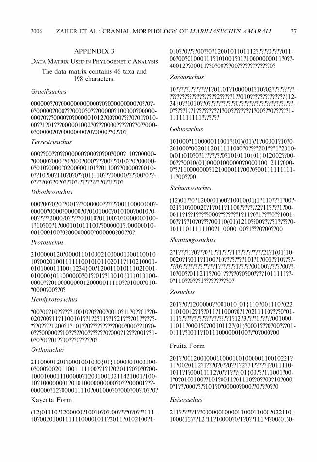
APPENDIX 3
DATA MATRIX USED IN PHYLOGENETIC ANALYSIS
The data matrix contains 46 taxa and198 characters.
Gracilisuchus
000000??0?000000000000?0?000000000?0??0?-0?00000?000???0000?0???00000?100000?00000-000?0???0000?0?000001012?00?00????0?01?010-00??1?01???000001002?0???0000?????0??0??000-0?00000?0?00000000?0?0000??0??0?
Terrestrisuchus
000??00??0??000000?000?0?00?000?110?00000-?00000?000??0?000?000????00???010??0?00000-0?010?0000?0200000101??01100??00000?0010-0??10?00?110?0?0??(01)110???00000????00?0??-0????00??0?0???0???????????0?????0?
Dibothrosuchus
000?00?020??001???000000??????00110000000?-00000?0000?00000?0?0101000?010100?0010?0-00?????2000?0?????01010?01100?0?0000000100-1?10?00?1?000101011100??000001??00000010-001000100?0?00000000?000000?00??0?
Protosuchus
2100000120?0000110100021000001000100010-10?00201001111110010101102011?110210001-010100011100{1234}00?120011010111021001-010000{01}000000?01??01??10010{01}010100-0000???0100000000120000011110??01000?010-?0000?00??0?
Hemiprotosuchus
?00?00?10??????10010?0??00?0010?11?0??01??0-020?00?11?1100101??1?2?11??1?21????01??????-???0????1200?1?101??0??????????000?000??10?0-0???00000??10?????00???????0?000?12???001??1-0?0?00?01??00???0?????0?
Orthosuchus
21100001201?0001001000{01}1000001000100-0?000?002011001111100??1?1?02011?0?0?0?00-1000100011100000?120010010211421001?100-10?100000001?01010000000000?0???00001???-000000?12?000011110?001000?0?000?00??0??0?
Kayenta Form
(12)01110?1200000?10010?0??00????0?0???111-10?002010011111100001011?2011?0102100?1-
010??0????00??0?1200101101112?????0????011-00?00?01000111?101001?01?10000000011?0??-40012??00011??0?00???00?????????????0?
Zaraasuchus
10?????????????1?01?01?1000001?10?02?????????-???????????????????2?????1??010??????????????{12-34}0??1010??0???????????0??????????????????????-0?????1??1?????????1?00????????1?00???0??????1-1111111111???????
Gobiosuchus
101000?110000011001?(01)(01)?1?00001?10?0-201000?0020112011111000?0????201???1?2010-0(01)010?0?1???????0?1010110{01}012002??00-00???0010(01)00001000000?00001001211?000-0???110000000?121000011?00?0?00111111111-11?00??00
Sichuanosuchus
(12)01??0?1200(01)00?10010(01)1?110???1?00?-021?10?00020?1?011?1100???????2?11????1?00-0011?1??1?????000????????1?11?0?1????0??1001-00??1??10?0????00110(01)1210??00?????1?????0-10111011111100?110000100?1???0?00??00
Shantungosuchus
2?1????1?0???0?1??1????11??????????21?1(01)10-0020?1?011?1100?10????????101?1?000??10????-???0??????????????1???????1?????00100??????00??-10?00??011211??001?????0?0?00????1011111??-0?110??0???1??????????0?
Zosuchus
201??0?1200000??001010{01}110?001110?022-11010012?1??011?11000?0?1?0211110????0?01-111????????????????????1?12?3????1?????001000-11011?0001?0?0010112?(01)?0001???0?00???01-0111??1011?10111000000100???0?000?00
Fruita Form
201??001200100010000100100000110010221?-11?0020112?1???0?0??0??1?2?31?????1?011110-1011?1?00011112?0??1???{01}00???1?1001?00-1?0?0100100??101?0011?01110??0??00?10?000-0?1???000????101?0?00000?000??0???0??0
Hsisosuchus
211??????1??000000100001100011000?022110-1000(12)??12?11?10000?0?1?0??111?4?00(01)0-
2006 ZAHER ET AL.: CRANIAL MORPHOLOGY OF MARILIASUCHUS AMARALI 37
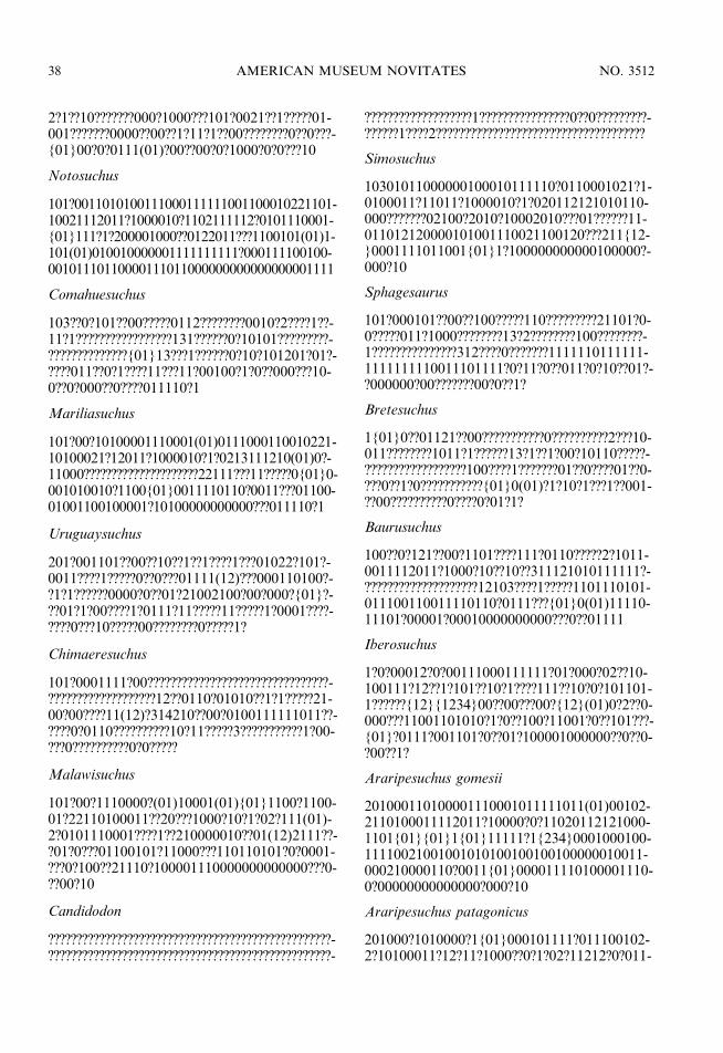
2?1??10???????000?1000???101?0021??1?????01-001???????0000??00??1?11?1??00????????0??0???-{01}00?0?0111(01)?00??00?0?1000?0?0???10
Notosuchus
101?0011010100111000111111001100010221101-10021112011?1000010?1102111112?0101110001-{01}111?1?200001000??0122011???1100101(01)1-101(01)010010000001111111111?000111100100-00101110110000111011000000000000000001111
Comahuesuchus
103??0?101??00?????0112????????0010?2????1??-11?1?????????????????131??????0?10101?????????-??????????????{01}13???1??????0?10?101201?01?-????011??0?1????11???11?00100?1?0??000???10-0??0?000??0????011110?1
Mariliasuchus
101?00?10100001110001(01)0111000110010221-10100021?12011?1000010?1?0213111210(01)0?-11000?????????????????????22111???11?????0{01}0-001010010?1100{01}0011110110?0011???01100-01001100100001?10100000000000???011110?1
Uruguaysuchus
201?001101??00??10??1??1????1???01022?101?-0011????1?????0??0???01111(12)???000110100?-?1?1??????0000?0??01?21002100?00?000?{01}?-??01?1?00????1?0111?11?????11?????1?0001????-????0???10?????00????????0?????1?
Chimaeresuchus
101?0001111?00????????????????????????????????-???????????????????12??0110?01010??1?1?????21-00?00????11(12)?314210??00?0100111111011??-????0?0110??????????10?11?????3???????????1?00-???0??????????0?0?????
Malawisuchus
101?00?1110000?(01)10001(01){01}1100?1100-01?22110100011??20???1000?10?1?02?111(01)-2?0101110001????1??210000010??01(12)2111??-?01?0???01100101?11000???110110101?0?0001-???0?100??21110?100001110000000000000???0-??00?10
Candidodon
??????????????????????????????????????????????????-??????????????????????????????????????????????????-
???????????????????1????????????????0??0?????????-??????1????2?????????????????????????????????????
Simosuchus
10301011000000100010111110?0110001021?1-0100011?11011?1000010?1?020112121010110-000???????02100?2010?10002010???01??????11-011012120000101001110021100120???211{12-}0001111011001{01}1?100000000000100000?-000?10
Sphagesaurus
101?000101??00??100?????110?????????21101?0-0?????011?1000????????13?2????????100????????-1???????????????312????0???????1111110111111-1111111110011101111?0?11?0??011?0?10??01?-?000000?00???????00?0??1?
Bretesuchus
1{01}0??01121??00???????????0??????????2???10-011????????1011?1??????13?1??1?00?10110?????-??????????????????100????1???????01??0????01??0-???0??1?0???????????{01}0(01)?1?10?1???1??001-??00??????????0????0?01?1?
Baurusuchus
100??0?121??00?1101????111?0110?????2?1011-0011112011?1000?10??10??311121010111111?-????????????????????12103????1?????1101110101-011100110011110110?0111???{01}0(01)11110-11101?00001?00010000000000???0??01111
Iberosuchus
1?0?00012?0?00111000111111?01?000?02??10-100111?12??1?101??10?1????111??10?0?101101-1??????{12}{1234}00??00???00?{12}(01)0?2??0-000???11001101010?1?0??100?11001?0??101???-{01}?0111?001101?0??01?100001000000??0??0-?00??1?
Araripesuchus gomesii
201000110100001110001011111011(01)00102-2110100011112011?10000?0?11020112121000-1101{01}{01}1{01}11111?1{234}0001000100-11110021001001010100100100100000010011-000210000110?0011{01}000011110100001110-0?00000000000000?000?10
Araripesuchus patagonicus
201000?1010000?1{01}000101111?011100102-2?10100011?12?11?1000??0?1?02?11212?0?011-
38 AMERICAN MUSEUM NOVITATES NO. 3512

{01}1??1?1??????????1000??0111100???01???0?-01?01101?010000??100110102?0??01????0??{0-1}1000111?0100001110?000000000000000?00-0?10
Lomasuchus
201????1211?00?11000101111??110001022?10-10001??12??1?100??1??1??2?21?????00??0(12)1-1????????????????????1???00???00?????0?00???1?1-10?00???00011?0??1??0??????010???0?11??10??0-1?1000??11000??0???0?0?0?10
Peirosaurus
201?011??1??00??????10?1????????0???2?10?????-??????????????????????1??????????(12)1???????????-?????????????000????????????0????1???0???????0?1-??????????????(01)??????0??????????????00??1????-??????0???????
Theriosuchus
20110111110100110000110111100110011?21-1010001?11?01111000?????1?20211?41001010-101101111100011112001001010002?00?10?1-10110(01)001?1100?00?0?00100??01??0?00??1-0100000?11?010??01?10000??0000??????0???0-?10
Alligatorium
?0??????1?0000?1000010?111??0?100?1????0??0-0??11??1??1000???????20?1????00101?101?0111-11000???1?00100???????????10??1???????????????-????0???????????????????????0??????????????????0?-?????????????????
Goniopholis
203?1211110010111000100111?0010001002?1-01000?1112011?1010?10?1?021312?4100{01}0-(12)02011?1??1??0?00?1200?11?000002100010-?1101101??101100?000010010001?1???000011-0020000011001000011110?01000000000000?0-00?10
Eutretauranosuchus
203????1?10010111000100111?00?0001001110-?000?1112011?1010??0?1?0?121204?00001020-111???1??0??0?1?????????000???00?????0?101???-?110???????0??00???1???0????10?2???001?0??000-?1?110?01?0000000???0?00??10
Pelagosaurus
202?1111110011020101000000000000(01)100-211010000001101111001001?10001200?30000-020000110111?0000001200011101?00???10???-????1?1??????0000??010010?0010???00????0001-000011201000011000001?0000000???0??00?10
Teleosauridae
(02)02?11111100110201001000000000001100-21?01000?001101111001011?1?00120003?000?-200002101111?0000?12000101011?0???10??01-001101??1011000011000010100?0??0000??100-01000011?010?0111000101000000000000??00?-10
Metriorhynchidae
(02)02?12110100111201011000?00000001100-21?0?000?001101111001011?1?001200?300010-200002101?11?0000??????0??012?0???100?0100-1101??1011?000??000010102?0???000????0010-00011?01000?1100000100000000???00?00?10
Sokotosuchus
2?2???1101??10????001001???101001?012?1????-??1112?11?1?11??0???1?1?0?????????01?????????-???????????????????????????????1????????????????0-??0????????????????????0?????????????????????????-????????????
Dyrosauridae
002??1?101?010?11?00100011?1010011012?10-101001112011?1011?10?10101302?3?00??2?00-0????????0?00??????1???????????????????1????????-????????0??0???????????????021?0001????0??????0-0??1?0000000???0??00?10
Pholidosaurus
212?111101??11?11101100111?00100010?211?-100001112111?101??10?100?1311?300???2?0??-?11?1???0??0??2?0?????????????????????1????1?11-0?????0?0010???????????????0?1?0001???10?001?-100?010???0??????0???0?10
Bernissartia
203??21111??00111000?00111?001000?002????-?0001112?11?10100?0?1???1?1??410010102011-?1?11??020021110110100000??00????????1????-1????????0??0?10???01???0????1?12000001????0?-?????00??10000????0??0???0?10
2006 ZAHER ET AL.: CRANIAL MORPHOLOGY OF MARILIASUCHUS AMARALI 39

Hylaeochampsa
00???????11???11????1?01???0????0?002?1?1011-?????????101??1??1?????10???????????????????????-???????????????????0???????10????????????0??0???-???0????????????2??00????????????????????????????-?0???0?1?
Borealosuchus
203?1211110010111000100111?001000100211-010111111211111010010?110?1310031000110-?011111111113111?110?00?000002110?100100-?101??11110??000000010001?1???0000110?20?-00011001000011100001000000000000???0?10
Gavialis
212?12111100111111011011111001000100211-0101101112011110110101110{01}131003100-012000001111110131112111100?000002110?1-
00100?101??121100?00000001000101?1?00001-?0?20?00011001000011100001?00000000000??-00?10
Crocodylus
203012111100(01)0111000102111100100010021-10?01111112011110100101110{01}13100310001-0010121111110131112021100?00000211001001-00?101??121100?0000000100110101100001?0?20-000011001000011100001000000000000?000?10
Alligator
203112?101?0001110001021111001000?0021101-01111112011110100101110{01}03120310001002-01211111111311120211?0?000002110010010011-01??111000?00000001001(12)01011000011(01)12-00000110010000111000010000000000000000?10
This paper meets the requirements of ANSI/NISO Z39.48-1992 (Permanence of Paper).
Complete lists of all issues of the Novitates and the Bulletin are available at World Wide
Web site http://library.amnh.org/pubs. Inquire about ordering printed copies via e-mail from
[email protected] or via standard mail from: American Museum of Natural History,
Library—Scientific Publications, Central Park West at 79th St., New York, NY 10024. TEL:
(212) 769-5545. FAX: (212) 769-5009.
40 AMERICAN MUSEUM NOVITATES NO. 3512