Embed Size (px)
Citation preview
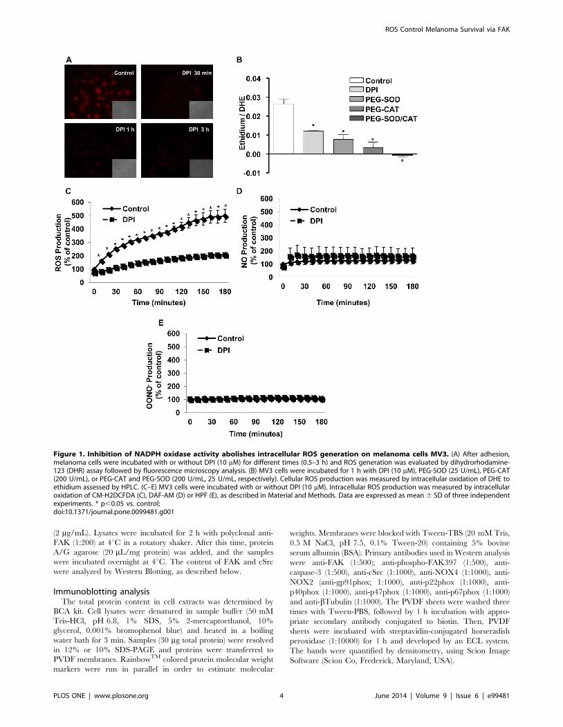
Redox Modulation of FAK Controls Melanoma Survival - Roleof NOX4Cristiane Ribeiro-Pereira1, Joao Alfredo Moraes1, Mariele de Jesus Souza1, Francisco R. Laurindo2, Maria
Augusta Arruda1,3, Christina Barja-Fidalgo1*
1 Laboratory of Cellular and Molecular Pharmacology, Department of Cell Biology, IBRAG, Universidade do Estado do Rio de Janeiro, Rio de Janeiro, RJ, Brazil, 2 Laboratory
of Vascular Biology, Instituto do Coracao, Universidade de Sao Paulo, Sao Paulo, SP, Brazil, 3 Vice-Diretoria de Ensino, Pesquisa e Inovacao, Farmanguinhos, Fiocruz, Rio de
Janeiro, RJ, Brazil
Abstract
Studies have demonstrated that reactive oxygen species (ROS) generated by NADPH oxidase are essential for melanomaproliferation and survival. However, the mechanisms by which NADPH oxidase regulates these effects are still unclear. In thiswork, we investigate the role of NADPH oxidase-derived ROS in the signaling events that coordinate melanoma cell survival.Using the highly metastatic human melanoma cell line MV3, we observed that pharmacological NADPH oxidase inhibitionreduced melanoma viability and induced dramatic cellular shape changes. These effects were accompanied by actincytoskeleton rearrangement, diminished FAKY397 phosphorylation, and decrease of FAK-actin and FAK-cSrc association,indicating disassembly of focal adhesion processes, a phenomenon that often results in anoikis. Accordingly, NADPHoxidase inhibition also enhanced hypodiploid DNA content, and caspase-3 activation, suggesting activation of theapoptotic machinery. NOX4 is likely to be involved in these effects, since silencing of NOX4 significantly inhibited basal ROSproduction, reduced FAKY397 phosphorylation and decreased tumor cell viability. Altogether, the results suggest thatintracellular ROS generated by the NADPH oxidase, most likely NOX4, transmits cell survival signals on melanoma cellsthrough the FAK pathway, maintaining adhesion contacts and cell viability.
Citation: Ribeiro-Pereira C, Moraes JA, Souza MdJ, Laurindo FR, Arruda MA, et al. (2014) Redox Modulation of FAK Controls Melanoma Survival - Role of NOX4. PLoSONE 9(6): e99481. doi:10.1371/journal.pone.0099481
Editor: Masuko Ushio-Fukai, University of Illinois at Chicago, United States of America
Received August 19, 2013; Accepted May 15, 2014; Published June 9, 2014
Copyright: � 2014 Ribeiro-Pereira et al. This is an open-access article distributed under the terms of the Creative Commons Attribution License, which permitsunrestricted use, distribution, and reproduction in any medium, provided the original author and source are credited.
Funding: This work was supported by CNPq (www.cnpq.br), CAPES (www.capes.gov.br), FAPERJ (www.faperj.br) and Sub-reitoria de Pos-Graduacao e Pesquisa(SR-2/UERJ - www.sr2.uerj.br). M.A. Arruda is a L9Oreal-UNESCO-ABC For Women In Science National Fellowship – 2008 awardee. The funders had no role in studydesign, data collection and analysis, decision to publish, or preparation of the manuscript.
Competing Interests: The authors have declared that no competing interests exist.
* E-mail: [email protected]
Introduction
Melanoma arises from the malignant transformation of
pigment-producing cells (melanocytes), and its incidence has
increased in many countries, being a prominent worldwide public
health challenge [1–3].
Development of skin cancer is a multistage process mediated by
different cellular, biochemical and molecular changes, involving
the activation of several anti-apoptotic and pro-survival signaling
pathways. Though, a critical step in melanoma biology is its ability
to overcome anchorage dependency, acquiring ‘vertical growth
phase’ (VGP) properties, enabling these cells to enter the deeper
dermis rather than growing only in or adjacent to the epidermis.
VGP can therefore make melanoma cells competent to metastasis.
The metastatic ability of these cells is closely related to a
rearrangement on the integrin-coordinated signaling hierarchy
[4,5].
Epidemiological studies have demonstrated that the major risk
factors for melanoma relate to both environmental exposure and
genetic alterations. For example, melanoma incidence in white
populations has revealed an inverse correlation with latitude and
positive correlation with ultraviolet radiation (UVR) index [6–7].
Skin exposure to UVR generates ROS in excessive quantities [8].
However, rather than this occurring as a direct effect of UVR, it
has been shown that the observed ROS accumulation also relies
on ROS generated by highly specialized enzymatic systems [9].
ROS are classically referred as cytotoxic agents due to their
ability to oxidize biomolecules [10]. However, the direct cell
damage only occurs when their generation is greatly increased and
the antioxidant mechanisms are overwhelmed, a condition defined
as ‘‘oxidative stress’’ [11]. On the other hand, a growing body of
reports shows that rather than being hazardous molecules, ROS
are second messengers, able to modulate a number of signaling
pathways, many of them involved in tumor development [12,13].
Among all intracellular ROS-generating systems, the most
specialized one is a family of multimeric enzymes called NADPH
oxidase [14]. NADPH oxidase was primarily described in
neutrophils where they exert a critical role in innate immunity,
taking part in the killing of pathogens [15]. NADPH oxidase
activity was also detected in other cell types, involving other
homologous of the main membrane subunit, NOX. To date, five
NOXs have been described (NOX1 – NOX5) [16].
In non-phagocytic cells, NADPH oxidase activity leads to the
generation of ROS, which seems to modulate diverse intracellular
signaling pathways [17,18]. While in most cell types NADPH
oxidase-dependent ROS generation is triggered and/or stimulated
by agonists, many malignant cells constitutively produce ROS in
an augmented fashion [19,20]. Recent works have pointed to a
PLOS ONE | www.plosone.org 1 June 2014 | Volume 9 | Issue 6 | e99481
critical role for ROS generated by NADPH oxidase in the
initiation and progression phases of malignant cells development
[21–24]. For example, NOX4 and NOX5 activities have been
reported to be involved in the survival of human glioma,
melanoma, and prostate cancer cells [25–27]. These findings
suggest that the modulation of survival and proliferation signaling
by ROS plays a critical role in cancer development. However, the
mechanisms by which NADPH oxidase regulates these signaling
pathways are not fully understood.
In this study, we aimed to characterize the role of endogenously
produced ROS in MV3 cells, a highly aggressive human
melanoma cell line [28]. Using both pharmacological and gene
silencing approaches, we investigated the role of NADPH oxidase
activity on cell fate and signaling. Our findings strongly suggest
that constitutive NADPH oxidase activity stimulates FAK
signaling pathway and protects melanoma cells from death.
Furthermore, our data shed light on NOX4 as a novel
pharmacological target for the control of melanoma growth and
metastasis.
Material and Methods
Ethics StatementEthical approval was obtained from the Ethics Committee of
Pedro Ernesto Hospital, Rio de Janeiro State University (2786/
2010) and all volunteers gave written informed consent for
participation before enrollment in the study.
ReagentsHEPES, ethylenediaminetetraacetate (EDTA), bovine serum
albumin (BSA), penicillin, streptomycin, dihydrorhodamine 123
(DHR), phenylmethylsulfonyl fluoride (PMSF), benzamidine,
leupeptin, and soybean trypsin inhibitor (SBTI), sodium orthova-
nadate (Na3VO4), apocynin (4-acetovanillone), 3-(4,5-di-
methylthiazol-2-yl)-2-5-diphenol tetrazolium bromide (MTT),
pyrrolidine dithiocarbamate (PDTC), diphenyleneiodonium
(DPI), TRITC-labelled phalloidin, cycloheximide (CHX), ribonu-
clease A (RNase A), propidium iodide (PI), diethylenetriamine
pentaacetic acid (DTPA), RPMI-1640 medium, superoxide
dismutase conjugated to polyethylene glycol (PEG-SOD), catalase
conjugated to PEG (PEG-CAT) and sulforhodamine B were from
Sigma-Aldrich (St. Louis, MO). Triton X-100, Percoll, PVDF
membranes, RainbowTM were from GE Healthcare (San
Francisco, CA). Fetal bovine serum (FBS) was purchased from
Cultilab (Campinas, SP, Brazil). Dulbecco’s Modified Eagle
Medium (DMEM), Dihydroethidium (DHE), anti- p-FAK397
and Lipofectamine 2000 were obtained from Invitrogen (Carlsbad,
CA). DAF-FMDA, CM-H2DCFDA, HPF and JC-1 were
obtained from Molecular Probes (Carlsbad, CA). siRNA oligomers
for NOX4 (59-CCTCAGCATCTGTTCTTAACCTCAA-39) and
its scrambled sequence (Scramble) were obtained using BLOCK-
iTTM RNAi Designer (Invitrogen). Antibody anti-caspase-3 was
obtained from Cell Signaling. All other antibodies and protein A/
G agarose were from Santa Cruz Biotechnology (Santa Cruz, CA).
Streptavidin-conjugated FITC and Streptavidin-conjugated horse-
radish peroxidase were from Caltag Laboratories. ECL system
(SuperSignal West Pico chemiluminescent substrate kit) was from
Pierce Biotechnology (Rockford, IL, USA). High capacity cDNA
reverse transcripition kit RNeasy, RNeasy Mini kit were from
Qiagen, RQ1 RNase-Free DNase, the set of dN TP and RNasin
RNase inhibitor were purchased from Promega (Madison, WI).
Cell CultureMV3 human melanoma cell line [28] was a gift from Dr Cezary
Marcienkiewicz (Center for Neurovirology and Cancer Biology,
Temple University, PA, USA). MV3 cells were maintained in
DMEM enriched with 10% FBS, 3.7 g/L sodium bicarbonate,
5.2 g/L HEPES, 0.5 U/mL penicillin and 0.5 mg/mL strepto-
mycin at 37uC in a humidified atmosphere of 5% CO2. Cells were
grown to 80-90% confluence into 75 cm culture flasks and were
detached by brief treatment with 5 mM EDTA in Hank’s
balanced salt solution (HBSS).
Measurement of ROS generation in intact cells by DHRIntracellular ROS production by MV3 cells was measured by
oxidation of dihydrorhodamine 123 (DHR) to rhodamine by
H2O2 as previously described [29]. Briefly, MV3 cells were seeded
on 96-well plates at a density of 66103 cells/well and, after
overnight incubation, the medium was replaced by serum-free
medium containing DHR (final concentration 50 mM). Cells were
then washed with phosphate-buffered saline (PBS) before exam-
ination under an Olympus IX71 inverted microscope (Tokyo,
Japan) equipped for fluorescence.
Cellular ROS measurement by HPLCEthidium and 2-hydroxyethidium (EOH), the oxidation prod-
ucts of DHE, were separated by HPLC as described above [30].
Briefly, human melanoma (46105 cells) cells were grown in 6-well
dishes in DMEM medium supplemented with fetal bovine serum
(10%). After adhesion, melanoma cells were incubated with DPI
(10 mM) or SOD conjugated to polyethylene glycol (PEG-SOD,
25 U/mL), catalase conjugated to PEG (PEG-CAT, 200 U/mL),
or both PEG-CAT (200 U/mL) and PEG-SOD (25 U/mL) for
1 hour at 37uC in a 5% CO2 atmosphere. Cells were washed twice
with Krebs (0.5 mM CaCl2, 1.2 mM MgSO4, 4.9 mM KCl, 5.7
KH2PO4, 145 mM NaCl, 5.7 mM Na2HPO4 and glucose
5.5 mM pH 7.4) and incubated in Krebs, containing DTPA
(100 mM) at a final DHE concentration of 50 mM for additional
30 min. Cells were washed with Krebs, harvested in acetonitrile
(0.5 mL/well) and centrifuged (12,000 g for 10 min at 4uC).
Supernatants were dried under vacuum (Speed Vac Plus model
SC-110A, Thermo Savant) and pellets maintained at 270uC in
the dark until analysis. Samples were ressuspended in 60 mL in
solution A (water/10% acetonitrile/0.1% trifluoracetic acid) and
injected (50 mL) into HPLC system.
Real-time intracellular ROS productionMV3 melanoma cells (and MV3 cells silenced with siRNA
scramble or siRNA NOX4, when mentioned) were detached by
brief treatment with 5 mM EDTA in HBSS, collected by
centrifugation, ressuspended in fresh 10% FBS DMEM medium
and incubated overnight on 96-well black plates at a density of
66103 cells/well, at 37uC in a humidified atmosphere of 5% CO2.
The cells were washed three times with PBS, incubated in HBSS
at 37uC, 5% CO2 for 1 h. Cells were then loaded for 1 h with one
of the intracellular ROS detection probes depicted below (final
concentration 5 mM). Once loaded, cells were washed with PBS
and treated or not with DPI. Fluorescence intensity was assessed
throughout 3 h using in the microplate reader EnvisionTM.
DAF-FM DA assay. NO production was assessed by the
fluorescence emitted by oxidized DAF, an specific probe for NO
detection. Fluorescence was monitored at excitation and emission
wavelengths of 495 nm and 515 nm, respectively.
CM-H2DCF DA assay. ROS production was detected
through fluorescence emitted from DCF oxidation. This probe
ROS Control Melanoma Survival via FAK
PLOS ONE | www.plosone.org 2 June 2014 | Volume 9 | Issue 6 | e99481

detects ROS in general, being more selective to H2O2 and
peroxynitrite. Fluorescence was monitored at excitation and
emission wavelengths of 495 nm and 525 nm, respectively.
HPF assay. Peroxynitrite (ONOO-) production was deter-
mined by monitoring the fluorescence resulted from HPF
oxidation. This probe, which is selective for ONOO-, had its
fluorescence detected at excitation and emission wavelengths of
490 nm and 515 nm, respectively.
Cell viability assaysMTT assay. MV3 melanoma cells were detached by brief
treatment with 5 mM EDTA in HBSS, collected by centrifuga-
tion, ressuspended in fresh 10% FBS medium DMEM and placed
into 96-well plates at a density of 66103 cells/well. After adhesion,
cells were incubated in the presence or absence of DPI (0.1–
10 mM), Na3VO4 (0.1–3 mM), apocynin (1–10 mM) or PDTC
(0.1–1 mM), at 37uC in humidified 5% CO2. After 48 hours of
incubation or after 48, 72 or 96 hours of siRNA transfection,
MTT assay was performed as previously described [31]. Cells
were incubated with MTT (50 mg/well) in the dark at 37uC for
4 hours, when MTT is reduced to formazan crystals by viable
cells. After incubation, the formazan crystals were dissolved in
isoprapanol and the optical densitometry obtained using a
microplate reader (BIO-RAD) using a 570 nM filter. Results are
shown as percentage of control, of three independent experiments
performed in quintuplicate.
Sulforhodamine B assay. MV3 melanoma cells were
detached by brief treatment with 5 mM EDTA in HBSS, collected
by centrifugation, ressuspended in fresh 10% FBS medium
DMEM and placed into 96-well plates at a density of 66103
cells/well. After adhesion, cells were incubated in the presence or
absence of DPI (10 mM), apocynin (10 mM) or cycloheximide
(5 mM), at 37uC in humidified 5% CO2. After 48 hours of
incubation, or after 48, 72 or 96 hours of siRNA transfection, the
medium was removed, and cells were fixed with cold 10%
trichloracetic acid (TCA) for 1 hour at 4uC. Plates were washed 5
times with Milli-Q water and left to dry at room temperature.
Cells were stained with 0.4% of sulforhodamine B (w/v) in 1%
acetic acid (v/v) at room temperature for 10 minutes. sulforho-
damine B was removed, and the plates were washed 56with 1%
acetic acid before air-drying. Bound dye was solubilized with
10 mM unbuffered Tris-base solution, and plates were left on a
plate shaker for at least 10 minutes. Absorbance was measured in a
96-well plate reader (BIO-RAD), at 490 nm.
Immunocytochemistry and cytochemistry assaysFor immunofluorescence microscopy, MV3 cells were grown on
glass coverslips (9.06103 cells/well). After adhesion, MV3 mela-
noma cells were incubated in the presence or absence of DPI
(10 mM) for 30 min, 2 and 4 h at 37uC and 5% CO2 atmosphere.
Following treatments, cells were washed with ice-cold PBS and
fixed with PBS containing 4% paraformaldehyde/4% sucrose for
20 min at room temperature and permeabilized in PBS containing
0.2% Triton X-100 for 5 min. Then, cells were incubated with
PBS-BSA for 30 min. For detection of FAK, permeabilized cells
were incubated with anti-FAK antibody (1:200) overnight at 4uCand then sequentially incubated with goat anti-IgG Ab biotin-
conjugated for 1 h and streptavidin-conjugated FITC (1:200) for
1 h. Filamentous actin was stained with rhodamin-conjugated
phalloidin (1:1000) for 2 h at room temperature. Coverlips were
mounted onto microscope slides using a solution of 20 mM N-
propylgallate and 20% glycerol in PBS. Microscopic analysis of
FAK- and phalloidin-stained cells were carried out using a laser
scanning confocal microscope (Olympus - Fluoview version 3.3).
Apoptosis detectionHypodiploid DNA content analysis. MV3 cells were
detached by brief treatment with 5 mM EDTA in HBSS, collected
by centrifugation and placed into 6-well plates at a density of
56105 cells/well in 10% FBS medium. After adhesion, melanoma
cells were incubated in the presence or absence of DPI (10 mM),
apocynin (10 mM) or cycloheximide (5 mM) for 24 h. Thereafter,
cells detached with 5 mM EDTA in HBSS and collected by
centrifugation were ressuspended in PBS, fixed with ethanol 70%
for 2 h and incubated in PBS solution containing 200 mg/mL
RNase A, 0.001% Triton X-100, 20 mg/mL PI for 30 min at
room temperature. DNA contents in stained nuclei were analyzed
with FACScan (Becton Dickinson). A suspension of cells was
analyzed for each DNA histogram, and the percentage of cells in
G0/G1, S, and G2/M phases was determined using the WINMDI
program.
Experiments using RNA interferenceNOX4 gene expression was repressed using RNA interference
technology according to Lipofectamine 2000 manufacturer’s
protocol. MV3 cells were seeded in 6-well (2.56105/well, for
western blotting or qRTPCR) or 96-well (56103, for ROS
production kinectics analysis, MTT, sulforhodamine B and JC-1
assay plates. The transfection efficiency was determined with
qRT-PCR and Western blotting.
Assessment of mitochondrial transmembrane potential(JC-1)
The mitochondrial stability was measured by the use of the
cationic dye JC-1, which is incorporated to the mitochondrial
intermembrane space. The monomer (green) can polymerize
forming clusters known as J-aggregates (red) in a transmembrane
potential-dependent manner. Therefore, viable, non-apoptotic
cells exhibit a higher red/green ratio [32]. MV3 melanoma cells
were seeded overnight into 96-well black plates at a density of
56103 cells/well. Cells were then, treated for 24 h with scramble
siRNA or NOX4 siRNA. After that, cells were washed with PBS
and stained with JC-1 (10 mg/mL) for 30 min. Mitochondrial
transmembrane potential was monitored using EnvisionTM
multilable plate reader. Red fluorescence intensity was assessed
by excitation and emission at 560 nm and 595 nm, respectively
and green fluorescence intensity was detected by excitation and
emission at 485 nm and 535 nm, respectively. Mitochondrial
transmembrane potential was expressed as the ratio red/green
fluorescence.
Total cell extractsAfter the incubation, the cells were suspended in lysis buffer
(HEPES 20 mM, pH 7.9; glicerol 20% (v/v); NP-40 1% (v/v);
MgCl2 1 mM; EDTA 0.5 mM; EGTA 0.1 mM; DTT 0.5 mM,
Na3VO4 1 mM) and the following protease inhibitors: PMSF
(1 mM), aprotinin (2 mg/mL), leupeptin (2 mg/mL) and SBTI
(2 mg/mL).
ImmunoprecipitationAfter adhesion, MV3 cells (56105 cells) were incubated in the
presence or absence of DPI (10 mM) for 2 and 4 h at 37uC in a 5%
CO2 atmosphere. The cells were lysed with RIPA buffer (50 mM
Tris-HCl, pH 8.0, 150 mM NaCl, 1% Sodium Desoxicolate w/v,
EDTA 5 mM; 1% Triton X-100 v/v, 50 mM NaF, 1 mM
Na3VO4, 30 mM Na4P2O7, 10 mM Iodocetamide, 2 mM DTT)
supplemented with the following protease inhibitors: PMSF
(1 mM), aprotinin (2 mg/mL), leupeptin (2 mg/mL) and SBTI
ROS Control Melanoma Survival via FAK
PLOS ONE | www.plosone.org 3 June 2014 | Volume 9 | Issue 6 | e99481
(2 mg/mL). Lysates were incubated for 2 h with polyclonal anti-
FAK (1:200) at 4uC in a rotatory shaker. After this time, protein
A/G agarose (20 mL/mg protein) was added, and the samples
were incubated overnight at 4uC. The content of FAK and cSrc
were analyzed by Western Blotting, as described below.
Immunoblotting analysisThe total protein content in cell extracts was determined by
BCA kit. Cell lysates were denatured in sample buffer (50 mM
Tris-HCl, pH 6.8, 1% SDS, 5% 2-mercaptoethanol, 10%
glycerol, 0.001% bromophenol blue) and heated in a boiling
water bath for 3 min. Samples (30 mg total protein) were resolved
in 12% or 10% SDS-PAGE and proteins were transferred to
PVDF membranes. RainbowTM colored protein molecular weight
markers were run in parallel in order to estimate molecular
weights. Membranes were blocked with Tween-TBS (20 mM Tris,
0.5 M NaCl, pH 7.5, 0.1% Tween-20) containing 5% bovine
serum albumin (BSA). Primary antibodies used in Western analysis
were anti-FAK (1:500); anti-phospho-FAK397 (1:500), anti-
caspase-3 (1:500), anti-cSrc (1:1000), anti-NOX4 (1:1000), anti-
NOX2 (anti-gp91phox; 1:1000), anti-p22phox (1:1000), anti-
p40phox (1:1000), anti-p47phox (1:1000), anti-p67phox (1:1000)
and anti-bTubulin (1:1000). The PVDF sheets were washed three
times with Tween-PBS, followed by 1 h incubation with appro-
priate secondary antibody conjugated to biotin. Then, PVDF
sheets were incubated with streptavidin-conjugated horseradish
peroxidase (1:10000) for 1 h and developed by an ECL system.
The bands were quantified by densitometry, using Scion Image
Software (Scion Co, Frederick, Maryland, USA).
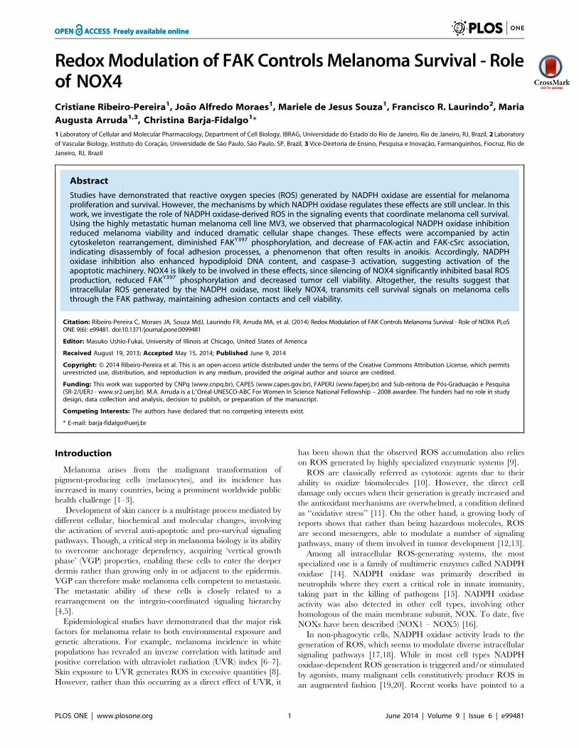
Figure 1. Inhibition of NADPH oxidase activity abolishes intracellular ROS generation on melanoma cells MV3. (A) After adhesion,melanoma cells were incubated with or without DPI (10 mM) for different times (0.5–3 h) and ROS generation was evaluated by dihydrorhodamine-123 (DHR) assay followed by fluorescence microscopy analysis. (B) MV3 cells were incubated for 1 h with DPI (10 mM), PEG-SOD (25 U/mL), PEG-CAT(200 U/mL), or PEG-CAT and PEG-SOD (200 U/mL, 25 U/mL, respectively). Cellular ROS production was measured by intracellular oxidation of DHE toethidium assessed by HPLC. (C–E) MV3 cells were incubated with or without DPI (10 mM). Intracellular ROS production was measured by intracellularoxidation of CM-H2DCFDA (C), DAF-AM (D) or HPF (E), as described in Material and Methods. Data are expressed as mean 6 SD of three independentexperiments. * p,0.05 vs. control;doi:10.1371/journal.pone.0099481.g001
ROS Control Melanoma Survival via FAK
PLOS ONE | www.plosone.org 4 June 2014 | Volume 9 | Issue 6 | e99481
Isolation of human neutrophilsHuman neutrophils were isolated from 0.5% EDTA treated
peripheral venous blood of healthy volunteers, using a four-step
discontinuous Percoll gradient [33]. Erythrocytes were removed
by hypotonic lysis. Isolated neutrophils (98% purity), estimated to
be at least 96% viable by trypan blue dye exclusion, were
ressuspended in RPMI-1640 medium.
RNA isolation and RT-PCRTotal RNA from MV3 melanoma cells (56105 cells), NGM
melanocytes (were obtained by cell bank of Rio de Janeiro) (56105
cells) and human neutrophils (26106 cells) were isolated using
RNeasy Mini kit. After DNase treatment (RQ1 RNase-Free
DNase), the mRNA was reverse transcribed using high capacity
cDNA reverse transcripition kit. Primers based on the sequence of
human p47phox (GeneBank accession nu NM_000265). The
following primers were used to amplify p47phox cDNA: sense, 59
– ATGAGCCTGCCCACCAAGAT - 39 (374–393), and anti-
sense, 59 – TCGAGGAAGGATGCTCCCAT - 39 (683–702).
The expected size of the p47phox PCR product was 328 bp. PCR
was performed with the following parameters: 95uC for 5 min for
1 cycle and 32 cycles of denaturation at 95uC for 45 s, annealing
at 58uC for 30 s, and elongation at 72uC for 30 s. The following
primers were used to amplify NOX4 cDNA: sense, 59 –
TCACAGAAGGTTCCAAGCAG - 39 (491–510), and antisense,
59- CTGTATTTTCTCAGGCGTGC - 39 (571–590). The
expected size of the NOX4 PCR product was 91 bp. PCR was
performed with the following parameters: One cycle of 95uC for
3 min followed by 35 cycles of denaturation at 95uC for 45 s,
annealing at 59uC for 45 s, and elongation at 72uC for 30 s.
GAPDH primers were used to validate the cDNA in each
reaction. PCR products were separated by 2% agarose gel
electrophoresis and visualized by UV exposure on transillumina-
tor.
For qPCR assay the PCR products were obtained using a
GeneAmo PCR System 2400 (Perkin Elmer). The Quantitative
real time PCR was performed in a Rotor gene Q using a SYBR-
green fluorescence quantification system (Qiagen) to quantify
amplicons. The standard PCR conditions were 95u for 5 minutes,
then 35 cycles at 95uC (5 s) and 60uC (10 s) followed by the
standard denaturation curve. Before normalizing the values we
performed DDCT in function of actin gene expression.
Statistical analysisStatistical significance was assessed by the two-tailed unpaired
Student’s t test. Data were log-transformed when required.
Differences were considered statistically significant when p#
0.05. The data were analyzed using GraphPad Prism version 5.00
for Windows (GraphPad Software, USA).
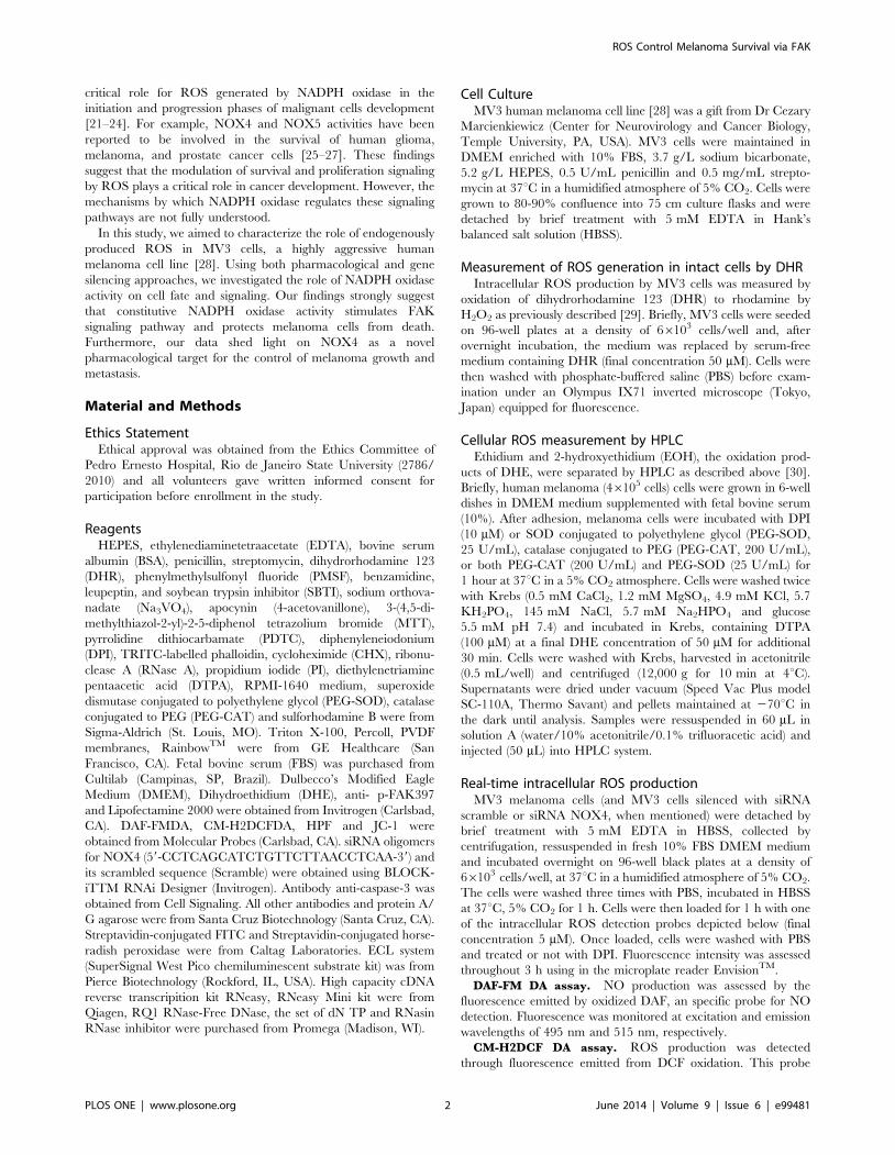
Figure 2. Inhibition of NADPH oxidase activity reduces survival of human melanoma cells MV3. (A–B) MV3 cells (66103) were incubatedfor 48 h with or without DPI (A; 0.1–10 mM) or apocynin (B; 1–10 mM), and MTT assay was performed as indicated in Material and Methods. (C) MV3cells (66103) were incubated for 48 h with or without DPI (10 mM), apocynin (10 mM) or Cycloheximide (5 mM). Subsequently, sulforhodamine-B assaywas performed as described in Materials and Methods. Results are shown as percentage of control and expressed as mean 6 SD of at least threeindependent experiments performed in quintuplicate. *p,0.05 vs. control.doi:10.1371/journal.pone.0099481.g002
ROS Control Melanoma Survival via FAK
PLOS ONE | www.plosone.org 5 June 2014 | Volume 9 | Issue 6 | e99481
Results
Constitutive ROS generation by MV3 melanoma cellsrequires NADPH oxidase activity
It has already been described that some melanoma cell lines can
produce intracellular ROS in a NADPH oxidase-dependent
manner [26]. We have observed, for the first time constitutive
intracellular ROS generation by the human melanoma cell line
MV3, using the dihydrorhodamine (DHR) assay. The non-
fluorescent DHR is oxidized to the fluorescent rhodamine,
indicating an intracellular accumulation of ROS (Fig. 1A). The
fluorescence intensity dramatically diminished when cells were
pre-incubated with the flavoprotein inhibitor DPI (10 mM;
Fig. 1A), which selectively inhibits NADPH oxidase activity in
the concentration range used in this study. Additionally, we have
also assessed intracellular ROS generation monitoring dihy-
droethidium (DHE) conversion to ethidium. Results shown in
Figure 1B suggest that superoxide and hydrogen peroxide are the
major reactive oxygen species produced by MV3 cells, since the
treatment with SOD and catalase impaired ethidium accumula-
tion. Furthermore, the inhibition by DPI confirms that ROS
generation by MV3 cells depends on NADPH oxidase activation
(Fig. 1B).
In order to determine which ROS are constitutively produced
by MV3 cells, we employed selective probes for different reactive
oxygen and nitrogen species. MV3 cells constitutively produce
considerable amounts of ROS, in a NADPH oxidase-dependent
manner (Fig. 1C) and low amounts NO, which was DPI-insensitive
(Fig. 1D). No detectable amounts of ONOO2 were generated by
MV3 cells in basal conditions (Fig. 1E). These results strongly
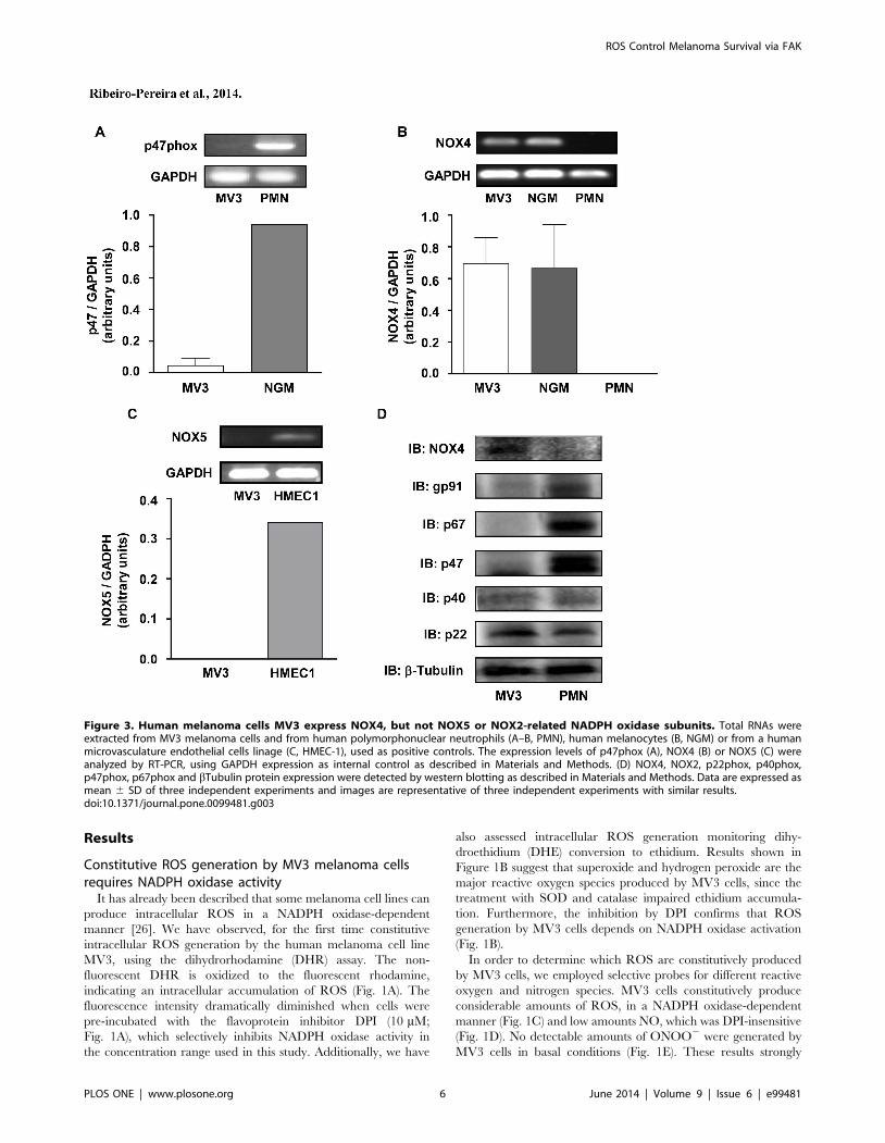
Figure 3. Human melanoma cells MV3 express NOX4, but not NOX5 or NOX2-related NADPH oxidase subunits. Total RNAs wereextracted from MV3 melanoma cells and from human polymorphonuclear neutrophils (A–B, PMN), human melanocytes (B, NGM) or from a humanmicrovasculature endothelial cells linage (C, HMEC-1), used as positive controls. The expression levels of p47phox (A), NOX4 (B) or NOX5 (C) wereanalyzed by RT-PCR, using GAPDH expression as internal control as described in Materials and Methods. (D) NOX4, NOX2, p22phox, p40phox,p47phox, p67phox and bTubulin protein expression were detected by western blotting as described in Materials and Methods. Data are expressed asmean 6 SD of three independent experiments and images are representative of three independent experiments with similar results.doi:10.1371/journal.pone.0099481.g003
ROS Control Melanoma Survival via FAK
PLOS ONE | www.plosone.org 6 June 2014 | Volume 9 | Issue 6 | e99481
suggest that NADPH oxidase activity is the major source of ROS
in resting MV3 melanoma cells.
NADPH oxidase-derived ROS are involved in MV3melanoma cells survival
As observed in other melanoma cell lines [26], MV3 melanoma
cells are highly sensitive to NADPH oxidase inhibition. DPI
inhibits cell survival in a concentration-dependent manner, as
assessed by MTT (Fig. 2A) and Sulforhodamine B (Fig. 2C) assays.
However, apocynin, an inhibitor of the cytosolic subunit p47phox
coupling to NOX2 [34] has no effect on MV3 survival (Fig. 2B
and 2C), indicating that the production of ROS by MV3 cells is
not related to NOX2 activity. Confirming the irrelevance of
NOX2 in these effects, MV3 cells do not express p47phox, which
is essential to NOX2 activity (Fig. 3A). We have also observed that
MV3 cells express high levels of NOX4 mRNA (Fig. 3B).
Furthermore, these melanoma cells do not express NOX5,
another NADPH oxidase isoform, which does not depend on
any of the classical cytosolic NADPH oxidase subunits and is
present in endothelial cells (Fig. 3C). We also analyzed NOX
subunits expression and we confirmed that MV3 expresses
negligible levels of p40phox, p47phox, p67phox and NOX2
(components of the NOX2 NADPH oxidase complex). On the
other hand, MV3 expresses NOX4 and p22phox (Fig. 3D). These
results indicated that neither NOX2 nor NOX5 contribute for
ROS production and strongly suggest that NOX4 is probably the
major source of endogenous ROS in MV3 melanoma cell line.
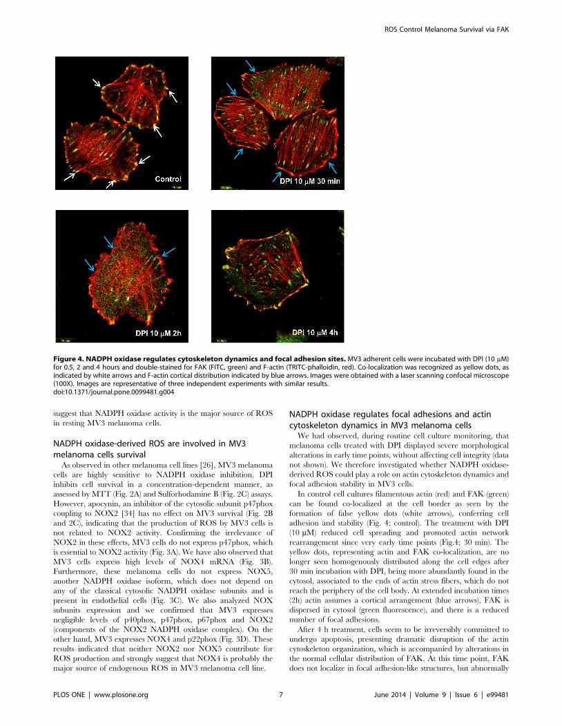
NADPH oxidase regulates focal adhesions and actincytoskeleton dynamics in MV3 melanoma cells
We had observed, during routine cell culture monitoring, that
melanoma cells treated with DPI displayed severe morphological
alterations in early time points, without affecting cell integrity (data
not shown). We therefore investigated whether NADPH oxidase-
derived ROS could play a role on actin cytoskeleton dynamics and
focal adhesion stability in MV3 cells.
In control cell cultures filamentous actin (red) and FAK (green)
can be found co-localized at the cell border as seen by the
formation of false yellow dots (white arrows), conferring cell
adhesion and stability (Fig. 4; control). The treatment with DPI
(10 mM) reduced cell spreading and promoted actin network
rearrangement since very early time points (Fig.4; 30 min). The
yellow dots, representing actin and FAK co-localization, are no
longer seen homogenously distributed along the cell edges after
30 min incubation with DPI, being more abundantly found in the
cytosol, associated to the ends of actin stress fibers, which do not
reach the periphery of the cell body. At extended incubation times
(2h) actin assumes a cortical arrangement (blue arrows), FAK is
dispersed in cytosol (green fluorescence), and there is a reduced
number of focal adhesions.
After 4 h treatment, cells seem to be irreversibly committed to
undergo apoptosis, presenting dramatic disruption of the actin
cytoskeleton organization, which is accompanied by alterations in
the normal cellular distribution of FAK. At this time point, FAK
does not localize in focal adhesion-like structures, but abnormally
Figure 4. NADPH oxidase regulates cytoskeleton dynamics and focal adhesion sites. MV3 adherent cells were incubated with DPI (10 mM)for 0.5, 2 and 4 hours and double-stained for FAK (FITC, green) and F-actin (TRITC-phalloidin, red). Co-localization was recognized as yellow dots, asindicated by white arrows and F-actin cortical distribution indicated by blue arrows. Images were obtained with a laser scanning confocal microscope(100X). Images are representative of three independent experiments with similar results.doi:10.1371/journal.pone.0099481.g004
ROS Control Melanoma Survival via FAK
PLOS ONE | www.plosone.org 7 June 2014 | Volume 9 | Issue 6 | e99481
accumulates at non-specialized sites of the cell membrane. Those
alterations precede cell detachment and consequent cell death.
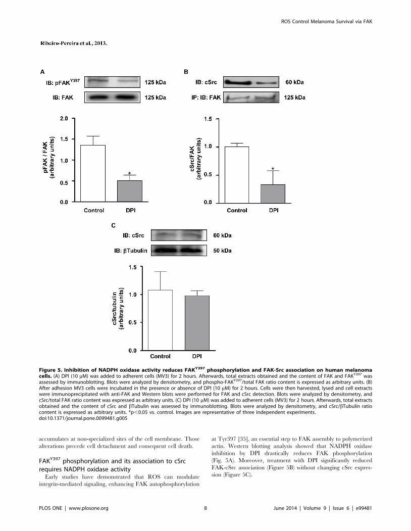
FAKY397 phosphorylation and its association to cSrcrequires NADPH oxidase activity
Early studies have demonstrated that ROS can modulate
integrin-mediated signaling, enhancing FAK autophosphorylation
at Tyr397 [35], an essential step to FAK assembly to polymerized
actin. Western blotting analysis showed that NADPH oxidase
inhibition by DPI drastically reduces FAK phosphorylation
(Fig. 5A). Moreover, treatment with DPI significantly reduced
FAK-cSrc association (Figure 5B) without changing cSrc expres-
sion (Figure 5C).
Figure 5. Inhibition of NADPH oxidase activity reduces FAKY397 phosphorylation and FAK-Src association on human melanomacells. (A) DPI (10 mM) was added to adherent cells (MV3) for 2 hours. Afterwards, total extracts obtained and the content of FAK and FAKY397 wasassessed by immunoblotting. Blots were analyzed by densitometry, and phospho-FAKY397/total FAK ratio content is expressed as arbitrary units. (B)After adhesion MV3 cells were incubated in the presence or absence of DPI (10 mM) for 2 hours. Cells were then harvested, lysed and cell extractswere immunoprecipitated with anti-FAK and Western blots were performed for FAK and cSrc detection. Blots were analyzed by densitometry, andcSrc/total FAK ratio content was expressed as arbitrary units. (C) DPI (10 mM) was added to adherent cells (MV3) for 2 hours. Afterwards, total extractsobtained and the content of cSrc and bTubulin was assessed by immunoblotting. Blots were analyzed by densitometry, and cSrc/bTubulin ratiocontent is expressed as arbitrary units. *p,0.05 vs. control. Images are representative of three independent experiments.doi:10.1371/journal.pone.0099481.g005
ROS Control Melanoma Survival via FAK
PLOS ONE | www.plosone.org 8 June 2014 | Volume 9 | Issue 6 | e99481
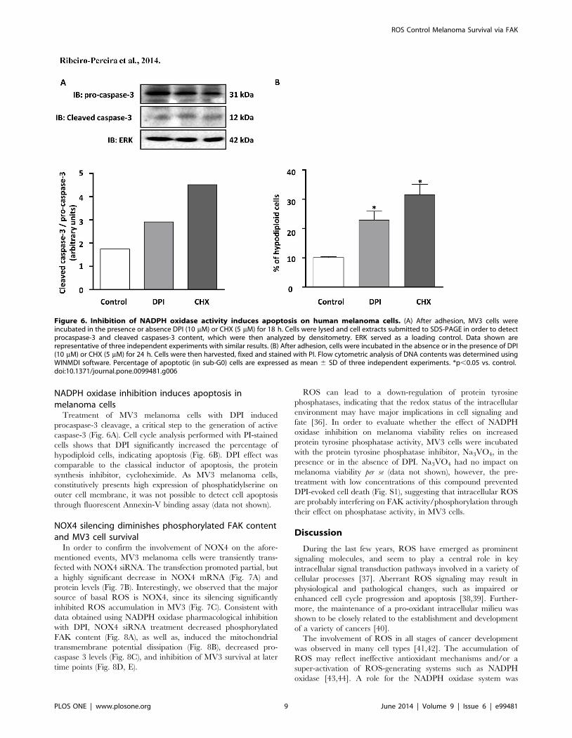
NADPH oxidase inhibition induces apoptosis inmelanoma cells
Treatment of MV3 melanoma cells with DPI induced
procaspase-3 cleavage, a critical step to the generation of active
caspase-3 (Fig. 6A). Cell cycle analysis performed with PI-stained
cells shows that DPI significantly increased the percentage of
hypodiploid cells, indicating apoptosis (Fig. 6B). DPI effect was
comparable to the classical inductor of apoptosis, the protein
synthesis inhibitor, cycloheximide. As MV3 melanoma cells,
constitutively presents high expression of phosphatidylserine on
outer cell membrane, it was not possible to detect cell apoptosis
through fluorescent Annexin-V binding assay (data not shown).
NOX4 silencing diminishes phosphorylated FAK contentand MV3 cell survival
In order to confirm the involvement of NOX4 on the afore-
mentioned events, MV3 melanoma cells were transiently trans-
fected with NOX4 siRNA. The transfection promoted partial, but
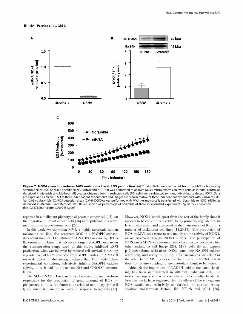
a highly significant decrease in NOX4 mRNA (Fig. 7A) and
protein levels (Fig. 7B). Interestingly, we observed that the major
source of basal ROS is NOX4, since its silencing significantly
inhibited ROS accumulation in MV3 (Fig. 7C). Consistent with
data obtained using NADPH oxidase pharmacological inhibition
with DPI, NOX4 siRNA treatment decreased phosphorylated
FAK content (Fig. 8A), as well as, induced the mitochondrial
transmembrane potential dissipation (Fig. 8B), decreased pro-
caspase 3 levels (Fig. 8C), and inhibition of MV3 survival at later
time points (Fig. 8D, E).
ROS can lead to a down-regulation of protein tyrosine
phosphatases, indicating that the redox status of the intracellular
environment may have major implications in cell signaling and
fate [36]. In order to evaluate whether the effect of NADPH
oxidase inhibition on melanoma viability relies on increased
protein tyrosine phosphatase activity, MV3 cells were incubated
with the protein tyrosine phosphatase inhibitor, Na3VO4, in the
presence or in the absence of DPI. Na3VO4 had no impact on
melanoma viability per se (data not shown), however, the pre-
treatment with low concentrations of this compound prevented
DPI-evoked cell death (Fig. S1), suggesting that intracellular ROS
are probably interfering on FAK activity/phosphorylation through
their effect on phosphatase activity, in MV3 cells.
Discussion
During the last few years, ROS have emerged as prominent
signaling molecules, and seem to play a central role in key
intracellular signal transduction pathways involved in a variety of
cellular processes [37]. Aberrant ROS signaling may result in
physiological and pathological changes, such as impaired or
enhanced cell cycle progression and apoptosis [38,39]. Further-
more, the maintenance of a pro-oxidant intracellular milieu was
shown to be closely related to the establishment and development
of a variety of cancers [40].
The involvement of ROS in all stages of cancer development
was observed in many cell types [41,42]. The accumulation of
ROS may reflect ineffective antioxidant mechanisms and/or a
super-activation of ROS-generating systems such as NADPH
oxidase [43,44]. A role for the NADPH oxidase system was
Figure 6. Inhibition of NADPH oxidase activity induces apoptosis on human melanoma cells. (A) After adhesion, MV3 cells wereincubated in the presence or absence DPI (10 mM) or CHX (5 mM) for 18 h. Cells were lysed and cell extracts submitted to SDS-PAGE in order to detectprocaspase-3 and cleaved caspases-3 content, which were then analyzed by densitometry. ERK served as a loading control. Data shown arerepresentative of three independent experiments with similar results. (B) After adhesion, cells were incubated in the absence or in the presence of DPI(10 mM) or CHX (5 mM) for 24 h. Cells were then harvested, fixed and stained with PI. Flow cytometric analysis of DNA contents was determined usingWINMDI software. Percentage of apoptotic (in sub-G0) cells are expressed as mean 6 SD of three independent experiments. *p,0.05 vs. control.doi:10.1371/journal.pone.0099481.g006
ROS Control Melanoma Survival via FAK
PLOS ONE | www.plosone.org 9 June 2014 | Volume 9 | Issue 6 | e99481
reported in a malignant phenotype of prostate cancer cell [45], on
the migration of breast cancer cells [46] and epithelial-mesenchy-
mal transition in melanoma cells [22].
In this work, we show that MV3, a highly metastatic human
melanoma cell line, also generates ROS in a NADPH oxidase-
dependent manner. The inhibition of NADPH oxidase by DPI, a
flavoprotein inhibitor that selectively targets NADPH oxidase in
the concentration range used in this study, inhibited ROS
production, what was followed by reduced cell survival, indicating
a pivotal role of ROS produced by NADPH oxidase in MV3 cell
survival. There is also strong evidence that DPI, under these
experimental conditions, selectively inhibits NADPH oxidase
activity, once it had no impact on NO and ONOO2 accumu-
lation.
The NOX2 NADPH oxidase is well known as the main isoform
responsible for the production of great amounts of ROS by
phagocytes, but it is also found in a variety of non-phagocytic cell
types, where it is mainly activated in response to agonists [47].
However, NOX4 stands apart from the rest of the family since it
appears to be constitutively active, being primarily regulated by its
level of expression and addressed as the main source of ROS in a
number of melanoma cell lines [14,26,48]. The production of
ROS by MV3 cells seems to rely mainly on the activity of NOX4,
as we observed through NOX4 siRNA. The participation of
NOX2 in NADPH oxidase-mediated effect was excluded once like
other melanoma cell linage [26], MV3 cells do not express
p47phox subunit (critical to NOX2-containing NADPH oxidase
activation), and apocynin did not affect melanoma viability. On
the other hand, MV3 cells express high levels of NOX4, which
does not require coupling to any cytosolic subunit to be active.
Although the importance of NADPH oxidase-mediated signal-
ing has been demonstrated in different malignant cells, the
molecular targets of their products have not been fully elucidated.
Previous works have suggested that the effects of the endogenous
ROS would rely exclusively on classical pro-survival, redox-
sensitive transcription factors, like NF-kB and AP-1 [26].
Figure 7. NOX4 silencing reduces MV3 melanoma basal ROS production. (A) Total mRNAs were extracted from the MV3 cells carryingscramble siRNA (Sc) or NOX4-specific siRNA (siRNA) and qRT-PCR was performed to analyze NOX4 mRNA expression, with actin as internal control asdescribed in Materials and Methods. (B) Lysates obtained from transfected cells (106 cells) were subjected to immunoblotting to detect NOX4. Dataare expressed as mean 6 SD of three independent experiments and images are representative of three independent experiments with similar results.*p,0.05 vs. Scramble. (C) ROS detection assay (CM-H2DCFDA) was performed with MV3 melanoma cells transfected with Scramble or NOX4 siRNA, asdescribed in Materials and Methods. Results are shown as percentage of Scramble of three independent experiments *p,0.05 vs. Scramble.doi:10.1371/journal.pone.0099481.g007
ROS Control Melanoma Survival via FAK
PLOS ONE | www.plosone.org 10 June 2014 | Volume 9 | Issue 6 | e99481
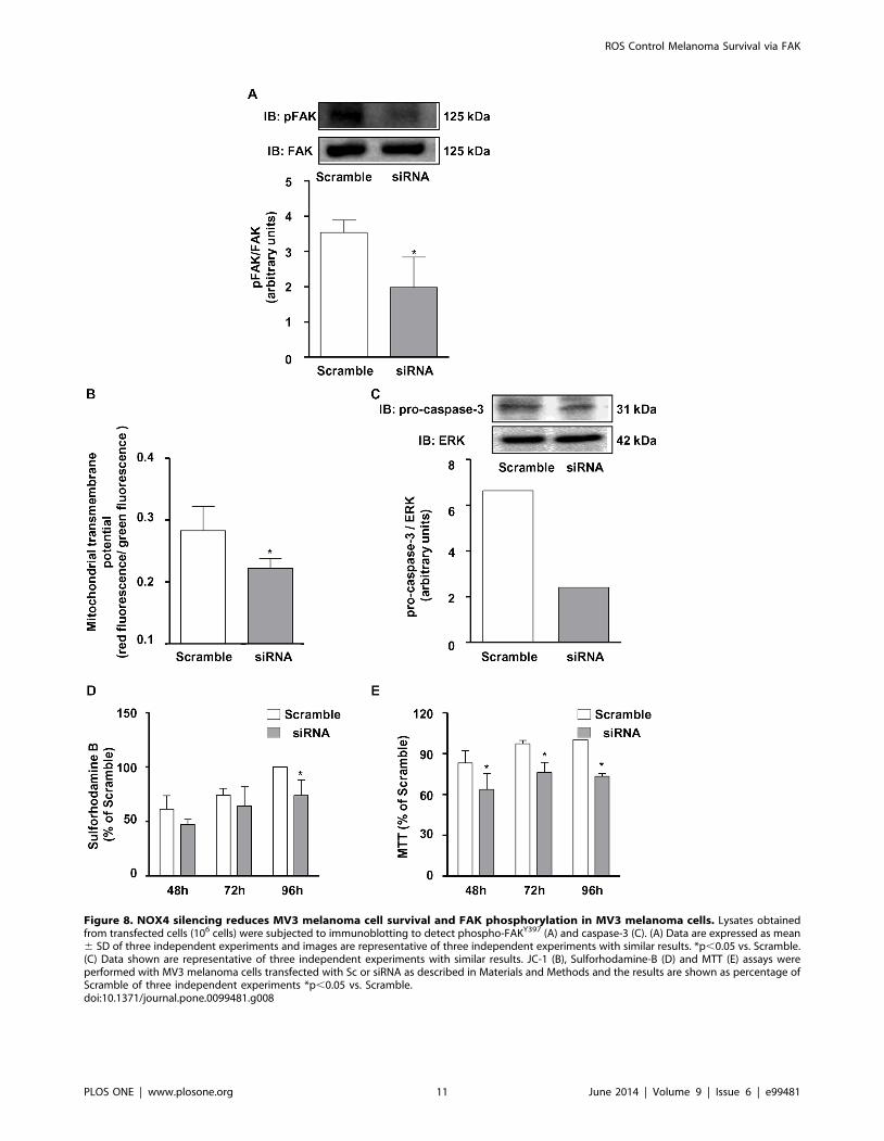
Figure 8. NOX4 silencing reduces MV3 melanoma cell survival and FAK phosphorylation in MV3 melanoma cells. Lysates obtainedfrom transfected cells (106 cells) were subjected to immunoblotting to detect phospho-FAKY397 (A) and caspase-3 (C). (A) Data are expressed as mean6 SD of three independent experiments and images are representative of three independent experiments with similar results. *p,0.05 vs. Scramble.(C) Data shown are representative of three independent experiments with similar results. JC-1 (B), Sulforhodamine-B (D) and MTT (E) assays wereperformed with MV3 melanoma cells transfected with Sc or siRNA as described in Materials and Methods and the results are shown as percentage ofScramble of three independent experiments *p,0.05 vs. Scramble.doi:10.1371/journal.pone.0099481.g008
ROS Control Melanoma Survival via FAK
PLOS ONE | www.plosone.org 11 June 2014 | Volume 9 | Issue 6 | e99481
However, monitoring MV3 cell cultures treated with DPI since
early time points (30 minutes) unveiled severe morphological
changes that preceded any alteration in cell viability. Those
changes provided a valuable clue, leading us to investigate
signaling pathways involved in cell adhesion.
Focal adhesions are points of interaction between integrins and
extracellular matrix (ECM) and draw together adhesion receptors,
as well as signaling and cytoskeletal proteins. They are critical to
maintaining cellular shape, survival, growth and migration [49].
The focal adhesion kinase is primarily activated during integrin-
mediated cell adhesion to ECM and to a lesser extent by growth
factors, bioactive lipids, neuropeptides, and ROS [35]. Autophos-
phorylation of FAK at Tyr397 residue induces its accumulation to
focal adhesion complexes and establishes a close connection
between integrins to actin cytoskeleton [50].
When MV3 melanoma cells are seeded on culture dishes, they
form a monolayer firmly attached to the substrate, displaying focal
adhesion points along the cell edge. However, NADPH oxidase
inhibition promoted reduction in cellular spreading, collapsing
focal adhesions cortical organization. Moreover, we observed the
formation of cortical polymerized actin ring, an indicative of cell
detachment.
As a key molecule in the transduction of integrin-mediated
signaling, FAK is critically involved in the development and
progression of cancer, regulating survival, proliferation, migration
and invasion [51]. Not surprisingly, highly aggressive melanoma
cell lines contained constitutive high levels of phosphorylated FAK
whereas the poorly aggressive melanoma cell lines did not [52].
MV3 melanoma cell line is characterized as highly metastatic [28]
and our results confirmed that this phenotype is linked to
constitutive high levels of FAK phosphorylated on tyrosine 397.
The phosphorylation of FAK at Tyr397 creates a high-affinity
binding site for the tyrosine kinase cSrc, which is then activated
[53]. The FAK-Src complex mediates cell migration [54],
proliferation [55] and survival [56]. Our data shows that high
levels of FAK are constitutively associated to Src in MV3
melanoma cells. However, the inhibition of NADPH oxidase
activity by DPI or NOX4 silencing significantly reduced FAKY397
phosphorylation and probably disrupted FAK-Src association.
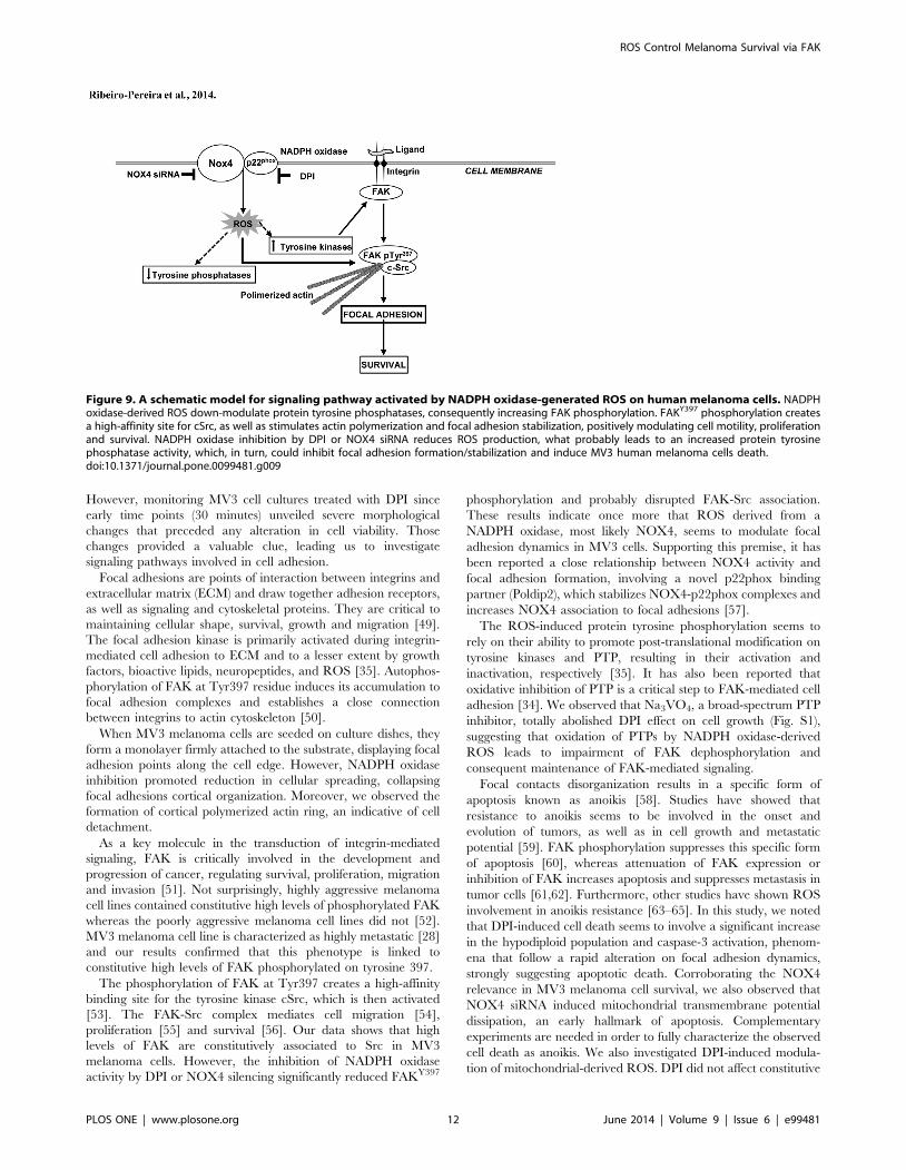
These results indicate once more that ROS derived from a
NADPH oxidase, most likely NOX4, seems to modulate focal
adhesion dynamics in MV3 cells. Supporting this premise, it has
been reported a close relationship between NOX4 activity and
focal adhesion formation, involving a novel p22phox binding
partner (Poldip2), which stabilizes NOX4-p22phox complexes and
increases NOX4 association to focal adhesions [57].
The ROS-induced protein tyrosine phosphorylation seems to
rely on their ability to promote post-translational modification on
tyrosine kinases and PTP, resulting in their activation and
inactivation, respectively [35]. It has also been reported that
oxidative inhibition of PTP is a critical step to FAK-mediated cell
adhesion [34]. We observed that Na3VO4, a broad-spectrum PTP
inhibitor, totally abolished DPI effect on cell growth (Fig. S1),
suggesting that oxidation of PTPs by NADPH oxidase-derived
ROS leads to impairment of FAK dephosphorylation and
consequent maintenance of FAK-mediated signaling.
Focal contacts disorganization results in a specific form of
apoptosis known as anoikis [58]. Studies have showed that
resistance to anoikis seems to be involved in the onset and
evolution of tumors, as well as in cell growth and metastatic
potential [59]. FAK phosphorylation suppresses this specific form
of apoptosis [60], whereas attenuation of FAK expression or
inhibition of FAK increases apoptosis and suppresses metastasis in
tumor cells [61,62]. Furthermore, other studies have shown ROS
involvement in anoikis resistance [63–65]. In this study, we noted
that DPI-induced cell death seems to involve a significant increase
in the hypodiploid population and caspase-3 activation, phenom-
ena that follow a rapid alteration on focal adhesion dynamics,
strongly suggesting apoptotic death. Corroborating the NOX4
relevance in MV3 melanoma cell survival, we also observed that
NOX4 siRNA induced mitochondrial transmembrane potential
dissipation, an early hallmark of apoptosis. Complementary
experiments are needed in order to fully characterize the observed
cell death as anoikis. We also investigated DPI-induced modula-
tion of mitochondrial-derived ROS. DPI did not affect constitutive
Figure 9. A schematic model for signaling pathway activated by NADPH oxidase-generated ROS on human melanoma cells. NADPHoxidase-derived ROS down-modulate protein tyrosine phosphatases, consequently increasing FAK phosphorylation. FAKY397 phosphorylation createsa high-affinity site for cSrc, as well as stimulates actin polymerization and focal adhesion stabilization, positively modulating cell motility, proliferationand survival. NADPH oxidase inhibition by DPI or NOX4 siRNA reduces ROS production, what probably leads to an increased protein tyrosinephosphatase activity, which, in turn, could inhibit focal adhesion formation/stabilization and induce MV3 human melanoma cells death.doi:10.1371/journal.pone.0099481.g009
ROS Control Melanoma Survival via FAK
PLOS ONE | www.plosone.org 12 June 2014 | Volume 9 | Issue 6 | e99481
mitochondrial ROS production, as evaluated using the specific
probe MitoSox (Fig. S2).
Taken together, our data strongly suggest that NADPH oxidase-
derived ROS convey cell survival signals in MV3 melanoma cells
through the persistent activation of the FAK pathway, probably
inhibiting protein tyrosine phosphatase activity. Our study
addresses, for the first time, FAK as an important target of
ROS-mediated signaling in melanoma cells, showing that FAK
phosphorylation and downstream events are highly sensitive to
NADPH oxidase-derived ROS. NADPH oxidase inhibition
promotes focal adhesions breakdown and cell death, (Fig. 9).
These findings shed light on a still underappreciated face of ROS
signaling in cancer cells and may corroborate to the development
of more selective and effective strategies in order to control
melanoma growth and metastatic colonization.
Supporting Information
Figure S1 Inhibition of tyrosine phosphatase activity reverts DPI
effect on melanoma survival. Cells (66103) were preincubated for
30 min in the presence or absence of increasing concentrations of
Na3VO4 (0.1–3 mM) and subsequently treated with DPI (10 mM)
for 48 hours. MTT assay was performed as described. Results are
shown as percentage of control and are expressed as mean 6 SD
of three independent experiments performed in quintuplicate. *p,
0.05 vs. control, **p,0.05 vs. DPI.
(TIF)
Figure S2 Inhibition of NADPH oxidase activity does not
abolish constitutive mitochondrial ROS generation on melanoma
cells MV3. MV3 cells were incubated with or without DPI
(10 mM). Mitochondrial ROS production was measured by
MitoSox probe oxidation. Data are expressed as mean 6 SD of
six independent experiments. * p,0.05 vs. control.
(TIF)
Acknowledgments
We would like to thank Genilson Rodrigues, Renata Turreta and Amanda
Lima-Resende for their excellent technical support, Carlos Bizarro for
confocal microscopy analysis, Denise Fernandes and Maria Aparecida
Bertoline for cellular ROS measurement by HPLC. We would also want to
express our gratitude to Mr. Andrew W. Bullock for kindly revising this
manuscript.
Author Contributions
Conceived and designed the experiments: CRP MAA JAM. Performed the
experiments: CRP JAM MJS. Analyzed the data: CRP JAM MAA.
Contributed reagents/materials/analysis tools: CBF MAA FRL. Wrote the
paper: CRP JAM.
References
1. Chin L (2003) The genetics of malignant melanoma: lessons from mouse andman. Nature reviews Cancer 3: 559–570.
2. Woodhead AD, Setlow RB, Tanaka M (1999) Environmental factors in
nonmelanoma and melanoma skin cancer. Journal of epidemiology Japan
Epidemiological Association 9: S102–14.
3. Carlson JA, Ross JS, Slominski A, Linette G, Mysliborski J, et al. (2005)Molecular diagnostics in melanoma. Journal of the American Academy of
Dermatology 52: 743–775; quiz 775–778.
4. Fidler IJ (2002) Critical determinants of metastasis. Seminars in Cancer Biology12: 89–96.
5. Braeuer RR, Zigler M, Villares GJ, Dobroff AS, Bar-Eli M (2011)
Transcriptional control of melanoma metastasis: the importance of the tumor
microenvironment. Seminars in Cancer Biology 21: 83–88.
6. Young C (2009) Solar ultraviolet radiation and skin cancer. Occupationalmedicine Oxford England 59: 82–88.
7. De Vries E, Arnold M, Altsitsiadis E, Trakatelli M, Hinrichs B, et al. (2012)
Potential impact of interventions resulting in reduced exposure to ultraviolet(UV) radiation (UVA and UVB) on skin cancer incidence in four European
countries, 2010–2050. The British journal of dermatology 167 Suppl: 53–62.
8. Poljsak B, Dahmane R (2012) Free radicals and extrinsic skin aging.
Dermatology research and practice 2012: 135206.
9. Cooper KL, Liu KJ, Hudson LG (2009) Enhanced ROS production and redoxsignaling with combined arsenite and UVA exposure: contribution of NADPH
oxidase. Free Radical Biology & Medicine 47: 381–388.
10. Darr D, Fridovich I (1994) Free radicals in cutaneous biology. The Journal of
investigative dermatology 102: 671–675.
11. Sies H (1991) Oxidative stress: from basic research to clinical application. TheAmerican Journal of Medicine 91: 31S–38S.
12. Block K, Gorin Y (2012) Aiding and abetting roles of NOX oxidases in cellular
transformation. Nature Reviews Cancer 12: 627–637.
13. Luo H, Yang Y, Duan J, Wu P, Jiang Q, et al. (2013) PTEN-regulated AKT/FoxO3a/Bim signaling contributes to reactive oxygen species-mediated
apoptosis in selenite-treated colorectal cancer cells. Cell death disease 4: e481.
14. Brieger K, Schiavone S, Miller FJ, Krause K-H (2012) Reactive oxygen species:
from health to disease. Swiss medical weekly 142: w13659. doi:10.4414/smw.2012.13659.
15. Babior BM, Lambeth JD, Nauseef W (2002) The neutrophil NADPH oxidase.
ArchBiochemBiophys 397: 342–344.
16. Kleniewska P, Piechota A, Skibska B, Goraca A (2012) The NADPH oxidasefamily and its inhibitors. Archivum Immunologiae et Therapiae Experimentalis
60: 277–294.
17. Santos CXC, Anilkumar N, Zhang M, Brewer AC, Shah AM (2011) Redox
signaling in cardiac myocytes. Free Radical Biology & Medicine 50: 777–793.
18. Weaver JR, Taylor-Fishwick D (2013) Regulation of NOX-1 expression in betacells: a positive feedback loop involving the Src-kinase signaling pathway.
Molecular and cellular endocrinology 369: 35–41.
19. Szatrowski TP, Nathan CF (1991) Production of large amounts of hydrogenperoxide by human tumor cells. Cancer Research 51: 794–798.
20. Mochizuki T, Furuta S, Mitsushita J, Shang WH, Ito M, et al. (2006) Inhibition
of NADPH oxidase 4 activates apoptosis via the AKT/apoptosis signal-
regulating kinase 1 pathway in pancreatic cancer PANC-1 cells. Oncogene 25:
3699–3707.
21. Hsieh C-H, Shyu W-C, Chiang C-Y, Kuo J-W, Shen W-C, et al. (2011)
NADPH oxidase subunit 4-mediated reactive oxygen species contribute to
cycling hypoxia-promoted tumor progression in glioblastoma multiforme. PLoS
ONE 6: e23945. doi:10.1371/journal.pone.0023945.
22. Liu F, Gomez Garcia AM, Meyskens FL (2012) NADPH Oxidase 1
Overexpression Enhances Invasion via Matrix Metalloproteinase-2 and
Epithelial-Mesenchymal Transition in Melanoma Cells. The Journal of
investigative dermatology: 1–9.
23. Du J, Nelson ES, Simons AL, Olney KE, Moser JC, et al. (2012) Regulation of
pancreatic cancer growth by superoxide. Molecular Carcinogenesis.
24. Zhou X, Li D, Resnick MB, Behar J, Wands J, et al. (2011) Signaling in H2O2-
induced increase in cell proliferation In Barrett’s Esophageal Adenocarcinoma
Cells. The Journal of pharmacology and experimental therapeutics 339: 218–
227.
25. Shono T, Yokoyama N, Uesaka T, Kuroda J, Takeya R, et al. (2008) Enhanced
expression of NADPH oxidase Nox4 in human gliomas and its roles in cell
proliferation and survival. International journal of cancer Journal international
du cancer 123: 787–792.
26. Brar SS, Kennedy TP, Sturrock AB, Huecksteadt TP, Quinn MT, et al. (2002)
An NAD(P)H oxidase regulates growth and transcription in melanoma cells.
American journal of physiology Cell physiology 282: C1212–24.
27. Brar SS, Corbin Z, Kennedy TP, Hemendinger R, Thornton L, et al. (2003)
NOX5 NAD(P)H oxidase regulates growth and apoptosis in DU 145 prostate
cancer cells. American journal of physiology Cell physiology 285: C353–C369.
28. van Muijen GN, Jansen KF, Cornelissen IM, Smeets DF, Beck JL, et al. (1991)
Establishment and characterization of a human melanoma cell line (MV3) which
is highly metastatic in nude mice. International journal of cancer Journal
international du cancer 48: 85–91.
29. Qin Y, Lu M, Gong X (2008) Dihydrorhodamine 123 is superior to 2,7-
dichlorodihydrofluorescein diacetate and dihydrorhodamine 6G in detecting
intracellular hydrogen peroxide in tumor cells. Cell Biol Int 32: 224–228.
30. Fernandes DC, Wosniak J, Pescatore LA, Bertoline MA, Liberman M, et al.
(2007) Analysis of DHE-derived oxidation products by HPLC in the assessment
of superoxide production and NADPH oxidase activity in vascular systems.
American journal of physiology Cell physiology 292: C413–22.
31. Van De Loosdrecht AA, Nennie E, Ossenkoppele GJ, Beelen RH, Langen-
huijsen MM (1991) Cell mediated cytotoxicity against U 937 cells by human
monocytes and macrophages in a modified colorimetric MTT assay. A
methodological study. Journal of Immunological Methods 141: 15–22.
32. Cossarizza A, Baccarani-Contri M, Kalashnikova G, Franceschi C (1993) A new
method for the cytofluorimetric analysis of mitochondrial membrane potential
using the J-aggregate forming lipophilic cation 5,59,6,69-tetrachloro-1,19,3,39-
tetraethylbenzimidazolcarbocyanine iodide (JC-1). Biochemical and biophysical
research communications 197: 40–45.
ROS Control Melanoma Survival via FAK
PLOS ONE | www.plosone.org 13 June 2014 | Volume 9 | Issue 6 | e99481
33. Dooley DC, Simpson JF, Meryman HT (1982) Isolation of large numbers of fully
viable human neutrophils: a preparative technique using percoll density gradient
centrifugation. Experimental Hematology 10: 591–599.
34. Aldieri E, Riganti C, Polimeni M, Gazzano E, Lussiana C, et al. (2008) Classical
inhibitors of NOX NAD(P)H oxidases are not specific. Current Drug
Metabolism 9: 686–696.
35. Chiarugi P, Pani G, Giannoni E, Taddei L, Colavitti R, et al. (2003) Reactive
oxygen species as essential mediators of cell adhesion: the oxidative inhibition of
a FAK tyrosine phosphatase is required for cell adhesion. The Journal of Cell
Biology 161: 933–944.
36. Chiarugi P (2005) PTPs versus PTKs: the redox side of the coin. Free Radical
Research 39: 353–364.
37. Poli G, Leonarduzzi G, Biasi F, Chiarpotto E (2004) Oxidative stress and cell
signalling. Current Medicinal Chemistry 11: 1163–1182.
38. Boonstra J, Post JA (2004) Molecular events associated with reactive oxygen
species and cell cycle progression in mammalian cells. Gene 337: 1–13.
39. Nogueira V, Park Y, Chen C-C, Xu P-Z, Chen M-L, et al. (2008) Akt
determines replicative senescence and oxidative or oncogenic premature
senescence and sensitizes cells to oxidative apoptosis. Cancer Cell 14: 458–470.
40. Afanas’ev I (2011) Reactive oxygen species signaling in cancer: comparison with
aging. Aging and disease 2: 219–230.
41. Sander CS, Chang H, Hamm F, Elsner P, Thiele JJ (2004) Role of oxidative
stress and the antioxidant network in cutaneous carcinogenesis. International
Journal of Dermatology 43: 326–335.
42. Weyemi U, Lagente-Chevallier O, Boufraqech M, Prenois F, Courtin F, et al.
(2012) ROS-generating NADPH oxidase NOX4 is a critical mediator in
oncogenic H-Ras-induced DNA damage and subsequent senescence. Oncogene
31: 1117–1129.
43. Valko M, Rhodes CJ, Moncol J, Izakovic M, Mazur M (2006) Free radicals,
metals and antioxidants in oxidative stress-induced cancer. Chemicobiological
interactions 160: 1–40.
44. Birben E, Sahiner UM, Sackesen C, Erzurum S, Kalayci O (2012) Oxidative
stress and antioxidant defense. The World Allergy Organization journal 5: 9–19.
doi:10.1097/WOX.0b013e3182439613.
45. Kumar B, Koul S, Khandrika L, Meacham RB, Koul HK (2008) Oxidative
Stress Is Inherent in Prostate Cancer Cells and Is Required for Aggressive
Phenotype: 1777–1785.
46. Klees RF, De Marco PC, Salasznyk RM, Ahuja D, Hogg M, et al. (2006)
Apocynin derivatives interrupt intracellular signaling resulting in decreased
migration in breast cancer cells. Journal of biomedicine & biotechnology 2006:
87246.
47. Drummond GR, Selemidis S, Griendling KK, Sobey CG (2011) Combating
oxidative stress in vascular disease: NADPH oxidases as therapeutic targets.
Nature Reviews Drug Discovery 10: 453–471.
48. Yamaura M, Mitsushita J, Furuta S, Kiniwa Y, Ashida A, et al. (2009) NADPH
oxidase 4 contributes to transformation phenotype of melanoma cells by
regulating G2-M cell cycle progression. Cancer Research 69: 2647–2654.
49. Hehlgans S, Haase M, Cordes N (2007) Signalling via integrins: implications for
cell survival and anticancer strategies. Biochimica et Biophysica Acta 1775: 163–180.
50. Mitra SK, Hanson DA, Schlaepfer DD (2005) Focal adhesion kinase: in
command and control of cell motility. 6: 56–68.51. Van Nimwegen MJ, Van De Water B (2007) Focal adhesion kinase: A potential
target in cancer therapy. 73: 597–609.52. Hess AR, Hendrix MJC (2006) Focal adhesion kinase signaling and the
aggressive melanoma phenotype. Cell cycle Georgetown Tex 5: 478–480.
53. Schaller MD, Hildebrand JD, Shannon JD, Fox JW, Vines RR, et al. (1994)Autophosphorylation of the focal adhesion kinase, pp125FAK, directs SH2-
dependent binding of pp60src. Molecular and Cellular Biology 14: 1680–1688.54. Mitra SK, Schlaepfer DD (2006) Integrin-regulated FAK-Src signaling in
normal and cancer cells. Current Opinion in Cell Biology 18: 516–523.55. Ding Q, Grammer JR, Nelson M, Guan J-L, Stewart JE, et al. (2005) p27Kip1
and cyclin D1 are necessary for focal adhesion kinase regulation of cell cycle
progression in glioblastoma cells propagated in vitro and in vivo in the scidmouse brain. The Journal of biological chemistry 280: 6802–6815.
56. Beausejour M, Noel D, Thibodeau S, Bouchard V, Harnois C, et al. (2012)Integrin/Fak/Src-mediated regulation of cell survival and anoikis in human
intestinal epithelial crypt cells: selective engagement and roles of PI3-K isoform
complexes. Apoptosis an international journal on programmed cell death 17:566–578.
57. Lyle AN, Deshpande NN, Taniyama Y, Seidel-Rogol B, Pounkova L, et al.(2009) Poldip2, a novel regulator of Nox4 and cytoskeletal integrity in vascular
smooth muscle cells. Circulation Research 105: 249–259.58. Frisch SM, Screaton RA (2001) Anoikis mechanisms. Curr Opin Cell Biol 13:
555–562.
59. Zhong X, Rescorla FJ (2012) Cell surface adhesion molecules and adhesion-initiated signaling: understanding of anoikis resistance mechanisms and
therapeutic opportunities. Cellular Signalling 24: 393–401.60. Grossmann J (2002) Molecular mechanisms of ‘‘detachment-induced apopto-
sis—Anoikis’’. Apoptosis an international journal on programmed cell death 7:
247–260.61. Duxbury MS, Ito H, Zinner MJ, Ashley SW, Whang EE (2004) Focal adhesion
kinase gene silencing promotes anoikis and suppresses metastasis of humanpancreatic adenocarcinoma cells. Surger 135: 555–562.
62. Liu G, Meng X, Jin Y, Bai J, Zhao Y, et al. (2008) Inhibitory role of focaladhesion kinase on anoikis in the lung cancer cell A549. Cell biology
international 32: 663–670.
63. Pani G, Galeotti T, Chiarugi P (2010) Metastasis: cancer cell’s escape fromoxidative stress. Cancer metastasis reviews 29: 351–378.
64. Giannoni E, Buricchi F, Grimaldi G, Parri M, Cialdai F, et al. (2008) Redoxregulation of anoikis: reactive oxygen species as essential mediators of cell
survival. Cell Death and Differentiation 15: 867–878.
65. Giannoni E, Fiaschi T, Ramponi G, Chiarugi P (2009) Redox regulation ofanoikis resistance of metastatic prostate cancer cells: key role for Src and EGFR-
mediated pro-survival signals. Oncogene 28: 2074–2086.
ROS Control Melanoma Survival via FAK
PLOS ONE | www.plosone.org 14 June 2014 | Volume 9 | Issue 6 | e99481


![NOX4 expression and distal arteriolar remodeling correlate ... · chronic PH and human idiopathic pulmonary arterial hypertension (PAH) [13–15]. Of importance, the NOX4 was a relevant](https://img.pdfslide.net/doc/110x75/5e07c488fd212f0268641ddf/nox4-expression-and-distal-arteriolar-remodeling-correlate-chronic-ph-and-human.jpg)